BMC Women's Health
Utilising synergism between the transverse abdominal and pelvic floor muscles at different postures in nulliparous women: a randomized case control study
--Manuscript Draft--
Manuscript Number: BMWH-D-20-01218R1
Full Title: Utilising synergism between the transverse abdominal and pelvic floor muscles at different postures in nulliparous women: a randomized case control study
Article Type: Research article
Section/Category: Women's health behaviors and health promotion
Funding Information: European Social Fund
(EFOP-3.6.1-16-2016-00008) BPthy Tímea Molnár
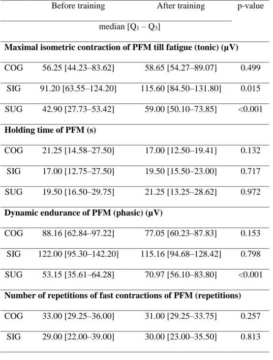
Abstract: Background To determine the effects of the pelvic floor muscle training (PFM-T) in combination with transverse abdominal muscle (TRA) activation (cPFM-T) in female urinary incontinence. Methods We enrolled nulliparous women in supine (SUG) (n = 22), sitting (SIG) (n = 19) and control (COG) (n = 14) groups. We performed the 8- week cPFM-T programme. We examined the effect of training on the parameters with the Kruskal–Wallis test, and the pairwise comparisons with the Mann-Whitney U-test and the Wilcoxon-rank test with the Bonferroni correction. Results Before training, 15 participants reported occasional urinary leakage. After cPFM-T seven participants reported that urinary leakage had disappeared. Maximal isometric contraction of the pelvic floor muscles (PFM) until fatigue improved significantly in the SUG (p < 0.001) and SIG (p = 0.015) and not significantly in the COG (p = 0.499). Holding time increased in the SUG (p = 0.972) and the SIG (p = 0.717), and decreased in the COG (p = 0.132). The dynamic endurance of the PFM improved significantly in the SUG (p <
0.001), but not in the SIG (p = 0.798) and the COG (p = 0.153). The number of maximal fast contractions within 1 minute increased in both the SUG (p < 0.001) and the SIG (p=0.813) and decreased in the COG (p = 0.257). Relaxation improved significantly in the SIG (p = 0.011). TRA thickness increased in both training groups.
Conclusions Slow-twitch fibres of the PFM can be trained effectively with PFM-T in both the body positions. Trial registration: This study was registered in the Hungarian National Healthcare Service Center: 019234/2014/OTIG Registered 07 April 2014 modification: 096623-002/2015/OTIG Registered 11 November 2015
https://www.aeek.hu The study was retrospectively registered in ClincalTrials.gov NCT04577872 Registered 22 September 2020. https://clinicaltrials.gov Corresponding Author: Suranyi Andrea, PhD
University of Szeged Szeged, HUNGARY Corresponding Author E-Mail: suranyi.andrea@yahoo.com Corresponding Author Secondary
Information:
Corresponding Author's Institution: University of Szeged Corresponding Author's Secondary
Institution:
First Author: Tímea Molnár
First Author Secondary Information:
Order of Authors: Tímea Molnár
Andrea Domján Mónika Szűcs
Order of Authors Secondary Information:
Response to Reviewers: Answer to Assistant Editor's comments:
1. Title Page
--We noticed that the corresponding author email provided on your title page is different to the one you indicated in the editorial submission. Please amend one so that they match.
I atteched the email, which is aaproved in the eitorial submission.
suranyi.andrea@yahoo.com
--Please ensure that in the list of authors on the title page, the authors’ full names are provided [first name last name], in order to match the list of authors in the submissions system.
The authors order is the same on the title page, than in in the submissions system. ( I added the accents to the names)
--Please remove the co-authors' email addresses from the title page of the manuscript.
Please note that only the corresponding author's email address should be provided.
I removed the co-authors’ email addresses.
2. Clinical Trials --CONSORT guidelines
In accordance with BMC Series editorial policies
(http://www.biomedcentral.com/submissions/editorial-policies#standards+of+reporting), could you please ensure your manuscript reporting adheres to CONSORT guidelines (http://www.consort-statement.org/) for reporting clinical trials. This is so your methodology can be fully evaluated and utilised. Please include a statement within your manuscript to indicate that your study adheres to CONSORT guidelines and include a completed CONSORT checklist as an additional file when submitting your revised manuscript.
We added the CONSORT checklist as additional file
Please complete the checklist in full by inserting the page number/paragraph and section of your manuscript which reports the information that meets the criteria of the checklist. For example “Methods, paragraph 2”. If a criterion is not applicable for your particular manuscript/study, we can accept “N/A”.
We completed the CONSORT checklist
Please note that checklists completed incorrectly will be returned for revision as we cannot progress your manuscript to peer review until the checklist has been completed.
3. Additional Files
--We note that you have cited Additional Files 2 and 3 in the main manuscript text.
Please ensure that these are uploaded with the manuscript, as they are currently missing from your submission.
It was a misstake, to indicate the figures and tables as ’additional files’.
Additional Information:
Question Response
Has this manuscript been submitted
before to this journal or another journal in No
Title
Utilising synergism between the transverse abdominal and pelvic floor muscles at different postures in nulliparous women: a randomized case control study
Authors
Tímea Molnár1,2, Andrea Domján1, Mónika Szűcs3, Andrea Surányi 4a, József Bódis 5a
1 University of Szeged, Faculty of Health Sciences and Social Studies, H-6726 Szeged Temesvári 31., Hungary, (during the study period)
2 Department of Physiotherapy University of Szeged, H-6725 Szeged Semmelweis u.6.
(present)
3 University of Szeged, Faculty of Medicine, Faculty of Science and Informatics, Department of Medical Physics and Informatics, H-6720 Szeged Korányi 9., Hungary, 4 University of Szeged, Faculty of Medicine, Department of Obstetrics and Gynaecology, Albert Szent- Györgyi Health Center, H-6725 Szeged Semmelweis 5., Hungary
5 University of Pécs, Faculty of Health Sciences, Doctoral School of Health Sciences, H-7621 Pécs Vörösmarty 4., Hungary
a: shared last authorship
Corresponding author:
Andrea Suranyi, email: suranyi.andrea@yahoo.com; gaspar-suranyi.andrea@med.u-szeged.hu -Tel.:+3662-545499 FAX: +3662-545711
Conflict of interest
The authors have no conflict of interests.
Statement:
Each author’s participation in the manuscript
Manuscript Click here to access/download;Manuscript;MS_SSMR-D-20-
01218rev1.docx Click here to view linked References
1 2 3 4 5 6 7 8 9 10 11 12 13 14 15 16 17 18 19 20 21 22 23 24 25 26 27 28 29 30 31 32 33 34 35 36 37 38 39 40 41 42 43 44 45 46 47 48 49 50 51 52 53 54 55 56 57 58 59
TM: project development, study design, ultrasound investigation, data collection, manuscript writing and editing
AD: project development, study design, manuscript revision MSz: data collection, statistical analysis,
AS: study design, ultrasound investigation, manuscript writing, editing and revision JB: project development, manuscript revision
Word count: 3849 Table count:3 Figure count:3
Abstract
Background: To determine the effects of the pelvic floor muscle training (PFM-T) in combination with transverse abdominal muscle (TRA) activation (cPFM-T) in female urinary incontinence.
Methods: We enrolled nulliparous women in supine (SUG) (n = 22), sitting (SIG) (n = 19) and control (COG) (n = 14) groups. We performed the 8-week cPFM-T programme. We examined the effect of training on the parameters with the Kruskal–Wallis test, and the pairwise comparisons with the Mann-Whitney U-test and the Wilcoxon-rank test with the Bonferroni correction.
Results: Before training, 15 participants reported occasional urinary leakage. After cPFM-T seven participants reported that urinary leakage had disappeared. Maximal isometric contraction of the pelvic floor muscles (PFM) until fatigue improved significantly in the SUG (p < 0.001) and SIG (p = 0.015) and not significantly in the COG (p = 0.499). Holding time increased in the SUG (p = 0.972) and the SIG (p = 0.717), and decreased in the COG (p =
1 2 3 4 5 6 7 8 9 10 11 12 13 14 15 16 17 18 19 20 21 22 23 24 25 26 27 28 29 30 31 32 33 34 35 36 37 38 39 40 41 42 43 44 45 46 47 48 49 50 51 52 53 54 55 56 57 58 59
0.132). The dynamic endurance of the PFM improved significantly in the SUG (p < 0.001), but not in the SIG (p = 0.798) and the COG (p = 0.153). The number of maximal fast contractions within 1 minute increased in both the SUG (p < 0.001) and the SIG (p=0.813) and decreased in the COG (p = 0.257). Relaxation improved significantly in the SIG (p = 0.011). TRA thickness increased in both training groups.
Conclusions: Slow-twitch fibres of the PFM can be trained effectively with PFM-T in both the body positions.
Trial registration: This study was registered in the Hungarian National Healthcare Service Center: 019234/2014/OTIG Registered 07 April 2014 modification: 096623-002/2015/OTIG Registered 11 November 2015 https://www.aeek.hu
The study was retrospectively registered in ClincalTrials.gov NCT04577872 Registered 22 September 2020. https:// https://clinicaltrials.gov
Keywords:
pelvic floor muscle training, transverse abdominal muscle, ultrasound measurement, urinary incontinence, vaginal surface electromyography
Brief summary
This physiotherapist-guided group training programme should be performed in both the supine and the sitting positions; this results in better and more cost-effective patient motivation.
Background
Worldwide, an estimated 20%–30% of young women have urinary incontinence (UI), making the frequency of this condition a fundamental problem [1].
According to Kegel, regular, specific strength training of the pelvic floor muscles (PFM) has a beneficial effect on female UI and pelvic organ prolapse [2].
1 2 3 4 5 6 7 8 9 10 11 12 13 14 15 16 17 18 19 20 21 22 23 24 25 26 27 28 29 30 31 32 33 34 35 36 37 38 39 40 41 42 43 44 45 46 47 48 49 50 51 52 53 54 55 56 57 58 59
The effectiveness of Kegel’s PFM-T exercises—classified by the International Consultation on Incontinence as level A evidence in Evidence Based Medicine [3, 4] has been proven in numerous randomised controlled trials.
Several research groups have reported a co-contraction between the deep abdominal muscles and the PFM [5, 6, 7, 8].
Together, these results suggest the necessity of involving a coordinated approach of the PFM and the deep abdominal muscles in the effective treatment of UI.
Several studies have revealed significantly higher resting PFM activity in unsupported sitting as compared to supported sitting and in a standing posture as compared to a supine position [8, 9, 10, 11].
Chmielewska[11] reported a significant a long-lasting contraction during unsupported sitting, due to the superior recruitment by the sitting posture of the sensorimotor control system to that by the supine position. They identified different coactivation patterns of the PFM and abdominal muscles during sitting to those during standing and lying.
Sufficient evidence that regular PFM-T improves the symptoms of incontinence has been reported [3, 12, 13]. However, only a few studies support the effect of the coordination of the diaphragm, deep abdominal muscles and PFM on incontinence [14, 15].
Current evidence on skeletal muscle training and other factors (UI, supervised PFM-T programme, e.t.c.) are known to have an impact on women’s participation in and adherence to PFM-T [3].
Here we aimed to investigate whether—based on trunk muscle synergism—the condition and functioning of the PFM would improve in the sitting and supine postures or in the control group during PFM-T with forced exhalation. We hypotized: the training position affect the muscle gymnastic performance.
1 2 3 4 5 6 7 8 9 10 11 12 13 14 15 16 17 18 19 20 21 22 23 24 25 26 27 28 29 30 31 32 33 34 35 36 37 38 39 40 41 42 43 44 45 46 47 48 49 50 51 52 53 54 55 56 57 58 59
Methods Participants
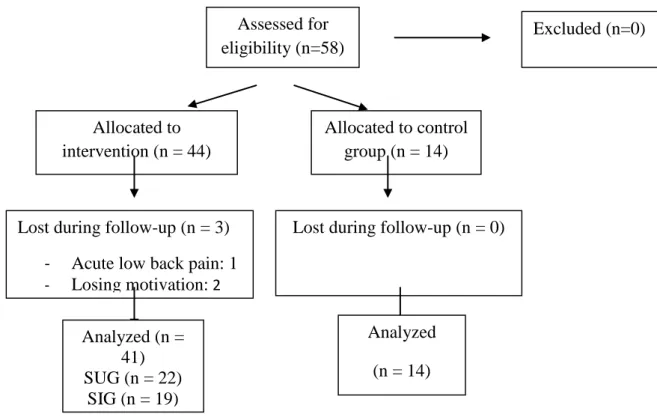
We performed our study at the Faculty of Health Sciences and Social Studies, University of Szeged, Hungary between October 1, 2016 and December 1, 2016. We enrolled 58 healthy, young (mean±SD: 21.27±1.46 years), nulliparous women in this trial. We recruited them through an online advertisement and then selected them for the training groups and the control group by T.M.(Additional file 3). The participants had no previous experience of PFM-T. We divided them into two study groups depending on the strength of the PFM and treated them with cPFM-T in both the supine position and the sitting position. The assessment comprised one phase: 3 repetitions of sustained 5-second voluntary PFM contraction with 10-second relaxation. We calculated the mean of 3 contractions and set the obtained values in ascending order. The 22 participants with lower muscle strength (under 60 V) comprised the SUG and the 22 participants with higher muscle strength (over 60 V) formed the SIG. Only 19 participants, however, completed the programme in the SIG (Additional file 3). We enrolled these 19 participants with higher PFM tension (over 60 V) in the SIG because the PFM must be stronger to resist gravity [16]. We created a control group (n = 14) (COG) comprising seven persons with a PFM tension under 60 V and seven individuals with a PFM tension over 60
V. The COG did not change their lifestyles and did not undergo PFM-T. We included in the study groups participants willing to participate in the study and able to contract the pelvic floor and TRA muscles correctly. Participants were required to maintain their everyday activities (attending lessons, sport activities, and so on). The local ethics committee (National Healthcare Service Center) approved the study (019234/2014/OTIG) and we received written, informed consent from all participants. Exclusion criteria were known neurological or rheumatological diseases and previous vaginal or abdominal surgery.
1 2 3 4 5 6 7 8 9 10 11 12 13 14 15 16 17 18 19 20 21 22 23 24 25 26 27 28 29 30 31 32 33 34 35 36 37 38 39 40 41 42 43 44 45 46 47 48 49 50 51 52 53 54 55 56 57 58 59
In the study, before and after the training programme, we used a self-administered questionnaire (Additional file 7), based on three validated questionnaires (the King’s health questionnaire, the Incontinence impact questionnaire and the Urogenital distress inventory) [17]. We included in the questionnaire risk factors for UI (obstetric history, height and weight, stress, physical activity level, sport, vaginal and abdominal surgery) and questions relating to the urinary tract and anal canal (involuntary urinary leakage, cystitis, constipation) and sexual activity (orgasm problems).
Objective measurements
Vaginal surface electromyography (vsEMG)
We measured changes in PFM activity with a vaginal surface electromyographic (vsEMG) instrument (FemiScan™ MultiTrainer™, Mega Electronics, Finland), which measures the electronic signals of PFM activity, using a sterile Periform™ intravaginal probe with vsEMG electrodes [7]—a pear-shaped device 8 cm in length and 3.4 cm in medial–lateral diameter at its peak width, but tapering at the introitus, with nickel detection surfaces on both sides and an indicator to help patients to perform the tasks correctly. A correct voluntary contraction of the PFM contributes to downward (posterior) movement of the indicator [18]. We positioned a reference electrode on the patient’s left forearm.
Procedure
We tested the participants in a lying position with the hips and knees flexed, feet resting on a plinth and knees supported to allow the hips and PFM to relax [7]. We instructed the volunteers on the proper placement of a FemiScan™ vsEMG probe in situ within the vagina. The same physiotherapist (T.M.) assessed all participants twice (at 0 weeks and at 8 weeks), at which times PFM activities were measured in a supine position. Before each measurement, the therapist instructed the participants how to perform PFM tasks. The participants were not
1 2 3 4 5 6 7 8 9 10 11 12 13 14 15 16 17 18 19 20 21 22 23 24 25 26 27 28 29 30 31 32 33 34 35 36 37 38 39 40 41 42 43 44 45 46 47 48 49 50 51 52 53 54 55 56 57 58 59
allowed to move the hips and lumbar spine. We displayed the vsEMG data as line graphs, thus providing visual feedback for the participants, and recorded the values in microvolts.
The participants performed three tasks:
1. PFM relaxation state for 30 seconds;
2. maximal isometric contraction till fatigue: one maximum voluntary tonic contraction of the PFM, held until fatigue, carried out once;
3. dynamic endurance: fast, sudden maximum voluntary phasic contractions of the PFM, performed for one minute.
Transabdominal ultrasound measurement
We performed the TRA measurements at the same time as the vaginal measurements and PFM tasks. We measured the degree of the change in the muscle thickness. We measured the thickness of the TRA because Madill and McLean [7] found that the synergistic coactivation between the TRA and the PFM was stronger than that between the rectus abdominis muscle (RA), the external oblique muscle (EO) and the PFM. Thus we decided that it was sufficient to measure the thickness of the TRA only, assessing it by ultrasound (US) visualisation (Z.ONARE™ SP/Musculoskeletal, 8 MHz, 35-mm curved linear array transducer) on the lateral abdominal wall. The same operator (A.S.), who is an expert in US evaluation, and was blind to the grouping of the patients, performed all imaging procedures. The technique of acquiring images of the TRA and the measurement techniques have been previously described [19]. She placed the centre of the transducer in the transverse plane just superior to the iliac crest in line with the mid-axillary line, performing the measurement of muscle thickness on the left side of the participant at rest and during the isometric tasks before and after cPFM-T. The participants were not allowed any movement of the hip and lumbar spine. We acquired the following recordings: (1) the thickness of the TRA during PFM relaxation, and (2) the TRA
1 2 3 4 5 6 7 8 9 10 11 12 13 14 15 16 17 18 19 20 21 22 23 24 25 26 27 28 29 30 31 32 33 34 35 36 37 38 39 40 41 42 43 44 45 46 47 48 49 50 51 52 53 54 55 56 57 58 59
thickness during maximal isometric contraction till PFM fatigue. We measured the thickness of the TRA at the point where the muscle was at its thickest.
Pelvic floor muscle training
A physical therapists (T.M.) supervised the training, and carried out the evaluation. We divided the participants into two study groups depending on the strength of the PFM and treated them with cPFM-T in both supine (n = 22) (SUG) and sitting (n = 19) (SIG) positions. We also set up one control group (n = 14) (COG). The treatment for the SUG and the SIG comprised 8 sessions, with a 1-hour cPFM-T session each week in a group and 15 minutes of individual home training, six times a week for a total of 8 weeks of treatment. Before the cPFM-T, we instructed all participants in the anatomy of the PFM and the lower urinary tract, the mechanism of continence and unsupported sitting posture. We discouraged the slumped sitting posture since unsupported sitting postures require greater PFM activity than supported sitting postures [9]. All training sessions comprised warming-up, gradual muscle strengthening and relaxation exercises. We found palpation and visualisation to be very powerful tools for re-educating muscles and very helpful, especially in the training of invisible muscles [20, 21]. During the training sessions, the physiotherapist taught awareness through palpation and visualisation. Any woman can feel both TRA and PFM activity at the medial anterior superior iliac spina (ASIS), because the initial intravaginal pressure is predominated by PFM activity and the later increase in pressure (up to 70% maximum pressure) is produced by the combined activation of the PFM, the RA, the internal oblique muscle (IO) and the TRA [22]. It is possible for anybody to feel the different levels of PFM contractions by TRA/PFM co-contraction at the medial ASIS (through the abdominal wall). The RA muscle had to be relaxed.
I. In the first 4 weeks of cPFM-T all study groups (n = 41) did exercises in the supine position with the hip and knees flexed, feet resting on the plinth, a hip width apart.
1 2 3 4 5 6 7 8 9 10 11 12 13 14 15 16 17 18 19 20 21 22 23 24 25 26 27 28 29 30 31 32 33 34 35 36 37 38 39 40 41 42 43 44 45 46 47 48 49 50 51 52 53 54 55 56 57 58 59
II. In the second 4 weeks the participants performed exercises, but while the SUG (n = 22) did exercises in the supine position with the hips and knees flexed, feet resting on the plinth, the SIG (n = 19) did exercises while sitting upright without support, feet resting on the plinth.
The Additional file 8 provides a detailed exercise regimen (based on the Sapsford’s method [23]) and progression, including prerequisites, exercise position, instructions, feedback and a home exercises set for each week.
Statistical analysis
We report our data as mean±SD or median [1st quartile–3rd quartile] and sample size for each parameter and study group. We examined the effect of training on the parameters with the Kruskal–Wallis test. We performed the pairwise comparisons with the Mann–Whitney U-test and the Wilcoxon-rank test with the Bonferroni correction. We performed all statistical analyses using R statistical programme (version 3.5.1, R Foundation for R statistical computing). We considered values of p < 0.05 to be statistically significant. A statistician (M. SZ) performed the evaluation.
Results
Questionnaire results
The participants were aged between 18 years and 25 years, with a body mass index (BMI) of 19.77 kg/m2–23.32 kg/m2. In Additional file 1 presents the questionnaire form and we list the characteristics of participants in a table (Table 1).
At the beginning of the training programme 15 participants (4 SUG, 8 SIG and 3 COG participants) complained of urinary leakage during stress (coughing, sneezing, laughing, nose blowing). After the training programme urinary leakage disappeared in 7 participants (3 SUG and 4 SIG participants), while the symptoms of the COG remained unchanged. All participants experienced the leakage of a few drops of urine, but rarely urinary loss (first degree SI).
1 2 3 4 5 6 7 8 9 10 11 12 13 14 15 16 17 18 19 20 21 22 23 24 25 26 27 28 29 30 31 32 33 34 35 36 37 38 39 40 41 42 43 44 45 46 47 48 49 50 51 52 53 54 55 56 57 58 59
Women with a sexual partner (n = 31) reported positive changes after the training programme in their sexual life.
20 participants (10 SUG, 8 SIG and 2 COG participants) complained of air flow to the vagina in unusual positions. After the training programme only 5 participants (2 SUG, 1 SIG and 2 COG participants) reported unchanged conditions.
6 participants (3 SUG and 3 SIG participants) suffered from constipation, which was in all cases resolved by the end of the training programme.
Electromyographic and ultrasound results
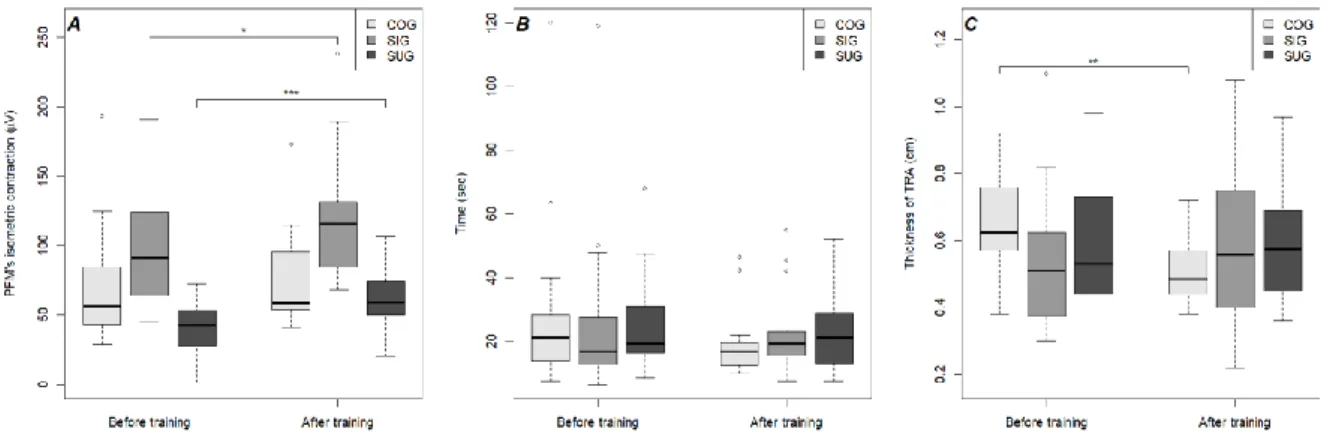
We present the data in Table 2 and Figure 2.The maximal isometric contraction of the PFM till fatigue improved significantly in both study groups; however, after cPFM-T, this increase was higher in the SUG. The tonic contraction of the PFM improved in the COG, but the improvement was not significant (p = 0.499) (Fig. 2. A). Although in both study groups, the holding time of the maximal isometric contraction of the PFM till fatigue and the thickness of the TRA during the maximal isometric contraction of the PFM till fatigue both increased, these changes were not significant (Fig. 2. B). The latter parameter decreased significantly in the COG (Fig. 2. C) (Table 2).
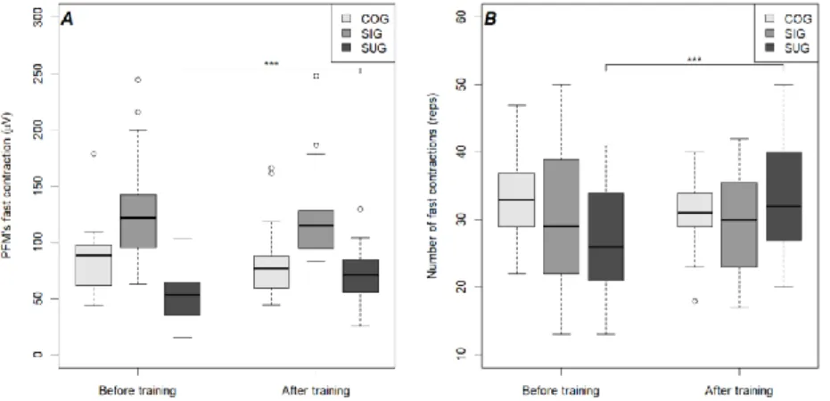
On the other hand, while the strength of the maximal fast contractions of the PFM within one minute increased significantly in the SUG, it decreased in the SIG and the COG; this decrease, however, was not significant (Fig. 3. A). In both study groups, the number of maximal fast contractions within a minute increased, but the increase was significant only in the SUG. This parameter decreased in the COG (Table 2, Fig. 3. B).
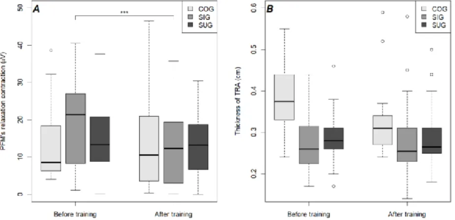
In both study groups, the values for relaxation of the PFM decreased but the decrease was significant only in the SIG (p = 0.011). This value increased in the COG (Fig. 4. A). The thickness of the TRA during relaxation of the PFM decreased in the SUG (p = 0.422) and the
1 2 3 4 5 6 7 8 9 10 11 12 13 14 15 16 17 18 19 20 21 22 23 24 25 26 27 28 29 30 31 32 33 34 35 36 37 38 39 40 41 42 43 44 45 46 47 48 49 50 51 52 53 54 55 56 57 58 59
COG (p = 0.209), but not in the SIG (p = 0.717). Neither of these changes were significant ( Fig. 4. B) ( Table 2).
Discussion
Our study demonstrated that an eight-week period of cPFM-T with forced exhalation, performed by young nulliparous women in both the supine posture and the sitting posture, using trunk muscle synergies, is effective in improving cases of incontinence.
Furthermore, the holding period, rapid reaction and resting function of the PFM—as well as the thickness of TRA—improved due to both the modification of body position and the exhalation technique. Improvements in the holding function of the PFM were significant in both the supine position and the sitting position, but improvements in the rapid reaction of the PFM were significant only in the supine position, while those in the resting function of the PFM were significant only in the sitting position. The thickness of the TRA improved in both the supine position and the sitting position.
Assessments of the PFM can be used to determine which structural or functional features are deficient, and to inform the design of subsequent training regimens to address these dysfunctions. A diversity of exercises, possibly tailored to the abilities of each woman, may be used and proposed training includes raising the number of repetitions of contractions [24]—a recommendation endorsed by our results.
Sapsford [23] advocated a new approach to the rehabilitation of urinary incontinence—motor relearning for diaphragmatic, deep abdominal muscles and the PFM rather than selective muscle strengthening.
Thompson et al. [25] suggested careful monitoring of Sapsford’s complex rehabilitation training, because abdominal muscles are more active than PFMs in symptomatic women. These results suggest that a coordinated approach involving both deep abdominal muscles and PFM is necessary.
1 2 3 4 5 6 7 8 9 10 11 12 13 14 15 16 17 18 19 20 21 22 23 24 25 26 27 28 29 30 31 32 33 34 35 36 37 38 39 40 41 42 43 44 45 46 47 48 49 50 51 52 53 54 55 56 57 58 59
The correct breathing technique is very important in PFM-T. The diaphragm is a respiratory muscle participating in the stabilisation of the lumbar spine by enhancing abdominal pressure [26], which in turn stabilises the lumbar spine. Hodges et al. [27] reported synergism between the diaphragm and the TRA. We also utilised this synergism by forced exhalation in our study.
Neumann and Gill [6] suggested that the activation of deep abdominal muscles is essential for an effective contraction of the PFM, because their continent participants were unable to contract the PFM effectively while maintaining relaxation of the TRA and the IO. Similarly, Madill and McLean [22] found that the patterns of abdominal muscle activity appear to occur due to voluntary PFM contractions in healthy continent women.
As a representation of vaginal closure force, the isometric contraction was considered to be greater in the supine than in the standing position. Subsequent studies, however, have determined that women are able to perform equally strong PFM contractions in either body position [7, 8, 11].
The PFM is a striated muscle, with two thirds of its fibres are type I (slow-twitch fibres), responsible for the resting tone of the levator muscle and one third of its fibres, type II (fast- twitch fibres), responsible for sudden, fast but powerful contractions. The activity of the slow fibres is necessary for the resting potential and the retention of urine and stool, while the fast fibres are responsible for resistance during sudden abdominal pressure [28]. We monitored the activity of these two fibre types (retention and rapid function) in our study with vsEMG measurements and trained both types in weaker and stronger PFM-T.
The fact that SUI happens most frequently in the upright position informed our study of the functional (static and dynamic) parameters occurring in everyday life. The holding function of the PFM is important for the patients—that is, they need to be able to get to the toilet in time before their urine starts leaking. The quality of breathing plays an important role in PFM training. It is necessary to teach participants the correct abdominal breathing technique which
1 2 3 4 5 6 7 8 9 10 11 12 13 14 15 16 17 18 19 20 21 22 23 24 25 26 27 28 29 30 31 32 33 34 35 36 37 38 39 40 41 42 43 44 45 46 47 48 49 50 51 52 53 54 55 56 57 58 59
activates the TRA. In our study, we activated the TRA and voluntary contraction of the PFM with strong exhalation techniques during both measurements and training. Using biofeedback (TRA ultrasound imaging and vsEMG curves of PFM) we visualised the co-contraction of the two muscles with the participants. Our vsEMG findings indicate that the static isometric contraction force of the PFM increased significantly in both study groups—and the holding time of static isometric contraction of the PFM increased slightly—with forced exhalation. In the COG, the static strength of the PFM increased slightly, but this increase was not significant and was characterised by a short retention time. The thickness of TRA during maximal isometric contraction of the PFM till fatigue improved in both training groups, but in neither group was this improvement significant. The COG claimed that this parameter decreased significantly because, during our study, they had spent much more time in a sitting position and were therefore in an enforced inactive lifestyle. Another study obtained similar results for musculus multifidus [29].
Hung et al. [14] 2010 investigated the effects of combining voluntary PFM- and deep abdominal muscle training in different body positions. The TRA activity was significantly greater in the sitting and standing positions than in the supine position. Chmielewska et al. [11] also reported that long-lasting contractions in the unsupported sitting position utilised the sensorimotor control system significantly compared with those in the supine position. However, in our study, cPFM-T in both the weaker PFM (in the SUG) and the stronger PFM (in the SIG) led to significant development in long-lasting contractions regardless of the body position of the measure.
Chmielewska et al. [11] measured the rapid activity of the PFM with vsEMG in the supine position and in the sitting position, finding no significant differences between the PFM average peak amplitudes in the investigated positions, while we found these values to be significantly improved in the supine position. In our study, only in the supine position did the strength and
1 2 3 4 5 6 7 8 9 10 11 12 13 14 15 16 17 18 19 20 21 22 23 24 25 26 27 28 29 30 31 32 33 34 35 36 37 38 39 40 41 42 43 44 45 46 47 48 49 50 51 52 53 54 55 56 57 58 59
repetition of dynamic fast contractions increase\ significantly; in the sitting position, changes in dynamic fast contractions were not significant. The fast activity of the PFM is responsible for resistance during sudden abdominal pressure. In the COG, dynamic strength and repetition decreased and did not manifest significant changes.
Capson et al. [8] and Chmielewska et al. [11] measured the relaxation tone of the PFM in the supine position and in the sitting position. They deduced that gravity forces increase the pressure on the PFM in the vertical position, increasing its tone and leading to a higher resting activity of the PFM in the sitting position than in the supine position. During our measures, the SIG participants found it easier to relax in the horizontal position. Based on our results, practising relaxation exercises can be beneficial not only in the horizontal position but also in the vertical position. Improvement in the relaxation ability of the PFM is also a beneficial result, because especially during urination, for a healthy, normal urination mechanism it is necessary to consciously relax the PFM. The relaxation state of the PFM improved significantly due to gravity forces only in the SIG.
We observed that the conditioning capabilities of the PFM improved in the SUG during the 8 week period of cPFM-T. The ability to sustain isometric contractions improved significantly, while holding time also improved, but this improvement was not significant. There was also a significant improvement in dynamic endurance and repetitions. Therefore we recommend a more intense strengthening of the TRA in the horizontal body position.
In the case of the SIG, the maximal isometric contraction of the PFM till fatigue also improved significantly, together with the holding time, but the latter improvement was not significant.
The dynamic endurance decreased and the number of repetitions improved slightly, but these changes were not significant. The relaxation state of the PFM improved significantly due to gravity forces. The resting tone of the TRA remained unchanged after 8 weeks. Thus, even more intense TRA activation is required in the vertical position, as a reliance on the enhanced
1 2 3 4 5 6 7 8 9 10 11 12 13 14 15 16 17 18 19 20 21 22 23 24 25 26 27 28 29 30 31 32 33 34 35 36 37 38 39 40 41 42 43 44 45 46 47 48 49 50 51 52 53 54 55 56 57 58 59
gravitational forces caused by the lumbopelvic posture or on the activated TRA induced by strong exhalation is insufficient to induce the necessary changes in the functioning of the TRA.
In the case of the COG, all parameters decreased—particularly the thickness of TRA during maximal isometric contraction of the PFM till fatigue, which decreased significantly—by reason of sedentary lifestyle.
We also established that young nulliparous women might also be affected by urinary leakage (27%) as confirmed by the results of Haslam et al. [1].
In the case of the SUG, both training exercises and measurements were performed in the supine position, whereas the SIG participants were measured in the lying position but performed all the exercises in the sitting position. Since SUI takes place most frequently in the vertical position, we should not measure and strengthen the static and dynamic functions of the PFM only in the supine position. We recommend that during cPFM-T, isometric and relaxation tasks should be performed both in the supine position and in the sitting position.
According to Sapsford et al. [5], the antigravity posture requires more intense PFM activity.
Furthermore, Chmielewska et al [11] reported a significant difference between the sustained 1- minute contraction of the PFM in the supine position and that in sitting position, while we found that the maximal isometric sustained contraction of PFM till fatigue significantly improved both in the supine position and in the sitting position.
Group training and individual training, according to recent research, are equally effective, and group training is more cost-effective without limitation[30]. Furthermore, due to differences in knowledge and behaviour, physiotherapist-guided training can help and motivate patients in persistent PFM-T [31].
Conclusions
1 2 3 4 5 6 7 8 9 10 11 12 13 14 15 16 17 18 19 20 21 22 23 24 25 26 27 28 29 30 31 32 33 34 35 36 37 38 39 40 41 42 43 44 45 46 47 48 49 50 51 52 53 54 55 56 57 58 59
TRA relaxion is easier in the horizontal position and strengthening is more effective in the horizontal body position. Physiotherapist-guided group training is more efficient.
The authors suggest that during PFM training the isometric tasks should be performed both in the supine position and in the sitting position, the dynamic endurance tasks of the PFM should be performed at the beginning of the training only in the supine position and more intensive strengthening of the TRA should be performed in the supine position to achieve maximal PFM contraction.
Abbreviations
ANOVA analysis of variance
ASIS anterior superior iliac spine
cPFM-T combined pelvic floor muscle training
COG control group
EO external oblique muscle
IO internal oblique muscle
PFM pelvic floor muscles
PFM-T pelvic floor muscle training
RA rectus abdominis muscle
SD standard deviation
SIG sitting group
SUI stress urinary incontinence
SUG supine group
TRA transverse abdominal muscle
RCT randomised controlled trial
1 2 3 4 5 6 7 8 9 10 11 12 13 14 15 16 17 18 19 20 21 22 23 24 25 26 27 28 29 30 31 32 33 34 35 36 37 38 39 40 41 42 43 44 45 46 47 48 49 50 51 52 53 54 55 56 57 58 59
UI urinary incontinence
US Ultrasound
vsEMG vaginal surface electromyography
Declarations
Ethics approval and consent to participate
The protocol and consent forms were approved by ethics committee of the Hungarian National Healthcare Service Center. The title of the ethics approval: Non-interventional study "Effect of PFM on Urinary Incontinence and Sexual Quality of Life". The number of the ethics approval:
019234/2014/OTIG Registered 07 April 2014.The name of the Ethics Committe: Scientific and Research Ethics Committee of the Health Science Council. The adress of the Ethics Committee:
Hungary, 1051 Budapest, Zrínyi street 3. Phone number: +36(1)8869329. E- mail: amd@ogyei.gov.hu. All participants provided written informed consent.
Consent for publication Not applicable.
Availability of data and materials
The datasets used and/or analysed during the current study are available from the corresponding author on reasonable request.
Competing interests
All financial and non-financial competing interests must be declared in this section.
Funding
1 2 3 4 5 6 7 8 9 10 11 12 13 14 15 16 17 18 19 20 21 22 23 24 25 26 27 28 29 30 31 32 33 34 35 36 37 38 39 40 41 42 43 44 45 46 47 48 49 50 51 52 53 54 55 56 57 58 59
The project has been supported by the European Union, co-financed by the European Social Fund. EFOP-3.6.1-16-2016-00008. The role of the funding body in the design of the study and collection, analysis, and interpretation of data and in writing the manuscript should be declared.
Authors’ contributions
Each author’s participation in the manuscript. All authors have read and approved the manuscript.
T M: project development, study design, ultrasound investigation, data collection, manuscript writing and editing
A D: project development, study design, manuscript revision M Sz: data collection, statistical analysis,
A S: study design, ultrasound investigation, manuscript writing, editing and revision J B: project development, manuscript revision
Acknowlegements Not applicable.
Author information Not applicable.
CONSORT guidelines
Our study adheres to CONSORT guidelines.
Legend to the tablesF
TABLE 1. Participant characteristics
1 2 3 4 5 6 7 8 9 10 11 12 13 14 15 16 17 18 19 20 21 22 23 24 25 26 27 28 29 30 31 32 33 34 35 36 37 38 39 40 41 42 43 44 45 46 47 48 49 50 51 52 53 54 55 56 57 58 59
TABLE 2. PFM activity while resting during tonic and phasic contractions in the COG, the SIG and the SUG, and the thickness of TRA during isometric contraction of PFMs until fatigue and in a relaxed state
Legend to the figures
FIGURE 1. Flow chart of study participants
FIGURE 2. (A) vsEMG values of the maximal isometric contraction of PFMs till fatigue; (B) holding time of maximal isometric contraction of PFMs till fatigue; (C) the thickness of TRA during maximal isometric contraction of PFMs till fatigue.
FIGURE 3. (A) EMG values of strength of maximal fast contractions within 1 min. (B) Number of maximal fast contractions within 1 min.
FIGURE 4. (A) Relaxation values for PFMs. (B) Thickness of TRA during relaxation of PFMs.
References
1. Haslam J The prevalence of stress urinary incontinence in women. Nurs Times.
2004;18;100(20):71–73.
2. Kegel AH Progressive resistance exercise in the functional restoration of the perineal muscles. Am J Obstet Gynecol. 1948;56(2):238–248.
3. Dumoulin C, Hay-Smith J Pelvic floor muscle training versus no treatment or inactive control treatments, for urinary incontinence in women. Cochrane Database Syst Rev.
2010;20;(1):CD005654.
4. Thüroff WJ, Abrams P, Andersson K-E, Artibani W, Chapple RC, Darke JM, Hampel C, Neisuis A, Schröder A, Tubaro A EAU Guidelines on urinary incontinence. Eur Urol.
2011;59:387–400.
1 2 3 4 5 6 7 8 9 10 11 12 13 14 15 16 17 18 19 20 21 22 23 24 25 26 27 28 29 30 31 32 33 34 35 36 37 38 39 40 41 42 43 44 45 46 47 48 49 50 51 52 53 54 55 56 57 58 59
5. Sapsford RR The pelvic floor. A clinical model for function and rehabilitation.
Physiotherapy. 2001;87:620–30.
6. Neumann P, Gill V Pelvic floor and abdominal muscle interaction: EMG activity and intra-abdominal pressure. Int Urogynecol J Pelvic Floor Dysfunct. 2002; 13(2):125–
132.
7. Madill SJ, McLean L Quantification of abdominal and pelvic floor muscle synergies in response to voluntary pelvic floor muscle contractions. J Electromyogr Kinesiol.
2008;18(6):955–964.
8. Capson AC, Nashed J, Mclean L The role of lumbopelvic posture in pelvic floor muscle activation in continent women. J Electromyogr Kinesiol. 2011;21(1)166–177.
9. Sapsford RR, Richardson CA, Stanton WR Sitting posture affects pelvic floor muscle activity in parous women: an observational study. Aust J Physiother. 2006;52(3):219–
222.
10. Sapsford RR, Richardson CA, Maher CF, Hodges PW Pelvic floor muscle activity in different sitting postures in continent and incontinent women. Arch Phys Med Rehabil.
2008;89(9):1741–1747.
11. Chmielewska D, Stania M, Sobota G, Kwasna K, Blaszczak E, Taradaj J, Juras G Impact of different body positions on bioelectrical activity of the pelvic floor muscles in nulliparous continet women. BioMed Res Int. 2015; doi: 10.1155/2015/905897.
12. Bø K, Mørkved S, Frawley H, Sherburn M Evidence for benefit of transversus abdominis training alone or in combination with pelvic floor muscle training to treat female urinary incontinence: a systematic review. Neurourol Urodyn. 2009;28:368–
373.
1 2 3 4 5 6 7 8 9 10 11 12 13 14 15 16 17 18 19 20 21 22 23 24 25 26 27 28 29 30 31 32 33 34 35 36 37 38 39 40 41 42 43 44 45 46 47 48 49 50 51 52 53 54 55 56 57 58 59
13. Nie XF, Ouyang YQ, Wang L, Redding SR A meta-analysis of pelvic floor muscle training for the treatment of urinary incontinence. Int J Gynaecol Obstet.
2017;138(3):250-255.
14. Hung H-C, Hsiao S-M, Chih, S-Y, Lin H-H, Tsauo J-Y An alternative intervention for urinary incontinence: Retraining diaphragmatic, deep abdominal and pelvic floor muscle function coordinated function. Man Ther. 2010;15:273–279.
15. Sriboonreung T, Wongtra-ngan S, Eungpinichpong W, Laopaiboon M Effectiveness of pelvic floor muscle training in incontinent women at Maharaj Nakorn Chiang Mai Hospital: a randomized controlled trial. J Med Assoc Thai. 2011;94:1–7.
16. Bø K, Finckenhagen HB Is there any difference in measurement of pelvic floor muscle strength in supine and standing position? Acta Obstet Gynecol Scand. 2003;82:1120–
1124.
17. Da Roza T, de Araujo MP, Viana R, Viana S, Jorge RN, Bø K, Mascarenhas T Pelvic floor muscle training to improve urinary incontinence in young, nulliparous sport students: a pilot study. Int Urogynecol J. 2012;23(8):1069–1073.
18. Bø K, Kvarstein B, Hagen R, Larsen S Pelvic floor muscle exercise for the treatment of female stress urinary incontinence: Validity of vaginal pressure measurements of pelvic floor muscle strength and the necessity of supplementary methods for control of correct contraction. Neurourol Urodyn. 1990;9:479–487.
19. Whittaker LJ, Warner BM, Stokes M Comparison of the sonographic features of the abdominal wall muscles and connective tissues in individuals with and without lumbopelvic pain. J Orthop Sports Phys Ther. 2013;43(1):11–19.
20. Carrière B Sensory awareness- feeling the pelvic floor. In: Carrière B editor. Fitness for the pelvic floor. 1st ed. Stuttgart-New York: Georg Thieme Verlag; 2002. p 24–30.
1 2 3 4 5 6 7 8 9 10 11 12 13 14 15 16 17 18 19 20 21 22 23 24 25 26 27 28 29 30 31 32 33 34 35 36 37 38 39 40 41 42 43 44 45 46 47 48 49 50 51 52 53 54 55 56 57 58 59
21. Shamsi M, Mirzaei M, HameiRad M Comparison of muscle activation imbalance following core stability or general exercises in nonspecific low back pain: a quasi- randomized controlled trial. BMC Sports Sci Med Rehabil. 2020;12:24 doi:10.1186/s13102-020-00173-0- eCollection 2020.
22. Madill SJ, McLean L Relationship between abdominal and pelvic floor muscle activation and intravaginal pressure during pelvis floor muscle contraction in healthy continent women. Neurol Urodyn. 2006;25:722–730.
23. Sapsford RR Rehabilitation of pelvic floor muscles utilizing trunk stabilization. Man Ther. 2004;9:3–12.
24. Dumoulin C, Glazener C, Jenkinson D Determining the pelvic floor muscle training regimen for women with stress urinary incontinence. Neurourol Urodyn. 2011;30:746–
753.
25. Thompson JA, O’Sullivan PB, Briffa NK, Neumann P Difference in muscle activation patterns during pelvic floor muscle contraction and valsalva manouevre. Neurourol Urodyn. 2006;25:148–155.
26. Kapandji IA The lumbar spine In: Kapandji IA editor. The physiology of the joints trunk and the vertebral column. London: Churchill Livingstone; 2008. p 84–141.
27. Hodges PW, Butler JE, Mckenzie DK, Gandevia SC Contraction of the human diaphragm during rapid postural adjustments. J Physiol. 1997;505(Pt 2):539–548.
28. Gosling JA, Dixon JS, Critchley HOD, Thompson SA A comparative study of the human external sphincter and periurethral levator ani muscle. Br J Urol. 1981;53:35-41.
29. Finta R, Nagy E, Bender T The effect of diaphragm training on lumbar stabilizer muscles: a new concept for improving segmental stability in the case of low back pain.
Pain Research. 2018. 11:3031–3045.doi: 10.2147/JPR.S181610.
1 2 3 4 5 6 7 8 9 10 11 12 13 14 15 16 17 18 19 20 21 22 23 24 25 26 27 28 29 30 31 32 33 34 35 36 37 38 39 40 41 42 43 44 45 46 47 48 49 50 51 52 53 54 55 56 57 58 59
30. Dumoulin C, Morin M, Mayrand MH, Tousignant M, Abrahamowicz M Group physiotherapy compared to individual physiotherapy to treat urinary incontinence in aging women: study protocol for randomized controlled trial. Trials. 2017;18(1):544.
31. Hay-Smith EJ, McClurg D, Frawley H, Dean SG Exercise adherence: integrating theory, evidence and behavior change techniques. Physiotherapy. 2016;102(1):7–9.
Additional file 1 :Questionnaire
Table1. Participant Characteristics
Age (years) 21.00 [20.00–22.00] (SUG)
21.00 [20.00–22.00] (SIG) 21.50 [21.00–23.00] (COG) Number of participants (positions) 22 (SUG)
19 (SIG) 14 (COG)
BMI (kg/m2) 21.85 [20.77–22.95] (SUG)
21.30 [20.00–22.20] (SIG) 22.00 [19.77–23.32] (COG) Number of participants doing regular
physical activity more than twice a week
9 (SUG) 9 (SIG) 5 (COG)
BMI SUM
(n = 55)
SUG (n = 22)
SIG (n = 19)
COG (n = 14)
Normal BMI 18-24 45 18 16 11
Overweight BMI 25-29 9 3 3 3
1 2 3 4 5 6 7 8 9 10 11 12 13 14 15 16 17 18 19 20 21 22 23 24 25 26 27 28 29 30 31 32 33 34 35 36 37 38 39 40 41 42 43 44 45 46 47 48 49 50 51 52 53 54 55 56 57 58 59