THESIS
BÓNAI ANDRÁS
UNIVERSITY OF KAPOSVÁR FACULTY OF AGRICULTURAL AND
ENVIRONMENTAL SCIENCES
2014
UNIVERSITY OF KAPOSVÁR
FACULTY OF AGRICULTURAL AND ENVIRONMENTAL SCIENCES
Institute of Physiology, Biochemistry and Animal Health
Head of Doctorate School:
Dr. Kovács Melinda
Corresponding Member of the Hungarian Academy of Sciences
Supervisor:
Dr. Kovács Melinda
Corresponding Member of the Hungarian Academy of Sciences
Co-Supervisor:
Dr. Sylvie Combes
PhD, Senior researcher of INRA-TANDEM
EXAMINATION OF THE EFFECTS OF CERTAIN FACTORS INFLUENCING CAECAL FERMENTATION IN
RABBITS
Written by:
BÓNAI ANDRÁS
KAPOSVÁR 2014
DOI: 10.17166/KE.2014.009
TABLE OF CONTENTS
1. INTRODUCTION 1
1.1. Antecedents 1
1.2. Objectives 4
2. REVIEW OF THE LITERATURE 5
2.1. Characteristics of the digestive tract and digestion of
rabbits 5
2.1.1. Microbial balance in the intestine 6 2.1.2. Characteristics of the caecal microbiota 8 2.1.3. Metabolic products of the caecal microbiota 11
2.1.4. Caecotrophy 12
2.2. Anatomical and digestive changes associated
with growth in rabbits 13 2.2.1. Growth and enzyme activity 13 2.2.2. Development of the caecal microbiota 15 2.3. Eubiosis and dysbiosis in the intestine 17 2.3.1. Some consequences of antibiotic use 20 2.3.2. Possibilities for the replacement of antibiotics 21
2.3.2.1. Probiotics 22
2.3.2.1.1. The application of Bacillus cereus 25
2.3.2.2. Prebiotics 27
2.3.2.2.1. Inulin 30
3. MATERIAL AND METHODS 33
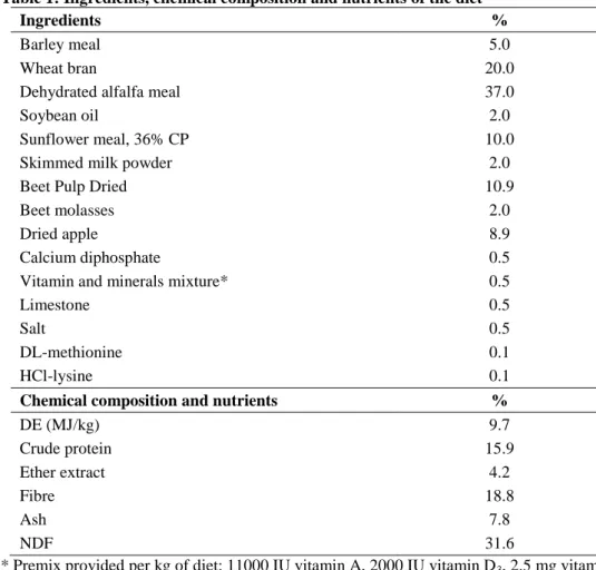
3.1. Effect of different weaning ages… 33 3.1.1. Experimental animals, housing and nutrition 33
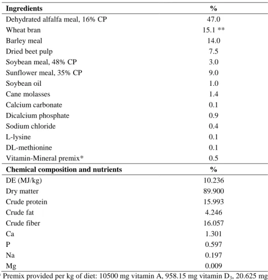
3.1.2. Samplings 35 3.2. Effect of Bacillus cereus var. toyoi… 36 3.2.1. Experimental animals, housing and nutrition 36
3.2.2. Samplings 38
3.3. Effect of inulin supplementation… 39 3.3.1. Experimental animals, housing and nutrition 39
3.3.2. Samplings 40
3.4. In vitro metabolism of inulin by rabbit microbiota 42
3.4.1. Experimental design 42
3.5. Laboratory analyses 43
3.5.1. Determination of feeds’ chemical composition 43 3.5.2. Determination of the pH-values 43 3.5.3. Microbiological culturing technics 43 3.5.4. Measurement of fibrolytic activity in caecal cont. 44 3.5.5. Determination of volatile fatty acids concentration 44 3.5.6. Molecular genetically investigation 45
3.5.6.1. DNA extraction 45
3.5.6.2. Characterisation of bacterial community
from caecal content samples 45 3.5.6.3. Determination of Bacteroides copy number using real time-PCR technology 46
3.6. Statistical analyses 48
3.6.1. Statistical analyses of molecular technics 49
4. RESULTS AND DISCUSSION 50
4.1. Effect of different weaning ages… 50 4.1.1. Body weight, milk and feed consumption 50 4.1.2. Age related development of organs 52
4.1.3. The pH-values, composition of the caecal
microbiota and short chain fatty acid content 56 4.2. Effect of Bacillus cereus var. toyoi… 61
4.2.1. Growth and health status 62
4.2.2. The pH-values, composition of the caecal
microbiota and volatile fatty acid content 63 4.3. Effect of inulin supplementation… 68 4.3.1. Live weight, feed intake and health status 68 4.3.2. Effect of inulin or medication on caecal microbiota. 70 4.3.3. DNA based qualitative analyses 75 4.3.4. DNA based quantitative analyses 79 4.4. In vitro metabolism of inulin by rabbit microbiota 81
4.5. General discussion 85
5. CONCLUSIONS AND RECOMMENDATIONS 87
6. NEW SCIENTIFIC RESULTS 91
7. SUMMARY 93
8. ACKNOWLEDGEMENTS 97
9. REFERENCES 98
10. SCIENTIFIC PAPERS AND LECTURES ON THE
SUBJECT OF THE DISSERTATION 117
10.1. Peer-reviewed papers published in foreign scientific
journals 117
10.2. Peer-reviewed paper published in Hungarian scientific
journal 117
10.3. Proceeding published in foreign language 118 10.4. Proceeding published in Hungarian language 119
11. OTHER PUBLICATIONS 120
11.1. Peer-reviewed papers published in foreign scientific
journals 120
11.2. Peer-reviewed paper published in Hungarian scientific
journal 120
11.3. Proceeding published in Hungarian language 120
12. CURRICULUM VITAE 121
LIST OF ABBREVIATIONS
ANOVA Analyses of Variance ADF Acid Detergent Fiber ADL Acid Detergent Lignin
BW Body Weight
CE Capillary Electrophoresis
CP Crude Protein
CFU Colony Forming Unit
d Day
DE Digestible Energy
DM Dry Matter
DNA Deoxyribonucleic Acid
FAO Food and Agriculture Organization GI Gastrointestinal
GLM General Lineral Model
h Hour
HRI Health Risk Index
ISO International Organization for Standardization LSD Least Significant Difference
NDF Neutral Detergent Fiber
nMDS non-metric Multi Dimensional Scaling PCA Principal Component Analysis
RDA Redundancy Analysis
RT - PCR Real Time - Polymerase Chain Reaction SSCP Single - Strand Conformation Polymorphism SCFA Short Chain Fatty Acid
1 1. INTRODUCTION
1.1. Antecedents
During the past 50 years, rabbit meat production of the world has quadrupled. In 2010, global rabbit meat production was approximately 1.7 million tons, and this production volume showed the following distribution by continent: Asia 48%, Europe 30%, America 17%, and Africa 5%.
According to data from the FAO (2012), the biggest rabbit meat producing countries of the world were China (668,980 tons), Italy (255,420 tons) and Venezuela (254,305 tons).
Based on data of the Hungarian Rabbit Product Council, in 1990 the quantity of rabbit meat production in Hungary was 33,468 tons (liveweight), of which 16,763 tons were exported. Over the past 20 years, both values have decreased to about one-third. Thus, in 2011 the quantity of rabbit meat production in Hungary was 10,300 tons (liveweight), of which 5090 tons were exported. Hungary has about 60–65 large rabbit farms with approximately 85000 does. At the two rabbit slaughterhouses (Baja and Lajosmizse) about 70–75 thousand rabbits are slaughtered weekly, in approximately equal proportions (Jurasko, 2013).
The rabbit sector of Hungary is unique in the world in that almost the entire quantity of meat from rabbit production for slaughter (the price of rabbit meat was 420–450 HUF/kg in 2012) is exported. The most important export markets of Hungary for rabbit meat are Italy, Switzerland and Germany.
The advantages to grow rabbits are as follow: rabbits have a high fertility rate with a rapid rate of growth, high feed conversion efficiency and early marketing age and high muscle-bone ratios; in addition, they require a small land area (Fernandez-Espla and O’Neil, 1993). The significance of
2
broiler rabbit production is increasing for both healthy nutrition of humans and economic reasons.
Rabbit meat is considered to be one of the healthiest meats because of its easy digestibility and excellent dietetic properties, e. g. high protein (20–
21%) and unsaturated fatty acids (oleic and linoleic acid; 60% of all fatty acids), potassium, phosphorus and magnesium concentrations and also low fat, cholesterol and sodium contents (Hermida et al., 2006). The rabbit meat is useful in human dietetics and recommended for consumption, e.g. to persons suffering of cardiovascular diseases (Hu and Willett, 2002).
According to Dalle Zotte (2002), the energy value of rabbit meat (427–849 kJ/100g of fresh meat) is similar to the values of commonly consumed meats, such as pig (418-1121 kJ/100g), beef (473-854 kJ/100g) and chicken (406-808 kJ/100g) (Dalle Zotte, 2002).
Diseases affecting the digestive tract cause major problems during the growing period of broiler rabbits. These diseases are often severe and may be fatal. About one-fourth of all mortality takes place in the period around weaning (at 2–6 weeks of age). This gives cause for concern from the animal welfare point of view and causes a substantial loss of profits to the rabbit growers. In order to minimise the losses, antibiotics are used on a wide scale, which results in a major food safety and human health risk.
From the middle of the past century, the development of intensive animal breeding made it necessary to use antibiotics as growth promoters.
Their use resulted in the improvement of many production parameters;
therefore, from the 1970s the use of antibiotic growth promoters became widespread all over the world. Besides its benefits, antibiotic use also has detrimental consequences. Methicillin-resistant Staphylococcus aureus (MRSA) is a significant infection-causing bacterium, since it gives rise to potentially life-threatening infections and also shows resistance to treatment
3
with the usual antibiotics. Hospital-acquired MRSA infections are associated with increased morbidity and mortality (Engemann et al., 2003). The incidence rate of MRSA is less than 1% in the Northern European countries while it exceeds 40% in the Southern and Western European countries. The proportion of human MRSA infections is decreasing in some countries but increasing in others (Anonymous, 2008).
Due to the risk that potentially pathogenic microbes develop resistance to antibiotics, the European Union has restricted the possibilities of using antibiotics. Antibiotics authorised for use in human medicine have been separated from those permitted for use in veterinary medicine. Finally, with effect from 1 January 2006, the growth-promoting use of all antibiotics applied in animal production, with the exception of anticoccidials, was banned on the territory of the European Union. This has resulted in an increased demand for alternative growth promoters (Ancsin et al., 2008).
This regulation applies to the animal production sector of Hungary as well, as Hungary has been a member of the European Union since 2004. Due to the restriction of antibiotic use in animal production, a decrease of the previously established yields has to be reckoned with, which has major economic implications. Rabbit producers have to face smaller profits due to the lower production yields and higher occurrence of deseases, and researchers have to find new solutions suitable for replacing the use of antibiotics. One way to this latter objective, it would be important to get to know the bacteria living in the digestive tract of rabbits and their metabolic processes as thoroughly as possible.
In association with a series of experiments underway at the Department of Physiology and Animal Hygiene, I studied the problems related to the development of diseases in the period around weaning in domestic rabbits, and looked for solutions suitable for replacing the use of antibiotics during
4
the growing period in rabbits. I determined the effect of weaning age on the implatation of digestive microbiota, and conducted experiments to determine the effects of probiotics and prebiotics added to the diet.
Our results may facilitate the reduction of antibiotic use during rabbit growing and, eventually, contribute to the full replacement of antibiotics by alternative solutions in rabbit production.
1.2. Objectives
1.) Determination of the effect of different weaning ages on the growth and certain parameters of digestion in rabbits.
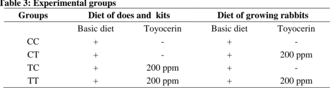
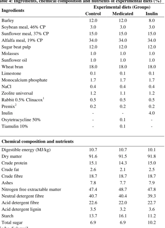
2.) Determination of the effect caused by the probiotic Bacillus cereus var. toyoi and by inulin, a compound having prebiotic effect, on the composition of the caecal microbiota and on caecal fermentation activity in the period around weaning in rabbits.
3.) Elaboration of a series of objective and complex biomonitoring methods to characterise the caecal microbiota and caecal fermentation processes of rabbits, and the use of these methods in the above-mentioned studies.
5 2. REVIEW OF THE LITERATURE
2.1. Characteristics of the digestive tract and digestion of rabbits During evolution, the digestive tract of the domestic rabbit has adapted to the herbivorous lifestyle. As a consequence, the volume of the stomach and caecum has increased considerably compare to the upper digestive tract.
The functioning of the rabbit’s upper digestive tract globally is the same as that of other monogastric domestic mammals. The speciality of the rabbit (and of Lagomorpha in general) lies in the dual function of the proximal colon (Gidenne and Fortun-Lamothe, 2002). Bacteria stimulate the immune system of rabbit, and bacteria enzymes ensure the degradation of forage fibers.
The rabbit stomach has a weak muscular layer and is always filled partially. Soft faeces is stored in the fundic region of the stomach after caecotrophy. The pH value depends on the region of stomach, in the fundus and in the cardiac-pyloric region it is about 1 and 5, respectively (Chamorro et al., 2007; Gomez-Conde et al., 2009).
In an adult rabbit the small intestine is approximately 3 m long, where the secretion of bile, digestive enzymes and buffers occurs. The pH value of the small intestine is close to 7 (Nicodemus et al., 2002).
Digestibility at the end of the ileum accounts for 80-100% of the total dietary amino acid and starch digestibility (Carabano et al., 2009).
The caecum is the largest digestive compartment of the rabbit (40%
of the whole digestive tract). It plays a key role in the digestion as a major site of fermentation (e.g. fibre degradation). For instance, in adult rabbits, SCFA absorption could represent 30% of the basal metabolism (Parker, 1976; Marty and Vernay, 1984).
6
The caecum is characterised by a weak muscular layer and content with a dry matter of 200 g/kg. The caecal contents are slightly acidic (pH 5.4–6.8) (Garcia et al, 2002). The capacity of the caecum is approximately 49 % of the total capacity of the digestive tract (Portsmouth, 1977). There is evidence that the caecal microbial activity plays a key role in the digestion and health of the rabbit (Gidenne, 1997).
The colon can be divided into the proximal and distal colon (approx.
35 and 80 cm long, respectively). The rear section of the distal colon acts as a pacemaker for the colon during the phase of hard faeces formation (Snipes et al., 1982).
2.1.1. Microbial balance in the intestine
The normal intestinal microbiota has characteristic composition which, however, differs by animal species, age and intestinal segment. It is constituted approximately by 400–500 species, in a total microbial count (log10 14 CFU/g chyme) in the intestinal tract of animals (Gedek, 1991). The digestive tract of healthy animals is colonised only by microbes typical of the given animal species and age. Between the host and the microflora a regulated state of equilibrium, called eubiosis, is developed (Szigeti, 1991).
Gouet and Fonty (1979) showed that 75% of the young rabbits do not harbour any flora in the stomach during the first 15 days of life, in spite of the existence of a slightly acidic pH (4.5 to 5.0). Barrier function of stomach is realized by fermentation end-product of Lactobacillus species. Number of pathogenic bacteria originated from feed is reduced by antiseptical lactic acid.
The number of bacteria is further reduced due to octanoic and decanoic acids of the rabbit doe milk in the gastrointestinal tract during the
7
suckling period (Canas-Rodrigues and Smith, 1966; Coloe et al., 1984;
Marounek et al., 1999, 2003).
Around the 3rd week of age, young rabbits start to consume solid feed, and caecotrophy begins as well. After weaning, the level of the gastric flora is log10 4 - 6 CFU/g of digesta (Raibaud et al., 1966).
Colonisation of the small intestine takes place faster than that of the stomach, and already in the first week of life the bacterial count in the small intestine is approx. 100 times higher than that in the stomach. Facultative anaerobic bacteria are still detectable in the small intestine before weaning but usually disappear from it thereafter (Gout and Fonty, 1979).
In the first week of life, the quantity of the caecal microbiota is log10
7 - 9 CFU/g of caecal content. After weaning, the count of facultative anaerobic bacteria decreases to log10 2 - 4 CFU/g of content (Ducluzeau, 1983). The number of obligate anaerobic bacteria becomes stabilised on the level of log10 9 - 10 CFU/g of digesta (Zomborszky-Kovács et al., 2000).
Those bacteria can participate in the constitution of the intestinal microflora which can attach to the surface of epithelial cells, and which are multiplicated at a faster rate than are removed by intestinal peristalsis (Ewing and Cole, 1994). One of the most important beneficial effects of the indigenous gastrointestinal microflora is to make the exogenous pathogenic bacteria more difficult to colonise the digestive tract (Berg, 1996).
Competitive exclusion is a complex inhibition process, which means that a non-pathogenic species predominates over a pathogenic one. Competition can be realised by differences in growth on a specific substrate, efficiency of mucosa colonisation, and production of substances inhibiting pathogen development (Hampson et al., 2001). The surface binding sites of intestinal epithelial cells can also be occupied by certain natural receptor analogues (e.g. oligosaccharides). In this way, enteropathogenic bacteria will not be
8
able to attach to the surface of intestinal epithelial cells and thus become members of the transient microbiota (Kelly and Tucker, 2005).
Bacteria can inhibit the growth of their competitors also by producing antimicrobial substances (Guarner and Malagelada, 2003).
Acting as antigen, the live and the already perished microbes stimulate the mucosal immunity of the digestive tract (GALT) and elicit enhanced antibody (sIgA) synthesis as well as T and B cell activity (Ewing and Cole, 1994).
2.1.2. Characteristics of the caecal microbiota
Bacteroides bacteria can be found in the caecal microbiota of calves, lambs and piglets. Their number decreases with age, and in adult animals they can no longer be detected (Smith and Crabb, 1961).
In caecum of newborn colt, there are detectable species of the Clostridium-, Enterococcus-, Enterobacter-, Lactobacillus-, Streptococcus-, Staphylococcus genus (Jullian et al., 1996). Initially, members of Enterococcus- and Enterobacteriaceae genus are dominant (Yuyama et al., 2004). Members of Bacteroidaceae, Clostridium, Enterobacteriaceae (log10
8.3 CFU/g) and Enterococcus (log10 7.7 CFU/g) were founded in faeces of three day old colt (Sakaitani et al., 1999).
Genus of Bacteroidaceae- and Lactobacillus is dominated in colt faeces, numbered log10 8.0 CFU/g digesta, from the 6th week of life. Number of Enterobacter-, Enterococcus-, and Staphylococcus bacteria is around log10 5.0 CFU/g (Jullian et al., 1996; Sakaitani et al., 1999).
In rabbits the bacteria number of Bacteroides is stabilised on a high level and they become constituents of the main microflora (Smith and Crabb, 1961). This has been confirmed by the results of Gouet and Fonty (1979), Boulahrouf et al. (1991) and Carabano et al. (2006) as well. However these
9
results have been queried with the introduction of molecular approach methodologies (Abecia et al., 2005; Combes et al., 2011).
According to Fuller and Lev (1964), caecal microbiota constituent characteristic of other domestic animal species, among others Bifidobacteria, can not be found in the rabbit caecum. Bornside and Cohn (1965), as well as Smith (1965) described a steady composition of the microbiota present in the digestive tract of adult domestic rabbits. This is characterised by the almost complete absence of lactobacilli, streptococci and Escherichia coli strains abundantly present in the gastrointestinal microflora of other species (Gouet and Fonty, 1973).
Although more than 74 strains of anaerobic bacteria have been isolated from the caecal mucosa of domestic rabbits, Bacteroides bacteria were regarded as the main constituents of the microbiota for a long time (Cheeke, 1987; Straw, 1988).
The cellulolytic bacterial community appears around 2 weeks after birth. This community was found to be dominated by Eubacterium cellulosolvens and Bacteroides spp. (Boulahrouf et al., 1991).
According to Gidenne (1997), the two main characteristics of the caecal microflora in domestic rabbits are its slow development and stabilization, simple composition. The latter means the dominance of Gram- negative, non-spore-forming, strictly anaerobic, rod-shaped bacteria.
According to Harcourt-Brown (2002), a typical feature of the rabbit intestinal microflora is the very simple composition of the facultative anaerobic flora. Up to the 14th day of life streptococci are dominant, but their number rapidly decreases after weaning. Enterobacteria can hardly be detected at all, and lactobacilli are almost entirely absent as well.
Microbiological culture methods can be used successfully for only a few groups of bacteria (Rastall and Maitin, 2002). According to Suau et al.
10
(1999), only 20–40% of bacteria living in the caecum can be cultured.
Thanks to the methodological developments involving the widely spreading genetic analyses and the experiments using such methods, the scope of information available on the caecal microbiota of rabbits is expanding fast.
By using dot-blot hybridisation with 16S rRNA targeted oligonucleotides probes, Bennegadi et al. (2003) stated that bacteria and archaea respectively represent 73% and 22% of the total microbial communities in the caecum at weaning, however this result was not confirmed nor by counting nor by methanogenic activity. Bacteria of the Flexibacter – Cytophaga – Bacteroidetes group and four cellulolytic bacterial strains (R. flavefaciens, R.
albus, F. succinogenes, F. intestinalis) were found.
Sequence and cluster analysis showed that more than 80% of the bacteria inhabiting the rabbit caecum are unknown, and bacteria belonging to the Prevotella/Bacteroidetes groups were not found either (Abecia et al., 2005). Subsequent, more accurate microbial genomic studies demonstrated that bacteria belonging to the Firmicutes (94%) and Bacteroidetes (4%) groups could be found in the bacterial community (Monteils et al., 2008).
The maximal density of the Bacteroides-Prevotella group was reached 3 weeks after birth (log10 10–11), but it decreased significantly thereafter (log10 9–10) (Combes et al., 2011).
Michelland et al. (2010) studied the bacterial ecosystem of the rabbit caecum by Capillary Electrophoresis Single-Strand Conformation Polymorphism (CE-SSCP) technique, and found that there were no marked individual differences among rabbits in terms of the bacterial community present in the caecal content, and that the bacterial community of the caecal content was more similar to that of the soft than that of the hard faeces. At the same time, the composition of the bacterial community and the amount
11
of bacteria were markedly influenced by changes in dietary fibre content (Michelland et al., 2011).
2.1.3. Metabolic products of the caecal microbiota
Although rabbits do not possess enzymes necessary for degrading fibre, their caecum contains large numbers of fibrolytic bacteria that adhere to the surface of dietary fibre substances and start to brake down them with the help of their enzymes. Cellulose, pectin, xylan and amylose molecules are broken down into their components. Released monosaccharide molecules are taken up by the microbes and used during fermentation. The end- products of this process are short-chain fatty acids (SCFA), which cannot be degraded further under anaerobic conditions.
Ammonia is used by bacteria, in combination with carbon chain produced from carbohydrate fermentation, to synthesise new amino acids for bacterial growth (Van Soest, 1994).
The caecal metabolism of nutrients is similar in rabbits to that can be shown in other herbivores, but the SCFA pattern exhibits some differences in rabbits, namely a predominance of acetate, followed by butyrate and then by propionate (Gidenne et al., 2008). In an average, the relative proportions of individual volatile fatty acids within the total volatile fatty acids are as follow: 75–85% acetic acid, 6–10% propionic acid and 8–17% butyric acid (Fortun-Lamothe and Gidenne, 2006; Combes et al., 2011). The propionic acid to butyric acid ratio decreases below 1 by 25–30 days of age (Zomborszky-Kovács et al., 2000). The ammonia concentration of the caecal content slightly decreases with age (Gidenne and Fortun-Lamothe, 2002).
Feed composition, such as dietary fibre, interacts with the digestive health of the young animal (Montagne et al., 2003) and particularly of the
12
growing rabbit after weaning (Gidenne, 2003). The ratio of butyric acid usually increases substantially with the decreasing ratio of fibre and starch (Gidenne, 1997). Higher ratios of digestible fibre will lead to a larger volume of caecal content. Microbial activity results in the production of more volatile fatty acids and less ammonia, which lower the pH of the caecal digesta between 15 and 42 days of age (Gidenne and Fortun- Lamothe, 2002).
From 15 to 50 days of age, the pH of the chyme decreases from 6.8 to 5.6 (Padilha et al., 1995). The pH of the caecal digesta decreases from 6.2 at 28 days of age to 5.8 at 70 days of age (Kimse et al., 2009; Combes et al., 2011), though to evaluate these results, it is necessary to consider the effect of diet composition, as well.
2.1.4. Caecotrophy
Young rabbits start to eat a notable amount of dry feed at 3 weeks of age (Lebas, 1997), in connection with which caecotrophy commences between 22 and 28 days of age (Orengo and Gidenne, 2007).
The caecum is starting to be filled by digesta and microbiota from 3 to 7 weeks of age, and its contents reach a peak of about 0.06 part to total body weight at 7–9 weeks of age (Padilha et al., 1995).
The co-ordinated operation of caecum and colon results in fractionation of the intestinal content. During this process, fermentable particles less than 0.1 mm in size with higher nutrient content become concentrated in the caecum while the less valuable feed particles exceeding 0.3 mm in size accumulate in the colon. Rabbits excrete the larger particles definitively in the form of hard faecal pellets (Lebas et al., 1997).
13
The motility of the basis of caecum and the proximal colon decreases during the formation of soft faeces (Ruckebush and Hornicke, 1977), because of the effect of prostaglandin F2α (Pairet et al., 1986).
Soft faecal pellets (small pellets of 5 mm size) covered by a mucous envelope from the proximal colon are taken directly from the anus and swallowed by the rabbit. They are stored (for 3–6 hours) in the fundic region of the stomach till digestion and absorption (Gidenne and Poncet, 1985).
The caecotroph is excreted according to a circadian rhythm.
Monophasic pattern of soft faeces excretion was showed by rabbits between 8:00 and 17:00. During the caecotrophy period, lasting from 7:00 to 9:00, there is an absence of hard faeces excretion and the feed intake is low (Carabano and Merino, 1996).
After weaning, soft faeces production linearly increases with age, reaching a maximum (25 g DM/day) at 63–77 days of age. Thereafter, this value is stabilised around 20 g DM/day (Gidenne and Lebas, 1987).
Some data on the chemical composition of soft faeces, expressed as average values, are as follow: dry matter (340 g/kg), crude protein (300 g/kg DM), crude fibre (180 g/kg DM). Compared to soft faeces, the hard faeces has higher values of dry matter (470 g/kg) and crude fibre (300 g/kg), however, its crude protein content is lower (470 g/kg) (Carabano et al., 1997). The soft faeces contains essential amino acids (e.g. lysine and threonine) (Garcia et al., 2004). The main benefit of caecotrophy is the utilisation of proteins, fatty acids and vitamins of bacterial origin, and the decrease of metabolic losses (Meyer et al., 2010).
14
2.2. Anatomical and digestive changes associated with growth in rabbits
2.2.1. Growth and enzyme activity
The anatomy of the digestive tract is stabilised by 9 weeks of age.
Between 3 and 11 weeks of age, the weight of organs is multiplied by 4 and 8 times for stomach and small intestine and for caecum and large intestine, respectively. Length is multiplied by 2 to 3 times for all digestive tract segments between 3 and 11 weeks of age (Lebas and Laplace, 1972; Alus and Edward, 1977; Xiccato et al., 2001).
In the first two weeks of life, newborn rabbits consume almost exclusively milk. The stomach can take up and store a large volume of milk.
Between 1 and 3 weeks of age, the daily milk intake of rabbit kits increases from 10 g to 30 g per capita (Gidenne and Lebas, 2006). Stomach weight increases from 17 days (3.6 g) to 35 days of age (10.4 g) (Orengo and Gidenne, 2007).
In that period, the pH value of the gastric digesta ranges around 4.5–
5.0. After the second week of life, when the milk production of the pregnant doe decreases, the young rabbits eat an increasing amount of solid feed.
Hydrochloric acid secretion of gastric glands is stimulated by solid feeding components, therefore the pH value of the stomach content decreases to values around 1.5–2.0 (Fortun-Lamoth and Gidenne, 2006).
In the stomach of young rabbits, the proteolytic activity is provided by rennin at birth (Henschel, 1972) and by pepsin from 2 weeks after birth (Dojana et al., 1998). The proteolytic activity of the pancreas increases with age (Marounek et al., 1995).
During the suckling period, gastric lipase represents most of the lipolytic activity of the whole digestive tract, whereas this activity is not detectable in the 3-month-old rabbit (Marounek et al., 1995). Intestinal
15
lactase activity is high until 25 days of age. The activity of sucrase and maltase rises until reaching the adult level (Gallois et al., 2008).
The weight of the pancreas increases greatly when the rabbit begins to eat solid feed (Lebas et al., 1971). Enzymatic activity of the rabbit pancreas increases progressively around days 21–24 of age, independently of the nature of diet (Corring et al., 1972). From 25 to 42 days of age, total enzymatic activity in the intestinal content strongly increases for chymotrypsin (5-fold), lipase (10-fold), amylase (17-fold) and maltase (11- fold), while trypsin activity is augmented only 2-fold between day 32 and day 42 (Gidenne et al., 2007).
Caecal fibrolytic activity is not detectable in young rabbits of 2 weeks of age. Cellulolytic activity improves progressively, reaches its maximum level around 35 days of age and stabilises thereafter. Xylanase and pectinolytic activity seems to increase between 10 and 24 weeks of age (Pinheiro et al, 2001). At 2 weeks of age, the amylolytic flora is active and stabilises between 15 and 49 days of age (Padilha et al, 1995).
The development of bacterial activity depends mainly on nutrients entering the caecum and consequently on dietary composition and digestibility (Gidenne and Fortun-Lamothe, 2002). The fibrolytic potential of the caecal flora is already high before weaning, even if fermentative activity remains weak (Gidenne et al., 2007).
A possible solution to reduce periweaning mortality may be the method of early weaning. As a result of early weaning, the weight of organs and organ contents increases, microbial colonisation is accelerated, fermentation activity is augmented and the maturation of the intestinal immune system becomes faster (Piattoni and Maertens, 1999; Gutierrez et al., 2002; Xiccato et al., 2003; Gallois et al., 2005; Carabano et al., 2008).
16
2.2.2. Development of the caecal microbiota
The stable microbial community of a given intestinal segment is established by bacteria having the ability to adapt to the conditions prevailing there and adhere to, and grow on, the surface cells of the intestinal mucosa. Numerous complex phenomena (bacterial motility, chemotactic as well as specific and nonspecific attraction) play a role in the mechanism of bacterial adhesion, during which the fimbria-type projections of the bacterium bind to the receptors of the intestinal epithelial cells. Colonisation requires the availability of properly defined receptor structures in a given animal species or even in individuals of a specific age or developmental status (Kelly and Tucker, 2005).
Colonisation means the constant presence and growth of a certain bacterial strain in a given segment of the digestive tract. In order to be regarded as a bacterium having colonised the intestine, a bacterial strain must be present in the intestinal microflora for a period of at least three weeks (Collignon and Adlerbecht, 2000).
Soon after birth, the microbiota must ultimately develop from a simple and unstable community into a complex and stable climax community in adulthood (Mackie et al., 1999).
According to the observations of Canas-Rodriguez and Smith (1966), the gastric microbiota is scanty in the first three weeks of life, which is probably attributable to the fatty acids of antibacterial effect present in the milk of the rabbit doe. This observation was supported by the findings of Marounek et al. (1999) as well.
The observations of Hudson et al. (1996) suggest that ingestion of the maternal faecal pellets may have an important role in the development of the intestinal microflora in newborn rabbits. According to these authors, in the second week of life rabbit pups start to intensively ingest the faecal pellets
17
left behind by the doe in the nest after nursing, and they eat some of the nest materials as well. The purpose of this phenomenon may be the quickest possible development of the intestinal microbiota. In addition, owing to their high fibre content the maternal faecal pellets provide an excellent growth medium for beneficial microbes capable of utilising dietary fibre.
Our previous studies (Kovács et al., 2002, 2004) have shown that Bacteroidetes bacteria are present in large numbers already when rabbit pups feed exclusively on milk and there is yet no fermentation in their caecum.
We also studied the effect of maternal faeces consumption and the nursing method on colonisation of the caecum with Bacteroidetes (Kovács et al., 2006). Colonisation by bacteria begins already on day 3, independently of the nursing method and access to maternal faeces. On the 2nd day of life, total germ count was still below log10 2 CFU/g of digesta, while on the 4th day of life it was already between log10 2 - 4 CFU/g of caecal content. In freely nursed rabbit pups and in those having access to maternal faeces, colonisation by Bacteroides took place more rapidly. Prevention of the ingestion of maternal faeces only delayed the development of normal intestinal microflora. The fact that Bacteroides bacteria could be cultured from the surface of the doe’s genital organs indicates that young rabbits could be infected by these bacteria already in the birth canal of does. Thus, colonisation of the intestine by the studied bacteria is not exclusively determined by the faeces in the nest environment. Therefore, it can be concluded that the characteristic components of the caecal bacterial ecosystem colonise the caecum already very early. During the period of exclusive milk feeding, their probable role is to develop the earliest possible protection against pathogens and to support intestinal development.
18
2.3. Eubiosis and dysbiosis in the intestine
The co-existence of the host animal and the intestinal microbiota in symbiosis, with the least possible burden posed to the intestinal immune system, can be defined as eubiosis. The intestinal microflora can be divided into the main, the satellite and the residual flora. In such a state of dynamic equilibrium, at least 90% of the total intestinal microflora is comprised by the main flora (including Bacteroides genus), 1–10% by the satellite flora (e.g. Enterococcus and E. coli), and less than 0.01% by the residual ones (e.g. members of the genera Salmonella, Campylobacter, Staphylococcus and Clostridium). Achieving and maintaining a state of eubiosis are of fundamental importance for keeping feed utilisation, production and health status on a desirable level. When the ratio of the satellite and residual flora to the main flora increases, eubiosis ceases and dysbiosis (dysbacteriosis) develops, which provides an increased burden on the intestinal immune system (Szigeti, 2003).
Dysbiosis can be induced by inadequate feed manufacturing technologies, poor hygienic condition of the feed, abruptly implemented feed changes, reduced intestinal motility, relative deficiency in gastric acid secretion, certain environmental stressors (e.g. weaning, temperature, transfer or regrouping), and pathogens taken up from the environment.
Young animals are especially susceptible to diseases triggered by disrupted balance of the microbiota, as initially the composition of the intestinal microflora is not stable yet. In the life of an animal, the days immediately after birth (the time of microflora development) and the time around weaning are the most critical periods with regard to the development of dysbiosis.
Weaning from the mother and the associated feed change represent a major stress for young animals. At that time, the composition of the
19
intestinal microflora detectably changes: the number of lactobacilli decreases while the counts of coliforms and Escherichia coli increase due to the lack of medium-chain fatty acids originated from the milk (Skrivanova and Marounek, 2006).
The pathogenic microbes (e.g. Clostridium-, Salmonella-, Campylobacter sp., E. coli) growing abundantly under such conditions can cause a wide variety of diseases resulting in an increased mortality rate. Of the different age groups, 4 to 6 weeks old rabbits are at the highest risk of developing digestive diseases.
After weaning, rabbit kits have to switch over to the exclusive consumption of solid feed. Although from about 3 weeks of age rabbit kits tend to eat also from the diet of their dam, and the adaptation of the intestinal microbiota takes time. The change of feed disrupts the balance of the microbial population that had until then been adapted to milk feeding. If the pathogenic bacteria find favourable conditions in the digestive tract and can adhere to the surface of the intestinal epithelial cells, they may propagate rapidly and cause disease in the host animal.
Depending on the original composition of the intestinal microflora, sometimes even the therapeutic doses of some broad-spectrum antibiotics (e.g. ampicillin and lincomycin) may disrupt the balance of the intestinal microflora.
In rabbits, the caecal microflora and the fermentation processes taking place in the caecum play a key role in digestion. In addition to other aetiological factors, disrupted balance of the gut flora (dysbiosis) may directly or indirectly contribute to the development of digestive disturbances or diseases. Digestive diseases may result in as high as 30–50% mortality in a rabbit herd, and the recovered animals markedly reduces performance (Lelkes and Chang, 1987).
20
The rearing losses occurring during meat rabbit production are largely attributable to diseases of the digestive tract and the mortality resulting from that (Gidenne and Fortune-Lamothe, 2002). About 25% of these losses occur in the period around weaning, i.e. between 18 and 50 days of age.
Disruption of eubiosis may result in enteritis, which is a collective term, as the clinical signs caused by different pathogens are very similar, with diarrhoea being their common feature. Digestive diseases in rabbits are often caused by pathogenic microorganisms and environmental factors, mainly feed change and inadequate keeping conditions such as poor hygiene and stress (Lelkes, 1986). Therefore, in rabbits nonspecific enteropathy is applied as a term for this process (Peeters et al., 1988), which is characteristically of multifactorial aetiology (Klis and Jansman, 2002).
The administration of antimicrobial agents is the most common practice to control digestive diseases, especially in farm animal productions (Chevance and Moulin, 2009).
2.3.1. Some consequences of antibiotic use
Regarding antibiotic use, a distinction should be made between the previously practiced subtherapeutic antibiotic application and the therapeutic use of antibiotics, which is permitted also at present.
Earlier, by feeding antibiotics at a subtherapeutic dose a marked (10–
25%) growth promotion and good production stability were achieved.
According to the supposition of Szigeti (2003) the feeding of antibiotics markedly reduces the number of pathogenic bacteria and the relative ratio of the satellite flora in the digestive tract, while the number of useful commensal bacteria is only slightly or not at all reduced, and their proportion may even increase.
21
Research so far indicates that continuous antibiotic use (e.g.
avoparcin and virginamycin) may result in the selection of antibiotic- resistant bacterial strains and their enrichment in the environment, which can cause infections in humans and/or animals (Chapin et al., 2005; Rönner et al., 2004; Fallon et al., 2004). According to Knopp et al. (2010), soil- dwelling bacteria possess a continually increasing number of antibiotic resistance genes. This increases the risk of antibiotic resistance genes being transferred from innocuous bacteria to disease-causing ones (e.g. methicillin- resistant Staphylococcus aureus).
Using denaturing gel electrophoresis, Abecia et al. (2004) studied the effects by certain antibiotics added to the feed on the caecal ecosystem, and found that chlortetracycline and tiamulin caused major changes in the caecal microflora.
Digestive disorders and especially epizootic rabbit enteropathy (ERE) adversely affected the health status of rabbit farms and increased the preventive use of antibiotics (Duperray et al., 2003).
One of the European Union fundamental principles is that European consumers have the right to safe food. The legislation of the European Community and the scope of the ‘farm to fork’ quality assurance principle includes the conditions of animal production and, thus, also the composition of feed.
The European Union (Directive 2001/82/EC of the European Parliament and of the Council) made provisions on prohibiting the use of antibiotics as growth promoters (Casewell et al., 2003), and this ban came into force on 1 January 2006. As Hungary is a member of the European Union, antibiotic supplementation of the feed of farm animals for preventive or growth promotion purposes is not permitted in Hungary either. Regulation (EC) No 470/2009 of the European Parliament and of the Council, as well as
22
Commission Regulation (EU) No 37/2010 specified the maximum residue limits of antibiotics permitted in foodstuffs of animal origin.
2.3.2. Possibilities for the replacement of antibiotics
The restrictions of antibiotic use in recent years have made it an urgent task to replace antibiotics with other products having probiotic or antimicrobial effects. According to Szigeti (2003), long-term efforts will be needed before antibiotics increasing both the safety of production and the production yields can be replaced with full-value alternatives.
Probiotics and prebiotics can change the composition of the intestinal microbiota and stabilise the health status of animals (Williams and Newbold, 1996; Bosi et al., 2001; Medina et al., 2002).
2.3.2.1. Probiotics
Probiotics are feed additives containing viable microorganisms, which ensure the healthy functioning of the digestive tract that can modulate the activities of the digestive microbiota in order to improve the health or performance of the host (Marteau et al., 2002). They do not destroy pathogenic bacteria, but provides a barrier function against them by preventing their development and colonisation (Maertens et al., 1992).
According to Abbott (2004), the possible mechanisms of action of probiotics are as follow: they increase the metabolic activity of the intestine, modify the composition of the intestinal microbiota through the competitive exclusion of pathogenic bacteria, modify the structure and function of the intestinal epithelium, and stimulate the immune system.
Probiotic microorganisms have been used in animal feed since the end of the 1980s and their application as feed additives is started to be regulated in 1993 (Council Directive 70/524/EEC) in animal nutrition. After
23
a transition period, which ended in the year 2000, each microbial strain has to be assessed by the EU committees and authorised by a Commission Regulation.
As probiotic microorganisms usually do not colonise the digestive tract permanently, probiotic supplementation of animal feeds should be provided on a continuous basis (Szigeti, 2003).
According to Fuller (1989), probiotics increase the absorption of minerals and reduce the excretion of ammonia and urea into the environment. On the other hand the biological effects of probiotics are highly dependent on the microorganism strain used, their ability to maintain the metabolic activity in the digestive environment and their cellular concentration.
Probiotic germs are microorganisms usually having enhanced ability for bioregulation, which belong to bacteria (e.g. to the genera Lactobacillus, Bifidobacterium, Streptococcus, Enterococcus and Bacillus) or fungi (e.g. to the genera Saccharomyces and Arxyozyma). In rabbit production, some commercial probiotic preparations mainly containing lactobacilli (Probiocin, Benebac, Probios) or bacilli (Toyocerin®, BioPlus 2B®) have already been used. Lactic acid producing bacteria (LAB, e.g. S. faecium, L. acidophilus) are able to resist the acidic environment, but lactic acid has bactericidal function for other bacteria such as E. coli (Gippert et al., 1992). However, in the rabbit digestive tract, lactobacilli do not represent a large part of the microbial flora (Yu and Tsen, 1993; Linaje et al., 2004).
The advantage of spore-forming bacteria (like B. licheniformis and B.
subtilis) is that they are able to survive the pelletisation process (Bosch, 1995). Supplementation of the diet of young rabbits with such bacteria was found advantageous, as it reduced the mortality and sanitary risk during the fattening period under summer conditions (Kustos et al., 2004).
24
A probiotic product containing the mixture of Streptococcus faecium, Lactobacillus acidophilus and Saccharomyces cerevisiae (1.25 g/litre of drinking water) reduced the incidence of enteritis by 50% (Hollister et al., 1989). When supplemented to the diet at the rate of 0.5, 1.0 and 1.5 g/kg of feed), the same product had no influence on the growth performance of rabbits (Ismail et al., 2004).
When mixed into the diet at a rate of 0.5 g/kg, a probiotic product containing Lactobacillus acidophilus bacteria (log10 8.9 CFU/g) improved the feed conversion ratio, increased the number of cellulolytic bacteria and decreased the ammonia content of the caecum (Amber et al., 2004).
Enterococci are part of the autochthonous gut flora of man and animals and the efficacy of their probiotic application was already demonstrated by many authors (Franz et al., 2003; Laukova et al., 2003;
Simonova et al., 2008). Enterococci colonise the digestive tract of rabbits and belong to the lactic acid bacteria, which are often show probiotic character (Salminen et al., 1998). Nevertheless, this genus is not dominant member of intestinal tract. Laukova et al. (2006) reported sufficient survival and antimicrobial effects of an enterocin A producing Enterococcus faecium strain in rabbits.
Bacillus subtilis did not improve the growth or the health status of growing rabbits (Cristofalo et al., 1980; Lambertini et al., 1990) but combined with B. licheniformis, both the growth rate and the feed conversion ratio were enhanced (Zoccarato et al., 1995; Bonanno et al., 1999).
Dietary supplementation with a combination of B. subtilis and B.
licheniformis (log10 5.1 CFU/g) at 400 mg/kg diet had no effect on weight gain or feed conversion ratio, but decreased the mortality and morbidity risk (Kustos et al., 2004).
25
In their experiment conducted with does and their litters, Maertens et al. (1992) supplemented the diet with a substance containing Bacillus (strain CIP 5832) spores (log10 6 CFU/g), and showed an increase in weaning weights. According to Vörös and Gaál (1992), the daily weight gain, feed conversion or mortality rate of rabbits were not modified by dietary supplementation with the same strain (0.01%).
Supposed mechanism of action for live Saccharomyces cerevisiae is that yeast cells survive the process of digestion and during their passage through the digestive tract they bind onto the surface of E. coli bacteria (Maertens and De Groote, 1992). The yeast used as feed additive had a favourable effect on the growth and health status of rabbits (Maertens and Ducatelle, 1996). For the same reason, mannan oligosaccharides (MOS) derived from the outer cell wall of S. cerevisiae could be used as a prebiotic.
In contrast, according to the results of Jerome et al. (1996), the growth performance of rabbits was not improved by live yeast (log10 6 CFU/g feed), oxytetracycline (200 ppm) or both combined, respectively. Similarly, diet supplementation with 10 g/kg S. cerevisiae (log10 7 CFU/g DM) did not greatly modify the caecal biotope and microbiota of caecal content in rabbits (Kimse et al., 2012).
When we started our studies according to the EEC (2001), on the territory of the European Union only two probiotics were authorised for use in rabbit diets: Bacillus cereus var. toyoi (Toyocerin®, EC no. E-1701) and Saccharomyces cerevisiae (Biosaf®, EC no. E-1702) (Pinheiro et al., 2007).
2.3.2.1.1. The application of Bacillus cereus
Bacillus cereus is a rod-shaped, Gram-positive bacterium 3–5 μm × 1 μm in size. It forms an endospore of ellipsoidal shape and belongs to the
26
Bacillaceae family. According to its relationship with oxygen, it is a facultative anaerobic bacterium (Adams and Moss, 2000).
The temperature optimum of growth of B. cereus is 28–35 °C, the minimum pH value for activity is 5–6, and the minimum water activity value is 0.95. This bacterium can be found in the soil and water, on the vegetation and it commonly occurs among the transient intestinal microbes in humans (Williams et al., 2009).
Some B. cereus strains can produce an enterotoxin that may cause vomiting and diarrhoea (Granum and Lund, 1997). In an experiment conducted by Williams et al. (2009), isolated ileal loops were injected with Toyocerin® (feed additive containing B. cerus) and B. toyoi preparations containing up to log10 10 spores/ml or concentrated up to 100-fold and observed the signs of enterotoxicity for up to 25 h. No adverse effects or signs of enterotoxicity were reported in the ileal loops of the rabbit.
European Commission authorized the use of the Bacillus cereus var.
toyoi (NCIMB 40112/CNCM I-1012) product belonging to the group of microorganisms as a feed additive for broiler chickens and fattening rabbits.
Subsequently, this feed additive product was included in the Community register of feed additives (2006), in harmony with Article 10(1) of Regulation (EC) No 1831/2003 of the European Parliament and of the Council.
The product designated Toyocerin® contains viable Bacillus cereus var. toyoi (NCIMB 40112/CNCM I-1012) spores in a minimum concentration of log10 10 CFU/g. This microbe is not a genetically modified organism.
Most of the experiments performed with B. cereus-containing products in several animal species demonstrated an increase in Lactobacillus
27
counts in the small intestine and a decrease in coliform counts in the large intestine (SCAN, 2001).
In piglets, Toyocerin® treatment resulted in a significant increase in body weight gain as compared to the control group (Taras et al., 2005;
Schierack et al., 2009). In a long-term feeding trial in cattle, Toyocerin® did not cause any adverse effects when administered at log10 9.3 spores/kg of body weight/day for 18 months. Increasing Toyocerin® level (log10 5, 6 and 6.7 Bacillus toyoi spores/g feed) caused a significant reduction in E. coli numbers, resulted in the complete prevention of diarrhoea and provided a favourable effect on live weight gain in rabbits (Hattori et al., 1984).
According to Nicodemus et al. (2004), the inclusion of 200 ppm Toyocerin® in the diet of lactating rabbit does improved reproduction (numerical productivity increased by 19%), and litter weight (by 7,6% at 25 day weaning age). Supplementation of the diet with 200 ppm Toyocerin® (concentration: log10 9 Bacillus cereus var. toyoi spores/g) significantly increased the weight gain, improved the feed conversion and reduced morbidity. No significant effect was observed with a higher inclusion rate in the diet (1000 ppm) (Trocino et al., 2005).
Toyocerin® increased feed intake by does with litters between day 18 of lactation and weaning. The weight of the kits at weaning was higher (by 4,9% and 5,6%) in the probiotic groups (Toyocerin 200 and T 1000) than in the control group. The increase in fertility could be due to the better health status of females. The probiotic product reduced the mortality of kits. The addition of 0.2 g Toyocerin/kg feed had effects similar to those obtained with an inclusion level of 1 g/kg (Pinheiro et al. (2007).
Dietary supplementation with 1000 ppm of Toyocerin® did not affect the growth and the feed conversion ratio, but significantly reduced the
28
mortality and the sanitary risk index of rabbits during the fattening period (Pascual et al., 2008).
Analysis of the complete genome sequence showed that the strain harbours all of the genes coding for non-haemolytic and haemolytic enterotoxins. Toyocerin strain has the capacity to elaborate functional toxins (EFSA, 2012).
2.3.2.2. Prebiotics
Prebiotics are food ingredients that selectively stimulate the growth and metabolic activity of one or a limited number of bacterial species of the intestinal microflora (usually of species belonging to the genus Lactobacillus and/or Bifidus), thus improving host health and well-being (Cummings et al., 2001; Roberfroid, 2001).
According to Quigley (2008), the prebiotics are oligosaccharides that are not absorbed or digested in the small intestine of animals, but fermented by the intestinal microflora. They increase the number of beneficial micro- organisms, while repressing the harmful bacteria.
According to the definition proposed by Gibson et al. (2004, 2005), prebiotics are nutrients selectively digestible in the colon, which pass the small intestine in chemically unchanged form and stimulate the growth of beneficial microorganisms in the colon.
Prebiotic oligosaccharides are defined as non-digestible food ingredients that stimulate selectively the growth and/or activity of potentially health-enhancing intestinal bacteria (Flickinger et al., 2003)
Prebiotics primarily stimulate the growth of bifidobacteria, thus supporting the prevention and elimination of intestinal infections of exogenous and endogenous origin (Szigeti, 2003).
29
According to Roberfroid (2000), the use of prebiotics reduces the risk of osteoporosis, type II diabetes, insulin resistance and associated obesity in humans. Prebiotics may also have importance in tumour prevention.
According to Reddy et al. (1997), the number of aberrant crypts induced by colonic carcinogens (e.g. azoxymethane and dimethylhydrazine) was significantly lower in rats fed inulin. In some cases, prebiotics were found to stimulate the degradation of endogenous carcinogens (e.g. sialomucin) by the intestinal microflora (Cassidy et al., 1990). Prebiotics depressed the growth of tumours in rodents (Taper et al., 1997). According to the results of a study, β-(2-1)-fructans stimulate the apoptosis of colonic epithelial cells, which is a significant anti-cancer effect (Hughes and Rowland, 2001).
Of the prebiotics, oligosaccharides (mannan, fructose, glucose oligosaccharides) already have a wide range of applications in animal production as well.
The addition of mannan oligosaccharide (MOS) to the diets resulted in better intestinal integrity and had a protective effect against common pathogens (Guedes et al., 2009). It is possible to bind the mannose receptors of some pathogenic bacteria (e.g. E. coli and Salmonella enteritidis) in order to prevent their attachment to the intestinal mucosa (Spring et al., 2000). At a concentration of 1 g/kg, MOS induced a significant reduction in the mortality rate in rabbits under critical conditions involving an episode of ERE. Mannan oligosaccharide (1 g/kg diet) could be used as an alternative to antibiotics during rabbit growth, and led to positive effects on body weight, nutrient digestibility and the fermentative activity of the caecal microbial population (Bovera et al., 2010).
The performance parameters of growing rabbits fed gluco- oligosaccharide- (GOS) supplemented diets did not significantly differe from those fed a diet without GOS supplementation (Gidenne, 1995; Peeters et al.,
30
1992). However, Gidenne (1995) observed a significant increase in morbidity (30%) and mortality (24%) caused by GOS supplementation. The morbidity was 18% and mortality was 15% during the fatterining period in the control group.
According to Morisse et al. (1993), a fructose-oligosaccharide- (FOS) supplemented (0.25%) diet of rabbits decreased the number of E. coli O103, caecal pH and NH3, while it increased SCFA production and improved the liveweight. Fructose oligosaccharide added at a level of 0.24% to rabbit diet improved the weight gain, and increased the pH of caecal content (Aguiler et al., 1996). On the other hand, FOS (at 0.34% of diet) had no effect on growth performance (Lebas, 1996) and SCFA production or on microbes causing diarrhoea (FOS at 0.36% of diet, Maurao et al., 2004).
2.3.2.2.1. Inulin
Inulin is a fibrous substance commonly occurring in plants; a fructose polymer. It was first isolated by Rose in 1804. It can be found in the rhizome and subterranean parts of plants belonging to the families Liliaceae, Compositae, Amarillidaceae and Gramineae (e.g. dahlia, dandelion, artichoke, squill and beans; Szabó and Szabó, 2003). In human nutrition, the commonest sources of inulin are wheat, onion, banana, garlic and leek (Van Loo et al., 1997). The source for the large-volume industrial production of inulin is the chicory (Cichorium intybus). Native inulin is treated with inulinase enzyme which breaks it down into short-chain fructans. These are mainly oligofructans with an average polymerisation degree of 5 monosaccharide unites. Long-chain fructans are produced by a physical separation technology, as it undergoes colloidal dissolution in warm water;
then, when it is cooled down, its main mass is precipitated again in the form of fine granules. Because of its β(2-1) bond, this structure resists digestion in
31
the upper segments of the digestive tract but it undergoes fermentation in the large intestine (Nines, 1999).
Inulin easily undergoes acidic hydrolysis, which takes place already as a result of prolonged boiling in diluted acetic acid or even in water. The acidic hydrolysis of inulin produces a large amount of D-fructose. In addition, D-glucose is also produced, in a proportion of approx. 6%. During methylation, each hexose component takes up three methyl groups, and inulin is converted into trimethylinulin. The acidic hydrolysis of trimethylinulin yields 3,4,6-trimethyl-D-fructose (91%), 1,3,4,6-tetramethyl- D-fructose (3.2%), tetramethyl-D-glucose (2.2%) and trimethyl-D-glucose (3.6%). These products can be separated quantitatively by extraction and chromatographic methods. The products of hydrolysis do not include dimethyl derivatives, and thus the possibility of branching is excluded.
According to enzyme cleavage experiments, not only fructose but also sucrose (saccharose) is produced during the hydrolysis of inulin (Bruckner, 1961).
Inulin is a polydisperse molecule, in which the fructose units constituting the linear branch are linked to one another by β (2-1) bonds, and a terminal glucose molecule is linked to the end of the chain with an α (1-2) bond. The chemical formula of pure inulin is (C6H10O5)n. The molecular weight of inulin is 6000 approximately.
Studies have shown that approx. 85–90% of ingested inulin (degree of polymerisation: 10–60) reaches the colon (Bach and Hessov, 1995).
Inulin is easily fermentable in the large intestine and is practically undetectable in the faeces (Molis et al., 1996). According to in vivo human studies, the fermentation of inulin selectively stimulates the growth of bifidobacteria (Madsen, 2001). The mechanism of selectivity involves general factors including a reduction of colonic pH and the formation of
32
metabolites inhibiting the growth of some bacteria while stimulating the growth of probiotic bacteria and exerting an antibiotic effect (Roberfroid, 2001).
Numerous medical studies have demonstrated the bifidogenic effect of inulin-type fructans in humans (Roberfroid, 1998; Van Loo et al., 1999).
In these studies, the increase in the numbers of bacteria was measured in the stool of human volunteers whose diet was supplemented with different levels of prebiotics. Despite the fact that after inoculation of the stool with prebiotics the number of bifidobacteria changed, the results were inconsistent and the dose-effect curve was inconclusive (Roberfroid, 1998).
According to Cummings et al. (2001), further studies are needed to determine the duration of action of prebiotics before the use of fructo- oligosaccharides as protective nutrients can be considered.
Roberfroid et al. (1998) as well as Roberfroid and Delzennem (2000) obtained sufficient evidence that inulin-type fructans can be used as prebiotics owing to their chemical structure.
Increasing levels (3%, 6% and 9%) of chicory inulin in the diet reduced the levels of enterobacteria and Enterococcus spp. in the descending colon and the rectum of pigs before slaughtering. The number of lactic acid producing bacteria (LAB) was not affected by the dietary treatments (Kjos et al., 2010). According to Yasuda et al. (2006) the supplemental dietary inulin was mainly degraded in their caecum in young pigs.
33 3. MATERIAL AND METHODS
Four experiments were conducted to answer the questions of objectives. All research protocols were reviewed by the Animal Use and Care Administrative Advisory Committee and approved by the Agricultural Administrative Authority (Protocol No. 00618/007/SOM/2003).
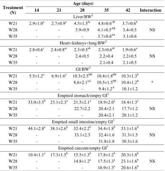
Besides investigating different parameters of growth and digestion, depending on the aim of the respective experiment, composition of the caecal microbiota and its fermentation activity were examined in each experiment.
Most of data have been published. The reference of the relevant publication is indicated in each chapter.
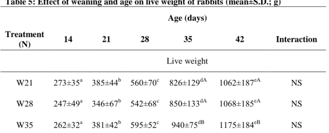
3.1. Effect of different weaning ages on production and digestive tract (published in ANIMAL 6:(6) pp. 894-901., 2012)
The aim of the study was to examine the effect of weaning on growth and certain parameters of the digestive tract in rabbits to assess the risk of early weaning for higher morbidity which can be attributed to presumably less developed digestive system.
3.1.1. Experimental animals, housing and nutrition
Pannon White does and their kits were housed in flat-deck cages (85x55cm), whereas after weaning the growing rabbits were housed in two- level wire mesh cages (two kits per cage, 84 cages per treatment) in a closed building. Average temperature ranged from 21oC to 29oC, the light was on between 05:00 and 21:00h, and the farm had overpressure air ventilation.