C A S E R E P O R T Open Access
A case report of isolated distal upper extremity weakness due to cerebral
metastasis involving the hand knob area
András Folyovich1, Viktória Varga1,2, György Várallyay3, Lajos Kozák3, Mária Bakos4, Erika Scheidl1,6, Katalin Anna Béres-Molnár1, Zita Kajdácsi5and Dániel Bereczki6*
Abstract
Background:Unilateral weakness of an upper extremity is most frequently caused by traumatic nerve injury or compression neuropathy. In rare cases, lesion of the central nervous system may result in syndromes suggesting peripheral nerve damage by the initial examination. Pseudoperipheral hand palsy is the best known of these, most frequently caused by a small lesion in the contralateral motor cortex of the brain. The‘hand knob’area refers to a circumscribed region in the precentral gyrus of the posterior frontal lobe, the lesion of which leads to isolated weakness of the upper extremity mimicking peripheral nerve damage. The etiology of this rare syndrome is almost exclusively related to an embolic infarction.
Case presentation:We present the case of a 70-year-old male patient with isolated left sided upper extremity weakness and clumsiness without sensory disturbance suggesting a lesion of the radial nerve. Nerve conduction studies had normal results excluding peripheral nerve damage. Neuroimaging (cranial CT and MRI) detected 3 space occupying lesions, one of them in the right precentral gyrus. An irregularly shaped tumor was found by CT in the left lung with multiple associated lymph node conglomerates. The metastasis from this mucinous tubular adenocarcinoma with solid anaplastic parts to the‘hand knob’area was responsible for the first clinical sign related to the pulmonary malignancy.
Conclusions:Pseudoperipheral palsy of the upper extremity is not necessarily the consequence of an embolic stroke. If nerve conduction studies have normal results, neuroimaging–preferably MRI–should be performed, as lesion in the hand-knob area of the precentral gyrus can also be caused by a malignancy.
Keywords:Isolated distal upper extremity weakness, Hand knob, Brain metastasis
Background
Isolated upper extremity weakness is predominantly at- tributed to an injury of the peripheral nervous system.
Weakness resulting from damage of the radial, ulnar, or median nerve are most commonly due to traumatic in- jury or compression neuropathy; however, it may de- velop as a symptom in association with other conditions including amyotrophic lateral sclerosis, cervical radiculo- pathy, or thoracic outlet syndrome. Isolated weakness of the upper extremity is rare in central nervous system
damage. In such cases lesions of the motor cortex or of the downstream corticospinal tract are responsible for the clinical signs. The ‘hand knob’ area is a circum- scribed region of the motor cortex in the precentral gyrus of the posterior frontal lobe [1], the lesion of which leads to isolated weakness of the contralateral upper extremity mimicking peripheral nerve damage.
This rare symptom is almost exclusively related to an underlying cortical infarction. The lesion presents in a misleading symptomatology, raising the suspicion of per- ipheral neuropathy, which may pose a challenge in the differential diagnosis.
Pseudoperipheral paresis of the upper extremity is known as a rare stroke syndrome. Cortical hand knob
* Correspondence:bereczki.daniel@med.semmelweis-univ.hu
6Department of Neurology, Semmelweis University, Balassa u. 6., H- 1083, Budapest, Hungary
Full list of author information is available at the end of the article
© The Author(s). 2018Open AccessThis article is distributed under the terms of the Creative Commons Attribution 4.0 International License (http://creativecommons.org/licenses/by/4.0/), which permits unrestricted use, distribution, and reproduction in any medium, provided you give appropriate credit to the original author(s) and the source, provide a link to the Creative Commons license, and indicate if changes were made. The Creative Commons Public Domain Dedication waiver (http://creativecommons.org/publicdomain/zero/1.0/) applies to the data made available in this article, unless otherwise stated.
strokes represent less than 1% of all hospitalized ische- mic strokes in single center surveys. The reported rate was 8 out of 815 (0.98%) in a Turkish study over 1,5 years [2], whereas a 5-year survey in Germany [3] found that 29 out of 3499 patients (0.8%) had a stroke of the cor- tical hand knob area. In a 5-year study in Norway, out of 866 patients with acute ischemic stroke 6 (0.7%) had a single lesion in the hand knob region [4]. In our study of 25 cases over 10 years we estimated the rate of hand knob strokes to be 0.35% [5]. Although the hand knob area certainly may be affected by intracerebral tumors like glioblastoma (see Fig.1 of Krieg et al. [6]), the inci- dence of malignant tumors in the background of isolated hand knob lesions have not been established.
Case report
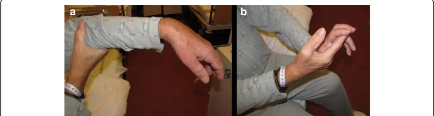
The 70-year-old male patient was admitted to the De- partment of Neurology with isolated left upper extremity weakness and clumsiness. He had wrist drop and was unable to grip with the fingers. He complained of no sensory loss or numbness. His past medical history in- cluded long-term oral anticoagulation due to recurrent lower extremity deep vein thrombosis, glaucoma, and a non-significant aortic valve stenosis, with vascular risk fac- tors including a 5-year history of treated hypertension, hypercholesterolemia, and a history of non-significant sten- osis of the left anterior descendent coronary artery. On ad- mission, no abnormality could be detected by physical examination, apart from the neurological signs, which in- cluded a wrist drop on the left side with decreased grip strength (Fig. 1). Pronation and wrist dorsiflection were lost, as well as the ability to form a ring with the thumb and the index. The reflexes of the left upper extremity were slightly brisk, with not pathological reflexes present and no sensory deficits. Laboratory parameters were without ab- normal findings except for a slightly elevated fasting blood glucose level. Cranial CT revealed contrast-enhancing, irregularly shaped lesions with diameters of 7, 10, and 9 mm (in the temporal, parietal, and frontal lobes, respect- ively), surrounded by perifocal edema. Carotid duplex
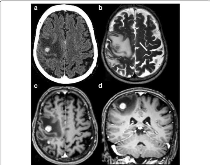
ultrasonography did not demonstrate signs of focal athero- sclerotic plaques, circulatory disturbance or stenosis on ei- ther side. In accordance with the findings of the CT scan, the cranial MRI confirmed the tumor in the right precen- tral gyrus, corresponding to the ‘hand knob’, accompanied by further similar lesions posteriorly in the parietal and the temporal lobes, and in the left frontal lobe (Fig.2). Electro- myography and nerve conduction studies did not support a peripheral origin underlying the paresis. The neuropsycho- logical examination revealed a deficit in the Luria three-step test as a single alteration, which performance could, however, be remarkably improved by verbal clues.
Chest X-ray performed as part of the search for primary tumor revealed no abnormality. Non-contrast and contrast-enhanced chest CT detected an irregularly shaped mass in segment 10 of the left lung, with inhomogeneous enhancement of the contrast agent and multiple associated lymph node conglomerates, suggesting lung cancer as the primary tumor. Abdominal ultrasonography did not detect malignancy. The patient received palliative steroid therapy, with no improvement in the paresis during the observation period. Based on the recommendation of the tumor board, the patient was transferred to the Department of Pulmonol- ogy for bronchoscopic tissue sampling. The verification of the diagnosis by this means, however, was unsuccessful.
The diagnostic process had to be suspended due to an acute bleeding duodenal ulcer, and the patient received blood transfusion and local hemostatic treatment in the De- partment of Surgery. In association with the antibiotic treatment, the patient developed pseudomembranous col- itis caused by Clostridium difficile. In addition to the per- sisting colitis, fever occurred accompanied by hemoptysis and coughing. The subsequent chest X-ray did not con- firm pneumonia. Despite the applied antibiotic and oxygen therapy, respiratory failure developed, and the patient passed away 2 months after the onset of the neurological symptom. The autopsy and the histo- pathological analysis identified an adenocarcinoma (mucinous tubular adenocarcinoma with solid anaplas- tic parts) both in the brain and the lung; (Fig.3).
Fig. 1Isolated central weakness of the left upper extremity. Wrist drop (a) and decreased handgrip strength (b)
Folyovichet al. BMC Cancer (2018) 18:947 Page 2 of 5
Fig. 2Contrast-enhanced axial CT (a), T2W axial MR (b), contrast-enhanced T1W axial (c) and coronal (d) MR. Intensive contrast enhancement (11 mm) in the right precentral gyrus, corresponding to the‘hand knob’s area, with large perifocal edema. The left‘hand knob’is normal (arrow).
Four other smaller enhancing metastases in the brain (not shown)
Fig. 3Pulmonary malignancy (mucinous tubular adenocarcinoma with anaplastic solid parts), 400× magnification, Hematoxylin & Eosin + alcian blue (a). Cerebral metastasis (mucinous tubular adenocarcinoma with anaplastic solid parts), 200×, magnification, Hematoxylin & Eosin + alcian blue (b)
Discussion and conclusions
Isolated upper extremity weakness is rarely caused by a lesion localized to the central nervous system. As the clinical signs suggest peripheral neuronal disease, the usual workup of such patients starts with nerve conduc- tion studies. With normal results of the electrophysio- logical tests, another etiology should be searched. An injury to a circumscribed territory of the precentral gyrus (primary motor cortex) referred to as‘hand knob’
results in clinical signs mimicking peripheral nerve le- sion that is called “pseudoperipheral” palsy. According to the Penfield-Rasmussen map, primary motor neurons innervating the upper extremity are localized in the lower third of the dorsolateral surface of the precentral gyrus [1, 7]. This region of the precentral gyrus is a knob-like structure, reminiscent of an epsilon or an omega in the axial functional MRI scans, whereas re- sembling a hook in the sagittal scans [8]. The etiology behind this rare symptom is almost exclusively reported to be a cortical infarction [3, 9–14], with hardly any ref- erence to alternative causes [15]. Intraoperative cortical stimulation, functional MRI and navigated transcranial magnetic stimulation studies clearly identified the region in the precentral gyrus of the posterior frontal lobe which is associated with hand movements, therefore should be saved during neurosurgical interventions, like resections of primary and metastatic brain tumors in the perirolandic area [6,16,17].
Our case confirms that circumscribed damage to the hand knob resulting in isolated distal upper extremity weakness may be caused not only by cerebral ischemia but also by a brain metastasis. The isolated weakness of the upper extremity was the first and only symptom that initiated the diagnostic process, eventually leading to the diagnosis of a primary pulmonary malignancy. The clin- ical diagnosis was confirmed by autopsy and histopatho- logical examination.
Although it has been suggested that patients with iso- lated hand palsy without alternative explanations for per- ipheral damage should be aggressively treated for acute ischemic stroke [18], pseudoperipheral palsy of the upper extremity is not necessarily the consequence of an ische- mic cerebrovascular lesion. If nerve conduction studies have normal results, neuroimaging should be performed, as a lesion in the hand-knob area of the precentral gyrus can also be caused by a malignancy affecting this region.
As CT misses the hand knob lesion in a considerable rate, MRI should be the preferred neuroimaging method.
Acknowledgements
DB was supported by the Higher Education Institutional Excellence Program of the Ministry of Human Resources of the Government of Hungary.
Funding
This research did not receive any specific grant from funding agencies in the public, commercial, or not-for-profit sectors.
Availability of data and materials
The original material (confidential patient record) is available at Szent Janos Hospital, Department of Neurology, Budapest, Hungary.
Authors’contributions
AF: patient examination, clinical management, medical record preparation, clinical neurological supervision, first draft of manuscript, concept, approval of final manuscript. VV: patient examination, clinical management, approval of final manuscript. GV: MRI neuroimaging, interpretation of results, manuscript preparation, critical review and approval of final manuscript. LK:
MRI neuroimaging, critical review and approval of final manuscript. MB: CT neuroimaging, critical review and approval of final manuscript. ES:
electrophysiological examination, interpretation of electrophysiological results, critical review and approval of final manuscript. KAB-M: neurological management, manuscript preparation, approval of final manuscript. ZK:
pulmonological supervision of the case, critical review and approval of final manuscript. DB: concept, critical review of initial versions of the manuscript, preparation of the final version of the manuscript.
Ethics approval and consent to participate
This case presentation did not involve any specific intervention therefore no ethics approval was needed. Data were collected retrospectively for this manuscript after the death of the patient. As only the usual diagnostic practice was followed, no specific consent was needed from the patient. The usual consent requested from patients for routine hospital care is
documented in the patient’s medical records.
Consent for publication
The outcome unfortunately was fatal, therefore the written consent to publish was given by a relative. The signed consent form is available on request.
Competing interests
The authors declare that they have no competing interests.
Publisher’s Note
Springer Nature remains neutral with regard to jurisdictional claims in published maps and institutional affiliations.
Author details
1Department of Neurology and Stroke Center, Szent János Hospital, Budapest, Hungary.2Department of Neurology, Uzsoki Hospital, Budapest, Hungary.3MR Research Centre, Semmelweis University, Budapest, Hungary.
4Department of Diagnostic Radiology, Szent János Hospital, Budapest, Hungary.5Korányi National Institute for Tuberculosis and Pulmonology, Budapest, Hungary.6Department of Neurology, Semmelweis University, Balassa u. 6., H- 1083, Budapest, Hungary.
Received: 20 February 2018 Accepted: 26 September 2018
References
1. Yousry T, Schmid U, Alkadhi H, Schmidt D, Peraud A, Buettner A, Winkler P.
Localization of the motor hand area to a knob on the precentral gyrus.
Brain. 1997;120:141–57.
2. Celebisoy M, Ozdemirkiran T, Tokucoglu F, Kaplangi DN, Arici S. Isolated hand palsy due to cortical infarction: localization of the motor hand area.
Neurologist. 2007;13:376–9.
3. Peters N, Müller-Schunk S, Freilinger T, Düring M, Pfefferkorn T, Dichgans M.
Ischemic stroke of the cortical“hand knob”area: stroke mechanisms and prognosis. J Neurol. 2009;256:1146–51.
4. Alstadhaug KB, Sjulstad A. Isolated hand paresis: a case series. Cerebrovasc Dis Extra. 2013;3:65–73.
5. Orosz P, Szőcs I, Rudas G, Folyovich A, Bereczki D, Vastagh I. Cortical hand knob stroke: report of 25 cases. J Stroke Cerebrovasc Dis. 2018;27:1949–55.
6. Krieg SM, Sabih J, Bulubasova L, Obermueller T, Negwer C, Janssen I, Shiban E, Meyer B, Ringel F. Preoperative motor mapping by navigated transcranial magnetic brain stimulation improves outcome for motor eloquent lesions.
Neuro-Oncology. 2014;16:1274–82.
Folyovichet al. BMC Cancer (2018) 18:947 Page 4 of 5
7. Zeller D, Su-Yin D, Stefan K, Biller A, Bartsch A, Saur D, Bendszus M, Rieckmann P, Toyka KV, Classen J. Functional role of ipsilateral motor areas in multiple sclerosis. J Neurol Neurosurg Psychiatry. 2011;82:578–83.
8. Gass A, Szabó K, Behrens S, Rossmanith C, Hennerici M. A diffusion- weighted MRI study of acute ischemic distal arm paresis. Neurology. 2001;
57:1589–94.
9. Back T, Mrowka M. Infarction of the“hand knob”area. Neurology. 2001;57:
1143.
10. Hall J, Flint A.“Hand knob”infarction. J Neurol Neurosurg Psychiatry. 2008;
79:406.
11. Paciaroni M, Caso V, Milia P, Venti M, Silvestrelli G, Palmerini F, Nardi K, Micheli S, Agnelli G. Isolated monoparesis following stroke. J Neurol Neurosurg Psychiatry. 2005;76:805–7.
12. Terao Y, Hayashi H, Kanda T, Tanebe H. Discrete cortical infarction with prominent impairment of thumb flexion. Stroke. 1993;24:2118 2120.
13. Uncini A, Caulo M, Ferretti A, Tartaro A, Ranieri F, Di Lazzaro V. Isolated shoulder palsy due to cortical infarction: localisation and
electrophysiological correlates of recovery. J Neurol Neurosurg Psychiatry.
2007;78:100–2.
14. Vastagh I, Kéri A, Heornyák C, Kovács T, Várallyay G, Barsi P, Rudas G, Bereczki D. Ischemic stroke of the“hand knob”area–6 years and 12 cases.
Cerebrovasc Dis. 2012;33(Suppl 2):164–5.
15. Tanuiguchi M, Kato A, Ninomiya H, Hirata M, Cheyne D, Robinson SE, Maruno M, Saitoh Y, Kishima H, Yoshimine T. Cerebral motor control in patients with gliomas around the central sulcus studied with spatially filtered magnetoencephalography. J Neurol Neurosurg Psychiatry. 2004;75:
466–71.
16. Krieg SM, Picht T, Sollmann N, Bährend I, Ringel F, Nagarajan SS, Meyer B, Tarapore PE. Resection of motor eloquent metastases aided by preoperative nTMS-based motor maps-comparison of two observational cohorts. Front Oncol. 2016;6:261.https://doi.org/10.3389/fonc.2016.00261.
17. Fang S, Liang J, Qian T, Wang Y, Liu X, Fan X, Li S, Wang Y, Jiang T.
Anatomic location of tumor predicts the accuracy of motor function localization in diffuse lower-grade gliomas involving the hand knob area.
AJNR Am J Neuroradiol. 2017;38:1990–7.
18. Siniscalchi A, Lochner P, Perrotta P, Rizzuto S, De Sarro G, Gallelli L. Isolated hand palsy in National Institutes of Health stroke scale (NIHSS): is it useful?
West J Emerg Med. 2018;19:524–6.