Received 29 Dec 2015|Accepted 10 Feb 2016|Published 1 Apr 2016
Neutrophil-specific deletion of the CARD9 gene expression regulator suppresses
autoantibody-induced inflammation in vivo
Tama´s Ne´meth1,2, Krisztina Futosi1, Cassian Sitaru3, Ju¨rgen Ruland4 & Attila Mo´csai1,2
Neutrophils are terminally differentiated cells with limited transcriptional activity. The biological function of their gene expression changes is poorly understood. CARD9 regulates transcription during antifungal immunity but its role in sterile inflammation is unclear. Here we show that neutrophil CARD9 mediates pro-inflammatory chemokine/cytokine but not lipid mediator release during non-infectious inflammation. Genetic deficiency of CARD9 suppresses autoantibody-induced arthritis and dermatitis in mice. Neutrophil-specific deletion of CARD9 is sufficient to induce that phenotype. Card9/ neutrophils show defective immune complex-induced gene expression changes and pro-inflammatory chemokine/cytokine release but normal LTB4production and other short-term responses.In vivodeletion of CARD9 reduces tissue levels of pro-inflammatory chemokines and cytokines but not LTB4. The CARD9-mediated signalling pathway involves Src-family kinases, Syk, PLCg2, Bcl10/Malt1 and NFkB. Collectively, CARD9-mediated gene expression changes within neutrophils play important roles during non-infectious inflammationin vivoand CARD9 acts as a divergence point between chemokine/cytokine and lipid mediator release.
DOI: 10.1038/ncomms11004 OPEN
1Department of Physiology, Semmelweis University School of Medicine, 1094 Budapest, Hungary.2MTA-SE ‘Lendu¨let’ Inflammation Physiology Research Group of the Hungarian Academy of Sciences and Semmelweis University, 1094 Budapest, Hungary.3Department of Dermatology, University Hospital Freiburg and BIOSS Centre for Biological Signalling Studies, 79104 Freiburg, Germany.4Department of Clinical Chemistry and Pathobiochemistry, Technical University of Munich, 81675 Munich, Germany. Correspondence and requests for materials should be addressed to A.M.
(email: mocsai.attila@med.semmelweis-univ.hu).
N
eutrophils are short-lived, terminally differentiated cells that play critical roles in antimicrobial host defence and also contribute to autoimmune and other non-infectious inflammatory processes1–3. Unlike the long-lived macrophages and dendritic cells, neutrophils have limited transcriptional activity but instead show robust short-term effector functions such as phagocytosis, respiratory burst, degranulation or NET release. The dissociation of neutrophil function from gene expression is best exemplified by the fact that anuclear neutrophils that have expelled their DNA through NETosis are still capable of performing various in vivo antimicrobial functions4. Based on their short lifespan, limited transcriptional activity and robust short-term effector functions, neutrophils are generally believed to be simple effector cells of the immune and inflammatory reaction.However, neutrophils have also been shown to be able to upregulate pro-inflammatory gene expression and to release various chemokines and cytokines5,6. Those non-conventional functional responses may indicate a more general role of neutrophils in the orchestration of the immune/inflammatory response1,3,6. Unfortunately, it is still unclear whether inflammation-related gene expression changes in neutrophils (and the resulting chemokine/cytokine production) are just evolutionary remnants from the macrophage-related origin of these cells, or they play an important functional role during the in vivoinflammation process. This uncertainty is primarily due to the fact that none of the currently availablein vivomodels allow suppression of gene expression changes in such a manner that it is both selective for neutrophils and it also retains other functional responses of neutrophils intact.
Caspase recruitment domain-containing protein 9 (CARD9) is an intracellular adapter protein primarily expressed in myeloid- lineage cells and couples C-type lectin receptors to NFkB- mediated gene expression7. CARD9 plays a critical role in host defence against fungal pathogens in both mice8–10 and humans11,12, and it is also involved in immunity against other microbial infections7,13. In addition to its antimicrobial function, human genetic studies have also linked CARD9 to highly prevalent human diseases of non-infectious origin such as inflammatory bowel disease14–17, ankylosing spondylitis18,19, rheumatoid arthritis20 or IgA nephropathy21. However, it is still unclear whether CARD9 indeed participates in non-infectious inflammation and if so, what are the cellular and molecular pathways involved. In addition, though the analysis of CARD9 function has so far focused on dendritic cells and macrophages, CARD9 is also present in neutrophils12,22 and the ImmGen database23 indicates that neutrophils express the highest level of CARD9 within the immune system.
Unfortunately, the role of CARD9 in neutrophils is still very poorly understood.
Autoantibody-induced sterile inflammation is an important component of autoimmune disease pathogenesis. Its experimental models24–26 mimic important aspects of human rheumatoid arthritis, bullous pemphigoid and epidermolysis bullosa acquisita.
Autoantibody-induced inflammation is mediated by sequential activation of lipid (LTB4), cytokine and chemokine cascades27. Neutrophils are critically involved in autoantibody-induced sterile inflammation2,28 and we have previously shown that autoantibody-induced inflammation is mediated by signalling through Src-family kinases, Syk and PLCg2 (refs 29–32).
However, it is at present unclear how signalling downstream of those receptor-proximal molecules triggers lipid, chemokine and cytokine release.
The lack of knowledge on the contribution of neutrophil gene expression to in vivo inflammation, on the role of CARD9 in non-infectious inflammation and neutrophil function and on
how receptor-proximal signalling molecules are coupled to inflammatory mediator release, prompted us to investigate the role of CARD9 in autoantibody-mediated in vivoinflammation models. Our results indicate that CARD9 mediates autoantibody- induced inflammation by acting as a divergence point down- stream of receptor-proximal signalling molecules triggering chemokine and cytokine but not lipid mediator (LTB4) release.
Importantly, lineage-specific studies revealed that those functions are primarily linked to CARD9 expression within neutrophils, indicating a critical contribution of neutrophil gene expression and chemokine/cytokine release to the overall in vivo inflammatory reaction.
Results
The role of CARD9 in autoantibody-induced arthritis.
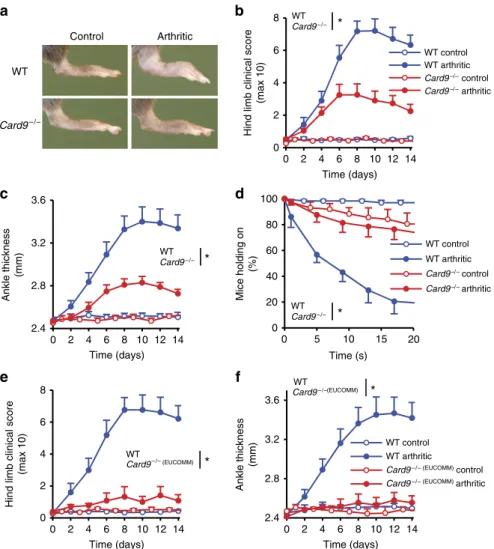
To test the role of CARD9 in non-infectious inflammation, we tested the effect of CARD9 deficiency on autoantibody-induced arthritis development in the K/BN serum-transfer model30. We have used two independent CARD9-deficient mouse strains (Supplementary Fig. 1a) that were homozygous either for the conventional Card9tm1Jrld knockout mutation (referred to as Card9/ mice) or for the so-called knockout- first33 Card9tm1a(EUCOMM)Hmgu mutation (referred to as Card9/ (EUCOMM) mice). CARD9 was absent from bone marrow (Supplementary Fig. 1b) and spleen (Supplementary Fig. 1c) cell lysates of both mutant strains. As indicated in Fig. 1a, Card9/ mice showed substantially reduced signs of arthritis development. Quantification of the visible clinical signs (Fig. 1b;
P¼0.0028; two-way analysis of variance (ANOVA)), ankle thickening (Fig. 1c; P¼0.0047; two-way ANOVA) and the ability of the mice to hold on to the bottom of a wire grid (Fig. 1d; P¼210–4; two-way ANOVA) all confirmed highly significant protection from disease development. Similarly, Card9/ (EUCOMM) mice were also strongly protected from visible signs of arthritis (Fig. 1e; P¼6.610–4; two-way ANOVA) and ankle thickening (Fig. 1f; P¼0.0038; two-way ANOVA). Therefore, CARD9 plays a major role in the development of autoantibody-induced arthritis.
We have also generated bone marrow chimeras by transplant- ing CARD9-deficient bone marrow cells to lethally irradiated wild-type recipients. Similar to intact mice, Card9/ bone marrow chimeras also showed partial protection from arthritis development in the K/BN serum-transfer model (Supplementary Figs 2a–d; P values between 9.110–11 and 1.6104; two-way ANOVA), indicating a role for CARD9 in a radiosensitive hematopoietic compartment.
The role of CARD9 in autoantibody-induced skin blistering.
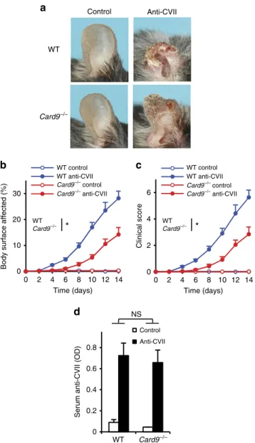
We have also tested the effect of CARD9 deficiency on dermatitis triggered by systemic administration of antibodies against collagen VII (a mouse model of the human blistering skin disease epidermolysis bullosa acquisita). Card9/ mice were sub- stantially, though not completely protected from the visible skin disease (Fig. 2a), which was also confirmed by quantitative analysis of the body surface area affected (Fig. 2b; P¼0.017;
two-way ANOVA) and the overall disease severity score (Fig. 2c;
P¼0.022; two-way ANOVA). This was not due to the reduced circulating autoantibody concentration since serum levels of collagen VII-specific IgG were similar in wild-type and Card9/ mice (Fig. 2d;P¼0.95; two-way ANOVA).
Role of CARD9 in leukocyte recruitment and migration.
Flow cytometric analysis of myeloid cells in the synovial infiltrate (Fig. 3a,b) revealed reduced accumulation of neutro- phils (P¼0.0041; Student’s t-test) and macrophages (P¼0.026;
Student’s t-test) at the site of inflammation in Card9/ mice subjected to K/BN serum-transfer arthritis. To reveal whether that reduction was due to a cell-autonomous migration defect, we generated mixed bone marrow chimeras carrying CD45.1- expressing wild type, along with CD45.2-expressing wild type or Card9/ hematopoietic tissues (WT: WT and WT:Card9/ chimeras, respectively). Those chimeras were subjected to K/BN serum-transfer arthritis and the ability of myeloid cells from the different genotypes to infiltrate the inflamed synovium was tested by comparing the ratio of CD45.1-expressing and CD45.2-expressing cells in the peripheral blood and synovial infiltrate. As shown in Fig. 3c and Supplementary Fig. 3a, no significant difference between the percentage of CD45.2-positive neutrophils in the blood and synovium was observed in any of the mixed bone marrow chimeras irrespective of the genotype (wild type or Card9/) of the CD45.2-positive population (P¼0.80 for normalized data shown in Supplementary Fig. 3a;
Student’s t-test). Similarly, the percentage of CD45.2-positive monocytes in the blood was always similar to that of CD45.2- positive macrophages in the synovium, irrespective of the geno- type of the CD45.2-positive cells (Fig. 3d and Supplementary Fig. 3b; P¼0.56; Student’s t-test). Those results indicated that
Card9/ myeloid cells did not have an intrinsic migration defectin vivo.
We also tested the intrinsic migratory capacity of neutrophils in in vitrotranswell assays. As shown in Fig. 3e, theCard9/ mutation did not affect the migration of neutrophils towards fMLP (P¼0.71; two-way ANOVA), MIP-2 (CXCL2; P¼0.53;
two-way ANOVA) or LTB4 (P¼0.92; two-way ANOVA) as chemoattractants either.
Taken together, Card9/ myeloid cells show partially reduced accumulation at the site of inflammation despite no cell-autonomous migration defect.
Decreased chemokine and cytokine but not LTB4levelsin vivo.
To resolve the apparent contradiction between partially reduced accumulation but normal intrinsic migratory capacity of Card9/ myeloid cells, we hypothesized that CARD9 was required for the generation of an appropriate inflammatory (chemoattractant) tissue microenvironment. Therefore, we tested the presence of various pro-inflammatory mediators in the syno- vial infiltrate. As shown in Fig. 4a,b, theCard9/mutation led to a strong reduction of the levels of various chemokines acting on myeloid cells, including KC (CXCL1; P¼0.011; two-way
WT
Card9–/–
WT Card9–/–
Card9–/– control Card9–/– arthritic Control Arthritic
Hind limb clinical score (max 10)
WT control WT arthritic
Card9–/– control Card9–/– arthritic WT control WT arthritic
Card9–/– (EUCOMM) control Card9–/– (EUCOMM) arthritic Card9–/–(EUCOMM)
WT control WT arthritic
*
WT Card9–/– *
WT Card9–/– *
WT
Card9–/– (EUCOMM)
WT
*
*
Time (days) 0 2 4 6 8 10 12 14 8
6 4 2 0
Hind limb clinical score (max 10)
Time (days) 0 2 4 6 8 10 12 14 8
6 4 2 0
100 80 60 40 20 0
0 5 10 15 20
Time (s) Mice holding on (%)
0 2 4 6 8 10 12 14 Time (days) 3.6
3.2
2.8
2.4 Ankle thickness (mm) 0 2 4 6 8 10 12 14
Time (days) 3.6
3.2
2.8
2.4 Ankle thickness (mm)
a b
d c
e f
Figure 1 | CARD9 is important for the development of autoantibody-induced arthritis.Intact wild type (WT),Card9/(a–d) orCard9/(EUCOMM) (e,f) mice were injected with BN (control) or K/BN (arthritic) serum i.p. on day 0. Arthritis development was followed by photographing on day 7 (a), clinical scoring of the hind limbs (b,e), ankle-thickness measurement (c,f) and an articular function test (hanging on a wire grid; (d)). Images are representative of, and quantitative data show mean and s.e.m. from 8 to 9 control and 12 to 13 arthritic serum-treated individual mice per group from four independent experiments (a–d) or 5 to 6 control and 8 to 9 arthritic serum-treated mice per group from three independent experiments (e,f).
(d) Results from functional test performed 12 times on each mouse between days 7–10. *Po0.05 (two-way ANOVA); see the text for actualPvalues.
ANOVA), MIP-1a (CCL3; P¼0.0013; two-way ANOVA) and MIP-2 (CXCL2; P¼0.027; two-way ANOVA), as well as the IL-1bcytokine (P¼2.510–5; two-way ANOVA). Interestingly, however, theCard9/ mutation did not affect the level of the lipid chemoattractant LTB4at the site of inflammation (Fig. 4c;
P¼0.77; two-way ANOVA). Those results likely explain the substantially but not completely reduced accumulation of myeloid cells in the inflamed synovium.
Neutrophil CARD9 is required forin vivoinflammation. The importance of neutrophils in autoantibody-induced inflamma- tion28,34 and the apparent role of CARD9 in a radiosensitive hematopoietic compartment (Supplementary Fig. 2) raised the possibility that CARD9 expressed in neutrophils was required for the autoantibody-induced arthritis and dermatitis response. The availability of the Card9tm1a(EUCOMM)Hmgu allele that allows further manipulation towards lineage-specific deletion provided a unique opportunity to test the functional relevance of CARD9 specifically within the neutrophil lineage.
Using FRT-mediated manipulation of the Card9tm1a(EU-
COMM)Hmgu knockout-first allele (Supplementary Fig. 4), we have generated mice carrying the Card9tm1c(EUCOMM)Hmgu
(referred to as Card9flox) floxed allele and bred those mice to MRP8-Cre35 transgene-expressing mice to generate MRP8- CreþCard9flox/flox (referred to as Card9DPMN) mice with lineage-specific deletion of CARD9 specifically in the neutrophil compartment (Supplementary Fig. 4). As shown in Fig. 5a, FACS- sorted Card9/ neutrophils had no detectable CARD9 expression, whereas the level of CARD9 was strongly reduced (though not completely absent) in similarly sorted Card9DPMN neutrophils. However, in agreement with prior studies showing high selectivity of the MRP8-Cre transgene35,36, theCard9DPMN mutation did not affect CARD9 expression in bone marrow- derived macrophages (Fig. 5b). Those results confirmed the effective and specific deletion of CARD9 from Card9DPMN neutrophils.
We next tested the effect of the Card9DPMN mutation on K/BN serum-transfer arthritis. As shown in Fig. 5c, both the Card9/ (P¼1.410–6; two-way ANOVA) and the Card9DPMN (P¼410–4; two-way ANOVA) mutations caused a substantial but incomplete reduction of the clinical arthritis score.
Similarly, the ankle-thickening response (Fig. 5d) was reduced in both Card9/ (P¼4.310–4; two-way ANOVA) and Card9DPMN(P¼0.0069; two-way ANOVA) mutants. Importantly, there was practically no difference between the effect of neutrophil- specific and complete CARD9 deficiency in either assay readouts tested (Pvalues between 0.47 and 0.56; two-way ANOVA).
We have also subjected the same mutants to the autoantibody- induced skin blistering disease model. As shown in Fig. 5e, both the Card9/ (P¼4.610–6; two-way ANOVA) and the Card9DPMN (P¼1.710–5; two-way ANOVA) mutants were significantly but not completely protected from disease develop- ment defined as the body area affected. Similarly, the clinical score (Fig. 5f) was reduced in both Card9/ (P¼1.610–4; two-way ANOVA) and Card9DPMN (P¼7.510–5; two-way ANOVA) mutants. Again, the two mutant strains showed practically identical phenotypes in both assay readouts tested (Pvalues between 0.17 and 0.69; two-way ANOVA).
Taken together, the defective arthritis and dermatitis develop- ment in Card9/ mice is mostly due to the role of CARD9 within the neutrophil compartment.
Reduced neutrophil chemokine/cytokine but not LTB4release.
Given the in vivo role of CARD9 within neutrophils, we next tested variousin vitrofunctions ofCard9/neutrophils. Those experiments focused on immune complex-induced activation as anin vitromodel of autoantibody-inducedin vivoactivation.
AlthoughCard9/ neutrophils lacked the CARD9 protein (Supplementary Fig. 5a), they expressed normal levels of the neutrophil maturation marker Ly6G (Supplementary Fig. 5b), and showed normal expression of cell surface FcgRII/III and FcgRIV (Supplementary Fig. 5c).
CARD9 deficiency did not affect immune complex-induced short-term functional responses of neutrophils such as superoxide
Control
Control Anti-CVII
Anti-CVII WT
Card9–/–
Card9–/– control
Card9–/–
Card9–/–
Card9–/– anti-CVII WT anti-CVII WT control
Card9–/– control Card9–/– anti-CVII WT anti-CVII WT control
WT
WT
* Card9–/–
WT *
30
20
10
0
0 2 4 6 8 10 12 14 Time (days)
0 2 4 6 8 10 12 14 Time (days)
Body surface affected (%) Clinical score
6
4
2
0
NS
0.8 0.6 0.4 0.2 0
Serum anti-CVII (OD)
a
b c
d
Figure 2 | CARD9 in autoantibody-induced skin blistering disease.
Blistering skin disease was triggered in wild type (WT) andCard9/ intact mice or bone marrow chimeras by systemic injection of collagen VII- specific (anti-CVII) antibodies. Skin disease was followed by photographing on day 14 (a) and clinical assessment of the total body surface affected (b) and the overall disease severity (c). The serum titre of anti-CVII antibodies was tested on day 6 by ELISA (d). Representative images (a) or mean and s.e.m. (b–c) from 7 to 9 control (2 intact and 5–7 bone marrow chimeric) and 13–14 anti-CVII-treated (2 intact and 11–12 bone marrow chimeric) mice per genotype from four independent experiments are shown. No difference between intact and chimeric mice of the same hematopoietic genotype was observed (not shown). (d) Mean and s.e.m.
from 4 control and 12–13 anti-CVII-treated mice from five independent experiments are shown. *Po0.05; NS, statistically not significant (two-way ANOVA); see the text for actualPvalues.
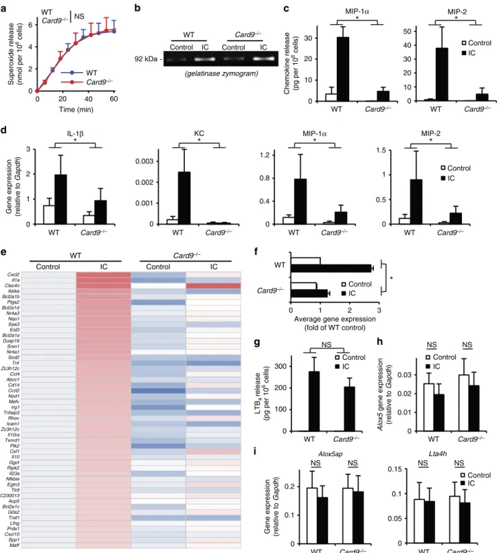
release (Fig. 6a;P¼0.79; two-way ANOVA) or the exocytosis of gelatinase-containing granules (Fig. 6b). However, CARD9 deficiency markedly reduced the immune complex-induced release of MIP-1a (P¼0.0079; two-way ANOVA) and MIP-2 (P¼1.210–7; two-way ANOVA) chemokines from neutrophils (Fig. 6c). As shown in Fig. 6d,Card9/ neutrophils also showed strong reduction of the immune complex-induced expression of the genes encoding for IL-1b(P¼0.018; two-way ANOVA), KC (P¼6.010–16; two-way ANOVA), MIP-1a(P¼9.610–7; two- way ANOVA) and MIP-2 (P¼6.210–8; two-way ANOVA).
We have also performed complementary DNA (cDNA) micro- array analysis to test whether the reduced upregulation of chemokine and cytokine genes reflected a generalized defect of immune complex-induced gene expression (Fig. 6e,f; Supplementary Table 1). As shown in the heat map of the 50 most highly upregulated genes (Fig. 6e), CARD9 deficiency led to an overall decrease of immune complex-induced upregulation of neutrophil
gene expression, including expression of various genes expressing pro-inflammatory mediators. The average changes of the expression of the genes shown in Fig. 6e has also been summarized in Fig. 6f, which indicated a 78% reduction of immune complex-induced gene expression inCard9/ cells (Po10-17; two-way ANOVA).
In contrast to the various chemokines and cytokines, the immune complex-induced release of LTB4from neutrophils was not affected by theCard9/ mutation (Fig. 6g;P¼0.39; two- way ANOVA). In addition, the expression ofAlox5that encodes for 5-lipoxigenase, the rate-limiting enzyme of LTB4synthesis, is not triggered by immune complex stimulation in wild-type neutrophils (P¼0.56; one-way ANOVA) and it is not affected by the Card9/ mutation either (Fig. 6h). The expression of Alox5apandLta4h, the other two components of LTB4synthesis, was also not altered by immune complex stimulation (P¼0.49 and 0.64, respectively; one-way ANOVA) or CARD9 deficiency (Fig. 6i).
8
6
4
2
0
PMN in synovium (104 cells per paw) MΦ in synovium (104 cells per paw)
* *
Control Arthritic
Control Arthritic
WT Card9–/– WT Card9–/–
5 4 3 2 1 0
a b
Control Control Control
WT : WT
WT Card9–/– WT Card9–/– WT Card9–/–
WT : Card9–/–
CD45.2+ PMN in synovium (% of total PMN)
CD45.2+ PMN in blood (% of total PMN) 75
75 50
50 25
25 0
0
WT : WT WT : Card9–/–
CD45.2+ MΦ in synovium (% of total MΦ)
CD45.2+ monocytes in blood (% of total monocytes) 75
75 50
50 25
25 0
0
NS NS NS
15 fMLP
10
5
0 Transmigrated cells (%)
25 20 15 10 5 0
MIP-2 LTB4
40
30
20
10
0
c d
e
Figure 3 | CARD9 plays no direct role in myeloid cell migration to the site of inflammation.Wild type (WT) andCard9/ mice were subjected to K/BN serum-transfer arthritis (a–d), the ankle area or the front paw was flushed on day 4 and the number of neutrophils (a) or monocytes/
macrophages (b) was determined by flow cytometry. Mixed bone marrow chimeras with CD45.1-expressing wild type (WT) and CD45.2-expressing WT or Card9/ hematopoietic cells were subjected to K/BN serum-transfer arthritis andin vivoaccumulation of neutrophils (c) and monocytes/
macrophages (d) was determined by flushing the synovial area on day 4, followed by flow cytometric analysis of the ratio of CD45.1 and CD45.2- expressing cells in peripheral blood and the synovial infiltrate. In (c,d), each dot represents an individual mouse. (e)In vitromigration of WT andCard9/ bone marrow neutrophils towards the indicated chemoattractants in a fibrinogen-coated Transwell system. Graphs represent mean and s.e.m. from 5–14 (a) or 6–16 (b) mice per group from five independent experiments. (e) Data represent mean and s.e.m. of three to five independent experiments. *Po0.05;
NS, statistically not significant (Student’st-test (a,b), two-way ANOVA (e)); see the text for actualPvalues.
Taken together, CARD9 is required for immune complex- induced gene expression changes and release of various chemokines and cytokines but not LTB4 from neutrophils.
Together with the defective inflammatory response in neutro- phil-specific CARD9 deletion mutants (Fig. 5), these results also provide strong evidence for the in vivo role of gene expression changes within the neutrophil compartment.
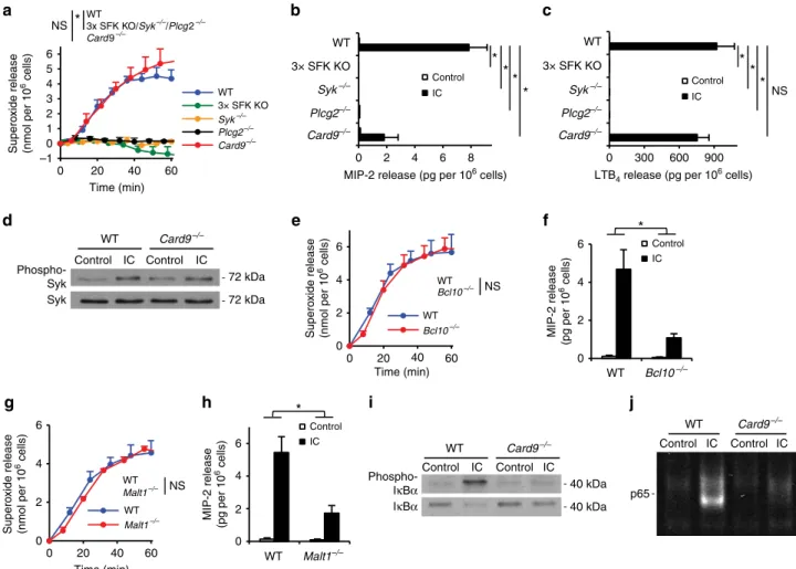
Upstream and downstream signalling components. Having established a critical role for CARD9-mediated neutrophil gene expression changes and chemokine/cytokine release, we finally aimed to characterize the signalling components upstream and downstream of CARD9 in neutrophils. We and others have pre- viously shown that myeloid Src-family kinases (Hck, Fgr and Lyn)32, Syk31,37and PLCg2 (refs 29,30) are required for immune complex- induced neutrophil functions and the K/BN serum-transfer arthritis, supposedly by forming a receptor-proximal signalling cluster. We therefore compared immune complex-induced functional responses of neutrophils lacking those molecules with responses of Card9/ cells. As shown in Fig. 7a, Hck/ Fgr/Lyn/ (3 SFK KO), Syk/ and Plcg2/ neutrophils showed complete defect in immune complex-induced superoxide production (Pvalues between 7.910–5and 1.610–4; two-way ANOVA), whereas Card9/ cell showed a normal response (P¼0.73; two-way ANOVA). Similarly, as shown in Fig. 7b,c, whileHck/Fgr/Lyn/,Syk/ andPlcg2/ neutrophils showed complete defect in immune complex-induced MIP-2 and LTB4 release (P values between 2.910–4 and 9.410–3; two-way ANOVA),Card9/ neutrophils displayed a strong but incomplete reduction of MIP-2 release (P¼0.027; two- way ANOVA) and no significant reduction of LTB4 release (P¼0.38; two-way ANOVA). The Card9/ neutrophil phenotype was not due to defective receptor-proximal signalling
since Card9/ neutrophils showed normal Syk phosphorylation on activation by immobilized immune complexes (Fig. 7d). Those results suggest that CARD9 functions downstream of the receptor- proximal Src-family/Syk/PLCg2 complex during immune complex- induced neutrophil activation.
During fungal infection, CARD9 coordinates anti-fungal host defence in cooperation with the downstream adapter Bcl10 (ref. 7). Therefore, we tested neutrophil functions and arthritis development in the absence of Bcl10. Bcl10/ neutrophils lacked the Bcl10 protein (Supplementary Fig. 6a) but expressed normal levels of Ly6G (Supplementary Fig. 6b) and Fcg-receptors (Supplementary Fig. 6c). On immune complex challenge, Bcl10/ neutrophils showed normal superoxide release (Fig. 7e; P¼0.76; two-way ANOVA) but defective release of the MIP-2 chemokine (Fig. 7f;P¼0.0057; two-way ANOVA). As shown in Supplementary Fig. 7, the Bcl10/ mutation also caused partial protection from the clinical signs (P¼0.065; two- way ANOVA) and ankle-thickening response (P¼0.015; two- way ANOVA) during K/BN serum-transfer arthritis. The CARD9-Bcl10 complex is thought to signal through the Malt1 paracaspase during antifungal immunity7. As shown in Fig. 7g,h, Malt1/ neutrophils produced normal levels of superoxide on immune complex activation (P¼0.87; two-way ANOVA) but their MIP-2 release was substantially reduced under identical conditions (P¼0.0092; two-way ANOVA). The similarity between the Card9/, Bcl10/ and Malt1/ phenotypes suggests that Bcl10 and Malt1 participates in signalling downstream of CARD9 during immune complex-induced neutrophil activation.
We have also explored how CARD9 is coupled to upregulation of pro-inflammatory gene expression. CARD9 deficiency strongly reduced immune complex-induced phosphorylation and degradation of IkBa (Fig. 7i) and the translocation of NFkB
30
20 10
0
WT Card9–/– WT Card9–/– WT Card9–/–
WT Card9–/–
WT Card9–/–
Control Arthritic
Control Arthritic Control
Arthritic
40 8
6
4 2
0
60
40
20
0
150
100
50
0
150
100
50
0
KC MIP-1α MIP-2
* * *
* NS
Chemokine concentr. (pg per paw) LI-1β concentration (pg per paw) LTB4 concentration (pg per paw)
a
b c
Figure 4 | CARD9 is required for the development of the inflammatory environment.(a–c) Wild type (WT) orCard9/ mice were subjected to K/BN serum-transfer arthritis as described above and the synovial area was flushed on day 4. The cell-free supernatants of the synovial infiltrates were probed using commercial ELISA assays for the indicated chemokines (a), cytokine (b) and the lipid mediator LTB4(c). Graphs represent mean and s.e.m.
from 4–9 (a), 3–5 (b) or 3–4 (c) mice per group from three independent experiments. *Po0.05; NS, statistically not significant (two-way ANOVA); see the text for actualPvalues.
p65 to the nucleus (Fig. 7j), indicating that CARD9 triggers upregulation of inflammatory gene expression through activation of the canonical NFkB pathway.
Taken together, the CARD9 signalling pathway respon- sible for chemokine/cytokine production by neutrophils during inflammation in vivo is reminiscent of the signa- lling pathway utilized by C-type lectin receptors and consists of a receptor-proximal Src-family/Syk/PLCg2 module triggering NFkB activation through the CARD9/Bcl10/Malt1 complex.
Discussion
Our results indicate that the CARD9 protein expressed within the neutrophil compartment plays an important role in chemokine and cytokine but not LTB4release during autoantibody-induced sterile inflammation. To our knowledge, these are the first experiments showing that neutrophil-specific abrogation of agonist-induced gene expression changes (without affecting other neutrophil functions) inhibitsin vivoinflammatory reactions and therefore provide the most direct evidence so far for an important in vivo role of inflammation-induced gene expression changes within the neutrophil lineage.
Although neutrophils have long been known to be critical effector cells of antimicrobial host defence, more recent studies indicated that they are also capable of releasing pro-inflammatory mediators5,6. Unfortunately, direct evidence for the role of neutrophil gene expression or neutrophil-derived chemokines/
cytokines has been missing. A number of prior studies reported a critical role for neutrophils inin vivoinflammatory reactions2,28 and a recent study has shown that neutrophil-specific deletion of Syk blocked arthritis development in the K/BN serum-transfer model37. Unfortunately, since those approaches not only blocked neutrophil-derived gene expression but also other functional responses of those cells, the specific effect of gene expression changes could not be assessed. Other attempts utilized complementation approaches using mixed bone marrow chimeras or isolated bone marrow neutrophils27,38,39. Unfortunately, all those approaches had major limitations related to the limited specificity of theGfi1/strain used or the limited purity and altered activation status of ex vivo isolated bone marrow-derived neutrophils. Our results with the Card9DPMN mutants (Fig. 5) provide the first direct evidence using lineage-specific deletion that the products of neutrophil gene expression coordinate thein vivoinflammatory response without affecting conventional neutrophil functions such as reactive
CARD9 - Actin -
WT Card9–/– Card9ΔPMN
CARD9 - Actin-
WT
WT control Card9–/– control Card9ΔPMN control WT arthritic Card9–/– arthritic Card9ΔPMN arthritic
WT control Card9–/– control Card9ΔPMN control WT anti-CVII Card9–/– anti-CVII Card9ΔPMN anti-CVII WT
Card9–/–
Card9–/–
Card9ΔPMN
Card9ΔPMN
62 kDa 42 kDa
NS WT
Card9–/–
Card9ΔPMN NS
WT Card9–/–
Card9ΔPMN NS
WT Card9–/–
Card9ΔPMN NS
* *
* *
* * * *
0 2 4 6 8 10 12 14 Time (days)
0 2 4 6 8 10 12 14 Time (days)
0 2 4 6 8 10 12 14 Time (days) 0 2 4 6 8 10 12 14
Time (days)
Hind limb clinical score (max 10) Ankle thickness (mm) Clinical score
Body surface affected (%) 8 6 4 2 0
30
20
10
0
6
4
2
0 3.4
3
2.6
2.2
a b
d c
e f
Figure 5 | Neutrophil-specific CARD9 deletion attenuates autoantibody-induced inflammation.(a,b) CARD9 expression was detected by western blot in cell-sorted bone marrow-derived neutrophils (a) or cultured macrophages (b) of wild type,Card9/orCard9DPMNanimals. (c,d) Wild type (WT), Card9/ orCard9DPMNbone marrow chimeras were injected with BN (control) or K/BN (arthritic) serum i.p. on day 0. Arthritis development was followed by clinical scoring of the hind limbs (c) and ankle-thickness measurement (d). Blistering skin disease was triggered in wild type (WT),Card9/ orCard9DPMNintact mice or bone marrow chimeras by systemic injection of collagen VII-specific (anti-CVII) antibodies. Skin disease was followed by clinical assessment of the total body surface affected (e) and the overall disease severity (f). The blots are representative of three to four independent experiments. Quantitative data show mean and s.e.m. from 7 control and 8 to 9 arthritic serum-treated individual mice per group from three
independent experiments (c,d) or 3 to 4 control and 4 anti-CVII-treated mice per group from two independent experiments (e,f). *Po0.05 (two-way ANOVA); NS, not significant; see the text for actualPvalues.
oxygen species (ROS) release. Our overall study also suggests that neutrophil-derived LTB4is capable of driving a clinically evident, though substantially reduced level of neutrophil-mediated
inflammation even in the absence of neutrophil-derived chemokines and cytokines. Those results together provide substantial novel insight into thein vivorole of neutrophil gene
(gelatinase zymogram)
Cxcl2 Il1a Clec4n Ikbke Bcl2a1b Ptgs2 Bcl2a1d Nr4a3 Nqo1 Saa3 Eid3 Bcl2a1a Dusp16 Srxn1 Nr4a1 Sod2 Tnf Zc3h12c Ccl4 Abcc1 Cd14 Ccrl2 Nod1 Mefv Irg1 Tnfaip3 Rhov Icam1 Zc3h12c Il10ra Txnrd1 Plk2 Csf1 Il10 Ggct Ripk2 Il23a Nfkbie Egln3 Tlr9 C230013 Acp5 Bcl2a1c G0s2 Traf1 Lfng Prdx1 Cxcl10 Spp1 Maff
6 4 2 0
WT
WT Card9–/–
Card9–/–
WT Card9–/– WT Card9–/–
WT Card9–/–
WT Card9–/–
NS
92 kDa
Control IC Control Control
IC IC
Control IC Control IC
MIP-1α MIP-2
* *
0 20 40 60
Time (min)
Superoxide release (nmol per 106 cells) Chemokine release (pg per 106 cells)
30 20 10 0
50 40 30 20 10 0
WT Card9–/–
Control IC MIP-2
*
1.5
1
0.5
0 WT Card9–/–
MIP-1α
*
1.2 0.8 0.4 0 WT Card9–/–
KC
*
0.003 0.002 0.001 0 WT Card9–/–
IL-1β
*
Gene expression (relative to Gapdh) 3
2
1
0
a b c
d
e
WT
Card9–/– Control
IC
*
0 1 2 3
f
Average gene expression (fold of WT control)
WT Card9–/– WT Card9–/–
NS NS NS
Control IC
Control 300 IC
200 100 0
0.03 0.02 0.01 0
LTB4 release (pg per 106 cells) Alox5 gene expression (relative to Gapdh)
g h
Alox5ap
WT Card9–/–
WT Card9–/–
NS NS
NS NS
Control IC
Gene expression (relative to Gapdh)
Lta4h 0.15
0.1
0.05
0 0.2
0.1
0
i
Figure 6 | CARD9 is required for neutrophil gene expression changes and chemokine/cytokine release.Wild type (WT) andCard9/neutrophils were placed on immobilized immune complex (IC) surfaces. Superoxide release (a) was followed by a spectrophotometric assay, while gelatinase degranulation was determined by gelatinase zymography (b). Cytokine, chemokine and lipid mediator levels in cell-free supernatants were determined after an incubation for 6 h (cytokines) or 1 h (LTB4) using ELISA assays (c,g). Gene expression changes were followed by quantitative PCR (d,h,i) or by Affymetrix Microarrays (e,f). (f) The average changes of the expression of the 50 most highly upregulated genes shown in (e). Kinetic curves inashow mean and s.e.m. of six independent experiments. Control data points were subtracted. (b) Representative of three independent experiments. Graphs (c,d,f–
i) show mean and s.e.m. from three to five independent experiments, while the heat map inerepresents the colour-coded mean of three independent experiments. *Po0.05; NS, statistically not significant (two-way ANOVA except forhandiwhere one-way ANOVA was used); see the text for actualP values.
expression and neutrophil-derived mediators during the inflammatory response.
The role of CARD9 has so far mainly been tested in functional responses of dendritic cells and macrophages, myeloid cell types primarily implicated in the organization of an adaptive immune response. Though we have observed that Card9/ macro- phages also show defective chemokine but not ROS release (Supplementary Fig. 8), our lineage-specific studies indicate that, at least in our experimental system, the effect of CARD9 deficiency is primarily due to the CARD9 expression in neutrophils. This is further supported by the presence of normal levels of CARD9 inCard9DPMNmacrophages.
A crucial question related to the interpretation of our lineage- specific deletion studies is the specificity of the MRP8-Cre transgene. The MRP8 (S100A8) protein is also expressed in other myeloid cells such as macrophages, and therefore Cre-mediated deletion of target genes could have been predicted also in macrophages. However, the fact that CARD9 is expressed normally in Card9DPMNbone marrow-derived (Fig. 5b) macro- phages indicates that this is not the case. Indeed, the original
description of the MRP8-Cre transgene35 indicated high specificity towards neutrophils and detailed recent studies have also shown negligible deletion of floxed genomic sequences in MRP8-Cre transgene-positive macrophages or dendritic cells36,37. This high specificity may be due to the fact that MRP8 is likely the most abundant cytoplasmic protein in neutrophils and the ImmGen database23indicates that it is expressed at 20–100-fold higher levels in neutrophils than in any other myeloid lineage cell types. Those results together provide convincing evidence that the in vivophenotypes of theCard9DPMNmutants are due to specific deletion of CARD9 from the neutrophil lineage.
Although the role of CARD9 in antifungal and other antimicrobial immunity is well established7, its role in non- infectious inflammation is poorly understood. The protection of Card9/ mice from autoantibody-induced inflammatory reactions and the defective responses of Card9/ cells to immune complex stimulation indicate a critical role for CARD9 also in non-infectious inflammatory processes. This issue is particularly important given the association of CARD9 polymorphisms with rheumatoid arthritis, inflammatory bowel
6 5 4 3 2 1 0
0 20 40 60
–1
Time (min)
Superoxide release (nmol per 106 cells) Superoxide release (nmol per 106 cells) MIP-2 release (pg per 106 cells)
NS * WT3x SFK KO/Syk–/–/Plcg2–/–
Card9–/–
WT
WT WT
WT
3× SFK KO
3× SFK KO
Syk–/–
Syk–/–
Plcg2–/–
Bcl10–/–
Plcg2–/–
Card9–/–
Card9–/–
Card9–/–
Control IC
* *
*
*
* *
**
0 2 4 6 8
MIP-2 release (pg per 106 cells)
WT 3× SFK KO Syk–/–
Plcg2–/–
Card9–/–
Control
IC NS
NS
0 300 600 900 LTB4 release (pg per 106 cells)
Control IC Control IC Phospho-
Syk Syk
72 kDa 72 kDa
6 4 2 0
0 20 40 60
Time (min) WT
WT Bcl10–/–
Bcl10–/–
6 4
2 0
Control IC
a b c
d e f
Time (min) Superoxide release (nmol per 106 cells)
NS 6
4
2
0
0 20 40 60
WT Malt1–/–
WT Malt1–/–
g
MIP-2 release (pg per 106 cells)
*
Control 6 IC
4 2 0
WT Malt1–/–
h
Card9–/–
Phospho- IκBα
WT
Control IC Control IC IκBα
40 kDa 40 kDa
i
p65
Card9–/–
WT
Control IC Control IC
j
Figure 7 | Upstream and downstream components of the CARD9 signalling pathway.(a–c) Wild type (WT),Hck/Fgr/Lyn/ (3 SFK KO), Syk/,Plcg2/orCard9/neutrophils were placed on immobilized immune complex (IC) surfaces. Their superoxide release (a), MIP-2 levels (b) and LTB4(c) release were determined. (d) WT andCard9/neutrophils were plated on IC surfaces for 10 min, the phosphorylation and the total amount of Syk in cell lysates was determined by western blot after immunoprecipitation. (e–h) Bcl10/ and Malt1/ neutrophils showed similar functional phenotypes on IC surfaces likeCard9/ neutrophils: they had intact superoxide release (e,g) and defective chemokine production (f,h). (i) WT and Card9/ neutrophils were stimulated for 20 min; the phosphorylation and degradation of IkBawas followed by western blotting from whole cell lysates.
(j)IC-activated WT andCard9/neutrophils were lysed after 20 min; the activation and nuclear translocation of NF-kB was determined by infrared dye- labelled NFkB consensus binding site probes. Kinetic curves ina,eandgshow mean and s.e.m. of three to four independent experiments. Control data points were subtracted. Graphs inb,c,fandhrepresent data from two to four independent experiments. (d,i,j) Representative of two to three independent experiments. *Po0.05; NS, statistically not significant (two-way ANOVA); see the text for actualPvalues.