EuroPRRS2012 Symposium
Understanding and combating PRRS in Europe COST Action FA902
PROCEEDINGS
10
th– 12
thOctober 2012
Grand Hotel Margaret Island
Budapest, Hungary
EuroPRRS2012 Symposium
Publishers
Szent István University Faculty of Veterinary Science, Budapest The Roslin Institute of the University of Edinburg
COST FA0902 Action
„Understanding and combating porcine reproductive and respiratory syndrome in Europe”
General sponsors
European Cooperation in he field of Scientific and Technical Research (COST Office)
Boehringer Ingelheim
Editors
Dr Gyula Balka Dr Tahar Ait-Ali ISBN 978-963-269-312-5Printed by
A/3 Printing and Publishing Services Ltd.
120 copies Budapest, Hungary
2012
EUROPRRS2012 SYMPOSIUM
Scientific Committee
Dr Tahar Ait –Ali, University of Edinburgh, UK
Dr Lars Erik Larsen, Technical University of Denmark, Denmark Professor Tomasz Stadejek, Warsaw University of Life Sciences, Poland Dr. Spyridon K. Kritas, Aristotle University of Thessaloniki, Greece Dr Gyula Balka, Faculty of Veterinary Science, Szent István University
Organizing Committee
Dr Tahar Ait –Ali, University of Edinburgh, UK
Dr Gyula Balka, Faculty of Veterinary Science, Szent István University Liz Brown, University of Edinburgh, UK
Damon Querry, University of Edinburgh, UK
CONTENT
Preface ... 7
Programme ... 9
Keynote address ... 15
Diagnostics of PRRS infection ... 19
Epidemiology and genetic diversity of PRRS infection ...57
Control and eradication of PRRS ... 93
PRRS immunology ... 109
List of participants ... 131
PREFACE
In recent decades there have been great improvements in pig production systems.
Genetics, nutrition, flow management, and building design have helped to improve production efficiency, profitability and animal welfare. However, infectious disease is still a major stumbling block on the road to sustainability of the pig industry. Thus, animal breeders and producers, veterinarians and researchers need to establish a coordinated approach to develop more effective means of controlling infectious diseases of farmed animal species. This requirement is driven not only by economic losses, but also by public pressure to reduce the prophylactic use of antimicrobials, to increase animal welfare and to improve food safety. Thus, control and eradication of endemic and infectious diseases are crucial to the development and sustainability of the pig industry and to satisfy consumer demand.
More than 20 years after its emergence, PRRS is still having major impact on pig health and welfare worldwide. Strategies based on husbandry, biosecurity and vaccination have been devised to eradicate the disease. However PRRS frequently re-emerges in farms after eradication, indicating that these control strategies are not effective. The difficulties of controlling PRRS are increased by the differences in, inadequacy or even absence of control strategies across many countries. Further research and greater coordination of research activities would inform the development of more effective control strategies.
EuroPRRSnet is the new European network COST FA902 dedicated to understand and combat PRRS disease. EuroPRRSnet is funded by COST. More than 20 European countries have joined this network. The aim of this network is to develop more effective multidisciplinary collaborative PRRS research centered on PRRSV epidemiology, immunopathology, vaccine development and harmonization of diagnostics tools. EuroPRRS2012 intends to gather PRRS researchers, veterinarians, pharmaceutical companies, pig breeding companies from Asia, USA and EU to debate on diagnostic and control of PRRS and to review the latest achievements in PRRS research.
In particular, it will address the key goals of:
•
Dissemination of the recent developments in PRRS research and development of new strategies•
Increase the global interaction between Europe, Asia and North America aiming at better understaning methods of detection and control of PRRS•
Development of adequate control methods to combat PRRSV in Eastern Europe and Asia.•
To train the next generation of young researchers in PRRS research, diagnosis and controlWe are grateful to all of you for joining the debates and making this event a reality. In particular we would like to present our sincere appreciation to the speakers who have accepted to present the latest of their research.
Finally, this conference would have never been possible without the exceptional sponsorship from Boehringer Ingelheim RCV GmbH, the Faculty of Veterinary Science, Szent István University, Budapest, Hungary, by the TÁMOP-4.2.2.B-10/1
„Development of a complex educational assistance/support system for talented students and prospective researchers at the Szent István University” grant, the János Bolyai Research Scholarship and the COST office and the university of Edinburgh. We are also grateful for participation of US and Chinese colleagues.
We wish the participants fruitful EuroPRRS2012 discussions and a warm welcome to Budapest.
Gyula Balka and Tahar Ait-Ali
PROGRAMME
Wednesday, 10 October, 2012
12:00 – 13:30 Lunch for the Management committee members (30 people) 13:30 – 15:00 Management committee meeting (30 people)
15:00 – 15:15 Coffee break
15:15 – 16:30 Management committee meeting (30 people) 16:30 – 18:00 Registration of participants
18:00 – 19:00 Opening ceremony Welcome Address:
Gyula Balka
Szent István University, Faculty of Veterinary Science, Budapest, Hungary
Tahar Ait-Ali
Coordinator of the COST FA0902 EuroPRRS.net
The Roslin Institute, University of Edinburgh, Roslin, Scotland Tamás Tuboly
Deputy Dean for Researces at Szent István University, Faculty of Veterinary Science, Budapest, Hungary
Keynote address:
Title: Development of vaccine vectors based on Coronavirus genomes to protect against PRRSV and SARS-CoV
Luis Enjuanes
National Center of Biotechnology – Spanish National Research Council, Campus Universidad Autonoma, Madrid, Spain
20:00 – 22:00 Dinner cocktail (80-120 people)
Thursday, 11 October, 2012
09:00 – 10:30 Session 1
Diagnostics of PRRS infection
Chairs: Lars Larsen, Katarzyna Podgórska
09:00 – 09:30 Keynote speech: Commercial and in house RT-PCR methods for detection of PRRSV. Design, performance and pittfalls.
Charlotte K. Hjulsager, DTU; Denmark 9:30 – 10:30 Four 15 min oral presentations:
1. Development of a random cloning approach for detection of diverse PRRSV isolates independent of specific primers.
Dr. ir. Peter Delputte, University of Gent, Belgium 2. Results of PRRSV proficidency tests (PCR and serology)
performed in the frame of two EU projects: Katarzyna Podgorska, Veterinary Research Institute, Pulawy, Poland
3. Detection of PRRSV in 218 field samples using six molecular methods: what we are looking for? Ivan Toplak, University of Ljubljana, Veterinary Faculty, Slovenia
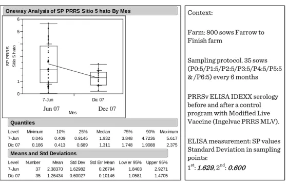
4. Monitoring tools in PRRSv sow herd stabilization programs, Jose R. Angulo, Boehringer Ingelheim Animal Health GmbH, Ingelheim am Rhein, Germany
Posters:
Evaluation of the new ADIAVET™ PRRS REALTIME PCR kit for screening of European and North American PRRS viruses.
Patrice Gracieux, ADIAGENE, Saint Brieuc, France
Reliable detection and typing of PRRSV using multiplex real-time RT- PCR. Christine Gaunitz, Labor Diagnostik GmbH Leipzig, Germany Development of a new IDEXX ELISA for the detection of PRRS antibodies in swine oral fluids. Sergio Lizano, IDEXX Laboratories, Inc., Westbrook, ME, USA
Optimization of oral fluid sampling for the detection of PRRSV TYPE 1. Robert Graage, Clinic for Swine, Vetmeduni Vienna
In situ hybridization to detect porcine reproductive and respiratory syndrome virus. Dinko Novosel, Croatian Veterinary Institut, Zagreb, Croatia
10:30 – 11:00 Coffee break with posters
11:00 – 13:00 Session 2
Epidemiology and genetic diversity of PRRS infection Chairs: Tomasz Stadajek, Jean-Pierre Frossard
11:00 – 11:30 Keynote speech: (keynote) Overview on Chinese PRRS situation Hanchun Yang, College of Veterinary, Medicine, China Agricultural University, Beijing 30'
11:30 – 12:30 Five 15 min oral presentations:
1. Porcine reproductive and respiratory syndrome virus: Antigenic and molecular diversity of British isolates and implications for diagnosis. Jean-Pierre Frossard, AHVLA - Weybridge, UK 2. Genetic diversity of Central and Eastern European PRRSV strains
(Austria, Czech Republic, Hungary, Serbia, Croatia, Romania).
Balka Gyula, SzIU, FVS, Budapest, Hungary
3. The spread of porcine reproductive and respiratory syndrome virus in Ukraine from 2005 to july 2012 years. Olia Ivashchenko, Kyiv National University named after Taras Shewchenko
4. "Impact of genetic variation and geographic distribution of Porcine Reproductive and Respiratory Syndrome virus on infectivity and pig growth " Sara Botti, Parco Tecnologico Padano – CERSA, Lodi, Italy
5. Evidence of PRRSV recombination from field samples in Northeastern Italy, Giovanni Franzo, University of Padova, Legnaro, Italy
Posters:
New, non-conventional clinical manifestation of porcine reproductive and respiratory syndrome (PRRS) in Hungary. Sándor Kecskeméti, National Food Chain Safety Office Veterinary Diagnostic Directorate, Debrecen, Hungary
A serological survey on porcine reproductive and respiratory syndrome (PRRS) virus infection in Serbian wild boars population.
Tamas Petrovic, Scientific Veterinary Institute “Novi Sad”, Serbia PRRS virus sequence diversity in the Czech Republic, Jitka Janková, University of Veterinary and Pharmaceutical Sciences Brno,
Czech Republic
Molecular epidemiology of highly pathogenic PRRS in Vietnam 2007- 12, Nguyen Tung, National Center for Veterinary Diagnosis, Hanoi, Vietnam
PRRSV and other co-infecting agents in PMWS affected pigs, Anna Jackova, University of Veterinary Medicine and Pharmacy, Kosice, Slovakia
12:45 – 13:45 Lunch
13:45 – 15:15 Group discussion (joint epidemiology and diagnostics topics):
– Epidemiology topics discussion chaired by and Tomasz Stadejek and Jean-Pierre Frossard
– Diagnostics topics discussion chaired by Lars Larsen and Katarzyna Podgórska
15:15 – 15:45 Coffee break with posters
15:45 – 17:30 Session 3
Control and Eradication of PRRS
Chairs: Spyridon Krytas and Tom Duinhof
15:45 – 16:15 Keynote speech: Views on control and eradication of PRRS in Europe.
Anders Elvstrøm (Swine Practitioner, Denmark) Five 15 min oral presentations:
1. Experience on laboratory results on PRRSV control and eradication possibilities. Luc Mieli LDA22, Poufragan, France 2. PRRS in the Netherlands: Actual status, economic impact, planned
actions. Tom Duinhof Animal Health Service, Deventer, The Netherlands
3. Economic evaluation of two PRRSV-elimination strategies.
Maaike Gonggrijp, Animal Health Service, The Netherlands 4. PRRSV control in Denmark: Status and perspectives.
Charlotte Sonne Kristensen, Pig Research Centre, Danish Agriculture & Food Council, Denmark.
5. Evaluation of a local pilot PRRSv elimination program in Brittany.
Catherine Belloc, INRA, Nantes, France 18:00 – 19:00 Danube Boat Trip
19:30 –24:00 Gala Dinner (120 people)
Friday, 12 October 2012
09:00 – 11:30 Session 4
PRRS immunology
Chairs: Tahar Ait-Ali and Sara Botti
09:00 – 09:30 Keynote speech: TBC Armin Saalmüller, Clinical Immunology, Veterinary University of Vienna, Austria
09:30 – 10:00 Keynote speech: Immunopathological Consequences of PRRSV Infection Michael Murtaugh (University of Minnesota, St Paul, Minnesota, USA)
10:00 – 11:00 Four 15 min oral presentations:
1. Pig immune response to general stimulus and to Porcine Reproductive and Respiratory Syndrome Virus infection:
a meta-analysis approach, Bouabid Badaoui (Parco Tecnologico Padano – CERSA, Lodi, Italy)
2. Effect of infection of monocyte-derived dendritic cell infection with PRRSV on the expression of differentiation and activation antigens and the influence on T-cell activation,
Irene Rodríguez-Gómez, University of Córdoba, Spain 3. Regulatory T-cells in PRRS virus infection, Enric Mateu
(UAB, Spain)
4. M1 polarized macrophages are resistant to genotype 1 but not to highly pathogenic genotype 2 PRRSV, Obdulio García-Nicolás (University of Murcia, Spain)
Posters:
Putative microchimerism involvement in the pathogenesis of congenital PRRSV infection. Uladzimir Karniychuk, Laboratory of Virology, Ghent University, Belgium
Genetic variation in response to PRRS, Pramod K. Mathur, TOPIGS Research Center, Beuningen, Netherlands
11:00 – 11:30 Coffee break with posters
11:30 –13:30 Group discussion (joint control/eradication and immunology topics):
– Control and eradication discussion chaired by and Spyridon Krytas and Tom Duinhof
– Immunology discussion chaired by Tahar Ait-Ali and Sara Botti 13:30 –14:30 End of meeting and Lunch
Keynote address
DEVELOPMENT OF VACCINE VECTORS BASED ON CORONAVIRUS GENOMES TO PROTECT AGAINST PRRSV AND SARS-COV
Luis Enjuanes, Marta L. DeDiego, Jose, L. Nieto, Jose M. Jimenez, Jose A. Regla, Raul Fernandez, Martina Becares, Sonia Zuñiga, Aitor Nogales, Silvia Marquez, Fernando Almazan,
Pedro Mateos-Gomez, Lucia Morales and Isabel Sola
National Center of Biotechnology
Spanish National Research Council (CNB-CSIC).
Darwin 3. Campus Universidad Autonoma. Cantoblanco. Madrid. Spain
The induction of long-term protection against virus-induced diseases requires the development of modify attenuated viruses derived from the virus that we try to prevent.
In some cases, this is possible because the live modified virus does not have dominant components that interfere with the induction of a protective immune response, as I will describe for the severe and acute syndrome virus (SARS-CoV). In contrast, in other cases, such as for the porcine respiratory and reproductive syndrome virus (PRRSV), this is a less clear possibility as a consequence of the presence of many viral encoded proteins that inhibit the onset of a protective immune response on due time.
In these cases, protection could in principle be induced by the selective expression of the viral antigens involved in protection, using a vector that, ideally, should be specific for the animal species that we try to protect. Today, a comprehensive amount of data generated both in Europe, Asia and in the States have identified potential correlates of protection, but also viral proteins that counteract the onset of an effective immune response, which should be removed from the antigen used to elicit protection. Albeit the enormous progress made, the control of PRRSV is not fully possible jet, whereas, in principle, the control of other potential emerging disease such of SARS, seems to have a more successful outcome.
Relevant progress in the molecular basis of coronavirus replication, transcription, virus host-interaction, and on the engineering of coronavirus genomes as vaccine vectors, has been made in order to control viruses with high relevance in animal and human health. The progress of our laboratory has had significant impact in: (i) the development of reverse genetics systems to engineer coronavirus genomes as vaccine vectors; (ii) the control of coronavirus replication and transcription to increase protective antigen expression levels; and (iii) the identification of genes involved in the virulence of transmissible gastroenteritis virus (TGEV) and SARS-CoV to generate modified live attenuated viruses.
We have shown that coronavirus transcription is guided mainly by the free energy released in the base pairing between an RNA sequence motif located in the viral genome, and a complementary sequence synthesized during transcription. In addition, a transcription enhancer has been identified that is very useful to increase the expression levels of the protective antigens.
The projects on the molecular basis of TGEV and SARS-CoV virulence also led to the identification of genes responsible for virus attenuation. The mechanism involved in these processes showed that defined viral proteins were responsible for the regulation of inflammation induction by TGEV and SARS-CoV, by other respiratory viruses, and by drugs inducing inflammation by different mechanisms. Using information from these basic studies we have engineered the first recombinant vaccine that protects against SARS-CoV infection in three independent animal model systems. Also, significant progress has ben made in the development of a vaccine for PRRSV, although additional developments are still required in this case to overcome the partial stability of the heterologous genes in the vaccine vector. This limitation is being solved in collaboration with members of the PoRRScon EU consortium.
Vectored vaccines for PRRSV based on TGEV virus genome have been constructed.
These vectors expressed PRRSV M protein and GP5 mutants with altered glycosylation patterns, since it has been proposed that removal of the glycosylation sites could lead to the improvement of the immune response against PRRSV.
Vaccinated animals showed significant humoral responses against PRRSV GP5 and M proteins. Nevertheless, the immune response elicited by these vectors did not provide full protection. Two hypotheses may explain these results. The first one is the limited stability of GP5 expression, mainly due to GP5 protein toxicity, that resulted in a significant loss of GP5 expression. The second one is the presence of domains inducing negative regulatory T cells (Treg) in the expressed proteins. To improve recombinant TGEV (rTGEV) vector stability, a rTGEV co-expressing M protein and the 68 most N- terminal amino acids of the GP5 protein (GP5-ecto) was designed. This GP5 protein fragment included the epitopes eliciting the immune response against GP5-ecto protein, but lacking protein toxic domains or described negative Treg signals.
Protection could be increased by the co-expression of GP3 and GP4 PRRSV surface antigens.
To generate attenuated SARS-CoVs the molecular determinants contributing to its high virulence were studied. To this end, infectious cDNA clones encoding SARS-CoV lacking E gene (SARS-CoV-∆E) non-adapted or adapted to grow in mice were constructed, and the corresponding deletion virus mutants were rescued. Both types of SARS-CoV-∆E were attenuated in hamsters, conventional mice and transgenic mice expressing the SARS-CoV receptor mACE-2. The attenuation mechanism of SARS- CoV-∆E was studied by comparing the differential gene expression in cells infected with SARS-CoV with or without E gene. Stress response genes were preferentially upregulated during infection in the absence of gene E. Interestingly, the addition of E protein in trans reversed the increase of stress response genes observed in cells infected by SARS-CoV-∆E and also considerably reduced the cellular stress induced by respiratory syncytial virus, or by drugs eliciting cell stress by different mechanisms. In addition, SARS-CoV E protein down-regulated the unfolded protein response and limited cell apoptosis induced during the infection. Conventional mice infection with a mouse adapted SARS-CoV reproduced many aspects of the human disease.
Accordingly, a mouse adapted SARS-CoV-∆E with an attenuated phenotype in mice was engineered. The expression of proinflammatory cytokines was clearly reduced in SARS-CoV-∆E-infected cells and in virus-infected lungs compared to SARS-CoV- infected cells and lungs. Furthermore, a reduction in neutrophil migration to sites of lung inflammation was observed in mice infected with mouse-adapted SARS-CoV-∆E, probably contributing to the lower degree of inflammation detected in conventional mice, hamsters and transgenic mice expressing the SARS-CoV receptor, hACE-2. This reduction of inflammation in the absence of E gene most likely contributed to the attenuation of SARS-CoV-∆E. This deletion mutant virus provided protection against challenge with homologous and heterologous pathogenic SARS-CoV strains in hamsters and transgenic mice. Furthermore, the mouse adapted ∆E virus mutant provided complete long-term protection against a virulent mouse adapted SARS-CoV in both young and old Balb/c mice, indicating that SARS-CoV-∆E is a very promising vaccine candidate.
In conclusion, the engineering of species specific coronavirus genomes may be very useful to elicit protection against coronavirus induced diseases, and also as vaccine vectors to protect against heterologous viruses. Nevertheless, in the case of PRRSV, additional studies to define the correlates of protection and the stability of their expression is still needed.
Session 1
Diagnostics of PRRS infection
Chairs:
Lars Erik Larsen, Katarzyna Podgórska
COMMERCIAL AND IN-HOUSE RT-PCR METHODS FOR DETECTION OF PRRSV.
DESIGN, PERFORMANCE AND PITFALLS
Charlotte Kristiane Hjulsager
National Veterinary Institute; Technical University of Denmark – ckhj@vet.dtu.dk
Abstract
RT-PCR is a widely used method for detection of PRRSV, because it is a rapid, sensitive and highly specific detection tool. However, the profound diversity and rapid evolution of PRRSV genomes complicates the development of highly sensitive and robust assays.
Several factors need to be taken into consideration when using RT-PCR for diagnosis of PRRSV.
Introduction
Porcine reproductive and respiratory syndrome virus (PRRSV) is an enveloped positive sense single-stranded RNA virus that belongs to the order Nidovirales, family Arteriviridae, genus Arterivirus (Cavanagh. 1997). PRRSV causes reproductive failure in sows and respiratory disease mainly in younger pigs (Rossow. 1998), and has major animal-welfare and economic impacts on swine industries world-wide. PRRSV isolates are divided into genotypes 1 or 2, also known as the European (EU) and North American (NA) types, respectively. Considerable genomic diversity exists both between and within genotypes, and this poses a major challenge to the design of adequate diagnostic RT-PCR detection methods.
Detection of PRRSV by RT-PCR
Reverse transcriptase PCR (RT-PCR) is used for detection of the genetic material (genome) of PRRSV in clinical specimens such as serum, lung tissue, oral fluid, semen etc. First the viral RNA is extracted from the sample material, often as total RNA preparation. A reverse transcriptase (RT) step converts the RNA to cDNA, which is exponentially amplified by polymerase chain reaction (PCR). The result is visualised by agarose gel electrophoresis (conventional RT-PCR) or followed in real-time by the
emission of fluorescence during amplification (real-time RT-PCR). The amount of emitted fluorescence is proportional to the amount of PCR product generated. The RT and PCR steps can be performed in separate tubes (two-step RT-PCR), or in a single tube (one-step RT-PCR). Many assays for routine diagnostics use the convenient one- step format.
RT-PCR has been increasingly used as the preferred method for detection of PRRS virus, because it is a rapid, sensitive and highly specific detection tool. A vast number of conventional RT-PCR assays have been published (Van Woensel et al. 1994; Suárez et al. 1994; Christopher-Hennings et al. 1995; Oleksiewicz et al. 1998). Primers for PRRSV detection have also been included in a number of conventional multiplex RT-PCR assays for simultaneous detection of several pathogens (Giammarioli et al. 2008; Liu et al. 2011; Xu et al. 2012). In recent years, real-time RT-PCR has replaced conventional RT-PCR as the preferred tool for the routine detection of PRRS virus in diagnostic specimens. Several different technical platforms and assay chemistries have been employed, but most published assays rely on intercalating fluorescent dyes such as SYBR Green I (Lurchachaiwong et al. 2008; Martinez et al. 2008; Tian et al. 2010) or dual-labeled (Taqman) probes (Egli et al. 2001; Revilla-Fernandez et al. 2005;
Kleiboeker et al. 2005). More recently, an assay using the Primer Probe Energy Transfer (PriProET) chemistry has been published (Balka et al. 2009). This chemistry is particularly suitable for detection of diverse viruses, because signaling is less dependent on perfect match in probe region than for other probe based chemistries.
Many RT-PCR assays are designed to simultaneously detect PRRSV and discriminate between genotypes, because this provides important information for the control of disease.
RT-PCR is a very sensitive method, and special care should be taken to avoid cross- contamination of samples that may lead to the generation of false positive results, both in the field during sampling as well as in laboratory. The laboratory procedures should be organized to minimize potential contamination. In general, real-time RT-PCR is preferred over conventional RT-PCR for detection, because these assays are more sensitive and offers a reduced risk of laboratory cross-contamination because post amplification handling is avoided. Furthermore, real-time assays are quantitative.
Commercial RT-PCR assays
In addition to the wealth of published assays, conventional and real-time RT-PCR assays are commercially available. There are conflicting reports on the performance of these assays and the major drawback is that the primer sequences are not publicly exposed. However, some of the commercial assays have been extensively evaluated in order to obtain market authorization and their performance is being continuously
monitored in order to provide updates of reagents (Toplak et al. 2012; Harmon et al.
2012).
Assay design
The diagnostic performance of a given RT-PCR assay rely on several factors, however, some important general factors can be highlighted for an assay to be effective for determining the absence or presence of a pathogen. First of all it is essential that the primers and probes are designed to target a conserved region of the genome. This is somewhat problematic with PRRSV, being an RNA virus undergoing rapid evolution and with a high degree of genetic variability (Chang et al. 2002). Even minor mismatches between the target and the primers and probe can cause false-negative results (Klungthong et al. 2010). Many of the available diagnostic RT-PCR assays are targeting the conserved ORF7 region, however, sequence comparisons between and among type 1 and 2 viruses have revealed significant sequence variability in ORF7 (Stadejek et al. 2006; Yoon et al. 2012), and this may have profound negative impact on the sensitivity of the assays. It is also import to consider the structure predictions of the PCR target, because secondary structure formations during PCR may render the target poorly accessible to the primers (Peters et al. 2004). Most published RT-PCR assays include only one pair of primers and, in the case of real-time probe based assays, a single probe for each target. However, to enhance the detection of the genetically diverse population of PRRSV, it has become more common to include multiple primers and probes in a single assay (Harmon et al. 2012).
The RNA extraction procedure may also have a major impact on assay performance, and it is necessary to validate the extraction procedure for each kind of sample material to be tested – i.e. a protocol that works well with one sample material may not be equally applicable to another sample material (Christopher-Hennings et al. 2006). Oral fluid extracted from ropes chewed by pigs, is a recent addition to the variety of diagnostic specimens for PRRSV testing. The importance of assay validation is emphasized by the need to optimize the RNA extraction procedure for this sample material to avoid PCR inhibition (Chittick et al. 2011). Furthermore, to avoid false- negative results due to PCR inhibition or extraction failure, it is recommended to include extraction controls in the set-up (Revilla-Fernandez et al. 2005).
To reduce cost related or/and to increase the sampling size, pooling of samples is sometimes preferred, however this may have a negative influence on the sensitivity of testing (Rovira et al. 2007; Harmon et al. 2012).
Conclusions/Recommendations
A huge variety of different RT-PCR assays and protocols are used in public and private diagnostic laboratories throughout Europe. Only some of these assays have been
published, and validation data are in general not available. It is therefore of outermost importance, that research activities are launched with the focus to improve, validate, implement and standardizes the diagnostic procedures used in Europe and globally.
Recently, a ring trial to evaluate PRRSV real-time RT-PCR detection methods in several European laboratories showed, that none of the in-house or commercial assays tested, could identify all different PRRSV strains with an optimal analytical and diagnostic sensitivity (Wernike et al. 2012), and a combination of methods was recommended.
Furthermore, PRRSV is a single stranded RNA virus, which is prone to antigenic drift.
As described, studies have shown that some European countries such as Lithuania, Latvia, Belarus and Russia harbor exceptionally diverse EU-genotype PRRSV strains (Stadejek et al. 2008). Alignments of these strains with the primer and probe sequences of published PRRSV RT-PCR assays indicated that the majority of the assays would not be able to recognize these recent strains. Updated PRRSV sequence data from many parts of Europe is very sparse; therefore, it is not clear if various assays in diagnostic use will produce a substantial number of false negative results when field samples are tested. Thus, it is obvious that there is a need for surveillance programs with the aim to continuously monitor the drift of PRRSV by sequencing subsets of circulating strains and by building joint PRRSV databases with public accessibility. The sequencing should focus on, but not be limited to ORF5 and ORF7. Full genome sequence data are presently sparse in GenBank, but when more such sequences become available, it is the hope to identify a more conserved region than the widely used ORF7, which could be the target for sensitive and robust real-time RT PCR assays.
To maximize the diagnostic performance of RT-PCR assays, it is necessary to perform in-house validation of assays, including the RNA extraction procedures, for each diagnostic specimen. It is also very important to continuously check the applicability of primers and probes for their ability to detect new variants of PRRSV. This can be done by in silico analysis and by participation in ring trials.
References
1. Balka, G., Hornyak, A., Balint, A., Benyeda, Z., Rusvai, M., 2009. Development of a One-Step Real-Time Quantitative PCR Assay Based on Primer-Probe Energy Transfer for the Detection of Porcine Reproductive and Respiratory Syndrome Virus. J. Virol. Methods 158, 41-45.
2. Cavanagh, D., 1997. Nidovirales: A New Order Comprising Coronaviridae and Arteriviridae. Arch. Virol. 142, 629-633.
3. Chang, C.-., Yoon, K.-., Zimmerman, J.J., Harmon, K.M., Dixon, P.M., Dvorak, C.M.T., Murtaugh, M.P., 2002. Evolution of Porcine Reproductive and Respiratory Syndrome Virus during Sequential Passages in Pigs. Journal of Virology 76, 4750- 4763.
4. Chittick, W.A., Stensland, W.R., Prickett, J.R., Strait, E.L., Harmon, K., Yoon, K., Wang, C., Zimmerman, J.J., 2011. Comparison of RNA Extraction and Real-Time Reverse Transcription Polymerase Chain Reaction Methods for the Detection of Porcine Reproductive and Respiratory Syndrome Virus in Porcine Oral Fluid Specimens. Journal of Veterinary Diagnostic Investigation 23, 248-253.
5. Christopher-Hennings, J., Dammen, M., Nelson, E., Rowland, R., Oberst, R., 2006.
Comparison of RNA Extraction Methods for the Detection of Porcine Reproductive and Respiratory Syndrome Virus from Boar Semen. J. Virol.
Methods 136, 248-253.
6. Christopher-Hennings, J., Nelson, E.A., Nelson, J.K., Hines, R.J., Swenson, S.L., Hill, H.T., Zimmerman, J.J., Katz, J.B., Yaeger, M.J., Chase, C.C., 1995. Detection of Porcine Reproductive and Respiratory Syndrome Virus in Boar Semen by PCR.
Journal of Clinical Microbiology 33, 1730-1734.
7. Egli, C., Thur, B., Liu, L., Hofmann, M.A., 2001. Quantitative TaqMan RT-PCR for the Detection and Differentiation of European and North American Strains of Porcine Reproductive and Respiratory Syndrome Virus. J. Virol. Methods 98, 63- 75.
8. Giammarioli, M., Pellegrini, C., Casciari, C., De Mia, G.M., 2008. Development of a Novel Hot-Start Multiplex PCR for Simultaneous Detection of Classical Swine Fever Virus, African Swine Fever Virus, Porcine Circovirus Type 2, Porcine Reproductive and Respiratory Syndrome Virus and Porcine Parvovirus. Vet. Res.
Commun. 32, 255-262.
9. Harmon, K.M., Abate, S.A., Chriswell, A.J., Chittick, W.A., Strait, E.L., 2012.
Comparison of Two Commercial Real-Time Reverse Transcriptase Polymerase Chain Reaction Assays for Porcine Reproductive and Respiratory Syndrome Virus. J Swine Health Prod. 20, 184-184-188.
10. Kleiboeker, S.B., Schommer, S.K., Lee, S.M., Watkins, S., Chittick, W., Polson, D., 2005. Simultaneous Detection of North American and European Porcine Reproductive and Respiratory Syndrome Virus using Real-Time Quantitative Reverse Transcriptase-PCR. J. Vet. Diagn. Invest. 17, 165-170.
11. Klungthong, C., Chinnawirotpisan, P., Hussem, K., Phonpakobsin, T., Manasatienkij, W., Ajariyakhajorn, C., Rungrojcharoenkit, K., Gibbons, R.V., Jarman, R.G., 2010. The Impact of Primer and Probe-Template Mismatches on the Sensitivity of Pandemic Influenza A/H1N1/2009 Virus Detection by Real- Time RT-PCR. Journal of Clinical Virology 48, 91-95.
12. Liu, S., Zhao, Y., Hu, Q., Lv, C., Zhang, C., Zhao, R., Hu, F., Lin, W., Cui, S., 2011. A Multiplex RT-PCR for Rapid and Simultaneous Detection of Porcine Teschovirus, Classical Swine Fever Virus, and Porcine Reproductive and Respiratory Syndrome Virus in Clinical Specimens. J. Virol. Methods 172, 88-92.
13. Lurchachaiwong, W., Payungporn, S., Srisatidnarakul, U., Mungkundar, C., Theamboonlers, A., Poovorawan, Y., 2008. Rapid Detection and Strain Identification of Porcine Reproductive and Respiratory Syndrome Virus (PRRSV) by Real-Time RT-PCR. Lett. Appl. Microbiol. 46, 55-60.
14. Martinez, E., Riera, P., Sitja, M., Fang, Y., Oliveira, S., Maldonado, J., 2008.
Simultaneous Detection and Genotyping of Porcine Reproductive and Respiratory Syndrome Virus (PRRSV) by Real-Time RT-PCR and Amplicon Melting Curve Analysis using SYBR Green. Res. Vet. Sci. 85, 184-193.
15. Oleksiewicz, M.B., Botner, A., Madsen, K.G., Storgaard, T., 1998. Sensitive Detection and Typing of Porcine Reproductive and Respiratory Syndrome Virus by RT-PCR Amplification of Whole Viral Genes. Vet. Microbiol. 64, 7-22.
16. Peters, I.R., Helps, C.R., Hall, E.J., Day, M.J., 2004. Real-Time RT-PCR:
Considerations for Efficient and Sensitive Assay Design. J. Immunol. Methods 286, 203-217.
17. Revilla-Fernandez, S., Wallner, B., Truschner, K., Benczak, A., Brem, G., Schmoll, F., Mueller, M., Steinborn, R., 2005. The use of Endogenous and Exogenous Reference RNAs for Qualitative and Quantitative Detection of PRRSV in Porcine Semen. J. Virol. Methods 126, 21-30.
18. Rossow, K.D., 1998. Porcine Reproductive and Respiratory Syndrome. Vet. Pathol.
35, 1-20.
19. Rovira, A., Clement, T., Christopher-Hennings, J., Thompson, B., Engle, M., Reicks, D., Muñoz-Zanzi, C., 2007. Evaluation of the Sensitivity of Reverse-Transcription Polymerase Chain Reaction to Detect Porcine Reproductive and Respiratory Syndrome Virus on Individual and Pooled Samples from Boars. Journal of Veterinary Diagnostic Investigation 19, 502-509.
20. Stadejek, T., Oleksiewicz, M.B., Potapchuk, D., Podgorska, K., 2006. Porcine Reproductive and Respiratory Syndrome Virus Strains of Exceptional Diversity in Eastern Europe Support the Definition of New Genetic Subtypes. J. Gen. Virol. 87, 1835-1841.
21. Stadejek, T., Oleksiewicz, M.B., Scherbakov, A.V., Timina, A.M., Krabbe, J.S., Chabros, K., Potapchuk, D., 2008. Definition of Subtypes in the European Genotype of Porcine Reproductive and Respiratory Syndrome Virus:
Nucleocapsid Characteristics and Geographical Distribution in Europe. Arch.
Virol. 153, 1479-1488.
22. Suárez, P., Zardoya, R., Prieto, C., Solana, A., Tabarés, E., Bautista, J.M., Castro, J.M., 1994. Direct Detection of the Porcine Reproductive and Respiratory Syndrome (PRRS) Virus by Reverse Polymerase Chain Reaction (RT-PCR).
Archives of Virology 135, 89-99.
23. Tian, H., Wu, J., Shang, Y., Cheng, Y., Liu, X., 2010. The Development of a Rapid SYBR One Step Real-Time RT-PCR for Detection of Porcine Reproductive and Respiratory Syndrome Virus. Virol. J. 7, 90.
24. Toplak, I., Rihtaric, D., Hostnik, P., Grom, J., Stukelj, M., Valencak, Z., 2012.
Identification of a Genetically Diverse Sequence of Porcine Reproductive and Respiratory Syndrome Virus in Slovenia and the Impact on the Sensitivity of Four Molecular Tests. J. Virol. Methods 179, 51-56.
25. Van Woensel, P., Van Der Wouw, J., Visser, N., 1994. Detection of Porcine Reproductive Respiratory Syndrome Virus by the Polymerase Chain Reaction. J.
Virol. Methods 47, 273-278.
26. Wernike, K., Bonilauri, P., Dauber, M., Errington, J., Leblanc, N., Revilla- Fernandez, S., Hjulsager, C., Isaksson, M., Stadejek, T., Beer, M., Hoffmann, B., 2012. Porcine Reproductive and Respiratory Syndrome Virus: Interlaboratory Ring Trial to Evaluate Real-Time Reverse Transcription Polymerase Chain Reaction Detection Methods. J. Vet. Diagn. Invest. 24, 855-866.
27. Xu, X., Chen, G., Huang, Y., Ding, L., Li, Z., Chang, C., Wang, C., Tong, D., Liu, H., 2012. Development of Multiplex PCR for Simultaneous Detection of Six Swine DNA and RNA Viruses. J. Virol. Methods 183, 69-74.
28. Yoon, S.H., Kim, H., Park, B., Kim, H., 2012. Tracing the Genetic History of Porcine Reproductive and Respiratory Syndrome Viruses Derived from the Complete ORF 5-7 Sequences: A Bayesian Coalescent Approach. Arch.Virol.
CHARACTERIZATION OF A CIRCULATING PRRSV STRAIN BY MEANS OF RANDOM PCR CLONING AND FULL GENOME SEQUENCING
Jan Van Doorsselaere
1, Marc Geldhof
2, Hans J Nauwynck
2, and Peter L Delputte
2,31 Department of Health Care and Biotechnology, KATHO Catholic University College of South-West Flanders, Wilgenstraat 32, 8800 Roeselare, Belgium
2 Department Virology, Parasitology and Immunology, Faculty of Veterinary Medicine, Ghent University, Salisburylaan 133, 9820 Merelbeke, Belgium
3 Laboratory of Microbiology, Parasitology and Hygiene (LMPH), Department of Biomedical Sciences, Faculty of Pharmaceutical, Biomedical and Veterinary Sciences Antwerp University, Universiteitsplein 1, B-2610 Antwerp, Belgium peter.delputte@ua.ac.be
PRRS is a pig disease of major economic importance that causes respiratory and reproductive problems in pigs. Over the last years it has become clear that PRRSV heterogeneity continues to increase, which has a potential impact on diagnosis and strategies to counter this disease. The use of sequence-independent PCR techniques for the detection and characterization of PRRSV is a valid approach to overcome problems associated with the heterogeneity of this virus, but has not yet been testen on PRRSV. In this study, a random PCR cloning approach was used to evaluate if the PRRSV strain 07V063 of unknown genetic background that circulated on a Belgian farm could be characterized. Using this approach, 7305 bp of sequence data were generated, and the obtained sequence were distributed randomly across the genome.
With the obtained sequence data, strain-specific primers were developed, and this allowed to obtain the full length sequence (15014 nt) using RT-PCR. Phylogenetic relationships using ORF5 and ORF1a (NSP2) sequences showed that 07V063 was classified in type 1 subtype 1. Furthermore, 07V063 appeared genetically different from prototype Lelystad Virus (LV). The aa identity was between 87-93% with LV ORFs coding for structural proteins. Surprisingly, Nsp2 of strain 07V063 showed most
variation (81% identity compared to LV) with a deletion of 28 aa. This deletion was different from other deletions in this ORF that were previously described.
In conclusion, these results show that in theory, a random PCR cloning approach can be used for the characterization of new PRRSV strains of unknown genetic background.
RESULTS OF PRRSV PROFICIENCY TESTS (PCR AND SEROLOGY) PERFORMED
IN THE FRAME OF TWO EU PROJECTS
Katarzyna Podgórska
National Veterinary Research Institute, Swine Diseases Department, Partyzantow 57 Str, 24-100 Pulawy, Poland
Abstract
Present paper describes evaluation of PCR and serological methods based on two ring trials performed within a frame of two EU projects. The aim of the ring trials was to identify problems in the areas of molecular and serological diagnostic methods in partners’ laboratories and selection of the best-working assays.
Serological ring trial indicated that some ELISA methods are prone to false positive results. Based on the panel of samples used in the PCR ring trial, there is no commercial or in house PCR test with satisfactory analytical and diagnostic sensitivity that would simultaneously enable differentiation between two genotypes. The combination of several methods is highly recommended to assure optimal diagnosis of PRRSV infections. Moreover, applied methods should be constantly validated according to the currently circulating strains.
Introduction
Currently two genotypes of porcine reproductive and respiratory syndrome virus (PRRSV) are known – previously restricted to Europe genotype 1 (EU) and genotype 2 (NA) discovered in North America. Genotype 1 is further divided into subtypes 1, 2, 3 and 4. Most probably both genotypes evolved independently on two continents and their geographical distribution was initially limited. Nowadays both genotypes are detected in Europe, America and Asia. Limited genetic and antigenic similarity between these two genotypes might probably influence serological and molecular diagnostic methods for PRRS (11).
Present paper describes evaluation of PCR and serological methods based on two ring trials performed within a frame of two EU projects. The aim of the ring trials was to identify problems in the areas of molecular and serological diagnostic methods in partners’ laboratories and selection of the best-working assays. Two ring trials including PCR methods and serological assays were organized. PCR ring trial was extended to participants of the two international projects PoRRSCon (6 participants) and EPIZONE (7 participants). The same panel of samples including 28 RNA templates of PRRSV strains representing all genetic subtypes of PRRSV was used.
PRRSV strains used in the ring trials were obtained from the collection of the Friedrich-Loeffler-Institut, Isle of Riems, Germany, or the National Veterinary Research Institute, Pulawy, Poland. Participants used from 1 to 4 PCR methods to analyze obtained samples. The results of the EPIZONE ring trial were recently described by Wernike et al. (11). Present manuscript summarizes results of both PCR ring trials as well as serological ring trial performed within PoRRSCon project.
PCR ring trial. The panel of RNA samples used in the ring trial included two series of dilutions of genotype 1 subtype 1 PRRSV strains and one set of serial dilutions of high pathogenic HP-PRRSV strain of genotype 2 from China. Additionally three other strains of genotype 2 and three strains of genotype 1 subtype 1, as well as samples representing subtypes 2, 3 and 4 classified within genotype 1 and three negative samples were included. Samples were distributed between 13 participants and analysed with 17 PCR assays available in partners’ laboratories. Four commercially available kits and 13 in house tests were applied. Four in house tests were pan-specific and detected all PRRSV genotypes, two other methods were specific for genotype 2 only. Seven remaining methods had an advantage of differentiation between genotype 1 and 2. Four in house tests were based on end-point analysis, others were real-time assays based mostly on TaqMan probes except one SYBR Green I method.
High variability of results was observed between applied methods. Most often problems were false negative results, especially regarding Eastern European strains of genotype 1 (subtypes EU-2, -3 and -4), but also low sensitivity regarding strains of genotype 1 subtype 1 or Chinese strain HP-PRRSV.
Only two of four commercial PCR kits detected all Eastern European PRRSV strains.
However, those kits had lower sensitivity regarding subtype EU-1 strains or HP- PRRSV strain. One of remaining commercial kits did not detect any strains of subtypes EU-2, -3 and -4, and the last one detected only some of them, depending on the laboratory.
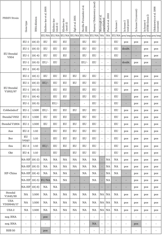
Similar discrepancies were observed also regarding in house PCR assays (Table 1).
Most of the tests except Kleiboeker and Kleiboeker (mod-2) detected all strains belonging to subtypes EU-2, -3 and -4. However, some methods had relatively low sensitivity regarding detection of EU-1 strains. Strain EU Stendal V954 was not
detected at all by method based on Egli et al. (1). Common problem was also low sensitivity in detection of high pathogenic PRRSV strain, which was completely missed by one of methods specific to genotype 2.
Serological ring trial. Serological part of the PoRRSCon ring trial consisted of testing of 9 serum samples. The panel of samples included sera obtained from pigs naturally infected with strains of genotype 1 (subtypes 1, 2 and 3), strains of genotype 2 and three samples from herds free from PRRSV. One of the negative samples was found before to give false positive results in some ELISA tests. Two participants analysed samples with both ELISA and IPMA methods, others used only ELISA (5 participants) or only IPMA (1 participant).
The results of IPMA proved that all applied protocols had characterized good specificity and enabled detection of specific antibodies in all positive samples.
Obtained titres differed depending on applied strain and protocol used.
Five in house ELISA tests and three commercial kits were used in the ring trial. Two of in house tests had an additional benefit of discrimination between genotype 1 and 2.
Two commercial ELISA kits gave false positive results in two different negative samples. Additionally, false positive result was detected in one of the in house tests.
The best results were obtained equally with IDEXX X3, Ingenasa DR and in house ELISA test based on Stadejek et al. (8).
Conclusions
Based on the panel of samples used in the ring trial, there is no commercial or in house PCR test with satisfactory analytical and diagnostic sensitivity that would simultaneously enable differentiation between two genotypes. The combination of several methods is highly recommended to assure optimal diagnosis of PRRSV infections. Moreover, applied methods should be constantly validated according to the currently circulating strains.
Results of serological tests were more consistent. However, it was shown that some ELISA methods are prone to false positive results.
Acknowledgements/Funding
This work was supported by PoRRSCon (FP7/2007-2013, grant agreement n° 245141), EPIZONE (contract FOOD-CT-2006-016236) and a grant from Polish Ministry of Science and Higher Education financed based on the decision No. 808/N- COST/2010/0.
Tomasz Stadejek is thanked for excellent scientific expertise.
Recommendations and References
1. Egli et al. 2001. J Virol Methods 98: 63-75 2. Balka et al. 2009. J Virol Methods 158:41-45
3. Kleiboeker et al. 2005. J Vet Diagn Invest 17: 165-170 4. Oleksiewicz et al. 1998. Vet Microbiol 64: 7-22 5. Oleksiewicz et al. 2000. Virology 267:135-140
6. Revilla-Fernandez et al. 2005 Virol Methods 126:21-30 7. Sørensen KJ et al. 1998. Vet Microbiol. 28;60(2-4):169-77.
8. Stadejek T. et al. 2007. Medycyna Wet 63 (11): 1336-41 9. Suarez et al. 1994. Arch Virol 135:89-99
10. Truyen U. 2006. J Vet Med B Infect Dis Vet Public Health 53:68-74 11. Wernike K. et al. 2012. J Vet Diagn Invest 24 (5): 855-66
Table 1. Results of in house assays used in the PCR ring trial.
PRRSV Strain
Geno type RNA dilution Kleiboe-ker et al. 2005 Kleiboe-ker (mod-1) Kleiboe-ker (mod-2) Egli et al. 2001 Revilla-Fernandez et al. 2005 Suarez/ Oleksiewicz (mod) Murtaugh et al. unpublished Pesch et al. 2003 In house 1 In house 2 (end point) In house 3 (convent) In house 4 (SYBR Green I) Oleksiewicz et al. 1998 (convent) EU/NA EU/NA EU/NA EU/NA EU/NA EU/NA NA NA EU/NA pos/neg pos/neg pos/neg pos/neg
EU Stendal V954
EU-1 10(-2) EU EU EU - EU EU - - EU pos pos pos pos
EU-1 10(-3) EU EU EU - EU EU - - EU doubt. - pos pos
EU-1 10(-4) EU EU EU - EU EU - - EU - - pos pos
EU-1 10(-5) EU/- EU - - EU/- EU - - - doubt. pos pos -
EU-1 10(-6) - - - - - - - - - - - - -
EU Stendal V1952/97
EU-1 10(-1) EU EU EU EU EU EU - - EU pos pos pos pos
EU-1 10(-2) EU/- EU EU EU EU EU - - EU pos pos pos pos
EU-1 10(-3) - EU EU - EU EU - - EU pos pos pos pos
EU-1 10(-4) - EU EU - EU EU - - EU - pos pos pos
EU-1 10(-5) - EU/- - - - EU - - EU - pos pos pos
Cobbelsdorf EU-1 1:500 EU/- EU EU EU EU EU - - EU pos pos pos pos
Stendal V852 EU-1 1:500 EU EU EU - EU EU - - EU pos pos pos pos
Stendal V1904 EU-1 1:500 EU EU EU EU EU EU - - EU pos pos pos pos
Aus EU-2 1:10 - EU EU EU EU EU - - EU pos pos pos pos
Bor EU
(atyp) 1:10 - EU EU EU EU EU - - EU pos pos pos pos
Sza EU-3 1:10 EU/- EU EU EU EU EU - - EU pos pos pos pos
Okt EU-4 1:10 - EU - EU EU EU - - EU pos pos pos pos
HP-China
NA-HF 10(-2) NA NA NA NA NA NA - NA NA pos pos pos pos
NA-HF 10(-3) NA NA NA NA NA NA - NA NA pos pos pos pos
NA-HF 10(-4) NA NA NA - NA NA - NA NA - pos pos pos
NA-HF 10(-5) EU/NA NA NA - - NA - NA - - pos pos pos
NA-HF 10(-6) NA NA - - - - - - - - pos pos pos
Stendal
V1445/99 NA 1:500 NA NA NA NA NA NA NA NA NA pos pos pos pos
VD29949/17 USA NA 1:500 NA NA NA NA NA NA NA NA NA pos pos pos pos
USA 2 NA 1:500 NA NA NA NA NA NA NA NA NA pos pos pos pos
neg. RNA - - pos - - - - - - - - - - - -
neg. RNA - - - - - - - NA - - - - pos - -
RSB 50 - - pos - - - - - - - - - - - -
DETECTION OF PRRSV IN 218 FIELD
SAMPLES USING SIX MOLECULAR METHODS:
WHAT WE ARE LOOKING FOR?
Ivan Toplak
1, Marina Štukelj
2, Patrice Gracieux
3, Balka Gyula
4, Lars Larsen
5, Rolf Rauh
61 University of Ljubljana, Veterinary Faculty, Gerbičeva 60, SI-1115 Ljubljana, Slovenia e-mail: ivan.toplak@vf.uni-lj.si
2 University of Ljubljana, Veterinary Faculty, Cesta v mestni log 47, SI-1115 Ljubljana, Slovenia
3 ADIAGENE, 38 rue de Paris, 22000 Saint Brieuc, France
4 Faculty of Veterinary Science, István u. 2, H-1078 Budapest, Hungary
5 National veterinary institute, Technical University of Denmark, Bülowsvej 27, Copenhagen, Denmark
6 Tetracore Inc, 9901 Belward Campus Drive, Suite 300, Rockville, MD, 20850, USA
Keywords: PRRSV, diagnosis, real-time RT-PCR, sensitivity, field samples
Abstract
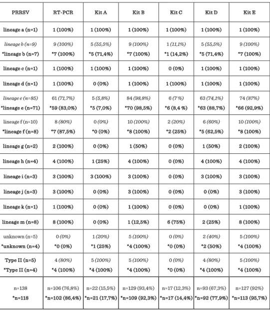
The purpose of this study was to determine the sensitivity of conventional RT-PCR protocol and five commercial real-time kits (kit A - E) used for the detection of porcine reproductive and respiratory syndrome virus (PRRSV). 218 field samples were collected between 2009 and 2011 from 50 PRRSV positive and 45 negative pig herds from Slovenia. According to determined 258 nucleotides long sequences (ORF7) 12 different lineages of Type I - subtype 1 (a=1, b=9, c=1, d=1, e=85, f=10, g=2, h=4, i=3, j=3, k=1, m=8) with 85.7-93.8 % nucleotide identity between lineages and four samples belong to Type II were identified. The highest sensitivity was observed with kit E (96,3%) and with kit B (94,5%), followed by conventional RT-PCR (87,8%) and kit D (82,1%), while the lowest sensitivity was observed with kit A (55,3%) and kit C (53,8%).
Reduced sensitivity was directly related to the some genetic lineages and RNA copy number in a sample. These findings emphasize that diagnostic PCR kits (conventional
and real-time) have to continuously follow the genetic evaluation of the PRRSV subtype viruses especially Type I and regularly update their primer and probes sequences.
Introduction
Porcine reproductive and respiratory syndrome virus (PRRSV) is the causative agent of porcine reproductive and respiratory syndrome (PRRS), which is a devastating multisystem infection of pigs, causing high economic loses in infected herds. The detected high heterogeneity among PRRSV isolates is likely to be the main obstacle for effective control of the disease. The isolates detected in Europe are heterogeneous with 72.2 - 90% homology and sequence reports for PRRSV originated from different countries such as Denmark, Italy, Czech Republic, Poland, Spain, Germany, Netherlands, Hungary, Belorussia and Slovenia supports the development of new diagnostics tolls (Oleksiewicz et al., 2000; Stadejek et al., 2002; Mateu et al., 2003;
Pesch et al., 2005; Indik et al., 2005; Stadejek et al., 2006; Balka et al., 2008; Toplak et al., 2012). Real-time RT-PCR technology using primers and TaqMan probes has successfully been used to detect the genotype and the quantity of PRRSV RNA (Egli et al., 2001; Chung et al., 2005). However, there are several potential problems with PRRSV detection because of the wide variability of the strains (Toplak et al., 2012). The main disadvantages are the design and the validation of specific functional probes to ensure the diagnostic sensitivity and specificity of the assay and the avoidance of false- negative results due to variability within the probe-binding site (Hughes et al., 2004).
This is the follow-up study after the detection of new strains in Slovenia and low sensitivity for two commercial real-time PCR kits and (Toplak et al., 2012). The sensitivity of six molecular methods was determined on 218 field samples.
Material and methods
218 field samples (serum, tissues) were collected between 2009 and 2011 from 50 PRRSV positive and 45 negative pig herds from Slovenia. Total viral RNA was extracted from original samples and stored in aliquots at < -60 °C until analysis.
Conventional RT-PCR with gel-electrophoresis and direct sequencing of RT-PCR products was performed as described previously (Donadeu et al., 1999; Toplak et al., 2012). For determination of the diagnostic sensitivity five commercially available real- time kits were used: TaqVet® PRRS Triplex PRRS-Genotyping (LSI, France), ADIAVET® PRRS Real-Time, (Adiagene, France), PRRS Real-Time Detection Kit (BioNote, Inc., Korea), NextGen Real-Time RT-PCR Target Specific Reagents for the Identification & Differentiation of North American & European PRRSV Viral RNA (Tetracore®, USA) and TaqMan® PRRSV Reagents and Controls one step qRT-PCR for NA and EU PRRSV RNA for detection differentiation of European/American PRRSV (Applied Biosystems, USA), named in following sentences as kit A, B, C, D and E were
tested. All field samples were analyzed with five commercial real-time RT-PCR kits and results of conventional RT-PCR were included into final comparison. Samples were tested using the same batch of RNA for different test, real-time PCR mix were prepared according to the instructions of producer of each kit.
Results
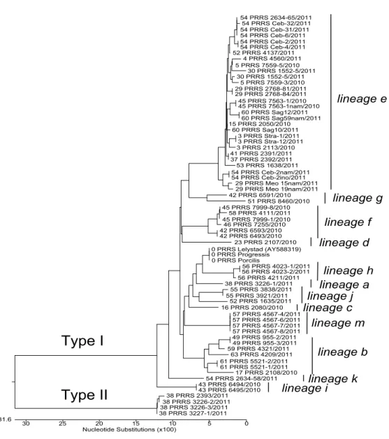
According to 258 nucleotides long sequences (258 nucleotides of ORF7, genome position 14.673 - 14.927 in the Lelystad virus, GenBank accession number M96262) determined for positive samples it was shown that 128 PRRSV positive samples collected in Slovenia belong to Type I (identification of 12 different lineages of EU subtype 1: a=1, b=9, c=1, d=1, e=85, f=10, g=2, h=4, i=3, j=3, k=1, m=8) with 85.7-93.8%
nucleotide identity between lineages (Figure 1). Five samples belong to Type II with 99.6-100% nucleotide identity between samples and reference strain VR-2332 (U87392). Five additional samples were identified as positive using one or more different real time kits, but were not detected as positive by conventional RT-PCR, thus for these samples the type of virus was not identified by sequencing (named in Table 1 as unknown type). The estimated copy number of PRRSV RNA for all 138 positive samples was determined according Ct value obtained with kit E using 10-fold dilutions of positive control with known copy number and estimated RNA copy numbers for samples was calculated from Ct values obtained in standard curve. The determined limit of detection for kit E is 40 copies of Type I or type II PRRSV RNA and this number was put as reference for determination of the sensitivity of other five kits.
20 out of 138 positive samples PRRSV was detected mainly with kit B and kit E which have the highest sensitivity, but according the obtained results of Ct value with kit E, sample contain less than 40 PRRSV RNA copies (in range between 1 and 25 copies).
Results for these 20 samples were excluded for determination of the sensitivity of all six methods. The highest sensitivity was observed with kit E (96,3%) and with kit B (94,5%), followed by conventional RT-PCR (87,8%) and kit D (82,1%), while the lowest sensitivity was observed with kit A (55,3%) and kit C (53,8%). Reduced sensitivity was directly related to the genetic lineages. For kits A and C we have observed problems with low sensitivity for almost all genetic lineages included in this study, thus this two kits are not suitable for testing field samples on our territory. The majority problems in kit D were observed with the detection of only 88,7% positive samples of lineage e, 71,4% of lineage b, 62,5% of lineage f, 50% of lineage g and 25% of lineage m. The results for kit D suggest that this kit have also higher limit of detection comparing to the kits B and E (Table 2). The results for conventional RT-PCR showed that this protocol is highly sensitive for the detection of different lineages of PRRSV, but have higher limit of detection to two real-time kits B and E. As a result of this lower sensitivity of RT- PCR, 12 samples of lineage e were not detected as positive. Kit B have almost perfect sensitivity, but have problem with the detection of seven samples from lineage m (12.5%) and one sample from lineage g (50%). Kit E has the highest sensitivity, but we observed that five samples from lineage e originated from the same farm were not
detected as positive. The percentages of detected positive samples for each lineage are presented in Table 1. The results with obtained Ct values for two field strains from lineage e (Meo15nam/2011 and Ceb2nam/2011) using 10-fold dilution and conventional RT-PCR together with five commercial real time kits (Kit A - Kit E) are presented in Table 2.
Discussion and conclusion
The study showed that the performances of commercial RT-PCR assays are highly dependent on the genetic make-up of the target viruses and RNA copy number in a sample. When compared the results of this study with findings of a previous study (Toplak et al., 2012) we showed great improvement for new version of kit B which was reconstructed by company after observing the poor results of previous study. Another commercial kit E have the highest sensitivity, but five positive samples of lineage e originated from the same herd were recognized as negative, suggesting that this strain can not be detected, even in high copy number in sample. Nevertheless, some commercial PCR kits failed to detect more detected lineages in this study or only some specific genetic lineages of PRRSV circulating in Slovenia. This high genetic diversity of detected strains on small territory is probably the results of importation of infected pigs from many different locations. Thus, these findings emphasize that it is crucial that diagnostics laboratories and the manufactures of diagnostic PCR kits (conventional and real-time) continuously follow the genetic diversity of PRRSV in area continuing the evaluation of diagnostics test for especially Type I PRRSV subtype viruses and regularly update their primer and probes sequences.