Investigation of internal dynamics modulated by local interactions as a biological regulatory
mechanism in globular proteins
Thesis excerpt
Bertalan Kovács 2019
Pázmány Péter Catholic University
Roska Tamás Doctoral School of Sciences and Technology Supervisor: Dr. Zoltán Gáspári
2
1 Introduction
1.1 The N-terminal PDZ1-2 tandem of PSD-95
The postsynaptic density protein 95 (PSD-95) plays key roles in a wide variety of biological processes. It is involved in clustering membrane receptors in the postsynaptic density, serves as an adaptor protein in signal transduction and regulates the threshold potential inducing long-term potentiation and depression. (Kim and Sheng 2004; Lee and Zheng 2010; Sheng and Sala 2001).
Its two N-terminal PDZ domains form an independent structural and dynamical unit termed PDZ tandem.
The diversity of binding partners of PSD-95 is perplexing. It is unclear how it discriminates between its many possibly ligands and how the supramodular reorientation of the PDZ1-2 tandem is regulated, which allows for the interaction with binding partners located in different layers of the postsynaptic density.
Our understanding of the ligand specificity of the PDZ domain is evolving. It turned out that the canonical binding mode, traditionally categorized into three classes, does not explain the fine-tuned affinity of the PDZ domain towards many possible ligands with a sufficient level of detail.
Beyond the GLGF binding motif and the hydrophobic core stabilizing the C- terminal hydrophobic (usually valine) residue, more and more emphasis is put on the role of the β2-β3 loop which interacts with upstream residues of the ligand (Mostarda, Gfeller, and Rao 2012). The dynamics of this loop were proven to decrease upon ligand binding both in fast and slow timescales.
(Fuentes, Der, and Lee 2004).
In general, the function of PDZ tandems is different from the simple sum of the two constituting domains. However, the extent of the interdomain interaction in the PSD-95 PDZ1-2 tandem, which would indicate some kind of synergy between the two domains, is barely noticeable. NMR experiments have shown that the complexed form of PDZ1-2 exhibits increased interdomain dynamics compared to the free form (Wang et al. 2009). Furthermore, many studies have attempted to determine the supramodular structure of the PDZ1-2 tandem, but the outcome of these partially contradicted each other in terms of the relative orientation of the two domains as well as the possible location of
3
the binding partners relative to PSD-95. Therefore, the determinative interdomain interactions stabilizing the supramodular structure and their connection to the presence of the binding partner remain to be identified.
1.2 Parvulin-type peptidyl-prolyl cis-trans isomerases
Peptidyl-prolyl cis-trans isomerases (PPIase) or rotamases catalyze the cis-trans isomerization of a peptide bond preceding a proline residue (Hanes 2015). They are ubiquitous enzymes found in all cellular compartments and take part in various biological processes, such as protein folding, cell-cycle regulation, apoptosis or gene transcription. They are the therapeutic target in the treatment of several diseases because of their frequently witnessed overexpression in diseased cells, even to the extent that their presence might be regarded a marker of the illness. All of these biological functions put PPIases in the center of the stage of cellular biology (Göthel and Marahiel 1999; Lu et al. 2007).
Even though they catalyze a relatively simple chemical reaction, their exact catalytic mechanism is still unknown. It is generally accepted however, that the catalysis occurs through a twisted amide transition state where there is no breaking and reforming of the peptide bond.
In parvulin-type rotamases, the binding pocket is surrounded by a small and a large lobe. Several pieces of evidence indicate that two conserved histidine residues found in the large lobe, and a hydrogen bonding network formed by five residues – including the two histidine residues, and a further cysteine and two serine residues – play an important role in the catalysis. It turned out however, that the double histidine-mutant form as well as the cysteine point mutant remain catalytically active (Bailey et al. 2008; Terada et al. 2001). Furthermore, only minimal chemical shift change was detected upon ligand binding which makes it unlikely that these residues directly interact with the ligand. These results suggest that the conserved histidine residues and the formation of the hydrogen bonding network play a structural-dynamical role, rather than directly taking part int the catalytic mechanism.
Pin1-type parvulins act in a phosphorylation-dependent manner: the residue preceding the proline must be a phospho-threonine or phospho-serine.
These parvulins contain, in addition to the catalytic domain, an N-terminal WW domain, the role of which is not fully understood. Since point mutants, in which the interaction between the two domains is hindered, are inactive, it was
4
proposed that the WW domain allosterically regulates the dynamics of the catalytic domain. (Olsson et al. 2016).
1.3 Dynamic structural ensembles
In the past two decades the approach has become more and more widespread, and eventually generally accepted, according to which the dynamic nature of proteins is a determining factor in their biological function.
Among the atomic-level structure determination methods, nuclear magnetic resonance (NMR) spectroscopy is the method of choice when investigating internal motions of proteins occurring on different timescales. Fast (ps-ns) internal motions include the rotation of side chains and the dynamics of the backbone. To indirectly observe these motions, measuring the amid 15N relaxation times is the most frequently used approach. The Lipari-Szabo model- free analysis of relaxation data yields the general order parameters (S2) which reflect how restrained the fluctuation of each relaxation vector is, or more intuitively, how wide the cone covered by the tumbling of the amide bond is.
𝑆2= 1 indicates complete rigidity, whereas 𝑆2= 0 hints isotropic fluctuation where the bond vector points to any direction with equal probability.
Structural models treating the protein molecules like rigid bodies have been amended, or even replaced by structural dynamic ensembles, which take into account their internal motions. In this approach, none of the individual structures are expected to comply with the experimental parameters, rather than the ensemble as a whole. For most – though not all – parameters, this is achieved by making the average of the back-calculated parameters comply with the experimental values, thus acknowledging that the measured values always reflect a time and ensemble average. As a result, not only the S2 order parameters derived from relaxation data, but any measured NMR-parameter can be interpreted in an ensemble-based approach.
5
2 Methods
In order to generate structural ensembles corresponding to experimentally determined parameters, I ran externally restrained molecular dynamics (MD) simulations. In this approach, the simulated molecules are forced by and extra energy term to comply with the observed parameters:
𝐸𝑡𝑜𝑡= 𝐸𝐹𝐹+ 𝐸𝑟𝑒𝑠𝑡𝑟
where 𝐸𝐹𝐹 is the energy term deriving from the force field, and 𝐸𝑟𝑒𝑠𝑡𝑟 is the penalty term.
In the MD simulations, I applied 1H-1H NOE distances and S2 order parameters as external restraints according to the MUMO protocol (minimal under-restraining, minimal over-restraining) (Best and Vendruscolo 2004;
Richter et al. 2007). To avoid overfitting, the order parameters are applied to the entire ensemble, where NOEs are averaged in a pairwise manner.
Experimental chemical shifts were only used for validating the structural ensembles but were not applied during the simulations.
Restraints applied during the MD simulations of PDZ1-2 tandem were only used to restrict intradomain motions. This was achieved by the local fitting algorithm of the S2-restraints, where the independent reorientation of the individual domains was not restricted. Similarly, the applied experimental 1H-
1H NOE restraint set also did not include any intradomain distance restrains.
To investigate the parvulin-type cis-trans isomerases, I carried out analyses on structural ensembles generated previously for three parvulin-type rotamases (SaPrsA, TbPin1 and CsPinA). These ensembles were also generated according to the MUMO protocol, using S2 order parameters and 1H-1H NOE distances as external restrains.
The most predominant internal motions were identified with principal component analysis (PCA). In case of the parvulins, this was carried out on the 89 residue-long consensus sequence for the three investigated proteins, and on the 53 residue-long consensus sequence for the set including experimental structures. For the PDZ1-2 tandem, intradomain motions were analyzed for both domains. Also, to identify motions corresponding to the relative displacement of the two domains, a subsequent PCA was carried out on the PDZ2 domain, after superimposing the ensembles on a common PDZ1 template.
6
3 New scientific results
3.1 First thesis group: results regarding the PDZ1-2 tandem
Thesis 1.1: I generated structural ensembles of the PDZ1-2 tandem of PSD-95 reflecting its experimentally measured dynamic behavior.
I generated three ensembles of the PDZ1-2 tandem: one of its free, and two of its complexed form (Table 1). In all three ensembles, NOE restraints were applied to restrict intradomain 1H- 1H distances, and one of the complexed ensembles was further restrained with S2 order parameters. The generated ensembles were compared to the original PDB ensemble.
Table 1: correlation between experimentally determined and back-calculated parameters resulting from the generated PDZ1-2 tandem ensembles, and the intradomain backbone RMSDs of the PDZ domains.
Ensemble Size Chemical shift correl.
S2 correl. RMSD
N H PDZ1 PDZ2
Original (PDB) 20 0.83 0.41 0.10 0.57 ± 0.01 0.62 ± 0.04 Free 1448 0.85 0.55 0.22 0.73 ± 0.01 0.66 ± 0.01 Complexed 1448 0.85 0.55 0.16 0.75 ± 0.02 0.58 ± 0.01 Restrained (S2) 1448 0.85 0.55 0.88 0.67 ± 0.01 0.63 ± 0.02
By applying external S2 restraints, a remarkable increase can be achieved in their correlation without compromising the correspondence to the experimental amid N and H chemical shifts relative to the original ensemble.
The intradomain backbone RMSD values determined for the PDZ domains are within the range expected from a well folded, globular protein.
7
Thesis 1.2: Using principal component analysis, I proved that the opening-closing motion of the binding site of PDZ domains changes upon ligand binding.
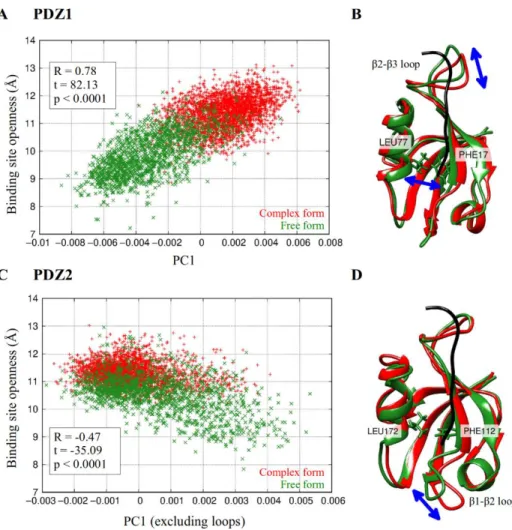
Figure 1: investigation of interdomain motions of the PDZ1-2 tandem. The openness of the binding site is measured by the distance between the Cα atoms of residues Leu77 and Phe77 in PDZ1, and Leu172 and Phe112 in PDZ2. A,C: distribution of the free and complexed ensembles along the first principal component and the openness of the binding site. Correlation is indicated.
B,D: the internal motion represented by the first principal component. The structures correspond to the two extreme conformations, with the largest fluctuations marked.
8
Principal component analysis of the intradomain motions proved that interaction with the ligand influences the distribution of the ensemble along the first principal component. This principal component represents the opening- closing motion of the hydrophobic core and the displacement of the β1-β2 loop in both domains, and additionally, the displacement of the β2-β3 loop in PDZ1 domain (Figure 1). The motion is present in the free and complexed PDZ domains as well, but the complexed ensemble covers a significantly smaller region along this motion than the free one. This effect is somewhat more pronounced in PDZ1 than in PDZ2 domain. It corresponds to the experimental observation according to which ligand binding causes rigidification of the β2- β3 loop both on fast and slow timescales (Fuentes et al. 2004).
Thesis 1.3: the structural ensembles of PDZ1-2 tandem generated by me cover the entire supramodular conformational space, in which the complexed form exhibits restricted dynamics relative to the free one. The tightly packed possible supramodular orientations can be separated into 7 clusters, which form distinct interdomain interfaces.
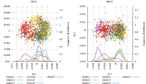
Principal component analysis of the PDZ2 domain followed by superimposing the ensembles on a common PDZ1 template yields information on motions reflecting the relative displacement of the two domains. The first two principal components can be converted into polar coordinates, the angular component of which represents the interdomain torsion angle, and the radial component represents the distance between the two domains.
The presence of the ligand strongly increases the interdomain conformational space covered by the ensemble, allowing for supramodular rearrangements with a larger amplitude (Figure 2A). Thus, I succeeded in creating an atomic-level model to explain one of the initial starting points of the analysis, i.e. that the complexed PDZ1-2 tandem exhibits a significantly increased interdomain dynamics relative to the free form.
Those supramodular conformers with a considerable amount of interdomain interactions (termed tightly packed structures) can be clustered into 7 subensembles, which are well separated along the first two interdomain principal components (Figure 2B). Since these clusters differ in the relative
9
orientation of the two domains, they also form interdomain interaction between different regions. 3 and 4 regions of the PDZ1 and PDZ2 domains, respectively, are involved in forming the interdomain interface (Figure 2C,D). Due to steric and other geometrical constraints (stemming for example from the shortness of the linker), only a part of the mathematically possible interface-combinations are allowed to occur.
Figure 2: investigation of interdomain motions in the PDZ1-2 tandem. A: principal component analysis of the PDZ2 domain, followed by the superimposition of the ensembles on a common PDZ1 template. Distribution along the first two principal components are plotted. B: clusters generated from the tightly packed conformations. C: splitting the surface of the PDZ domain in 5 distinct regions. D: the interdomain interface in each cluster, with the relative orientation of the two ligands marked.
10
Thesis 1.4: I compared the generated ensembles to experimental structures of PDZ1-2 available in public database. The conformational space covered by the generated ensembles includes the experimentally determined structures of PDZ1-2 tandem. The clusters resulting from the tightly packed conformations in our ensembles anticipate the existence of not yet observed, but otherwise geometrically allowed supramodular structures.
The generated ensembles include almost all of the previously determined structures, even though they partially contradict each other in terms of the supramodular orientation. Apart from the FRET-structure, which was refined in a later study, all structures are within the conformational space covered by the generated ensembles. Furthermore, each one of the tightly packed experimental structures belongs to one of the 7 clusters (Figure 3).
Figure 3: available experimental structures of the PDZ1-2 tandem plotted together on a common interdomain PCA plot with the generated clusters.
Interestingly, one of the crystal structures of PDZ1-2 (PDB: 6SPV for the free, 6SPZ for the complexed form) was published after completing the present analyses. Even though its supramodular conformation does not correspond to
11
any of the previously observed structures, it was covered by my ensembles, nevertheless. Based on both the interdomain orientation and the interdomain interface, it clearly belongs to cluster 6. Similarly, the existence of other supramodular structures can be anticipated which are harder to observe because of their lower weight in the ensemble (e.g. clusters 1 and 7), but due to their role in maintaining the dynamic equilibrium, they might still have a relevant biological function.
Thesis 1.5: Based on the conclusions resulting from analyzing the ensembles, I proposed a mechanism of the binding partners of PDZ1-2 tandem regulating the supramodular dynamics. Binding partners exert their regulatory role on the supramodular dynamics of PDZ1-2 tandem through local interactions with the β2-β3 loop, which is also the region mostly responsible for forming the interdomain interface.
Figure 4: investigating the connection between the intra- and interdomain motions in PDZ1-2 tandem. The data points corresponding to the generated clusters are plotted along the first two principal components determined for the intradomain motions of both domains. The distribution of the clusters along the first principal component is also plotted.
12
Regions most frequently involved in the formation of the interdomain interface were identified in both PDZ domains. Strikingly, these regions coincide with those the internal dynamics of which are mostly modified upon ligand binding: the β1-β2 and β2-β3 loops in PDZ1, and the β1-β2 loop in PDZ2. Inspecting the interfaces formed by the clusters derived from the tightly packed conformations, it turns out that in all but one clusters these regions are involved in the interdomain interface.
The intra- and interdomain principal components are interdependent. The clusters derived from the tightly packed conformation implicitly express a diversity in the interdomain conformational state, and these clusters are not evenly distributed along the intradomain principal component (Figure 4).
In each cluster, a different hydrogen-bonding pattern is formed between the upstream (-3 – -7) residues of the ligand the β2-β3 loop region of the PDZ domains. Since these clusters represent different interdomain orientations, this observation proves the connection between ligand binding and the supramodular dynamics.
13
3.2 Second thesis group: results regarding parvulin-type rotamases
Thesis 2.1: Using principal component analysis, I proved that the opening-closing motion is characteristic of parvulin-type rotamases. The three investigated parvulins sample distinct regions along this motion.
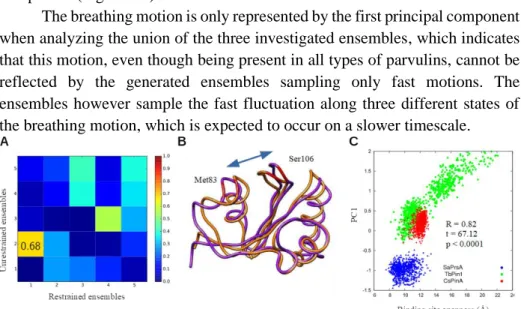
The first principal component resulting from the PCA analysis of the three parvulin-ensembles, representing the most predominant motion, corresponds to the opening-closing motion of the binding pocket, termed breathing motion. The three ensembles occupy distinct regions along the first principal component (Figure 5).
The breathing motion is present both in the restrained and unrestrained ensembles, but in the latter ones it is only represented by the second principal component (Figure 5A).
The breathing motion is only represented by the first principal component when analyzing the union of the three investigated ensembles, which indicates that this motion, even though being present in all types of parvulins, cannot be reflected by the generated ensembles sampling only fast motions. The ensembles however sample the fast fluctuation along three different states of the breathing motion, which is expected to occur on a slower timescale.
Figure 5: connection between the first principal component of the restrained parvulin ensembles and the openness of the binding site. A: distribution of the three ensembles along the openness of the binding site and the first principal component. B: the most predominant motion represented by the first principal component. The two structures reflect the extreme positions. C: overlap between the principal components of the restrained and unrestrained ensembles.
14
Thesis 2.2: I compared the generated ensembles to publicly available experimental structures. Using principal component on their consensus sequence, I proved that displacement of the hinge region connecting the small and large lobes is also a motion characteristic of parvulin-type rotamases. This region is responsible for the interaction with the WW domain, if present.
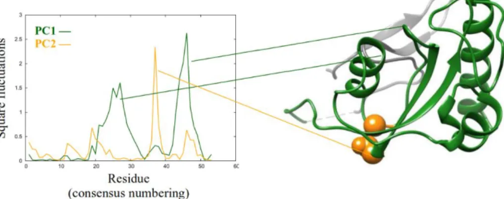
A principal component analysis was carried out on the generated ensembles combined with 100 experimentally determined structures of parvulins, following a multiple structure alignment. The second most predominant motion turned out to be the displacement of the hinge region facilitating the breathing motion (i.e. the joining point of the small and large lobes) (Figure 6).
The three generated ensembles are also separated quite well along this principal component. This means they not only occupy distinct regions along the breathing motion, but also along the displacement of the hinge region: these two motions, therefore, are connected. The WW containing and WW-less parvulins are also slightly separated along the second principal component, corresponding to the fact that this is the region responsible for the interaction between the catalytic and WW domain.
Figure 6: square fluctuations of the first two principal components, resulting from the principal component analysis carried out on the investigated ensembles combined with 100 experimentally determined structures of parvulins. On the right side, Pin1 is shown with those regions highlighted which are mostly displaced in the first two motions.
15
Thesis 2.3: I proved that the protonation state of the hydrogen-bonding network influences the internal dynamics of the large lobe in parvulin- type rotamases.
The two conserved histidine residues, part of the hydrogen-bonding network, reorient in an interdependent manner, as it can be concluded from the pKa values as well as from the side-chain torsion angles (Figure 7A,B). Furthermore, the first principal component resulting from the principal component analysis of the 5 residues involved in the hydrogen-bonding network correlates with the breathing motion (Figure 7C). Correlation does not mean causation on its own.
However, the ensembles are considerably separated along this principal component, as well as along the side-chain orientation angles. This difference can be explained with the difference in their protonation state. Both histidine residues in SaPrsA are mono-protonated, which allows for the formation of the complete hydrogen-bonding network. On the other hand, histidine residues in the other two parvulins are fully protonated, which only allows for a partial formation of the hydrogen bonding network. Consequently, of the three parvulin ensembles, it is SaPrsA the one with the least histidine side-chain variability.
Figure 7: A: absolute difference between the χ1 and χ2 side-chain torsion angles of the two histidine residues, plotted against each other. B: pKa values of the two histidine residues plotted against each other. C: the first principal components resulting from the principal component analysis of the hydrogen bonding network and the three investigated ensembles, plotted against each other.
16
Thesis 2.4: Based on the conclusions resulting from analyzing the structural ensembles, I proposed a general model for the regulation mechanism of parvulin-type rotamases. The opening-closing motion of the binding pocket is modulated on the one hand by the protonation state of the hydrogen-bonding network, on the other hand by the WW domain, if present, through the motion of the hinge region. The different activity of the distinct parvulin-types can be explained by the differences in the modulation of the dynamics of the opening-closing motion of the binding pocket.
Based on the presented results, a general model is proposed for the mechanism of parvulin-type rotamases. The dynamics and extent of the breathing motion determine the ligand selectivity and regulates the catalysis. The breathing motion, in turn, is modulated by two further factors: the interaction with the WW domain through regulating the motions of the hinge region and the protonation state of the hydrogen-bonding network in the large lobe.
The proposed mode of regulation is in correspondence with a number of previous experimental findings. Molecular dynamics simulations of Pin1 showed that the dynamics of the loops surrounding the binding pocket depend on the interaction with the WW domain, as well as on the interaction between the ligand and both the catalytic and WW domain (Guo, Pang, and Zhou 2015;
Olsson et al. 2016). My conclusions regarding the hydrogen-bonding network correspond to the proposed dynamic hydrogen-bond formed between the two histidine residues, which modulates the dynamics of the large lobe (Barman and Hamelberg 2014). Results similar to ours were obtained for one FKBP-type rotamase: the dynamics of the binding site, once again, play an important role in ligand binding and catalysis (Quistgaard et al. 2016). All this considered, such mechanism can very likely be a general mechanism for fine tuning the biological activity of proteins.
17
4 Publications serving as a basis for the present work
4.1 Research papers
Bertalan Kovács, Nóra Zajácz‐Epresi, Zoltán Gáspári (2019): “Ligand‐
dependent Intra‐ and Interdomain Motions in the PDZ12 Tandem Regulate Binding Interfaces in Postsynaptic Density Protein‐95.” FEBS Letters 594(5):887–902.
https://doi.org/10.1002/1873-3468.13626
András Czajlik*, Bertalan Kovács*, Perttu Permi, Zoltán Gáspári (2017):
“Fine-Tuning the Extent and Dynamics of Binding Cleft Opening as a Potential General Regulatory Mechanism in Parvulin-Type Peptidyl Prolyl Isomerases.” Scientific Reports 7. 44504.
https://doi.org/10.1038/srep44504
4.2 Further publications
Dániel Dudola, Bertalan Kovács, Zoltán Gáspári (2017): “CoNSEnsX+
Webserver for the Analysis of Protein Structural Ensembles Reflecting Experimentally Determined Internal Dynamics.” Journal of Chemical Information and Modeling, 57(8), 1728–1734.
https://doi.org/10.1021/acs.jcim.7b00066
Dániel Dudola, Bertalan Kovács, Zoltán Gáspári (2020): “Evaluation and Selection of Dynamic Protein Structural Ensembles with CoNSEnsX+.”
In: Structural Bioinformatics, (Gáspári Zoltán ed.) New York (NY), United States of America: Springer US, (2020) pp. 241-254.
https://doi.org/10.1007/978-1-0716-0270-6_16 ISBN: 978-1-0716-0270- 6
Additionally, 16 further presentations on 8 national and 8 international conferences.
18
5 References
Bailey, Melanie L., Brian H. Shilton, Christopher J. Brandl, and David W.
Litchfield. 2008. “The Dual Histidine Motif in the Active Site of Pin1 Has a Structural Rather than Catalytic Role.” Biochemistry 47(44):11481–89.
Barman, Arghya, and Donald Hamelberg. 2014. “Cysteine-Mediated Dynamic Hydrogen-Bonding Network in the Active Site of Pin1.” Biochemistry 53(23):3839–50.
Best, Robert B., and Michele Vendruscolo. 2004. “Determination of Protein Structures Consistent with NMR Order Parameters.” Journal of the American Chemical Society 126(26):8090–91.
Fuentes, Ernesto J., Channing J. Der, and Andrew L. Lee. 2004. “Ligand- Dependent Dynamics and Intramolecular Signaling in a PDZ Domain.”
Journal of Molecular Biology 335(4):1105–15.
Göthel, S. F., and M. A. Marahiel. 1999. “Peptidyl-Prolyl Cis-Trams Isomerases, a Superfamily of Ubiquitous Folding Catalysts.” Cellular and Molecular Life Sciences 55(3):423–36.
Guo, Jingjing, Xiaodong Pang, and Huan-Xiang Xiang Zhou. 2015. “Two Pathways Mediate Interdomain Allosteric Regulation in Pin1.” Structure 23(1):237–47.
Hanes, Steven D. 2015. “Prolyl Isomerases in Gene Transcription.” Biochimica et Biophysica Acta 1850(10):2017–34.
Kim, Eunjoon, and Morgan Sheng. 2004. “PDZ Domain Proteins of Synapses.”
Nature Reviews Neuroscience 5(10):771–81.
Lee, Ho-Jin, and Jie J. Zheng. 2010. “PDZ Domains and Their Binding Partners:
Structure, Specificity, and Modification.” Cell Communication and Signaling : CCS 8:8.
Lu, Kun Ping, Greg Finn, Tae Ho Lee, and Linda K. Nicholson. 2007. “Prolyl Cis-Trans Isomerization as a Molecular Timer.” Nature Chemical Biology 3(10):619–29.
Mostarda, Stefano, David Gfeller, and Francesco Rao. 2012. “Beyond the Binding Site: The Role of the Β2 - Β3 Loop and Extra-Domain Structures in PDZ Domains.” PLoS Computational Biology 8(3).
Olsson, Simon, Dean Strotz, Beat Vögeli, Roland Riek, and Andrea Cavalli.
2016. “The Dynamic Basis for Signal Propagation in Human Pin1-WW.”
Structure (London, England : 1993) 24(9):1464–75.
Quistgaard, Esben M., Ulrich Weininger, Yonca Ural-Blimke, Kristofer Modig, Pär Nordlund, Mikael Akke, and Christian Löw. 2016. “Molecular Insights into Substrate Recognition and Catalytic Mechanism of the
19
Chaperone and FKBP Peptidyl-Prolyl Isomerase SlyD.” BMC Biology 14(1):82.
Richter, Barbara, Joerg Gsponer, Péter Várnai, Xavier Salvatella, and Michele Vendruscolo. 2007. “The MUMO (Minimal under-Restraining Minimal over-Restraining) Method for the Determination of Native State Ensembles of Proteins.” Journal of Biomolecular NMR 37(2):117–35.
Sheng, Morgan, and Carlo Sala. 2001. “PDZ Domains and the Organization of Supramolecular Complexes.” Annual Review of Neuroscience 24(1):1–29.
Terada, Tohru, Mikako Shirouzu, Yasuhiro Fukumori, Fumihiro Fujimori, Yutaka Ito, Takanori Kigawa, Shigeyuki Yokoyama, and Takafumi Uchida. 2001. “Solution Structure of the Human Parvulin-like Peptidyl Prolyl Cis/Trans Isomerase, HPar14.” Journal of Molecular Biology 305(4):917–26.
Wang, Wenning, Jingwei Weng, Xu Zhang, Maili Liu, and Mingjie Zhang.
2009. “Creating Conformational Entropy by Increasing Interdomain Mobility in Ligand Binding Regulation: A Revisit to N-Terminal Tandem PDZ Domains of PSD-95.” Journal of the American Chemical Society 131(2):787–96.