Basic
1944
Nikolett Lupsa et al. DOI: 10.1002/eji.201847552 Eur. J. Immunol. 2018. 48: 1944–1957Tissue immunology and leukocyte trafficking Research Article
Skin-homing CD8 + T cells preferentially express GPI-anchored peptidase inhibitor 16, an inhibitor of cathepsin K
Nikolett Lupsa
1,2, Barbara ´Ersek
1,3, Andor Horv´ath
1, Andr´as Bencsik
1, Eszter Lajk´o
1, P´alma Sill´o
4,
´Ad´am Oszvald
1, Zolt´an Wiener
1, P´eter Rem´enyi
5, G´abor Mikala
5, Tam´as Masszi
5,6, Edit I Buz´as
1,2and Zolt´an P´os
11Department of Genetics, Cell and Immunobiology, Semmelweis University, Budapest, Hungary
2Hungarian Academy of Sciences, Semmelweis University Immunoproteogenomics Extracellular Vesicle Research Group, Budapest, Hungary
3Office for Research Groups Attached to Universities and Other Institutions, Hungarian Academy of Sciences, Budapest, Hungary
4Department of Dermatology, Venereology and Dermatooncology, Semmelweis University, Budapest, Hungary
5Department of Hematology and Stem Cell Transplantation, St. Istvan and Saint Laszlo Hospital, Budapest, Hungary
63rd Department of Internal Medicine, Semmelweis University, Budapest, Hungary
This study sought to identify novel CD8+ T cell homing markers by studying acute graft versus host disease (aGvHD), typically involving increased T cell homing to the skin and gut. FACS-sorted skin-homing (CD8β+/CLA+), gut-homing (CD8β+/integrinβ7+), and reference (CD8β+/CLA–/integrinβ7–) T cells were compared in patients affected by cutaneous and/or gastrointestinal aGVHD. Microarray analysis, qPCR, and flow cytome- try revealed increased expression of peptidase inhibitor 16 (PI16) in skin-homing CD8+ T cells. Robust association of PI16 with skin homing was confirmed in all types of aGvHD and in healthy controls, too. PI16 was not observed on CLA+ leukocytes other than T cells. Induction of PI16 expression on skin-homing T cells occurred independently of vitamin D3. Among skin-homing T cells, PI16 expression was most pronounced in memory-like CD45RO+/CD127+/CD25+/CD69−/granzyme B− cells. PI16 was confined to the plasma membrane, was GPI-anchored, and was lost upon restimulation of memory CD8+T cells. Loss of PI16 occurred by downregulation of PI16 transcription, and not by Phospholipase C (PLC)- or Angiotensin-converting enzyme (ACE)-mediated shedding, or by protein recycling. Inhibitor screening and pull-down experiments confirmed that PI16 inhibits cathepsin K, but may not bind to other skin proteases. These data link PI16 to skin-homing CD8+ T cells, and raise the possibility that PI16 may regulate cutaneous cathepsin K.
Keywords:CD8+T cell rGvHD rHoming rPI16 rSkin
Additional supporting information may be found online in the Supporting Information section at the end of the article.Correspondence:Dr. Zolt ´an P ´os
e-mail: pos.zoltan@med.semmelweis-univ.hu
Introduction
Precise regulation of CD8+T cell homing to distinct tissues is a prerequisite for mounting immune responses against pathogens that have narrow tissue tropism. It is well established that regula- tion of T-cell homing utilizes complex regulatory circuits involving unique combinations of chemokines and adhesion molecules, each identifying different tissues for circulating T cells. Homing is heav- ily implicated in the maintenance of immunity against antigen challenge occurring in different anatomical compartments, espe- cially in barrier tissues, such as the gut and the skin [1]. On the other hand, T-cell homing to distinct anatomical compartments is also involved in the development of several immunologic disorders or adverse immune reactions restricted to given tissues or organs.
This has been demonstrated in multiple animal models and human clinical studies, among others in ulcerative colitis (UC) [2, 3], multiple sclerosis (MS) [4, 5], Crohn’s disease (CD) [6, 7], and has also been suggested to be involved in acute graft versus host disease (aGvHD). We believe that aGvHD is of particular interest in terms of CD8+ T-cell homing to the gut and skin, for several reasons.
Acute GvHD is one of the most frequent, potentially fatal, short-term adverse events of allogeneic hematopoietic stem cell transplantation (aHSCT) [8]. The disease is typically associated with massive, graft-mediated cytotoxic tissue damage, character- istically restricted to a limited set of organs, typically to the skin and the gut [9, 10]. Notably, some patients suffer from either cutaneous or gastrointestinal (GI) aGvHD, while others display both manifestations simultaneously. Tissue damage in both cuta- neous and GI aGvHD occurs upon invasion of the skin and gut by CD8+ T cells [9] displaying canonical skin-homing (CLA+) and gut-homing integrinβ7+(ITGβ4β7+) phenotypes, respectively [11, 12]. It has been shown that rapid shifts in the numbers of these activated subsets in the blood of aHSCT patients precede the two forms of the disease, and hence may be of diagnostic and prognostic value [11–13]. Furthermore, proof of concept studies showed that blockade of CD8+T cell homing pathways may allevi- ate respective tissue damage in mouse models of aGVHD [14, 15].
Indeed, current phase 1 and phase 2 clinical trials with two monoclonal antibodies, vedolizumab and natalizumab, targeting ITGβ4 or ITGβ4β7, approved for the treatment of UC/CD and CD/MS, respectively, are currently exploring whether the same approach may work for the treatment of GI aGVHD in humans, as well [16, 17]. Although less well documented, there is some evi- dence that the homing preferences of activated CD4+Th and Treg cells are also altered in aGvHD. Activated CD4+Th cells become enriched in damaged organs [18]. In contrast, Treg cells home to tissues affected by aGvHD in diminished numbers [19, 20].
Taken together, aHSCT patients diagnosed with cutaneous and/or GI aGvHD provide a unique opportunity for the in-depth comparison of naturally activated human CD8+T cells homing to the gut and skin, for several reasons: (i) in aGvHD, CD8+T cells home to both the skin and the gut, enabling a direct comparison of the two cell types in the same human disease, without the skewing effect of different disease states, (ii) these phenomena may occur
simultaneously, enabling the comparison of these cells within the same host, (iii) CD8+ T cells actively home to these organs and are not only attracted by local inflammation, (iv) full maturation of skin and gut-homing effector CD8+ T cells takes place, and (v) effector mechanisms mediated by skin- and gut-homing CD8+ T cells are fundamental to the disease process.
In this study, we compared skin-and gut-homing CD8+T cells of aGvHD patients by gene expression profiling, flow cytometry, and functional bioassays, and confirmed our findings on CD8+ T cells of healthy blood donors, as well. We propose that a novel marker, peptidase inhibitor 16 (PI16), is associated with the skin- homing CD8+T cell subset in both aGVHD patients, and healthy individuals. Based on these results, we propose that PI16 is linked to human skin-homing T cells in both inflammatory and steady- state conditions.
Results
Skin-homing CD8+T cells overexpress PI16 mRNA in patients affected by aGvHD
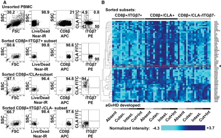
Skin- and gut-homing CD8+ T cell subsets were isolated from four groups of aHSCT patients developing (i) cutaneous aGvHD, (ii) GI aGvHD, (iii) both cutaneous and GI aGvHD simulta- neously, or (iv) none (see Supporting Information Table 1 for detailed patient information). PBMCs of aHSCT patients were sorted into skin-homing Ly/LIVE/DEADR–/CD8β+/CLA+, gut-homing Ly/LIVE/DEADR–/CD8β+/ITGβ7+, and reference Ly/LIVE/DEADR–/CD8β+/CLA–/ITGβ7– cytotoxic T cell subsets by three-way FACS sorting (Fig. 1A). Next, skin-homing, gut- homing, and reference CD8+ T cells were analyzed by compar- ative gene expression profiling. Transcriptome analysis revealed 62 genes differentially expressed between the three CTL subsets (Fig. 1B, two-way repeated measures [RM] ANOVA with false discovery rate [FDR]<0.01, see Supporting Information Table 2 for detailed results). As gene set enrichment analysis was unable to identify known gene sets differentially expressed depending on CTL homing preferences (p>0.05), we next set out to ana- lyze individual genes. Among the most significant differences, we selected PI16 for further analysis (Fig. 1B, marked with asterisk to the right) based on others’ earlier publications [21, 22].
PI16 expression is a hallmark feature of skin-homing CD8+T cells regardless of organ involvement in aGvHD
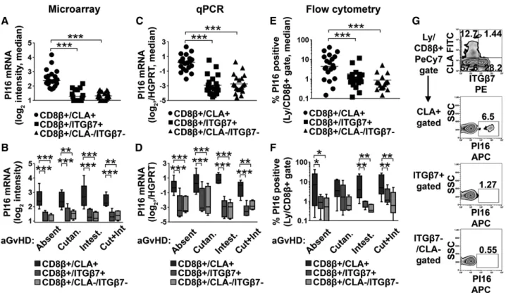
Microarray data suggested that PI16 mRNA showed more than nine times higher expression in skin-homing CD8β+/CLA+T cells than in gut-homing, or reference CTLs of aHSCT patients (Fig. 2B, one-way RM ANOVA,p<0.0001). We analyzed whether this phe- nomenon was restricted to any of the four patient groups, that is, linked to any given type of CTL-mediated organ damage in aGvHD, or GvHD in general (Fig. 2B). We found that overexpression
1946
Nikolett Lupsa et al. Eur. J. Immunol. 2018. 48: 1944–1957Figure 1. Comparative gene expression profiling of skin-homing, gut-homing, and reference CD8+T cells of aHSCT patients developing no aGvHD, cutaneous aGvHD, gastrointestinal GvHD, or both aGvHD manifestations simultaneously. (A) Gating strategy and representative three-way FACS sorting of gut-homing Ly/Live/CD8β/ITGβ7+, skin-homing Ly/Live/CD8β/CLA+, and reference Ly/Live/CD8β/ITGβ7–/CLA–cytotoxic T cells, isolated from PBMCs of aHSCT patients. Numbers indicate percent gated cells. One experiment representative of five independent experiments with n=4 donors for each group,n=20 donors total. (B) FACS-sorted gut-homing, skin-homing, and reference cytotoxic T cells (as indicated at the top) of aHSCT patients developing no aGvHD, cutaneous aGvHD, intestinal aGvHD, or both aGVHD manifestations simultaneously (as indicated at the bottom), analyzed by gene expression profiling (n=5 for each possible T cell subset/disease group combination). Heatmap displays 62 differentially expressed genes, identified by two-way RM ANOVA (FDR<0.01), analyzing sources of variance introduced by CD8+T cell homing subsets, respective organ damage, and possible interactions between these two factors. Columns represent individual samples; rows correspond to genes. Colors display expression below (pale blue), or above (dark blue) the mean. For clarity’s sake, data shown are centered and SD-normalized.
To the right, asterisk marks peptidase inhibitor 16 (PI16). Combined results of three independent experiments withn=4,n=8, andn=8 donors for each group,n=20 total.
of PI16 by skin-homing CLA+ CD8+T cells occurred in all four patient groups (Fig. 2B, two-way RM ANOVA, p < 0.0001) regardless of the type of organ involvement in aGVHD (p>0.05).
In addition, the data suggested that PI16 overexpression was not restricted to aGVHD, as aHSCT patients with no aGvHD also dis- played increased expression of PI16 in skin-homing CD8+T cells.
These results were also confirmed by qPCR (Fig. 2C, 2D two-way RM ANOVA,p< 0.0001). Flow cytometry also confirmed that PI16 protein positive cells were enriched in the CLA+subset (on average 10.9-times) compared with reference CTLs (Fig. 2E–G).
Furthermore, PI16+ cells were significantly more frequent (on average 5.2-times) among skin-homing than among gut-homing CD8+T cells (Fig. 2E, two-way RM ANOVA,p=0.0004). This observed pattern held true in all patient groups regardless of the type of organ involvement (Fig. 2F, two-way RM ANOVA, p=0.9388), which is in line with the RNA data. Again, observed organ damage did not appear to have an influence on the pattern of PI16 expression (Fig. 2F, two-way RM ANOVA,p=0.9204), even
if, in the cutaneous aGvHD group, between-subset comparisons did not reach formal significance (Fig. 2F,p=0.2266,p=0.4859, respectively). These observations conclusively suggested that PI16 is associated with skin-homing CD8+T cells, the expression of which may be largely independent of the development of aGvHD, or the exact type of organ involvement in aGvHD.
Robust association of PI16 protein with skin-homing CD8+T cells in both health and disease
As the expression of PI16 appeared to be independent of aGvHD, we next went on to test whether aHSCT was required at all for the induction of PI16 expression on skin-homing T cells. To this end, we analyzed skin-homing, gut-homing, and reference CTLs of healthy blood donors in a similar fashion. We found PI16 expression patterns matching those observed in aHSCT, with even more pronounced differences of PI16 expression between cell
Figure 2. Testing the link among PI16, CD8+T cell homing subsets, and aGvHD organ involvement by microarray gene expression profiling, qPCR, and flow cytometry. (A, C, and E [top]) PI16 expression in skin-homing CD8β/CLA+, gut-homing CD8β/ITGβ7+, and reference CD8β/ITGβ7–/CLA– cytotoxic T cells of aHSCT patients, as analyzed by microarray, qPCR, and flow cytometry, respectively (n=15 each, median) is shown. One-way RM ANOVA with FDR correction; stars indicate significance in pairwise comparisons: ***p<0.001 (B, D and F [bottom]) PI16 expression among the same three cytotoxic T cell subsets, also accounting for possible differences introduced by CD8+T cell-mediated organ damage, that is, in patients with no aGvHD, cutaneous aGvHD, intestinal aGvHD, or both manifestations of aGvHD. (n=5 for each possible T cell subset/disease group combination;
box and whiskers plots with median). Two-way RM ANOVA with FDR correction; stars indicate significance in pairwise comparisons: *p<0.05,
**p<0.01, ***p<0.001. (G [far right] representative flow cytometry data of a single patient). Numbers indicate percent gated cells. Combined results of three experiments withn=4,n=8, andn=8 donors for each group,n=20 total.
subsets among healthy individuals compared with aHSCT patient.
On average, 63% of the skin-homing T cells of healthy blood donors stained positive for PI16 protein; 13.3-times more than gut-homing T cells (4.74%), and 7.5-times more than reference (8.43%) CTLs (Fig. 3A, two-way RM ANOVA,p<0.0001). These data suggested that the presence of PI16 on skin-homing T cells is a distinctive feature of this subset in both health and disease, that is, it is not restricted to, or induced by either aGvHD or aHSCT.
Also, it was apparent that the difference is more pronounced among healthy individuals, than in aHSCT patients (irrespective of them being affected by, or remaining free of aGvHD). For these reasons, we used healthy donors for the further analysis of PI16.
PI16 is a marker associated with skin-homing CD8+
T cells but not other CLA+leukocytes
Considering that CLA is frequently expressed by a wide variety of leukocytes beside CD8+T cells, we first sought to clarify if PI16 expression is linked to CLA expression, or if it is a restricted fea- ture typical to skin-homing T cells. Flow cytometry confirmed that PI16 over-expression was restricted to CLA+T CD8+ and CD4+
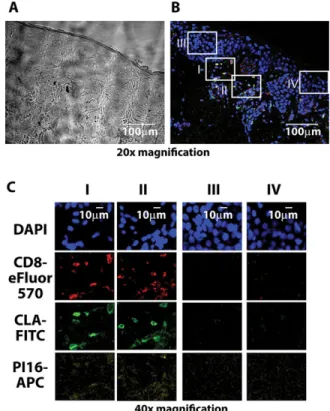
T cells, as CLA+monocytes or NK cells expressing CLA did not display PI16 in appreciable numbers (Fig. 3B). This suggests that expression of PI16 is not mechanistically linked to that of CLA in various leukocytes; but instead, PI16 is specifically linked to skin- homing T cells. We also found that within the skin-homing CD8+ T cell subset, PI16 positive cells were more frequently CD45RO+, CD127+, CD25+, CD69–, and granzyme B cells (Fig. 3C), than their PI16 negative counterparts, while there was no difference in the case of some other CD8+T cell markers (IFN-γ, CD40L, not shown). These data indicate that PI16 production is most intense in memory-like, resting skin-homing CD8+T cells and is less char- acteristic for recently activated effector cytotoxic T cells actively producing effector molecules. Finally, we tested if PI16 expression was maintained in skin-homing T cells after entering the human skin, like that of CLA [23], or became downregulated upon enter- ing the target organ, like in the case of integrinβ4 on gut-homing T cells [24]. We found that PI16-expressing CD8+CLA+T cells can be found in the healthy human skin (Fig. 4). Spatial distribution of PI16 in the skin was similar to others’ earlier observations [25]:
that is, weak or absent in the epidermis, scattered in the dermis.
We found that scattered dermal PI16 positivity showed clear colo- calization with cutaneous CD8+/CLA+T cells (Fig. 4).
1948
Nikolett Lupsa et al. Eur. J. Immunol. 2018. 48: 1944–1957Figure 3. Evaluation of PI16 as a marker associated with skin-homing CLA+T cells of healthy individuals. (A) Frequency of PI16 positive cells among skin-homing CD8β/CLA+, gut-homing CD8β/ITGβ7+, and refer- ence CD8β/ITGβ7–/CLA–cytotoxic T cells of healthy blood donors. Gating strategy and representative data for each analyzed cytotoxic T cell sub- set are shown to the left. One-way RM ANOVA with FDR correction is shown to the right (one experiment,n=7, median): stars indicate signif- icance in pairwise comparisons: ***p<0.001. (B) PI16 expression in dis- tinct PBMC subsets expressing the canonical skin-homing T cell marker CLA; typical patterns of PI16 expression in CLA+blood cytotoxic T cells, CLA+helper T cells, CLA+NK cells, and CLA+monocytes of healthy blood donors (n=3, representative data). (C) Comparative analysis of PI16−and PI16+ skin-homing CD8+ T cells of healthy blood donors (n=6, box and whiskers plot with median). Pairedt-test: **p<0.01 and ***p<0.001. Each panel shows one experiment representative of six (panel A) or two (panels B and C).
Figure 4. Spatial distribution of PI16 protein and skin-resident CD8+ T cells in the skin. Analysis of the localization of CD8+skin-homing T cells and PI16 in healthy human skin. (A and B) Phase contrast and composite fluorescent images of a representative human skin section analyzed by immunohistochemistry is shown. (C) Selected regions of (B) sections I–II represent dermal, sections III–IV epidermal staining with DAPI, CD8-, CLA-, and PI16-specific antibodies, as indicated. Shown are results of one experiment representative of four, one sample each.
Skin-homing CD8+T cells display a
membrane-bound, GPI-anchored PI16 protein
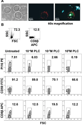
PI16 is a protein with largely unknown functions that, originally, was described as a soluble serum protein released by prostate cells [26]. However, it has been reported that the protein may appear on the surface of Treg cells [22], and this is also in line with our flow cytometry data. By performing confocal microscopy on sorted CD8+T cells, we found that the majority of PI16 protein in CD8+T cells were indeed confined to the plasma membrane (PM), showing a similar distribution to, but no clear colocaliza- tion with, CLA (Fig. 5A), within the PM. Recently, it has been confirmed that PI16 protein expressed by murine fibroblasts is GPI-anchored [27]. To test the means of PI16 linkage to the PM of human CD8+T cells, we exposed CD8+T cells to a bacterial PI-PLC, capable of detaching GPI-anchored proteins from the PM.
We found that upon exposition of CD8β+/PI16+ T cells to PI- PLC, PI16+ staining showed a rapid, dose-dependent reduction (Fig. 5B), similar to that of CD59, a known GPI-anchored protein, but unlike CD8β, which does not have a GPI anchor, and hence remained unaffected. These data demonstrate that human skin homing CLA+T cells express a membrane bound, GPI-anchored form of PI16.
Figure 5. Analysis of the subcellular distribution of PI16 protein in skin- homing CD8β+T cells. (A) Analysis of the subcellular localization of PI16 in skin-homing CD8+T cells. Phase contrast, monochrome, and three-color fluorescent microscopy of MACS-sorted CD8+T cells are shown. Blue, CD8; green, CLA; and red, PI16. Representative subcellular distribution of PI16 in a typical, skin-homing, CD8β+/CLA+/PI16+T cell is displayed in the cell at the center. One experiment representative of three independent experiments. (B) Investigation of human PI16 as a possible GPI-anchored PM protein of T cells. Assessing cell surface PI16 signal after PI-PLC-mediated in vitro digestion of GPI-anchors. CD59 (a known GPI-anchored PM protein) and CD8β(having no GPI anchor) serve as positive and negative controls, respectively. Numbers indicate percent positivity of T cells upon exposure to various concentrations of PI-PLC, as shown. One experiment representative of two independent experiments.
Induction of PI16 occurs independently of canonical pathways imprinting the skin-homing phenotype
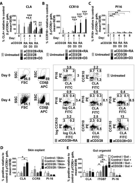
We next analyzed whether known mechanisms responsible for imprinting the skin- and gut-homing phenotype in activated na¨ıve CD8+ T cells could also invoke or suppress PI16 expression on these cells. In two seminal papers, it was shown that exposure of in vitro activated blood CD8+T cells to DC-released vitamin D (D3) and retinoic acid (RA) was capable of inducing some (although not all) skin- and gut-homing markers, respectively, in a mutu- ally exclusive fashion [28, 29]. Using this approach, we found that although D3 and RA were capable of affecting expression
of the skin-homing markers CLA (Fig. 6A, two-way RM ANOVA, p=0.0049) and CCR10 on activated CD8+T cells (Fig. 6B, two- way RM ANOVA,p=0.0015), PI16 remained unaffected by D3 and RA, only reacting on T-cell activation (Fig. 6C). We next tested whether expression of PI16 on skin-homing T cells may be induced independently of DC-mediators, for example, by local epithelia. It has been suggested that skin epithelial cells may imprint activated skin-homing T cells to express CLA and CCR8 via soluble medi- ators that reach skin-draining lymph nodes and activated T cells via afferent lymphatics [30]. Considering this evidence, we ana- lyzed if indirect coculture of CD8+T cells during their activation with live skin explants and gut organoids, containing live epithe- lia, promotes or inhibits PI16 expression. We found that coculture of CD8+ T cells with skin explants provided some support for the induction of a skin-homing phenotype in terms of CLA and CCR8 expression, partly in an activation-dependent manner (Fig.
6D). However, PI16 expression was not promoted by cocultivation with skin explants (Fig. 6D, two-way RM ANOVA,p>0.05). Also, expression of PI16 remained unaffected by the presence of intesti- nal organoids (Fig. 5E, two-way RM ANOVA,p>0.05). Hence, induction of PI16 on skin-homing CD8+T cells seems to be inde- pendent of known factors inducing other skin-homing CD8+T cell biomarkers, and particularly those affecting CLA expression.
Skin-committed CD8+T cells maintain PI16 expression until their activation
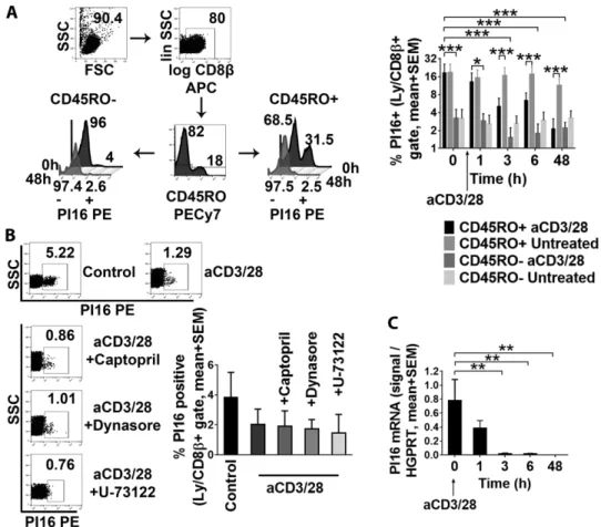
As PI16 expression was most typical for skin-homing, non-na¨ıve, memory-like CD45RO+/CD8+T cells (Fig. 3C), and stimulation of peripheral PI16+T cells resulted in rapid downregulation of this marker (Fig. 6C), we next tested whether reactivation of memory- like T cells leads to loss of PI16. We found that reactivation of CD45RO+ T cells by a-CD3/CD28 led to an almost complete elimination of PI16 from the cell surface within 48 h (Fig. 7A, two-way RM ANOVA, p = 0.0002). In marked contrast, PI16 was barely present on bona fide na¨ıve CD45RO−/CD8+ T cells, and did not react to activation either (Fig. 7A). Next, we tested the mechanism of PI16 removal from the surface of reactivated skin-homing memory-like CD8+T cells. Considering (i) that bac- terial PLC was capable of detaching PI16 from the surface of skin- homing T cells and (ii) that PLC activation is an inherent compo- nent of T cell activation, we hypothesized that activation of the PLC pathway in T cells may be responsible for the removal of PI16 from the cell surface upon activation. Hence, we tested whether (i) inhibition of PLC activity by U-73122, (ii) inhibition of the angiotensin-converting enzyme, another possible GPI-removing enzyme by captopril, [31, 32], or (iii) inhibition of possible endo- cytic recycling of PI16 by Dynasore could inhibit loss of PI16 from the cell surface upon activation. We observed that loss of PI16 upon a-CD3/CD28 treatment could not be inhibited by PLC- or ACE-inhibitors, or by stopping endocytic protein recycling either (Fig. 7B). On the other hand, we found that skin-homing CD8+ T cells ceased to express PI16 mRNA post activation, closely fol- lowed by the loss of the protein from the PM (Fig. 7C). Overall,
1950
Nikolett Lupsa et al. Eur. J. Immunol. 2018. 48: 1944–1957Figure 6. Investigation of the induction of PI16 expression in skin-homing CD8β+T cells. (A–C) Impact of vitamin D3 (D3) and retinoic acid (RA) treatment on the expression of the canonical skin-homing markers CLA (panel A) and CCR10 (panel B), versus on that of PI16 (panel C) after ex vivo activation of peripheral blood CD8+T cells with a-CD3/CD28 (n=3, mean with SEM). Gating strategy and representative results obtained on days 0 and 4 are shown below. Results of one experiment representative of two experiments. (D) Expression of skin-homing markers CLA and CCR8, and that of PI16 on activated CD8+T cells after 4 days long coculture with skin organoids (n=3, mean with SEM) One experiment representative of two. (E) Expression of CLA, the gut-homing marker integrinβ7 (ITGβ7), and PI16 on activated CD8+T cells upon 4 days of coculture with gut organoids (n=3, mean with SEM). One experiment performed. For clarity, all bar charts display percent PI16 positivity compared to day 0 untreated controls as reference. Results of FDR-corrected two-way RM ANOVA are shown with stars indicating significance in pairwise comparisons: *p<0.05,
**p<0.01, ***p<0.001.
these data suggest that reactivated skin-committed CD8+memory- like T cells lose PM-bound PI16 by transcriptional downregulation, and not due to protein shedding or increased recycling of the pro- tein from the PM.
PI16 acts as a partial inhibitor of cathepsin K
Typically, reactivation of CD8+/CLA+/CD45RO+, skin-homing memory T cells occurs within the human skin, where a large
Figure 7. Analysis of the termination of PI16 expression in skin-homing CD8+/CD45RO+T cells. (A) PI16 protein expression of healthy blood donors’ CD8β+/CD45RO+and CD8β+/CD45RO–T cells before, and at various time points after ex vivo activation by anti-CD3/CD28 (n=3, mean with SEM). Two-way RM ANOVA with FDR correction: *p<0.05, **p<0.01, ***p<0.001 in pairwise comparisons. (B) Flow cytometry analyzing PI16 expression upon anti-CD3/CD28 treatment in the absence and presence of U-73122 (inhibitor of PLC-mediated shedding of GPI-anchored proteins), captopril (inhibition of ACE-mediated shedding), or Dynasore (inhibition of endocytic protein recycling,n=3; mean with SEM). (C) Kinetics of PI16 mRNA expression upon a-CD3/28-mediated activation of CD8β+T cells as assessed by qPCR (n=3, mean with SEM). FDR-corrected one-way RM ANOVA: in pairwise analyses, stars indicate significance: **p<0.01. Each dataset represents one experiment of two performed.
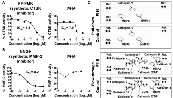
number of proteases, involved in various inflammatory processes and pathologies, are present [33, 34]. Considering that PI16 is a largely unknown, rather putative, peptidase inhibitor, we tested whether human PI16 was capable of suppressing the activity of skin proteases. Earlier it was suggested that mouse PI16 may sup- press murine cathepsin K [27], and that PI16 may inhibit MMP- 2 [35]. Our protease inhibitor assays (Fig. 8) demonstrated that, in line with others’ mouse data, human PI16 is a partial inhibitor of human cathepsin K (Fig. 8A). Human PI16 has an IC50compa- rable to synthetic inhibitors, but in line with others’ findings made in a murine system, it is not able to achieve full enzymatic inhibi- tion [27]. On the other hand, at least in our hands, human PI16 did not affect human MMP-2 activity (Fig. 8B). To further corroborate these findings, and also to identify other skin proteases possibly interacting with PI16, we next performed pull-down assays uti- lizing PI16 as a bait to find its partners in human skin protein lysates. Comparative analysis of the flow-through and the pulled down protein fractions showed that PI16 did not display high affin- ity against a large number of skin proteases, that is, cathepsins A, D, V, X, Z, P, kallikreins 5, 10, 11, CD26 (dipeptidyl peptidase-4), and MMP-2 (Fig. 8C), as none of them were specifically enriched
in the pulled-down fraction, or depleted from the flow-through by PI16. Taken together, these data suggest that human PI16 on skin-homing T cells may act as a partial inhibitor of cathepsin K in the human skin, but may not bind to other skin proteases with high affinity.
Discussion
This study shows that by analyzing human patients diagnosed with aGvHD, further insight may be gained into the mechanisms regulating cutaneous and GI CD8+T cell homing. Our data suggest that PI16 is a novel protein marker displaying robust association with the skin-homing CD8+T cell phenotype in both health and disease.
PI16 was first described as a protein released into the blood serum by prostate cells, and originally designated as prostate secretory protein 94-binding protein due to its high affinity for PSP94 [26]. It soon became clear that PI16 was expressed by multiple other, unrelated cell types, too, and the protein became incorporated into the CAP (cysteine-rich secretory proteins,
1952
Nikolett Lupsa et al. Eur. J. Immunol. 2018. 48: 1944–1957Figure 8. Testing human PI16 as an inhibitor of human matrix metalloprotease-2, cathepsin K, and other skin proteases involved in skin home- ostasis and cutaneous inflammation. (A and B) Relative activity of human cathepsin K (CTSK) and matrix metalloprotease-2 (MMP-2), respectively, in the presence of various concentrations of potent synthetic inhibitors and recombinant human PI16. Half-maximal inhibitory concentration (IC50), calculated by nonlinear regression, is shown in log10M concentration. Shown are the results of one experiment out of two for both CTSK and MMP-2, each data point representing a mean of duplicates. (C) Pull-down assay utilizing PI16-covered beads as baits, and PI16-free beads as controls performed to identify other human skin proteases selectively binding to recombinant human PI16. Distribution of various skin proteases in the pull-down versus the flow-through fractions in both cases, as analyzed by proteome profiling arrays. One experiment representative of two, one sample each.
antigen 5, and pathogenesis-related 1 proteins) superfamily [36].
All CAP proteins feature a highly conserved CAP domain, but they are rather heterogeneous due to additional, highly variable protein domains, which allow them to exert multiple, independent biological functions. These include involvement of CAP proteins into endocrine and paracrine regulation, regula- tion of matrix reorganization, but also fertilization, signaling, immunomodulation, and host defense.
Nevertheless, the function of the highly conserved core domain, the CAP domain itself, is poorly understood, and hence the com- mon function of such proteins is still a matter of speculation [37].
This issue becomes critical in the case of PI16, as it consists of nothing but the signature CAP domain and a rather long hinge region. Without solid knowledge on the function of CAP domains, deciphering the function(s) of PI16 remains challenging. In this regard, the hinge region itself is not particularly helpful either, as it lacks similarity to any known protein domains. Even though its very end is rich in hydrophobic amino acids, it has no confirmed intramembrane or intracellular domains [36]. Based on sequence analysis of the hinge, existence of a GPI anchor attached to the end of PI16 has been first suspected [26], then confirmed, in mice [27], but so far, not in humans. Based on sequence homology, PI16 was hypothesized to act as a protease inhibitor, and in line with this, it has been proposed that PI16 suppresses cathepsin K [27] and MMP-2 activity [35]. So far, these activities have only been shown
by studying murine cardiomyocytes and human cardiac endothe- lium, respectively, and have not been confirmed in humans or by independent research groups. To sum up, the exact biological roles and the functional properties of the CAP domain, most CAP family members, also including PI16, have, until now, remained largely obscure [36, 37].
This study demonstrates that human skin-homing CD8+T cells display a GPI-anchored, PM-bound form of PI16, and that PI16 expression is most pronounced in the CD45RO+memory compart- ment. Interestingly, earlier observations of others have already provided some, albeit very circumstantial, evidence supporting the notion that PI16 may be linked to cutaneous homing of T cells.
Among the extremely limited number of papers published on PI16, one has shown that PI16 is expressed by Tregs responding to CCL17 [22]. This is relevant to our findings because CCL17 is recognized by CCR4, which is a chemokine receptor on many skin-homing T cells. There is a large body of evidence confirming the involvement of CCL17 and CCR4 homing of T cells to the skin, especially under inflammatory conditions [38, 39], and mostly within the non-na¨ıve (effector/memory) T cell compartment [40].
Further corroborating this concept, in another paper, a genome- wide association study identified a polymorphism within the PI16 gene that predisposed to chronic skin inflammation [21].
We found that in healthy blood donors, the majority of CD8+/CLA+T cells expressed PI16, which was in sharp contrast
with the scarce PI16 positivity observed on other, CD8+/CLA− T cell subsets. However, expression of PI16 was neither complete within the skin-homing T cell subset, nor was it exclusive to the skin-homing phenotype. In short, PI16 is clearly associated to T cell homing, but it has limited sensitivity and specificity as a marker of all skin-homing T cells. We found that PI16 expression was most characteristic for non-na¨ıve CD45RO+, memory-like skin- homing CD8+T cells, and was less typical for recently activated (CD69+and Granzyme B+) effector cells homing to the skin. In addition, expression of PI16 was tightly controlled, and became virtually eliminated by CD45RO+ CD8+T cells upon their reac- tivation in vitro. Collectively, these data suggest that within the non-na¨ıve CD8+T cell compartment, PI16 expression may be less typical for recently activated, or reactivated, cells exerting effector functions. Interestingly, differences in PI16 expression in health and disease also suggested that PI16 expression became less pro- nounced once skin-homing T cells had been activated. As judged by flow cytometry, PI16 expression of skin-homing CD8+T cells was most prominent in healthy blood donors (having limited num- bers of activated skin-homing CD8+T cells), was less pronounced in aGvHD (polyclonal CD8+T cell activation), and was even less prominent in active cutaneous aGvHD (widespread activation of skin-homing CD8+T cells).
We also showed that PI16 disappeared from activated CD8+ T cells by means of transcriptional downregulation, and not by GPI anchor digestion and subsequent protein shedding. These obser- vations may allow speculation that the PI16 function(s) associated to skin-homing T cells are not related to release of this putative peptidase inhibitor into the extracellular space. They rather sug- gest that the function(s) of PI16 become(s) either dispensable or detrimental upon cognate activation of skin-homing CD8+T cells in the skin, for example, in cutaneous immune responses requiring local CTL intervention.
In this regard, it is interesting that both MMP-2 and cathepsin K, the two proposed targets of PI16 action, are critically involved in the regulation of skin tissue homeostasis. MMP-2 is an extracellu- lar protease and the altered activity of MMP-2 has sweeping conse- quences for the skin; MMP2 alters both the availability and activity of a plethora of signaling ligands and adhesion molecules affecting cutaneous integrity and local inflammation [41]. Cathepsin K, on the other hand, being capable of collagen and elastin digestion, is also linked to skin inflammation, matrix reorganization, and scar formation both as an intracellular lysosomal enzyme [42], and as a secreted, extracellular protease [43]. We found that similar to murine data, human PI16 was capable of interfering with human cathepsin K activity, acting as a partial inhibitor in the submicro- molar range. However, in our hands, recombinant PI16 was not able to suppress MMP-2 activity or pull-down MMP-2 from skin lysates, which conflicts others’ observations and hence the role of PI16 as a regulator of MMP-2 will need further analysis.
Taken together, our data indicate that resting human skin- homing CD8+T cells are instructed to display PI16, and maintain its expression until their reactivation. The possibility that reac- tivation of skin-committed CD8+ CLA+T cells may suppress an inhibitor (PI16) of a cutaneous inflammatory protease (cathep-
sin K) is intriguing. This observation may allow speculation that during T cell-mediated cutaneous immune responses, cutaneous CD8+T cells may affect skin homeostasis by stopping inhibition of select inflammatory proteases. Nevertheless, a more complete understanding of the role of PI16 in this context needs further studies, particularly studies focusing on the exact function of the CAP domain, the signature structural component of this elusive peptidase inhibitor.
Materials and methods
Tissues
Human skin and intestinal biopsies were obtained from patients of the Department of Dermatology, Venereology and Derma- tooncology, Budapest, Hungary, and the Uzsoki Hospital of the Semmelweis University, Budapest, Hungary, respectively. Biopsies were retrieved following IRB-approved protocols, after obtaining informed consent from all involved patients. Sections of biopsies used were evaluated by pathologists and declared disease-free.
Blood samples
Blood samples were collected from selected aHSCT patients (n=20, see Supporting Information Table 1) of the Department of Hematology and Stem Cell Transplantation, St. Istvan and St Laszlo Hospital, Budapest, Hungary for research purposes, after obtaining informed consent and IRB approval. aHSCT patients belonging to the following groups were recruited: (i) patients developing no acute GvHD following aHSCT, (ii) aHSCT patients with acute cutaneous GvHD, (iii) aHSCT patients with acute GI GvHD, or (iv) aHSCT patients with both acute and GI aGvHD (see Supporting Information Table 1 for details, n =5 each).
Samples from aHSCT patients without acute GvHD were collected after ca. 30 days post aHSCT (no GvHD group). Samples from aHSCT patients with aGvHD were collected at the time of diag- nosis, before any GvHD-specific treatment had been commenced (GvHD groups). In addition, control blood samples were also col- lected from healthy volunteers, as well (n=10).
PBMC isolation
Peripheral blood was collected by venipuncture in VACUETTER Acid Citrate Dextrose (ACD-A) tubes (BD Biosciences, San Jose, CA). Samples were stored and transported at room temper- ature (RT). PBMCs were isolated by density centrifugation using Histopaque-1077 (Sigma-Aldrich, St. Louis, MO) over LeucoSepTM centrifuge tubes (Greiner Bio-One, Kremsm¨unster, Austria). Median PBMC yield was 1.3×10⁶PBMC/mL blood; cell viability was>95% by trypan blue exclusion. Within 2.5±0.5 h of venipuncture, cells were frozen in 90% FBS/10% DMSO using a
1954
Nikolett Lupsa et al. Eur. J. Immunol. 2018. 48: 1944–1957Mr. Frosty freezing container (Thermo Fisher Scientific, Waltham, MA) at−80°C and transferred to liquid nitrogen until further use.
FACS sorting
FACS sorting was used to isolate skin- and gut-homing CD8+ T cells for microarray gene expression profiling. Frozen PBMC samples were thawed as described elsewhere [44]
and immediately stained with LIVE/DEADR Fixable Dead Cell Stain Near-IR (Thermo), anti-human CD8β-APC, anti- human Cutaneous Lymphocyte Antigen (CLA)-FITC, and anti- human Integrin β7 (ITGβ7)-PE antibodies (all from BD).
Three-way sorting of PBMCs was done on a BD FACSAria III sorter. Skin-homing Ly/LIVE/DEADR–/CD8β+/CLA+ T cells, gut-homing Ly/LIVE/DEADR–/CD8β+/ ITGβ7+ T cells, and Ly/LIVE/DEADR–/CD8β+/CLA–/ITGβ7–T cells, the latter serving as reference, were acquired with the help of the FACSDiva soft- ware (BD). Viability of sorted T cells was>95% by LIVE/DEADR staining. T cells were sorted directly into RLT Plus lysis buffer (Qiagen, Valencia, CA); and the cell lysates were subsequently frozen at−80°C.
Microarray gene expression profiling
Total RNA was extracted from FACS-sorted CD8+ T cell subsets using the RNeasy Plus Micro Kit (Qiagen). Sample integrity and yield were assessed on a 2100 Bioanalyzer using an RNA 6000 Pico Kit (Agilent Technologies, Santa Clara, CA). Three-thousand picograms of total RNA per sample was amplified in two rounds and Cy3-labeled by the Arcturus RiboAmp HS PLUS, Cy3 label- ing Kit (Thermo). Cy3-labeled amplified cRNA was hybridized to human GE 4x44K v2 whole-genome microarrays (Agilent) and scanned on an Agilent Microarray Scanner. Raw data were retrieved with the Feature Extraction software (Agilent). Batch effect was removed by distance weighted discrimination [45].
Analyzes were performed using the BRB-ArrayTools developed by Dr. Richard Simon and the BRB-ArrayTools Development Team.
The following genes were excluded from further analyzes: genes flagged as not detected, genes displaying batch adjusted signal intensities lower than log2=1, genes not detected in all samples of any experimental groups, and genes not displaying twofold change in at least one experimental group in either direction from the given gene’s median value. The remaining gene set (1981 genes) was analyzed by two-way RM ANOVA and Benjamini-Hochberg multiple testing correction (FDR<0.01) to identify differentially expressed genes. Microarray data have been deposited at GEO under accession number GSE103569.
qPCR
Total RNA was extracted and reverse transcribed from FACS- sorted T cell subsets as above. PI16 gene expression was assessed
with human PI16-specific TaqMan probes and HGPRT as inter- nal reference. cDNA was amplified on a 7900HT Fast Real-Time PCR machine (Thermo) using 2×Sensifast Probe HI-ROX mas- ter mix (Bioline, Taunton, MA). Results were calculated using the comparative CT (CT) method.
Flow cytometry
For flow cytometry, cells were stained with anti-human CLA-FITC, IFN-γ-FITC, integrinβ7-PE, PI16-PE, CCR10–PE, CCR4–PE, CCR8- PE, CD8β–APC, CD8β-PECy7, CD3-APC, CD59-FITC, CD14 PerCP- Cy5.5, CD56-APC (BD), granzyme-B–FITC, CD69–FITC (Biole- gend, San Diego, CA), CD127–FITC, CD25–PerCP-eFluor710, CD45RO PerCP-eFluor710 (Thermo), anti-human PI16-APC anti- bodies (Miltenyi Biotec, Bergisch Gladbach, Germany), and appro- priate isotype controls following standard protocols, as indicated.
Intracellular antigens were stained using Brefeldin-A and Fixa- tion/Permeabilization Solution (BD). Cells were analyzed on a FACSCalibur flow cytometer (BD). Data were evaluated using the FlowJo v10.1 software (Treestar, Ashland, OR).
MACS sorting
MACS sorting was applied for all CD8+T cell studies other than gene expression profiling. CD8+T cells were isolated from PBMCs of healthy blood donors using anti-human CD8βAPC antibodies (BD) and anti-APC multisort microbeads. Magnetic cell sorting was done on an AutoMACS Pro cell sorter, using the posselds pro- gram (Miltenyi Biotec). APC-multisort microbeads were released from sorted cells before any downstream assays have been com- menced with.
Immunocytochemistry
MACS-sorted CD8β+T cells were blocked with mouse and rat sera (Sigma-Aldrich) and stained with anti-human CLA–FITC, PI16–
PE, and CD8β-APC antibodies (BD). For APC detection, signal amplification was done using anti-APC-biotin and Streptavidin- APC (Biolegend). After staining, cytospin slides were prepared, and samples were covered with Fluoroshield, with DAPI histology mounting media (Sigma-Aldrich) overnight, at RT. Images were taken on an FV500 inverted laser scanning confocal microscope (Olympus, Center Valley, PA) at 60×magnification. Image analy- sis was performed with the Image J v. 1.8.0 112 software.
Immunohistochemistry
FFPE tissue sections (5μm) were prepared from healthy human skin biopsies. Antigen retrieval was done in sodium citrate buffer (pH 6.0) at+105°C for 30 min. Samples were blocked with 10%
FBS and stained with rat anti-human CLA-FITC (1:10, BD), mouse
anti-human CD8a (1:50, Santa Cruz, Dallas, TX), and rabbit anti- human PI16 (1:100, Novus, Littleton, CO) antibodies. Secondary antibodies used were anti-mouse IgG-eFluor570 (1:100 Thermo) and anti-rabbit IgG-APC (1:100, Thermo). Staining controls were done by omitting primary antibodies. Images were captured with an inverted confocal microscope (Olympus FV500) at 20×mag- nification and evaluated with Image J v. 1.8.0 112.
GPI anchor digestion
MACS-sorted CD8β+ T cells were washed twice with PBS, and 1×10⁶cells/500μL were left untreated or exposed to various doses ofBacillus cereusPI-PLC (Thermo), as indicated, for 1 h at RT. Cells were immediately stained and analyzed by flow cytom- etry, as indicated.
RA and 1,25-dihydroxivitamin D3 treatment
MACS-sorted CD8β+ T cells were seeded on 24-well plates at a density of 7 ×10⁵ cells/well, in glutamine-supplemented RPMI 1640 (Thermo), 10% human serum type AB, 1×Glutamax, 1%
penicillin/streptomycin (Sigma-Aldrich). Culture media and sera were pretested for assay performance in pilot studies. Cells were activated with Dynabeads CD3/CD28 (Thermo) at a cell:bead ratio of 1:3 for 2 days in the presence of 2.5 ng/mL recombinant human IL-12 (Thermo). After 2 days, cultures were supplemented with recombinant 12.5 ng/mL IL-2 (Thermo), split into three and treated with 10−8M RA, 10−8M 1,25-dihydroxivitamin D3 (D3) (Sigma-Aldrich), or vehicle only (0.1% ethanol). Every 2 days till day 10 post activation, cultures were split into two, supplemented with fresh chemicals and media, and analyzed by flow cytometry.
Coculture of activated CD8+T cells with skin explants and intestinal organoids
CD8+ T cell isolates were obtained and activated as above and maintained in the lower chambers of 24 well 0.4μm pore size tran- swell plates. CD8+T cells obtained from a single donor were split in four, and were either (i) left untreated, (ii) activated only, (iii) cocultivated with explants/organoids only, or (iv) activated and also cocultured with explants/organoids. For CD8+T cell cocul- ture with skin explant cultures, healthy skin biopsies were cut into three to four pieces and transferred to the upper chamber of the transwell plates on day 2. Fifty microliter of RPMI 1640 (Thermo), 10% human serum type AB, and 1% pen/strep (Sigma-Aldrich) were added to the upper chamber of each well, and skin biop- sies were maintained at the air–liquid interphase for four further days. For CD8+T cell coculture with intestinal organoids, healthy colon samples were processed as described elsewhere [46], with minor modifications. Briefly, colonic crypts were isolated by resus- pension of colon biopsies with a cold 2 mM EDTA release buffer.
Isolated crypts were embedded in 40μL matrigel (Corning, Corn-
ing, NY) and transferred to the upper chamber of transwell plates as above. After the matrigel sealed the chamber, 400 μL/well culture medium, consisting of advanced DMEM/F-12, 5% FBS, 1× Glutamax, 1 × N-2, 1× B-27 (all from Thermo), 10 mM HEPES, pen/strep, 1 mM acetylcysteine, 500 nM A-83-01, 10μM SB202190, 10 nM [Leu15]-Gastrin I (all from Sigma-Aldrich) 50 ng/mL h EGF, 100 ng/mL h noggin, 100 ng/mL m Wnt3 A (all from Peprotech, London, UK), and 500 ng/mL h R-Spondin1 (R&D Systems, Minneapolis, MN) was added on top of the solidified matrigel, and organoids were maintained for four further days.
Every 2 days till day 4 post activation, explant/organoid media was replaced, T cell cultures were split into two, supplemented with fresh chemicals and media, and analyzed by flow cytometry.
Pull-down assay and proteome profiling
Human skin samples, obtained as above, were immediately trans- ferred to a ProteoJet Mammalian Cell Lysis Reagent, (Thermo) supplemented with HaltTMProtease Inhibitors Cocktail (Thermo) and preincubated at+4°C for 30 min. Next, subcutaneous fat was removed and samples were cut in 1–3 mm pieces by sterile scalpels and homogenized with a Diax 100 homogenizer (Heidolph, Schwabach, Germany). Debris were removed by centrifugation.
Skin lysates were stored at−80°C until further use. For protein pull down, 10μg recombinant GST-tagged human PI16 (Abnova, Taipei, Taiwan) was dialyzed using Slide-A-LyzerTMDialysis Cas- settes (Thermo). After removal of excess glutathione, PI16 was bound to glutathione-linked agarose beads of the PierceTMGST Protein Interaction Pull-Down Kit (Thermo). Pull-down assay was performed with 100μg skin protein lysate, following the manufac- turer’s instructions. Proteome profiling was used to compare the following fractions in a pair-wise manner: (i) specific pull-down fraction (PI16-bound agarose beads), (ii) aspecific binding control (empty agarose beads), (iii) whole skin lysate before pull down, (iv) flow-through fraction after pull down. Skin proteases specifi- cally bound to PI16 in pull-down assays were identified using Pro- teome ProfilerTMHuman Protease Array Kits (R&D). PI16-bound skin proteases were identified by comparing the specific pull-down fraction with the aspecific binding control, and the whole skin lysate with the flow-through, respectively. Sample preparation, membrane incubation, and signal detection were all carried out following the manufacturer’s instructions. Densitometry was done using the FluorChem Alphaview software (Alpha Innotech, San Leandro, CA).
Protease inhibitor assays
Protease inhibitor assays were carried out using 25μg recombi- nant GST-tagged human PI16 (Abnova) protein, predialyzed on Slide-A-LyzerTM Dialysis Cassettes (Thermo). Various dilutions of dialyzed PI16 peptide, as indicated, were tested as putative inhibitors of cathepsin K and MMP-2 using a Cathepsin K Inhibitor Screening Kit and an MMP2 Inhibitor Screening Assay Kit,
1956
Nikolett Lupsa et al. Eur. J. Immunol. 2018. 48: 1944–1957respectively (both from Abcam, Cambridge, UK). Assays were done in duplicates, following the manufacturer’s instructions.
Cathepsin K assays were evaluated on a Labsystems Luminoskan reader (Thermo). Results of MMP-2 assays were obtained using a Labsystems Multiskan MS spectrophotometer (Thermo).
Statistical analysis
Statistical analysis was done using BRB Array Tools (microarray results) or the Graphpad software (all other analyses). Two-way RM ANOVA, one-way RM ANOVA, and pairedt-tests were used together with Benjamini–Hochberg FDR-correction for multiple testing and pairwise comparisons, as needed. Unless otherwise stated,p<0.05 (t-tests) or FDR<0.05 (all other tests) was con- sidered statistically significant.
General laboratory operation and raw data accessibility
These studies were conducted in an ISO 9001:2015-certified lab- oratory that operates under exploratory research principles. All studies were performed using standard operating protocols and research assays. FACS sorting and flow cytometry was performed following the Guidelines for the use of flow cytometry and cell sorting in immunological studies [47]. Methodology and results of T-cell assays were described in accordance with the MIATA guidelines [48]. Raw data can be provided per request.
Acknowledgments: This work was supported by the Hungarian National Research, Development and Innovation Office (OTKA K 116340, OTKA-NN 118018), by the ´UNKP-17-3-III-SE-22 New National Excellence Program of the Ministry of Human Capacities, by the International Centre for Genetic Engineering and Biotech- nology (ICGEB CRP/HUN16-04 EC) and by EFOP-3.6.3-VEKOP- 16-2017-00009. The authors would like to thank Katalin Szab´o- Taylor for proofreading the manuscript, and J´anos Matk´o for his help with FACS sorting and confocal microscopy.
Conflict of interest:The authors declare no financial or commer- cial conflict of interest.
References
1Masopust, D. and Schenkel, J. M., The integration of T cell migration, differentiation and function.Nat. Rev. Immunol.2013.13: 309–320.
2Podolsky, D. K.,Lobb, R.,King, N.,Benjamin, C. D.,Pepinsky, B.,Sehgal, P. and deBeaumont, M., Attenuation of colitis in the cotton-top tamarin
by anti-alpha 4 integrin monoclonal antibody.J. Clin. Invest.1993.92:
372–380.
3 Feagan, B. G.,Greenberg, G. R.,Wild, G.,Fedorak, R. N.,Pare, P.,McDon- ald, J. W.,Dube, R. et al.,Treatment of ulcerative colitis with a human- ized antibody to the alpha4beta7 integrin.N. Engl. J. Med.2005.352: 2499–
2507.
4 Yednock, T. A.,Cannon, C.,Fritz, L. C.,Sanchez-Madrid, F.,Steinman, L.
and Karin, N., Prevention of experimental autoimmune encephalomyeli- tis by antibodies against alpha 4 beta 1 integrin.Nature1992.356: 63–66.
5 Polman, C. H.,O’Connor, P. W.,Havrdova, E.,Hutchinson, M.,Kappos, L.,Miller, D. H.,Phillips, J. T. et al.,A randomized, placebo-controlled trial of natalizumab for relapsing multiple sclerosis.N. Engl. J. Med.2006.
354: 899–910.
6 Ghosh, S.,Goldin, E.,Gordon, F. H.,Malchow, H. A.,Rask-Madsen, J., Rutgeerts, P.,Vyhnalek, P. et al.,Natalizumab for active Crohn’s disease.
N. Engl. J. Med.2003.348: 24–32.
7 Kurmaeva, E.,Lord, J. D.,Zhang, S.,Bao, J. R.,Kevil, C. G.,Grisham, M.
B. and Ostanin, D. V., T cell-associated alpha4beta7 but not alpha4beta1 integrin is required for the induction and perpetuation of chronic colitis.
Mucosal. Immunol.2014.7: 1354–1365.
8 Hu, S. W. and Cotliar, J., Acute graft-versus-host disease following hematopoietic stem-cell transplantation.Dermatol. Ther.2011.24: 411–
423.
9 Blazar, B. R.,Murphy, W. J. and Abedi, M., Advances in graft-versus-host disease biology and therapy.Nat. Rev. Immunol.2012.12: 443–458.
10Reddy, P., Pathophysiology of acute graft-versus-host disease.Hematol.
Oncol.2003.21: 149–161.
11Chen, Y. B.,McDonough, S.,Chen, H.,Kennedy, J.,Illiano, C.,Attar, E.
C.,Ballen, K. K. et al.,Expression of alpha4beta7 integrin on memory CD8(+) T cells at the presentation of acute intestinal GVHD.Bone Marrow Transplant.2013.48: 598–603.
12Tsuchiyama, J.,Yoshino, T.,Saito, T.,Furukawa, T.,Ito, K.,Fuse, I. and Aizawa, Y., Cutaneous lymphocyte antigen-positive T cells may predict the development of acute GVHD: alterations and differences of CLA+T- and NK-cell fractions.Bone Marrow Transplant.2009.43: 863–873.
13Chen, Y. B.,Kim, H. T.,McDonough, S.,Odze, R. D.,Yao, X.,Lazo- Kallanian, S.,Spitzer, T. R. et al.,Up-regulation of alpha4beta7 integrin on peripheral T cell subsets correlates with the development of acute intestinal graft-versus-host disease following allogeneic stem cell trans- plantation.Biol. Blood Marrow Transplant.2009.15: 1066–1076.
14Petrovic, A.,Alpdogan, O.,Willis, L. M.,Eng, J. M.,Greenberg, A. S., Kappel, B. J.,Liu, C. et al.,LPAM (alpha 4 beta 7 integrin) is an important homing integrin on alloreactive T cells in the development of intestinal graft-versus-host disease.Blood2004.103: 1542–1547.
15Waldman, E.,Lu, S. X.,Hubbard, V. M.,Kochman, A. A.,Eng, J. M.,Ter- wey, T. H.,Muriglan, S. J. et al.,Absence of beta7 integrin results in less graft-versus-host disease because of decreased homing of alloreactive T cells to intestine.Blood2006.107: 1703–1711.
16Floisand, Y.,Lundin, K. E.,Lazarevic, V.,Kristiansen, J. D.,Osnes, L. T., Tjonnfjord, G. E.,Reims, H. M. et al.,Targeting integrin alpha4beta7 in steroid-refractory intestinal graft-versus-host disease.Biol. Blood Marrow Transplant.2017.23: 172–175.
17Tilg, H. and Kaser, A., Vedolizumab, a humanized mAb against the alpha4beta7 integrin for the potential treatment of ulcerative colitis and Crohn’s disease.Curr. Opin. Investig. Drugs2010.11: 1295–1304.
18Faaij, C. M.,Lankester, A. C.,Spierings, E.,Hoogeboom, M.,Bowman, E.
P.,Bierings, M.,Revesz, T. et al.,A possible role for CCL27/CTACK-CCR10 interaction in recruiting CD4T cells to skin in human graft-versus-host disease.Br. J. Haematol.2006.133: 538–549.
