https://doi.org/10.1177/1066896920954920 International Journal of Surgical Pathology 1 –7
© The Author(s) 2020 Article reuse guidelines:
sagepub.com/journals-permissions DOI: 10.1177/1066896920954920 journals.sagepub.com/home/ijs
Case Report
Introduction
The 2019 World Health Organization Classification of Breast Tumours defines pure invasive papillary carcinoma (IPC) as an infiltrating carcinoma completely composed of fibrovascular cores covered by neoplastic epithelium.1,2 IPC may present as pure invasive forms, or with an associ- ated invasive breast cancer (IBC) of no special type (NST), and may be associated with papillary ductal carcinoma in situ (DCIS).3 Pure IPC is extremely rare, representing approximately 0.5% of all IBCs.4
Liu et al published the first large clinicopathological cohort of IPC (284 cases) in 2013, and they found that the incidences of local recurrence, distant metastasis, and cancer-related death were relatively low with a signifi- cantly more favorable prognosis than IBC-NST.3 In 2016,
a SEER population-based study reported the largest clini- copathological cohort (524 cases).5 A lower grade, a smaller tumor size, reduced lymph nodes involvement, earlier stages, higher hormone receptors expression, and lower HER2 amplification rates were found for IPC com- pared with IBC-NST.5
1Santa Chiara Hospital, Trento, Italy
2Bács-Kiskun County Teaching Hospital, Kecskemét, Hungary
3University of Szeged, Albert Szent-Györgyi Clinical Centre, Szeged, Hungary
Corresponding Author:
Luca Cima, Pathology Unit, Department of Clinical Services, Santa Chiara Hospital, Largo Medaglie D’Oro n. 9, Trento 38122, Italy.
Email: lucacima85@gmail.com
Mixed Invasive Apocrine
Papillary/Micropapillary Carcinoma of the Breast: Another Brick in
the Triple-Negative Wall
Luca Cima, MD
1, Nicola Mirabassi, MD
1, Chiara Sartori, MD
1, Francesco Giuseppe Carbone, MD
1, Luca Morelli, MD
1,
Gábor Cserni, MD, PhD
2,3, and Mattia Barbareschi, MD, PhD
1Abstract
Pure invasive papillary carcinoma (IPC) is a rare subtype of breast carcinoma with good prognosis compared with classical invasive breast carcinoma (IBC) of no special type. The majority of IPC are estrogen receptor and progesterone receptor (ER/PR) positive and HER2 negative (luminal A-like). We report the case of a 72-year-old women who was referred to the Senology Clinic for a routine workup following surgery for an intraductal papilloma. The core needle biopsy (CNB) showed a lesion mainly composed of irregular papillae and micropapillae with apocrine epithelial cells of low-to-intermediate nuclear grade, without a myoepithelial cell layer within the papillae and at the periphery, as demonstrated with multiple immunostains. The diagnosis of apocrine papillary lesion of uncertain malignant potential was made. The subsequent lumpectomy showed an IBC with the same cyto-architectural features as the CNB. In addition, lymphovascular invasion and papillary/micropapillary apocrine in situ lesion were noted. Notably, the tumor was ER/PR and HER2 negative and strongly positive for androgen receptor. A final diagnosis of mixed apocrine papillary/
micropapillary carcinoma with triple-negative status was made. To the best of our knowledge, this is the first report of an IBC with these features. Breast pathologists should be aware of this entity when dealing with CNB samples characterized by a complex papillary lesion with apocrine atypia that lacks a myoepithelial cell layer on multiple immunostains. These lesions should be classified at least as of uncertain malignant potential based on the cyto-architectural features prompting a surgery for removal.
Keywords
breast carcinoma, papillary, micropapillary, apocrine, triple-negative, histological variant
papillary IBC have been described to date.
We report a unique case of a triple-negative breast car- cinoma with mixed papillary/micropapillary architecture and apocrine features. A detailed histopathological com- parison between the core needle biopsy (CNB) sample and the lumpectomy specimen (with sentinel lymph node biopsy) is also provided.
Materials and Methods Clinical Findings
A 72-year old woman was referred to the Senology Clinic for a routine workup after surgical treatment for an intra- ductal papilloma diagnosed 4 years before, in the same breast and in the same quadrant. Breast ultrasonography revealed the presence of an irregular 15-mm solid mass with regular contours in the left breast. The patient underwent CNB and subsequently completed excision of the lesion with lumpectomy and sentinel lymph node biopsy.
Sample and Immunohistochemistry
Tissue samples obtained were fixed in 10% formalin and embedded in paraffin. Paraffin-embedded tissue blocks were cut into 2 to 3 µm sections and stained with hema- toxylin and eosin (H&E).
Immunohistochemistry (IHC) was performed with the following antibodies on specifically serial sections: CK5, clone XM26 (Leica), 1:600 dilution; p63, clone 7JUL (Leica), 1:50 dilution; S-100, rabbit polyclonal serum (Leica), 1:300 dilution; calponin, clone 26AII (Leica), Ready To Use; smooth muscle actin, clone asm-I (Leica), 1:50 dilution; Ki-67, clone MM1 (Leica), Ready to Use;
estrogen receptor (ER), clone 6F11 (Leica), 1:100 dilution;
progesterone receptor (PR), clone 16 (Leica), 1:100 dilu- tion; androgen receptor, clone AR441 (Dako), 1:400 dilu- tion; GCDFP-15, clone 23A3 (Leica), 1:400 dilution; Her2, clone Her2 (Leica), Ready to Use; D240, clone D240 (Biocare), Ready to Use; in-house assembled calponin/p63 and CK5/p63 multiple stains were also used.
Digital Image Analysis
The Aperio ScanScope CS slide scanner was used at ×20 to digitize the Ki67-stained slide and to evaluate the Ki67 proliferation rate with the automatic nuclear segmentation method performed by the nuclear algorithm of the Spectrum Webscope platform.
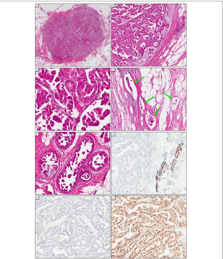
Four bioptic samples were obtained with ultrasound- guided CNB. H&E sections demonstrated a lesion mainly composed of irregular papillae with fibrovascular cores (Figure 1A and B), covered by epithelial cells often show- ing micropapillary outgrowths (without fibrovascular cores, like in micropapillary DCIS or micropapillary ovar- ian cancers) and sometimes displaying cribriform lumens.
The neoplastic epithelium was characterized by marked apocrine features and low-to-intermediate nuclear grade (Figure 1C). Less than 10% of the lesion showed a micro- papillary and solid architecture without a papillary back- ground structure, always with apocrine features. No clearly infiltrative foci were noted, although some were highly suspicious. Necrosis was absent. Mitoses were rare.
Myoepithelial (ME) cells were not detected on the H&E- stained slides and multiple stains for ME markers (CK5, S-100, p63, SMA, calponin, calponin/p63, and CK5/p63) were completely negative (Figure 1D). As the rare lack of myoepithelium in benign apocrine proliferations has been described, a final diagnosis of papillary lesion of uncertain malignant potential was made with a B3 classification according to Ellis8 and a complete removal of the lesion has been strongly indicated.
Pathological and Immunophenotypical Findings of the Lumpectomy
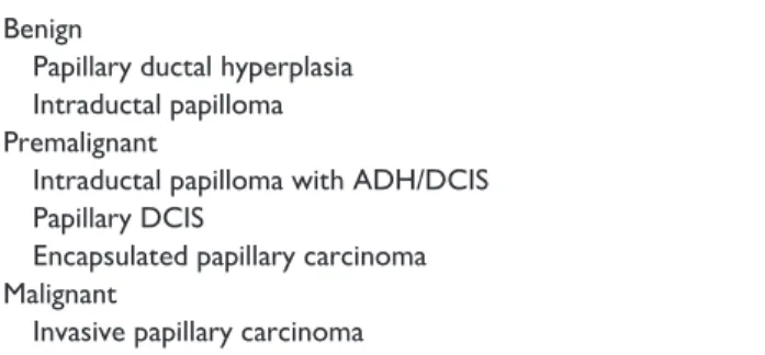
The patient underwent lumpectomy with sentinel lymph node biopsy. At gross examination, the breast mass was solid, well circumscribed with lobulated margins, and whitish with a diameter of approximately 1.5 cm. H&E sections showed that the lesion was surrounded by a thin and discontinuous pseudocapsule. The invasive tumor within this was composed of either anastomosing or branching papillary fronds with central fibrovascular cores. Coalescing luminal epithelial cells and sometimes cribriform structures (Figure 2A and B) or eye-catching micropapillary outgrowths or typical inside-out patterned cell groups were highlighted. At some places, the fibrovas- cular bands were clearly cores of papillary structures. At other places, it was difficult to decide whether they were septa between nests or cores of papillae with dissociated epithelium (Figure 2C). The micropapillary outgrowths and cribriform secondary lumens of the apocrine epithe- lium were also present in this specimen. The exact propor- tion where the structure was clearly papillary was difficult to estimate, but was obviously >10% and around 50% by 2 observers and around 90% by 2 other observers. The neoplastic cells showed apocrine atypia with a 2- to 3-fold
nuclear enlargement, hyperchromasia, and prominent/
multiple nucleoli (Figure 2C). Necrosis was absent and the mitotic activity was low (5 mitoses per 10 high-power fields). An Elston-Ellis grade 2 was assigned. Several foci of lymphovascular invasion into and beyond the pseudo- capsule were noted (Figure 2D). In addition, foci of cribri- form and papillary/micropapillary DCIS, the latter with apocrine atypia, were noted beyond the pseudocapsule in the surrounding breast tissue (Figure 2E).
On IHC, the calponin/p63 and the CK5/p63 multiplex stains showed a complete absence of the ME cell layer around the invasive component and its presence around the DCIS component. Ki67 proliferation rate evaluated with digital nuclear image analysis was 5.5%. The negative expression of ER, PR, and HER2 (Figure 2F and G) classi- fied biologically the invasive component as triple-negative, while diffuse positive expression of AR (Figure 2H) and GCDFP-15 showed the apocrine nature of both the inva- sive and in situ papillary/micropapillary components.
The final diagnosis was “mixed invasive apocrine papillary/micropapillary carcinoma associated with foci of cribriform and apocrine papillary/micropapillary DCIS.
The pathological TNM (tumor, nodes, and metastases) stage was T1c N0 (sn) M0, that is, anatomical stage IA and prognostic stage IB.
Follow-up and Treatment
No recurrence of the disease has been observed at the 6-month follow-up. After surgery, the patient received a whole breast irradiation consisting of 16 daily fractions of 265 cGy to a total dose of 4240 cGy. No adjuvant che- motherapy was given because of an anxiety-depressive syndrome.
Discussion
Pure IPC represents approximately 0.5% of IBC.3 Most commonly it is diagnosed in postmenopausal females and rarely in males.3 Mixed papillary carcinomas with <90%
obvious papillary structure are also rare. IPC usually have low-grade nuclei, low mitotic activity, and are ER and PR positive and HER2 negative.3,4 They carry a favorable prognosis compared with IBC-NST.3,4 To our knowledge, Figure 1. Core needle biopsy. The lesion has a complex architecture composed of irregular papillae with fibrovascular cores and micropapillae, 5×, hematoxylin and eosin (H&E; A), 10×, H&E (B). The irregular papillae and micropapillae are covered by cells with round nuclei with different size and shape, prominent nucleoli, and marked apocrine features, 20×, H&E (C). Note the micropapillary projections seen on the high-power view (C). Absence of calponin/p63 expression, 10×, immunohistochemistry (D).
Figure 2. Lumpectomy specimen. Well-circumscribed nodule surrounded by a thin and discontinuous pseudocapsule, 1×, hematoxylin and eosin (H&E; A). Papillary configuration of the lesion with thin irregular papillae and fibrovascular fronds lined by neoplastic cells with apocrine atypia, 5×, H&E (B), 20×, H&E (C). Note the invasive micropapillary pattern reflected by the inside-out reverted polarity of the tumor cells (C). Multiple foci of invasion in small lymphatic channels, with the tumor emboli also displaying the reverted polarity of micropapillary carcinoma 10×, H&E (D). Multiple foci of papillary/micropapillary ductal carcinoma in situ (DCIS) with apocrine features in the surrounding breast tissue, 10×, H&E (E). Absence of nuclear expression of estrogen receptor, 10×, immunochemistry (IHC; F). Absence of membrane expression of HER2, 10×, IHC (G). Intense nuclear expression of androgen receptor, 10×, IHC (F).
apocrine invasive micropapillary has not been described before. We report a unique case of mixed apocrine IPC with micropapillary features, which may represent a dis- tinct subtype of IPC, micropapillary carcinoma or apocrine carcinoma and a potential diagnostic pitfall on CNB.
Triple-negative IPC represent a subset of IPC (about 19% according to Liu et al),3 and apocrine differentiation is extremely rare. The only IPC with apocrine features that was reported in the literature has been described by Terzi et al and showed high nuclear grade, high mitotic activity, abundant necrosis, and remarkable lymphoplasmacytic stromal infiltrate; no lymphovascular invasion was noted.7
At variance from that case, ours displayed a low-to- intermediate nuclear grade, a low mitotic activity, and nei- ther stromal lymphoplasmacytic infiltrate nor necrosis. It was also characterized by lymphovascular invasion within and beyond the tumor border and in situ disease in the sur- rounding breast tissue composed of cribriform and micro- papillary/papillary DCIS, the latter with apocrine features.
Pure special type carcinomas require at least 90% of the tumor to be composed of the characteristic features. The current World Health Organization classification groups breast cancers according to several features; thus, it allows for dual classifications.2 This invasive carcinoma fulfills the 90% criterion of pure apocrine carcinoma, and on structural grounds, it is partly papillary and micropapillary and can be classified as mixed papillary and micropapil- lary. The excessive lymphovascular invasion is explain- able with this latter morphological pattern.
Our case provides additional evidence of the heterogene- ity of triple-negative breast cancers the spectrum of which has more histotypes than what is expected on the basis of published works.9 Apocrine lesions, for many pathologists, imply an ER and PR negative status by definition.10,11 Malignant apocrine tumors may be HER2 negative or posi- tive, the tumor being triple-negative in the first setting. The apocrine subtype of triple-negative breast cancers is also recognized on the basis of gene-expression profiles.11,12
The association between papillary lesions and apocrine changes represents a great challenge for breast patholo- gists. Papotti et al first reported areas of apocrine differen- tiation in papillary breast carcinomas with and without invasion.13 Apocrine changes in papillomas may be exten- sive and, in some cases, can be difficult to distinguish from low-grade DCIS.14,15 Apocrine differentiation may be prominent in papillary DCIS leading to classification as apocrine papillary DCIS.16,17 Eleven cases of apocrine- encapsulated papillary carcinoma have been reported,18-20 the latter published by Kovari et al was associated with IBC-NST with apocrine features.21 Benign apocrine lesions lacking myoepithelium, including papillary lesions that may mimic papillary carcinomas were described by Cserni in 200822 and in 2012,23 by Tramm et al in 2011,24 as well as Ha et al in 2018.25
Papillary breast lesions that may show apocrine changes are summarized in Table 1. To our knowledge, solid papil- lary carcinomas and tall cell carcinomas with reversed polar- ity with apocrine phenotype have not been described so far.
From a practical point of view, apocrine papillary lesions may represent a considerable exception to the general rule that absence of ME cells in breast lesions is usually consid- ered as a hallmark to separate invasive from in situ and malignant from benign lesions. In fact, benign and noninva- sive apocrine lesions may show a reduction and occasional complete loss of ME.24 The same concept was highlighted by Cserni et al, who reported 2 cases of apocrine intracystic papillary proliferation devoid of ME cells stressing again that even a complete lack of myoepithelium should not be equated with a diagnosis of malignancy. Given the impor- tance of ME cells, and given their sometimes unpredictable immunoprofile, it is important to use a combination of dif- ferent immunohistochemical ME markers.23 Combined immunostains, such as calponin/p63 and/or CK5/p63, may be useful in this context, especially as they may spare pre- cious bioptic material in small CNB samples. From another side, well-circumscribed papillary tumors, like encapsulated and solid papillary carcinomas, are staged as in situ carci- noma despite their lack of ME cells at the periphery of the structures involved.26,27
Breast pathologists should be aware that ME cells may be absent in some benign apocrine papillary lesions and thus should interpret this finding in light of the whole his- topathological picture.
In our case, the CNB showing a complex apocrine pap- illary proliferation with low-to-moderate cytological atypia, lack of ME layer demonstrated on multiple immu- nostains, and absence of clearly infiltrative foci was a diagnostic challenge. It could have been overdiagnosed or underdiagnosed as a malignant or a benign papillary apo- crine proliferation, respectively. Therefore, we believe that these lesions should be reported as papillary lesion of uncertain malignant potential, or suspicious of malignancy requiring surgical excision.
Table 1. List of Papillary Breast Lesions That May Show Apocrine Changes.
Benign
Papillary ductal hyperplasia Intraductal papilloma Premalignant
Intraductal papilloma with ADH/DCIS Papillary DCIS
Encapsulated papillary carcinoma Malignant
Invasive papillary carcinoma
Abbreviations: ADH, atypical ductal hyperplasia; DCIS, ductal carcinoma in situ.
curation. Gabor Cserni: Writing original draft, and writing review and editing. Mattia Barbareschi: Writing original draft, writing review and editing, and supervision.
Declaration of Conflicting Interests
The author(s) declared no potential conflicts of interest with respect to the research, authorship, and/or publication of this article.
Funding
The author(s) disclosed the following financial support for the research, authorship, and/or publication of this article: Internal funding from Department of Clinical Services has been used in part for study-related facilities.
Ethical Approval
All procedures performed were in accordance with the ethical standards of the Institutional and/or National Research Committee and with the 1964 Helsinki Declaration and its later amendments or comparable ethical standards.
Informed Consent
The patient provided consent for publication.
Trial Registration
Not applicable, because this article does not contain any clinical trials.
ORCID iD
Luca Cima https://orcid.org/0000-0002-3445-1622
References
1. MacGrogan G, Collins LC, Lerwill M, et al. Invasive papil- lary carcinoma. In: Lokuhetty D, White VA, Watanabe R, et al, eds. WHO Classification of Breast Tumours. 5th ed.
IARC Press; 2019:66-67.
2. Cserni G. Histological type and typing of breast carcinomas and the WHO classification changes over time. Pathologica.
2020;112:25-41.
3. Liu ZY, Liu N, Wang YH, et al. Clinicopathologic character- istics and molecular subtypes of invasive papillary carcinoma of the breast: a large case study. J Cancer Res Clin Oncol.
2013;139:77-84.
4. Wei S. Papillary lesions of the breast: an update. Arch Pathol Lab Med. 2016;140;628-643.
5. Zheng YZ, Hu X, Shao ZM. Clinicopathological characteris- tics and survival outcomes in invasive papillary carcinoma
adverse prognosis. Int J Surg Pathol. 2014;22:47-54.
7. Terzi A, Uner AH. An unusual case of invasive papillary carcinoma of the breast. Indian J Pathol Microbiol. 2012;
55:543-545.
8. Ellis IO, Humphreys S, Michell M, Pinder SE, Wells CA, Zakhou HD. Best Practice No 179. Guidelines for breast needle core biopsy handling and reporting in breast screen- ing assessment. J Clin Pathol. 2004;57:897-902.
9. Geyer FC, Pareja F, Weigelt B, et al. The spectrum of triple- negative breast disease: high- and low-grade lesions. Am J Pathol. 2017;187:2139-2151.
10. D’Arcy C, Quinn C. Apocrine lesions of the breast: part 1 of a two-part review: benign, atypical and in situ apocrine proliferations of the breast. J Clin Pathol. 2019;72:1-6.
11. D’Arcy C, Quinn C. Apocrine lesions of the breast: part 2 of a two-part review. Invasive apocrine carcinoma, the molecu- lar apocrine signature and utility of immunohistochemistry in the diagnosis of apocrine lesions of the breast. J Clin Pathol. 2019;72:7-11.
12. Provenzano E, Gatalica Z, Vranic S. Carcinoma with apo- crine differentiation. In: Lokuhetty D, White VA, Watanabe R, et al, eds. WHO Classification of Breast Tumours. 5th ed.
IARC Press; 2019:131-133.
13. Papotti M, Eusebi V, Gugliotta P, Bussolati G. Immuno- histochemical analysis of benign and malignant papillary lesions of the breast. Am J Surg Pathol. 1983;7:451-461.
14. Asirvatham JR, Falcone MM, Kleer CG. Atypical apocrine adenosis: diagnostic challenges and pitfalls. Arch Pathol Lab Med. 2016;140:1045-1051.
15. Mallick D, Aniruna A, Gon S, Ghosh G. Apocrine metapla- sia in intraductal papilloma with foci of DCIS: a friend or foe? Iran J Pathol. 2016;11:167-170.
16. Tavassoli FA, Norris HJ. Intraductal apocrine carcinoma:
a clinicopathologic study of 37 cases. Mod Pathol. 1994;7:
813-818.
17. Rakha EA, Ellis IO. Diagnostic challenges in papillary lesions of the breast. Pathology. 2018;50:100-110.
18. Seal M, Wilson C, Naus GJ, Chia S, Bainbridge TC, Hayes MM. Encapsulated apocrine papillary carcinoma of the breast—a tumour of uncertain malignant potential: report of five cases. Virchows Arch. 2009;455:477-483.
19. Laforga JB, Gasent JM, Sanchez I. Encapsulated apo- crine papillary carcinoma of the breast: case report with clinicopathologic and immunohistochemical study. Diagn Cytopathol. 2011;39:288-293.
20. Kuroda N, Fujishima N, Hayes MM, Moritani S, Ichihara S. Encapsulated papillary carcinoma, apocrine type, of the breast. Malays J Pathol. 2014;36:139-143.
21. Kovari B, Ormandi K, Simonka Z, Voros A, Cserni G.
Apocrine encapsulated papillary carcinoma of the breast: the first reported case with an infiltrative component. J Breast Cancer. 2018;21:227-230.
22. Cserni G. Lack of myoepithelium in apocrine glands of the breast does not necessarily imply malignancy. Histopathology.
2008;52:253-255.
23. Cserni G. Benign apocrine papillary lesions of the breast lacking or virtually lacking myoepithelial cells—poten- tial pitfalls in diagnosing malignancy. APMIS. 2012;120:
249-252.
24. Tramm T, Kim JY, Tavassoli FA. Diminished number or complete loss of myoepithelial cells associated with meta- plastic and neoplastic apocrine lesions of the breast. Am J Surg Pathol. 2011;35:202-211.
25. Ha T, Yim H, Park SY, Kang DK, Kim TH. Papillary apo- crine metaplasia of the breast mimicking papillary neoplasm:
a case report. J Korean Soc Radiol. 2018;78:103-106.
26. MacGrogan G, Collins LC, Lerwill M, et al. Encapsulated papillary carcinoma. In: Lokuhetty D, White VA, Watanabe R, et al, eds. WHO Classification of Breast Tumours. 5th ed.
IARC Press; 2019:60-62.
27. MacGrogan G, Collins LC, Lerwill M, et al. Solid papillary carcinoma (in situ and invasive). In: Lokuhetty D, White VA, Watanabe R, et al, eds. WHO Classification of Breast Tumours. 5th ed. IARC Press; 2019:63-65.