Microcystin-LR, a cyanobacterial toxin affects root development by changing levels of PIN proteins and auxin response in Arabidopsis roots
Csongor Freytag
a, Csaba M ath e
a, G abor Rig o
b, Tomasz Nodzy nski
c, Zolt an K onya
d, Ferenc Erd} odi
d, Agnes Cs epl} o
b, Erik P ozer
a, L aszl o Szabados
b, Adrienn Kelemen
a, G abor Vasas
a, Tam as Garda
a,*aUniversity of Debrecen, Faculty of Science and Technology, Department of Botany, Egyetem Ter 1., H-4032, Debrecen, Hungary
bBiological Research Centre, Institute of Plant Biology, Temesvari Krt 62, H-6726, Szeged, Hungary
cMendel Centre for Plant Genomics and Proteomics, Central European Institute of Technology (CEITEC), Masaryk University, Kamenice 5, 625 00, Brno, Czech Republic
dUniversity of Debrecen, Faculty of Medicine, Department of Medical Chemistry, Egyetem Ter 1., H-4032, Debrecen, Hungary
h i g h l i g h t s
Microcystin-LR (MCY-LR) is a strongly harmful cyanotoxin for many eukaryotes.
MCY-LR inhibited protein phosphatase activities in Col-0 Arabidopsis roots.
In consequence, PIN protein levels were altered within roots, affecting auxin levels.
MCY-LR altered root development without changing gravitropic bending in Col-0.
We discuss potential effects of such physiological changes in aquatic ecosystems.
a r t i c l e i n f o
Article history:
Received 20 January 2021 Received in revised form 2 March 2021 Accepted 3 March 2021 Available online 9 March 2021 Handling Editor: Willie Peijnenburg
Keywords:
Microcystin-LR Arabidopsis PIN efflux Carrier Auxin
Protein phosphatase PP2A Root development
a b s t r a c t
Microcystin-LR (MCY-LR) is a heptapeptide toxin produced mainly by freshwater cyanobacteria. It strongly inhibits protein phosphatases PP2A and PP1. Functioning of the PIN family of auxin efflux carriers is crucial for plant ontogenesis and their functions depend on their reversible phosphorylation.
We aimed to reveal the adverse effects of MCY-LR on PIN and auxin distribution in Arabidopsis roots and its consequences for root development.
Relatively short-term (24 h) MCY-LR treatments decreased the levels of PIN1, PIN2 and PIN7, but not of PIN3 in tips of primary roots. In contrast, levels of PIN1 and PIN2 increased in emergent lateral roots and their levels depended on the type of PIN in lateral root primordia. DR5:GFP reporter activity showed that the cyanotoxin-induced decrease of auxin levels/responses in tips of main roots in parallel to PIN levels.
Those alterations did not affect gravitropic response of roots. However, MCY-LR complemented the altered gravitropic response ofcrk5-1mutants, defective in a protein kinase with essential role in the correct membrane localization of PIN2. For MCY-LR treated Col-0 plants, the number of lateral root primordia but not of emergent laterals increased and lateral root primordia showed early development.
In conclusion, inhibition of protein phosphatase activities changed PIN and auxin levels, thus altered root development. Previous data on aquatic plants naturally co-occurring with the cyanotoxin showed similar alterations of root development. Thus, our results on the model plant Arabidopsis give a mechanistic explanation of MCY-LR phytotoxicity in aquatic ecosystems.
©2021 The Author(s). Published by Elsevier Ltd. This is an open access article under the CC BY license (http://creativecommons.org/licenses/by/4.0/).
1. Introduction
Microcystin-LR (MCY-LR) is a natural cyclic heptapeptide toxin synthetized non-ribosomally in several cyanobacterial genera
*Corresponding author.
E-mail address:garda.tamas@science.unideb.hu(T. Garda).
Contents lists available atScienceDirect
Chemosphere
j o u r n a l h o m e p a g e : w w w . e l s e v i e r . c o m / l o c a t e / c h e m o s p h e r e
https://doi.org/10.1016/j.chemosphere.2021.130183
0045-6535/©2021 The Author(s). Published by Elsevier Ltd. This is an open access article under the CC BY license (http://creativecommons.org/licenses/by/4.0/).
(Rastogi et al., 2014). It is known for a long time for its adverse effects on human and animal health as well as its deleterious effects in aquatic ecosystems (Campos and Vasconcelos, 2010). In general, MCY-LR has a double, although probably inter-related effect on eukaryotic cells: (i) it is a potent inhibitor of the serine-threonine protein phosphatases PP1 and PP2A as well as of the minor phos- phatases PP4, PP5 and PP6 (Fontanillo and K€ohn, 2018;MacKintosh et al., 1990;Mathe et al., 2016). It is known that the inhibitor binds covalently close to the active sites of the catalytic subunits of PP1 and PP2A, causing an irreversible inhibition (see Bouaïcha et al., 2019for an example). (ii) it is an inducer of reactive oxygen spe- cies (ROS) (Mathe et al., 2016,2019).
The “long” PIN-formed proteins (PIN1-4 and PIN7) are auxin efflux carriers (polar transporters) playing crucial roles in the dis- tribution and functioning of the plant hormone indole-3-acetic acid (IAA, the most abundant auxin): the proper tissue localization of IAA is important for normal growth and development including root tip meristem identity and lateral root formation (Adamowski and Friml, 2015; Barbosa et al., 2018). For roots, PIN1 is trans- porting IAA from the base towards root tip through the stele, while PIN2 is directing transport from the tip towards the base in the root epidermis and in both directions in root cortex. PIN3 and PIN7 are redistributors of IAA in the root cap columella and are present in root stele as well (Michniewicz et al., 2007a). PIN2, PIN3 and PIN7 are important for gravitropic responses (Adamowski and Friml, 2015; Barbosa et al., 2018; Michniewicz et al., 2007a). Besides PIN1 and PIN2, PIN3 and PIN7 are also involved in lateral root development-proper organization of pericycle cell divisions during initiation of lateral root primordia- and gravitropism (Benkova et al., 2003; Michniewicz et al., 2007a; Okumura et al., 2013).
Functioning and stability of PINs is regulated by post-translational modifications such as reversible phosphorylation for nearly all PINs and ubiquitination at least for PIN2 (Zwiewka et al., 2019).
The main partners of PIN reversible phosphorylation are PINOID kinase (PID, a serine-threonine kinase) and the serine-threonine protein phosphatase PP2A. PID can phosphorylate all PINs involved in this study (Ganguly et al., 2012;Barbosa et al., 2018).
CRK5 involved in this study is a member of the CDPK-related kinase family and it is important in the proper plasma membrane locali- zation of PIN2. The Arabidopsis loss-of function mutantcrk5-1is impaired in root gravitropic bending (Barbosa et al., 2018; Rigo et al., 2013). The reversible phosphorylation of PINs regulates their plasma membrane localization and their dephosphorylation will trigger internalisation and then recycling, usually to the opposite cell side (Kleine-Vehn et al., 2009;Yao and Xue, 2011).
Protein phosphatase inhibitors maintain the phosphorylated state of PIN1 caused by the activity of PID (Michniewicz et al., 2007b). Cantharidin and okadaic acid are strong inhibitors of PP2A activity that alter auxin transport and decrease auxin content in root tips. Cantharidin alters root development (Shin et al., 2005).
For MCY-LR, nothing was known to date on its effects on the fate of PINs and consequent auxin distribution. Since it is one of the most abundant naturally occurring cyanotoxins, the relevant studies are necessary. Related to this, in rice roots it decreased auxin levels, inhibiting root elongation and lateral root formation (Chen et al., 2013).
The aim of the present study was to give novel insights into the effects of a natural cyanotoxin on the levels of PIN proteins (PIN 1, 2, 3 and 7) and hence, of auxin (IAA) within Arabidopsis roots and the consequent alterations in root development and gravitropism. We discuss the possible environmental implications of these alterations.
2. Materials and methods
2.1. The purification of microcystin-LR
MCY-LR was purified from Microcystis aeruginosa BGSD 243 according toKos et al. (1995)modified byVasas et al. (2004). Cells were collected by centrifugation, then extracted as described. Pu- rification of cyanotoxin was performed on a DEAE Cellulose-52 (Whatman DE 52) column. The eluates containing MCY-LR were further purified by Toyopearl Size Exclusion Chromatography. The purity of the preparations was verified by HPLC (C-18 HPLC column, Supelcosil TM SPLC-18, 25 cm 10 mm, 5 mm). HPLC analyses were performed with a Shimadzu HPLC system equipped with a Shi- madzu SPD-M 10 AVP diode array. The distinctive peaks of the chromatogram were checked by LC-MS. MCY-LR was detected as [MþH]þatm/z995.5.
2.2. Plant material and MCY-LR treatments
TheArabidopsis thalianagenotypes used in this study were: Col- 0 (wild-type), Col-0 plants bearing PIN1:GFP, PIN2:GFP, PIN3:GFP (PIN1:PIN1-GFP, PIN2:PIN2-GFP and PIN3:PIN3-GFP) (Benkova et al., 2003;Xu et al., 2006;Zadníkova et al., 2010), DR5rev:GFP fusion constructs for the detection of tissue areas characterized by the presence of and responses to auxins (Friml et al., 2003b;
Zadníkova et al., 2010) and thecrk5-1loss of function mutant (Rigo et al., 2013).
Seeds were surface sterilized by rinsing two times for 10 min with 10% of a sodium-hypochlorite containing solution, then washedfive times for 5 min with sterile water. Seeds were trans- ferred to MS (Murashige and Skoog, 1962) basal medium supple- mented with Gamborg’s vitamins, 2% (w/v) sucrose (Molar, Budapest, Hungary) and 0.8% (w/v) Bacto-agar (Difco, Lawrence, KS, USA) (Gamborg et al., 1968;Murashige and Skoog, 1962). After a 48- h cold treatment, plates were placed in a tissue culture room (14/
10 h photoperiod, 22±2C, 60mmol m2s1photonflux density in the light period). After 5 days of germination, seedlings were treated with MCY-LR under the above specified physical laboratory conditions (seeNagy et al. (2018) for the method of treatments).
The study of gravitropic responses was performed with 3 days old seedlings.
2.3. Immunohistochemistry and histochemistry methods.
Microscopy
Seedlings bearing the GFP-fusion constructs (PIN:GFP and DR5rev:GFP) were examined with an Olympus FluoView 1000 confocal microscope (excitation wavelength: 488 nm, emission wavelength range: 500e530 nm;60 water-immersion UPLSAPO objective, with a numerical aperture of 1.2, acquisition software:
FV-1000). For the analysis of signal intensity, we constructed 2D projection images, then 2D heatmaps of these images were con- structed for a better visualization. Mean grey values (AIODs, Area Integrated Optical Densities) were calculated with the aid of Fiji (ImageJ-Win64) software after background subtraction of 2D heatmap images. For GFP signals in DR5rev:GFP plants, heatmaps from 3D images of confocal microscopic stacks for tips and differ- entiated segments of main roots were also constructed.“Differen- tiated root segments”or“-tissues”means the maturation zone of main (primary) root throughout this study. 3D Heatmaps were obtained with the aid of ZEN Blue 2.3 Lite software and showed local intensities and areas of GFP signals in root segments.
o et al. Chemosphere 276 (2021) 130183
The whole-mount immunolocalization of PIN1, PIN2 and PIN7 was performed by the use of InsituPro VSi pipetting robot as described previously (Friml et al., 2003a;Sauer et al., 2006). We fixed Col-0 main root tips with 4% (w/v) paraformaldehyde (PFA) in PBS (137 mM NaCl, 2.7 mM KCl, 10 mM Na2HPO4, 1.8 mM KH2PO4, 1 mM CaCl22H2O, 0.5 mM MgCl26H2O/pH 7.4 adjusted with HCl) for 40 min under vacuum. Samples were then washed with PBS/
0.1% Triton-X100 (3 15 min) and deionized water/0.1% Triton- X100 (3 15 min). Cell walls were digested with 2% Driselase (Sigma-Aldrich, St. Louis, Mo., USA) in PBS for 30 min at 37C and samples were washed with PBS/0.1% Triton-X100 (315 min). In the next step the seedlings were permeabilized with 10% DMSO/3%
IGEPAL in PBS for 30 min and washed with PBS/0.1% Triton-X100 (315 min). Samples were pre-incubated in 2% BSA/PBS for 1 h and incubated with primary antibody in 2% BSA/PBS for 4 h at 37C, then washed with PBS/0.1% Triton-X100 (515 min). Rabbit raised anti-PIN1, anti-PIN2 (Skokan et al., 2019) and anti-PIN7 (Doyle et al., 2019) were used as primary antibodies. Secondary antibody was Cy3 conjugated anti-rabbit IgG generated in sheep (Sigma- Aldrich, C2306). Incubation with secondary antibody was in 2%
BSA/PBS for 4 h at 37C, then samples were washed with PBS/0.1%
Triton-X100 (3 15 min) and deionized water (3 15 min).
Preparations were analyzed with a Zeiss LSM Z700 confocal mi- croscope with the conventional settings for Cy3 visualization. Data evaluation was performed by comparing the signal intensity of control plants with signal intensity of treated ones. In all cases, we worked with the same microscope settings that included laser in- tensity, gain, objective and magnification.
Reactive oxygen species (ROS) were detected in Col-0 roots ac- cording toGarda et al. (2016). We have used thefluorescent dye 20,70-dichlorofluorescein-diacetate (DCFH-DA, Sigma-Aldrich) at a concentration of 20mM. The dye is excitable at 450e480 nm. We used an Olympus Provis AX-70fluorescence microscope (Olympus, Tokyo, Japan) for observations and labelling intensities were quantified with ImageJ 64 software.
In all experiments described in this subchapter, at least five seedlings per treatment per experiment were used and four-five independent experiments were performed.
2.4. Investigation of root development and gravitropic responses
Gravitropic response assay was performed essentially as described byRigo et al. (2013)with modifications. Three days-old Arabidopsis Col-0 and crk5-1 mutant seedlings were used for treatments. For the study of gravitropic bending, seedlings were grown vertically on half-strength MS medium with 1% (w/v) su- crose, in contrast to Rigo et al. (2013), where reduced sucrose concentration (0.5%) was used. Afterwards, seedlings were trans- ferred to the same culture medium for control and 1mM MCY-LR treatments. The vertically grown seedlings were reoriented by 135, and the degree of root bending was recorded by scanning 24 h after rotation. The rate of root bending was determined by measuring the angle formed between the horizontal baseline and growth direction of root tip as described-that is, the angle between reoriented starting position and a 24-h position (Rigo et al., 2013).
At least 100 wild-type andcrk5-1seedlings were tested per each treatment in three separate experiments.
For the study of development of lateral root primordia and emergent laterals, non-gravistimulatedfive days old Col-0 seed- lings were MCY-LR treated for one and two days. PFA-fixed roots were labeled for chromatin with DAPI (see above, Immunohisto- chemistry and histochemistry), that showed clearly not only emergent laterals, but primordia as well. We considered as primordia all stages of lateral root development before their
emergence from the main (primary) root. Whole roots were scan- ned by microscopy for lateral root numbers. The distance between tips of main roots and the occurrence of thefirst lateral root pri- mordium was also measured. At least 200 seedlings were tested per each treatment and experiments were repeated four times.
2.5. The assay of protein phosphatase activities
Total protein phosphatase activity, which includes PP1 and PP2A activities in vivo, was measured as described previously (Garda et al., 2018; Mathe et al., 2013). Arabidopsis Col-0 whole roots were extracted with a buffer containing 50 mM Tris-HCl (pH 7.5), 0.1 mM EDTA, 0.2 mM EGTA, 0.1% (w/v)b-Mercaptoethanol, 1 mM PMSF (Sigma-Aldrich), 0.5% (v/v) protease inhibitor cocktail (Roche Applied Science, Indianapolis, USA). The protein content of extracts was determined according toBradford (1976) so that the assays were carried out with the same amounts of protein.32P-MLC20 (phosphorylated turkey gizzard 20 kDa myosin light chain labeled with 32P) was used as substrate. Specific phosphatase activities were given as pmol [32Pi] released mg protein1(Erd}odi et al., 1995) and presented as the percentage of control activities (100%). Protein phosphatase activity assays were performed in three parallel measurements per treatment and three separate experimental replicates.
2.6. Data analysis
All quantified data were plotted eplots are showing the mean±SE values-with the aid of Systat Sigma Plot 10.0®software except for data on gravitropic responses plotted with R studio version 1.2 with ggplot2 graphical package. Statistical significances for the differences between controls and treatments were studied by Mann-Whitney Rank Sum Test and t-tests. Differences were considered to be significant at P<0.05.
3. Results
Our preliminary studies employed the use of MCY-LR in a con- centration range of 1e10mM. Concentrations of 5mM and above induced cell lethality at long and even short term exposures, so they were omitted (data not shown). 1mM MCY-LR induced suffi- cient protein phosphatase inhibition after 24 h of exposure without inducing rapid cell death, therefore this treatment was used throughout our experiments. The above findings confirm the
“dualistic response”hypothesis for the effects of microcystins in plants (Corbel et al., 2015). The relatively short term treatments with 1mM MCY-LR allowed us to get insights into the PIN/auxin content-root development relationship.
3.1. Effects of MCY-LR on the levels of auxin efflux carriers (PIN1, PIN2, PIN3, PIN7)
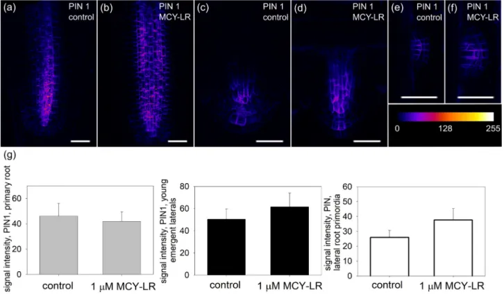
PINs were analyzed in PIN:GFP plants and by immunohisto- chemistry. Both approaches showed their normal polar localization and distribution at the subcellular and tissue level in roots of control Arabidopsis thaliana seedlings. For primary roots, PIN1 protein was normally localized in the stele and PIN2 protein in the cortex and epidermis (Figs. 1a and 2a,Supplementary Fig. S1/a, c, e).
Immunohistochemical labeling showed clear decreases of PIN levels after MCY-LR treatments at each test period. 1mM MCY-LR reduced the level of PIN1 and PIN2 proteins after one (Supplementary Fig. S1/b), two (Supplementary Fig. S1/d) andfive (Supplementary Fig. S1/f) days as compared to control plants. For PIN:GFP plants, MCY-LR decreased PIN1 and PIN2 levels after one
o et al. Chemosphere 276 (2021) 130183
day treatment as well (Figs. 1b and 2b). However, these decreases were non-significant (Figs. 1g and 2e/primary roots).
In contrast to primary roots, GFP signals for both PIN1 and PIN2 increased in emergent laterals in the presence of MCY-LR (Figs. 1d and 2d) and this increase was significant for PIN2:GFP (Fig. 2e/
emergent laterals). For lateral root primordia, 1mM MCY-LR induced a slight, but non-significant increase of PIN1:GFP signal (Fig. 1e, f, g/
lateral primordia). For PIN2:GFP, there was a minimal number of lateral root primordia in the young seedlings investigated, so we could not quantify this parameter. Data for lateral roots refer only to 24 h treatments with the cyanotoxin.
For PIN3:GFP plants, untreated primary roots showed strong GFP expression in root cap columella and weaker expression in stele (Fig. 3a). At relatively short term (24 h) MCY-LR exposures, GFP signals increased both in the columella and stele of primary root tips and for the stele, this change was significant (Fig. 3b, e/
primary root columella and stele). There was no sufficient number of PIN3:GFP emergent laterals to perform quantification. For lateral root primordia, MCY-LR had decreased PIN3:GFP signal in a sig- nificant manner (Fig. 3c, d, e/lateral root primordia). After seven days of exposure, 1 mM MCY-LR caused a decrease of PIN3:GFP signal in primary roots (data not shown).
Immunohistochemical labeling of PIN7 in primary roots showed its normal distribution with high levels in the stele and distal segments of root apex in control plants (Supplementary Fig. S2/a, a’, c, e, g). At 1mM MCY-LR treatments, the level of PIN7 was reduced in the stele at each test time (Supplementary Fig. S2/b, d, f, h) and distal segments of root apex after 24 h of exposure (Supplementary Fig. S2/b’).
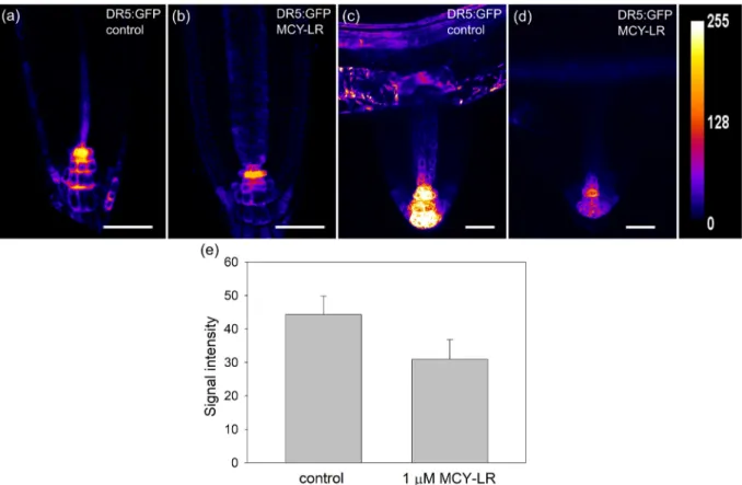
3.2. Effects of MCY-LR on auxin levels in roots
The levels of and responses to auxin in roots were analyzed by the examination of GFP signals inA. thalianaplants bearing the auxin responsive DR5rev:GFP construct. Quantification of 2D heat map projections of GFP signals showed non-significant, but consistent decreases of auxin levels/responses in tips of primary roots (Fig. 4a, b, e). To get more precise data on distribution of auxin content/response in root tissues, 3D heat map projections of GFP signals were constructed. These showed the distribution of auxin in the apex and differentiated segments of primary roots. For control plants, GFP signals were detected mostly in root cap colu- mella and stele of root tips (Supplementary Fig. S3/a, e) and in the subepidermal cell layers and stele of differentiated root segments (Supplementary Fig. S3/c, g). 24 h of treatment with 1mM MCY-LR induced decreases of intensities and areas of GFP signals within root tips (Supplementary Fig. S3/b) and differentiated root seg- ments (Supplementary Fig. S3/d). For the latter, auxin levels/re- sponses decreased well visibly in stele (Supplementary Fig. S3/d, arrowhead). For 48 h treatments with 1mM MCY-LR, auxin levels were reduced as well (Supplementary Fig. S3/f, h).
The 24-h treatment with 1mM MCY-LR resulted in a well visible decrease of GFP signal in emergent lateral roots of DR5rev:GFP plants (Fig. 4c and d). However, there were very few emergent lateral roots or lateral root primordia in the young DR5rev:GFP seedlings, thus we could not perform a thorough quantification of results here.
Fig. 1.The effects of MCY-LR on PIN1 content of primary and lateral roots. (a-f) Intensities of color-coded (heat-mapped) 2D maximum projections of CLSM images of PIN1:GFP signals from tips of non-gravistimulated roots show that 24 h treatment with 1mM MCY-LR modifies PIN levels in Arabidopsis. (a, b)- primary roots; (c, d)- emergent laterals; (e, f)- lateral root primordia. Heatmap scale is shown. Size scalebars: 50mm. (g) Quantification of PIN1:GFP signals from Arabidopsis root tips show differential effects of MCY-LR on PIN levels in primary vs. lateral roots at 24 h treatments. Signal intensity was calculated as AIOD (mean grey value, area integrated optical density/AIOD). Left: primary roots, centre:
emergent lateral roots, right: lateral root primordia. See Section2.3. of Materials and methods for sample numbers and Section2.6. for the methods of statistical analysis. (For interpretation of the references to color in thisfigure legend, the reader is referred to the Web version of this article.)
o et al. Chemosphere 276 (2021) 130183
3.3. Effects of MCY-LR on root gravitropic response and development
As the experiments with DR5rev:GFP plants revealed that MCY- LR interferes with auxin levels/responses, we became interested how this inhibitor can influence auxin-controlled responses such as root gravitropism or root development. MCY-LR did not affect the gravitropic response of Col-0 plants (Fig. 5a). Gravitropic bending was inhibited in the control crk5-1loss-of function mutant (see Introduction for the function of CRK5) (Fig. 5a): the percentage of plants showing maximum 90 bending increased and the per- centage of plants restoring 135bending decreased (Fig. 5a). These observations confirmed those ofRigo et al. (2013), even under different physical laboratory conditions and increased sucrose content of culture medium. Interestingly, the abnormal gravitropic response ofcrk5-1mutant was restored to a level close to control by MCY-LR (Fig. 5a).
For non-gravistimulated Col-0 plants, the effects of 1mM MCY- LR on the number of lateral root primordia and emergent lateral roots were time -dependent (Fig. 5b and c). MCY-LR treatment promoted lateral root initiation, since it increased significantly the number of primordia after two days of exposure. At this stage the number of primordia was nearly doubled as compared to the start of experiment, while in case of controls, development of primordia was relatively slow (Fig. 5b). For emergent laterals, their number decreased, but in a non-significant manner even after two days of exposure (Fig. 5c). This meant that development of lateral root
primordia was stimulated by the cyanotoxin, but this was not fol- lowed by the promotion of their further development.
MCY-LR inhibited the elongation growth of main roots in Col- 0 plants. 1mM MCY-LR reduced significantly the distance of the first lateral root primordium from the main root tip after 24 h treatments. This effect persisted after two days of treatments (Supplementary Fig. S4).
3.4. The effects of MCY-LR on ROS production and protein phosphatase activities in Col-0 roots
In Col-0 seedlings, MCY-LR did not alter ROS levels in primary root tips at 24 h treatments (Fig. 6a). Only a long-term (5 days) treatment with the cyanotoxin induced a significant increase of ROS levels (data not shown). In differentiated root segments ROS levels did not change significantly for up to three days of MCY-LR treatments (Fig. 6b), while they were decreased significantly after seven days of exposures with the phosphatase inhibitor (data not shown). The total serine-threonine protein phosphatase (PP1 and PP2A) activities were significantly inhibited (nearly 50% inhibition) by 1mM MCY-LR in whole root extracts of plants treated for 24 h (Fig. 6c).
4. Discussion
Several harmful effects of MCY-LR on plant cells/tissues are known. This includes alterations of cytoskeleton and chromatin Fig. 2.The effects of MCY-LR on PIN2 content of primary and lateral roots. (a-d) Intensities of color-coded (heat-mapped) 2D maximum projections of CLSM images of PIN2:GFP signals from tips of non-gravistimulated roots show that 24 h treatment with 1mM MCY-LR modifies PIN2 levels in Arabidopsis. (a, b)- primary roots; (c, d)- emergent laterals.
Heatmap scale is shown. Size scalebars: 50mm. (e) Quantification of PIN2:GFP signals from Arabidopsis root tips show differential effects of MCY-LR on PIN levels in primary vs.
lateral roots at 24 h treatments. Signal intensity was calculated as AIOD (mean grey value, area integrated optical density/AIOD). Left: primary roots, right: emergent lateral roots.
Asterisk shows a significant difference of MCY-LR treatments vs. controls. See Section2.3. of Materials and methods for sample numbers and Section2.6. for the methods of statistical analysis. (For interpretation of the references to color in thisfigure legend, the reader is referred to the Web version of this article.)
o et al. Chemosphere 276 (2021) 130183
organization, cell cycle regulation, cell death/tissue necrosis (Jambrik et al., 2011;Mathe et al, 2013,2016;Pappas et al., 2020).
Auxin as a plant growth regulator is crucial for root develop- ment in all vascular plants. Any perturbation of its local concen- tration will alter root and shoot development. Its distribution within the plant body depends largely on the proper functioning of PIN efflux carriers (Adamowski and Friml, 2015). Functioning of PINs is regulated by reversible phosphorylation (Friml et al., 2004;
Zwiewka et al., 2019). This paper shows how local amounts of PINs and in consequence auxin are altered in roots of Arabidopsis in the presence of MCY-LR, a potent protein phosphatase inhibitor.rcn1, pp2aa1,2, mutants lacking a functional“A”regulatory subunit of PP2A show alterations of primary and lateral root development as well as gravitropic response of roots (Michniewicz et al., 2007b;
Rashotte et al., 2001;Zhou et al., 2004) and as we will see, MCY-LR has several effects similar to this mutant phenotype.
The levels of PIN1, PIN2 and PIN7 decrease in the tips of Arabi- dopsis primary roots treated with MCY-LR. Plants bearing PIN:GFP constructs show a non-significant decrease, while the immuno- histochemistry approach shows a more dramatic change (Figs. 1e2 andSupplementary Figs. S1-S2). Interestingly cantharidin, another natural potent inhibitor of PP2A (MacKintosh and Diplexcito, 2009) did not alter PIN2 levels in root tips (Shin et al., 2005). For PIN3, MCY-LR has opposing effects since its level increases in primary root tips (Fig. 3). For lateral roots, the cyanotoxin has contrasting effects-levels of PIN1 and PIN2 increase and those of PIN3 (in lateral root primordia) decrease here (Figs. 1e3). This suggests that MCY-
LR induces redistribution of PINs in roots. The biochemical back- ground is the inhibition of protein phosphatase activity, while ROS levels do not change at short-term exposure to the cyanotoxin (Fig. 6). Thus, the potential side-effects of the cyanotoxin like ROS induction can be excluded during 24-h treatments and the changes in PIN/auxin levels can be directly related to the specific effects of MCY-LR on protein phosphatases. Changes of auxin levels/re- sponses are expected after such changes in the localization of PINs and indeed, plants bearing DR5rev:GFP constructs showed non- significant, but consistent decrease of auxin levels in tips of pri- mary roots (Fig. 4andSupplementary Fig. S3).
What is the consequence of these changes for root develop- ment? Again, the Arabidopsis mutants for PP2A scaffolding, regu- latory or catalytic subunits show that perturbation of PP2A activity or its subcellular/tissue localization inhibits longitudinal growth of primary roots, stimulates the formation of lateral root primordia but inhibit their elongation. This is partly a consequence of auxin mislocalization (Zhou et al., 2004; Michniewicz et al., 2007b;
Spinner et al., 2013). We observed very similar root developmental alterations after MCY-LR treatments. The distance between root tip and thefirst lateral root primordium decreased, which means the elongation of primary root was inhibited (Supplementary Fig. S4).
Meanwhile, development of lateral root primordia was stimulated and this was not followed by stimulation of lateral root emergence (Fig. 5b, c).
PIN2, PIN3 and PIN7 are important for the gravitropic response of roots and decrease of their levels inhibits gravitropic bending Fig. 3.The effects of MCY-LR on PIN3 content of primary and lateral roots. (a-d) Intensities of color-coded (heat-mapped) 2D maximum projections of CLSM images of PIN3:GFP signals from tips of non-gravistimulated roots show that 24 h treatment with 1mM MCY-LR modifies PIN3 levels in Arabidopsis. (a, b)- primary roots; (c, d)- lateral root primordia.
Heatmap scale is shown. Size scalebars: 50mm. (e) Quantification of PIN3:GFP signals from Arabidopsis root tips show differential effects of MCY-LR on PIN levels in primary vs.
lateral roots at 24 h treatments. Signal intensity was calculated as AIOD (mean grey value, area integrated optical density/AIOD). Left: primary roots, columella, centre: primary roots, stele, right: lateral root primordia. Asterisks show significant differences of MCY-LR treatments vs. controls. See Section2.3. of Materials and methods for sample numbers and Section2.6. for the methods of statistical analysis. (For interpretation of the references to color in thisfigure legend, the reader is referred to the Web version of this article.)
o et al. Chemosphere 276 (2021) 130183
Fig. 4.(a-d) Heat-mapped 2D maximum projections of CLSM images of DR5rev:GFP signals from tips of non-gravistimulated primary and lateral roots show that 1mM MCY-LR decreases auxin levels in Arabidopsis after 24 h of treatment. (a, b) - primary roots; (c, d) emergent laterals. Heatmap scale is shown. Size scalebars: 50mm. (e) quantification of DR5rev:GFP signals from Arabidopsis root tips show decreases in the presence of MCY-LR. Signal intensity was calculated as AIOD (mean grey value, area integrated optical density/AIOD). See Section2.3. of Materials and methods for sample numbers and Section2.6. for the methods of statistical analysis.
Fig. 5. (a)MCY-LR restores the abnormal gravitropic response ofcrk5-1mutants. Plants were gravistimulated as described byRigo et al. (2013). This Figure represents bending of primary roots after the 135reorientation (see Methods). Lower bending degrees than 135indicate abnormal gravitropic response. Complete root (re)bending is of 135(arrows).
(b, c)- Time- and dose dependent effects of MCY-LR on lateral root development in non-gravistimulated roots of Col-0 seedlings:(b)number of lateral root primordia;(c)number of emergent laterals. Asterisk on (b) indicates significant difference between controls and treatment. See Section2.4. of Materials and methods for sample numbers and Section2.6. for the methods of statistical analysis.
o et al. Chemosphere 276 (2021) 130183
(Adamowski and Friml, 2015;Michniewicz et al., 2007a). MCY-LR did not alter this response, probably because increase of PIN3 levels in primary root tips compensated for the decreases of PIN2 and PIN7. Interestingly, the cyanotoxin restored gravitropic response ofcrk5mutants (Fig. 5a). This mutant lacks a CDPK type protein kinase that contributes to PIN2 phosphorylation (Rigo et al., 2013). As a consequence, PIN2 levels in the transition zone of vertically grown roots decrease and the shift of PIN2 to asymmetric distribution during gravistimulation is delayed. This will result in the perturbation of gravitropic response in mutants (Rigo et al., 2013). The effect of MCY-LR may be caused by maintenance of PIN2 phosphorylated state by an alternative, yet unknown kinase and parallel inhibition of PIN2 dephosphorylation by a PP2A com- plex. This is an exciting issue that needs further investigation. In contrast to MCY-LR, cantharidin reduces gravitropic bending of roots, similarly to the Arabidopsisrcn1mutant, defective in the A regulatory subunit of PP2A (Rashotte et al., 2001). Other loss of function mutants of thepp2aaserieseagain, affecting A subunits- are also inducing altered gravitropism- and the collapse of root apical meristem (Xi et al., 2016). What can be the reason for the differential effects of cantharidin? The studies on cantharidin (otherwise known to be potent inhibitor of PP2A) mentioned in this paper did not include protein phosphatase activity assays and we can suppose it had differential effects to MCY-LR presented here in this respect.
What can be the environmental consequence of such alterations in PIN/auxin distribution? MCY-LR induces serious impairment in the development of primary and lateral roots of Phragmites aus- tralis, an aquatic plant naturally co-occurring with the cyanotoxin.
Long-term exposures to MCY-LR induce dramatic changes in root development ofP. australis(Mathe et al, 2007,2009). Root elon- gation is inhibited, radial expansion of primary root cells occurs instead. Lateral root primordia are formed abundantly at the vi- cinity of primary root tips (Mathe et al., 2009). Radial expansion of shoot tip cells is induced by MCY-LR in another aquatic plant, Ceratophyllum demersumunder laboratory conditions (Szigeti et al., 2010). Similar shoot alteration occurred inC. submersumnaturally co-existing with a MCY containing cyanobacterial bloom in a small Hungarian pond (Ujvarosi et al., 2019). This study shows that even relatively short-term cyanotoxin treatments induce inhibition of root elongation and promote initiation of lateral root development in Arabidopsis-related to altered levels of PINs and in consequence, auxins. Thus, the model presented here suggests that develop- mental alterations induced by the cyanotoxin in natural plant
communities originate from alterations of local auxin concentra- tions in roots.
To conclude, MCY-LR induces alterations of root development in the model plant Arabidopsis and this is correlated to changes in the levels of PIN auxin efflux carriers and of local auxin levels in roots.
These changes are related directly to the protein phosphatase inhibitory effects of the cyanotoxin at 24 h treatments. The present study is thefirst one to show such phytotoxic effects of a cyano- toxin. It contributes to a better understanding of phytotoxicity of a well-known cyanotoxin both under laboratory conditions and the real environment.
Funding
This work was supported by the Hungarian Scientific Grant NKFIH (National Research, Development and Innovation Office) K120638 for CM, K119647 for GV, K129104 for FE, NN118089 for LS, AC, RG and Ministry of Education, Youth and Sports of Czech Re- public under the projects CEITEC 2020 (LQ1601) for TN. TG and CF were supported by the EFOP-3.6.1-16-2016-00022 project co- financed by the European Union and the European Social Fund.
FC was supported by ÚNKP-20-3-II-DE-412, AK was supported by ÚNKP-20-2-I-DE-129, New National Excellence Program of the Ministry for Innovation and Technology from the source of the National Research, Development and Innovation Fund.
Availability of data and material
The authors declare that any experimental data for this manu- script are transparent and can be made visible at request.
Declaration of competing interest
The authors declare they have no competing conflict of interests.
Acknowledgements
We thank Jiri Friml for useful advices on the experimental approach and helping us in obtaining the PIN:GFP plants and Taras P. Pasternak for his advices regarding methodologies. We acknowledge Gy€orgy Vamosi, Gy€orgy Vereb, Laszlo Újlaky-Nagy and the Sandor Damjanovich Cell Analysis Core Facility at the Department of Biophysics and Cell Biology, Faculty of Medicine, University of Debrecen for helping us in the use of confocal Fig. 6.(a, b) The effects of 24 h MCY-LR treatment on ROS levels in root tips (a) and differentiated root tissues (b) visualized by the intensity of DCFH-DA staining. Signal intensity was calculated as AIOD (mean grey value, area integrated optical density/AIOD). (c) The effect of 1mM MCY-LR on total protein phosphatase activity at 24 h of exposure. Asterisk indicates significant difference of controls vs. treatments. See Section2.3. of Materials and methods for sample numbers and Section2.6. for the methods of statistical analysis.
o et al. Chemosphere 276 (2021) 130183
microscopy devices at UD. We also acknowledge the core facilities:
CELLIM of CEITEC supported by the Czech-BioImaging large RI project (LM2018129 funded by MEYS CR) and Plant Sciences Core Facility of CEITEC Masaryk University for their support with obtaining part of the scientific data presented in this paper.
Appendix A. Supplementary data
Supplementary data to this article can be found online at https://doi.org/10.1016/j.chemosphere.2021.130183.
Author contribution statement
C.F. performed experimental work and contributed to experi- mental design. C.M. participated in microscopy work and contrib- uted to writing of manuscript. G.R., A.C. and L.S. helped in performing work with CRK5:CRK5-GFP plants andcrk5mutants.
T.N. contributed to immunohistochemistry work and helped in experimental design. Z.K. and F.E. contributed with experimental design and work related to protein phosphatase activity assays. A.K.
and E.P. helped in histochemistry work and assay of lateral root development. G.V. purified MCY-LR. T.G. participated in design and execution of experiments and contributed to writing of manuscript.
References
Adamowski, M., Friml, J., 2015. PIN-dependent auxin transport: action, regulation, and evolution. Plant Cell 27, 20e32.https://doi.org/10.1105/tpc.114.134874.
Barbosa, I.C.R., Hammes, U.Z., Schwechheimer, C., 2018. Activation and polarity control of PIN-formed auxin transporters by phosphorylation. Trends Plant Sci.
23, 523e538.https://doi.org/10.1016/j.tplants.2018.03.009.
Benkova, E., Michniewicz, M., Sauer, M., Teichmann, T., Seifertova, D., Jürgens, G., Friml, J., 2003. Local, efflux-dependent auxin gradients as a common module for plant organ formation. Cell 115, 591e602.
Bouaïcha, N., Miles, C.O., Beach, D.G., Labidi, Z., Djabri, A., Benayache, N.Y., Nguyen- Quang, T., 2019. Structural diversity, characterization and toxicology of micro- cystins. Toxins 11, 714.https://doi.org/10.3390/toxins11120714.
Bradford, M.M., 1976. A rapid and sensitive method for the quantitation of micro- gram quantities of protein utilizing the principle of protein-dye binding. Anal.
Biochem. 72, 248e254.
Campos, A., Vasconcelos, V., 2010. Molecular mechanisms of microcystin toxicity in animal cells. Int. J. Mol. Sci. 11, 268e287.https://doi.org/10.3390/ijms11010268.
Chen, J., Zhang, H.-Q., Hu, L.-B., Shi, Z.-Q., 2013. Microcystin-LR-induced phytotox- icity in rice crown root is associated with the cross-talk between auxin and nitric oxide. Chemosphere 93, 283e293. https://doi.org/10.1016/
j.chemosphere.2013.04.079.
Corbel, S., Mougin, C., Martin-Laurent, F., Crouzet, O., Bru, D., Nelieu, S., Bouaïcha, N., 2015. Evaluation of phytotoxicity and ecotoxicity potentials of a cyanobacterial extract containing microcystins under realistic environmental concentrations and in a soileplant system. Chemosphere 128, 332e340. https://doi.org/
10.1016/j.chemosphere.2015.02.008.
Doyle, S.M., Rigal, A., Grones, P., Karady, M., Barange, D.K., Majda, M., Parízkova, B., Karampelias, M., Zwiewka, M., Pencík, A., Almqvist, F., Ljung, K., Novak, O., Robert, S., 2019. A role for the auxin precursor anthranilic acid in root gravi- tropism via regulation of PIN-FORMED protein polarity and relocalisation in Arabidopsis. New Phytol. 223, 1420e1432.https://doi.org/10.1111/nph.15877.
Erd}odi, F., Toth, B., Hirano, K., Hirano, M., Hartshorne, D.J., Gergely, P., 1995. Endo- thall thioanhydride inhibits protein phosphatases-1 and -2A in vivo. Am. J.
Physiol. Cell Physiol. 269, C1176eC1184. https://doi.org/10.1152/
ajpcell.1995.269.5.C1176.
Fontanillo, M., K€ohn, M., 2018. Microcystins: synthesis and structureeactivity relationship studies toward PP1 and PP2A. Bioorg. Med. Chem. 26, 1118e1126.
https://doi.org/10.1016/j.bmc.2017.08.040.
Friml, J., Benkova, E., Mayer, U., Palme, K., Muster, G., 2003a. Automated whole mount localisation techniques for plant seedlings. Plant J. 34, 115e124.https://
doi.org/10.1046/j.1365-313X.2003.01705.x.
Friml, J., Vieten, A., Sauer, M., Weijers, D., Schwarz, H., Hamann, T., Offringa, R., Jürgens, G., 2003b. Efflux-dependent auxin gradients establish the apicalebasal axis of Arabidopsis. Nature 426, 147e153.https://doi.org/10.1038/nature02085.
Friml, J., Yang, X., Michniewicz, M., Weijers, D., Quint, A., Tietz, O., Benjamins, R., Ouwerkerk, P.B.F., Ljung, K., Sandberg, G., Hooykaas, P.J.J., Palme, K., Offringa, R., 2004. A PINOID-dependent binary switch in apical-basal PIN polar targeting directs auxin efflux. Science 306, 862e865. https://doi.org/10.1126/
science.1100618.
Gamborg, O.L., Miller, R.A., Ojima, K., 1968. Nutrient requirements of suspension cultures of soybean root cells. Exp. Cell Res. 50, 151e158.
Ganguly, A., Lee, S.-H., Cho, H.-T., 2012. Functional identification of the phosphor- ylation sites of Arabidopsis PIN-FORMED3 for its subcellular localization and biological role. Plant J. 71, 810e823. https://doi.org/10.1111/j.1365- 313X.2012.05030.x.
Garda, T., Konya, Z., Freytag, C., Erd}odi, F., Gonda, S., Vasas, G., Szücs, B., M- Hamvas, M., Kiss-Szikszai, A., Vamosi, G., Mathe, C., 2018. Allyl-isothiocyanate and microcystin-LR reveal the protein phosphatase mediated regulation of metaphase-anaphase transition in Vicia faba. Front. Plant Sci. 9https://doi.org/
10.3389/fpls.2018.01823.
Garda, T., Konya, Z., Tandor, I., Beyer, D., Vasas, G., Erd}odi, F., Vereb, G., Papp, G., Riba, M., M-Hamvas, M., Mathe, C., 2016. Microcystin-LR induces mitotic spindle assembly disorders in Vicia faba by protein phosphatase inhibition and not reactive oxygen species induction. J. Plant Physiol. 199, 1e11.https://doi.org/
10.1016/j.jplph.2016.04.009.
Jambrik, K., Mathe, C., Vasas, G., Beyer, D., Molnar, E., Borbely, G., M-Hamvas, M., 2011. Microcystin-LR induces chromatin alterations and modulates neutral single-strand-preferring nuclease activity in Phragmites australis. J. Plant Physiol. 168, 678e686.https://doi.org/10.1016/j.jplph.2010.10.007.
Kleine-Vehn, J., Huang, F., Naramoto, S., Zhang, J., Michniewicz, M., Offringa, R., Friml, J., 2009. PIN auxin efflux carrier polarity is regulated by PINOID kinase- mediated recruitment into GNOM-independent trafficking in Arabidopsis.
Plant Cell 21, 3839e3849.https://doi.org/10.1105/tpc.109.071639.
Kos, P., Gorzo, G., Suranyi, G., Borbely, G., 1995. Simple and efficient method for isolation and measurement of cyanobacterial hepatotoxins by plant tests (sinapis alba L.). Anal. Biochem. 225, 49e53. https://doi.org/10.1006/
abio.1995.1106.
MacKintosh, C., Beattie, K.A., Klumpp, S., Cohen, P., Codd, G.A., 1990. Cyanobacterial microcystin-LR is a potent and specific inhibitor of protein phosphatases 1 and 2A from both mammals and higher plants. FEBS Lett. 264, 187e192.
MacKintosh, C., Diplexcito, J., 2009. Naturally occurring inhibitors of serine/threo- nine phosphatases. In: Bradshaw, R.A., Dennis, E.A. (Eds.), Handbook of Cell Signaling. Academic Press.
Mathe, C., Beyer, D., Erd}odi, F., Serf}oz}o, Z., Szekv€olgyi, L., Vasas, G., M-Hamvas, M., Jambrik, K., Gonda, S., Kiss, A., 2009. Microcystin-LR induces abnormal root development by altering microtubule organization in tissue-cultured common reed (Phragmites australis) plantlets. Aquat. Toxicol. 92, 122e130. https://
doi.org/10.1016/j.aquatox.2009.02.005.
Mathe, C., Beyer, D., M-Hamvas, M., Vasas, G., 2016. The effects of microcystins (cyanobacterial heptapeptides) on the eukaryotic cytoskeletal system. Mini Rev.
Med. Chem. 1063e1077.
Mathe, C., Garda, T., Freytag, C., M-Hamvas, M., 2019. The role of serine-threonine protein phosphatase PP2A in plant oxidative stress signalingdfacts and hy- potheses. Int. J. Mol. Sci. 18.
Mathe, C., M-Hamvas, M., Vasas, G., Suranyi, G., Bacsi, I., Beyer, D., Toth, S., Tímar, M., Borbely, G., 2007. Microcystin-LR, a cyanobacterial toxin, induces growth inhi- bition and histological alterations in common reed (Phragmites australis) plants regenerated from embryogenic calli. New Phytol. 176, 824e835.https://doi.org/
10.1111/j.1469-8137.2007.02230.x.
Mathe, C., Vasas, G., Borbely, G., Erd}odi, F., Beyer, D., Kiss, A., Suranyi, G., Gonda, S., Jambrik, K., M-Hamvas, M., 2013. Histological, cytological and biochemical al- terations induced by microcystin-LR and cylindrospermopsin in white mustard (Sinapis alba L.) seedlings. Acta Biol. Hung. 64, 71e85.https://doi.org/10.1556/
ABiol.64.2013.1.7.
Michniewicz, M., Brewer, P.B., Friml, J., 2007a. Polar Auxin Transport and Asym- metric Auxin Distribution, vol. 5. Arabidopsis Book.https://doi.org/10.1199/
tab.0108.
Michniewicz, M., Zago, M.K., Abas, L., Weijers, D., Schweighofer, A., Meskiene, I., Heisler, M.G., Ohno, C., Zhang, J., Huang, F., Schwab, R., Weigel, D., Meyerowitz, E.M., Luschnig, C., Offringa, R., Friml, J., 2007b. Antagonistic regulation of PIN phosphorylation by PP2A and PINOID directs auxinflux. Cell 130, 1044e1056.https://doi.org/10.1016/j.cell.2007.07.033.
Murashige, T., Skoog, F., 1962. A revised medium for rapid growth and bio assays with tobacco tissue cultures. Physiol. Plantarum 15, 473e497.https://doi.org/
10.1111/j.1399-3054.1962.tb08052.x.
Nagy, M., Keki, S., Racz, D., Mathur, J., Vereb, G., Garda, T., M-Hamvas, M., Chaumont, F., Boka, K., B€oddi, B., Freytag, C., Vasas, G., Mathe, C., 2018. Novel fluorochromes label tonoplast in living plant cells and reveal changes in vacuolar organization after treatment with protein phosphatase inhibitors.
Protoplasma 255, 829e839.https://doi.org/10.1007/s00709-017-1190-0.
Okumura, K., Goh, T., Toyokura, K., Kasahara, H., Takebayashi, Y., Mimura, T., Kamiya, Y., Fukaki, H., 2013. GNOM/FEWER ROOTS is required for the estab- lishment of an auxin response maximum for Arabidopsis lateral root initiation.
Plant Cell Physiol. 54, 406e417.https://doi.org/10.1093/pcp/pct018.
Pappas, D., Gkelis, S., Panteris, E., 2020. The effects of microcystin-LR in Oryza sativa root cells: F-actin as a new target of cyanobacterial toxicity. Plant Biol. J 22, 839e849.https://doi.org/10.1111/plb.13120.
Rashotte, A.M., DeLong, A., Muday, G.K., 2001. Genetic and chemical reductions in protein phosphatase activity alter auxin transport, gravity response, and lateral root growth. Plant Cell 13, 1683e1697.https://doi.org/10.1105/TPC.010158.
Rastogi, R.P., Sinha, R.P., Incharoensakdi, A., 2014. The cyanotoxin-microcystins:
current overview. Rev. Environ. Sci. Biotechnol. 13, 215e249.https://doi.org/
10.1007/s11157-014-9334-6.
Rigo, G., Ayaydin, F., Tietz, O., Zsigmond, L., Kovacs, H., Pay, A., Salchert, K., Darula, Z., Medzihradszky, K.F., Szabados, L., Palme, K., Koncz, C., Csepl}o,A., 2013. Inacti- vation of plasma membraneelocalized CDPK-RELATED KINASE5 decelerates
o et al. Chemosphere 276 (2021) 130183
PIN2 exocytosis and root gravitropic response inArabidopsis. Plant Cell 25, 1592e1608.https://doi.org/10.1105/tpc.113.110452.
Sauer, M., Balla, J., Luschnig, C., Wisniewska, J., Reinohl, V., Friml, J., Benkova, E., 2006. Canalization of auxinflow by Aux/IAA-ARF-dependent feedback regula- tion of PIN polarity. Genes Dev. 20, 2902e2911. https://doi.org/10.1101/
gad.390806.
Shin, H., Shin, H.-S., Guo, Z., Blancaflor, E.B., Masson, P.H., Chen, R., 2005. Complex regulation of Arabidopsis AGR1/PIN2-mediated root gravitropic response and basipetal auxin transport by cantharidin-sensitive protein phosphatases. Plant J.
42, 188e200.https://doi.org/10.1111/j.1365-313X.2005.02369.x.
Skokan, R., Medvecka, E., Viaene, T., Vosolsobe, S., Zwiewka, M., Müller, K., Skupa, P., Karady, M., Zhang, Y., Janacek, D.P., Hammes, U.Z., Ljung, K., Nodzynski, T., Petrasek, J., Friml, J., 2019. PIN-driven auxin transport emerged early in strep- tophyte evolution. Native Plants 5, 1114e1119.https://doi.org/10.1038/s41477- 019-0542-5.
Spinner, L., Gadeyne, A., Belcram, K., Goussot, M., Moison, M., Duroc, Y., Eeckhout, D., Winne, N.D., Schaefer, E., Slijke, E.V.D., Persiau, G., Witters, E., Gevaert, K., Jaeger, G.D., Bouchez, D., Damme, D.V., Pastuglia, M., 2013. A protein phosphatase 2A complex spatially controls plant cell division. Nat. Commun. 4, 1863.https://doi.org/10.1038/ncomms2831.
Szigeti, Z.M., Jambrik, K., Roszik, J., M-Hamvas, M., Tandor, I., Beyer, D., Vasas, G., Vereb, G., Suranyi, G., Mathe, C., 2010. Cytoskeletal and developmental alter- ations in Ceratophyllum demersum induced by microcystin-LR, a cyanobacte- rial toxin. Aquat. Bot. 92, 179e184. https://doi.org/10.1016/
j.aquabot.2009.11.003.
Ujvarosi, A.Z., Riba, M., Garda, T., Gyemant, G., Vereb, G., M-Hamvas, M., Vasas, G., Mathe, C., 2019. Attack of Microcystis aeruginosa bloom on a Ceratophyllum
submersumfield: ecotoxicological measurements in real environment with real microcystin exposure. Sci. Total Environ. 662, 735e745.https://doi.org/10.1016/
j.scitotenv.2019.01.226.
Vasas, G., Gaspar, A., Pager, C., Suranyi, G., Mathe, C., Hamvas, M.M., Borbely, G., 2004. Analysis of cyanobacterial toxins (anatoxin-a, cylindrospermopsin, microcystin-LR) by capillary electrophoresis. Electrophoresis 25, 108e115.
https://doi.org/10.1002/elps.200305641.
Xi, W., Gong, X., Yang, Q., Yu, H., Liou, Y.-C., 2016. Pin1At regulates PIN1 polar localization and root gravitropism. Nat. Commun. 7, 10430. https://doi.org/
10.1038/ncomms10430.
Xu, J., Hofhuis, H., Heidstra, R., Sauer, M., Friml, J., Scheres, B., 2006. A molecular framework for plant regeneration. Science 311, 385e388. https://doi.org/
10.1126/science.1121790.
Yao, H.-Y., Xue, H.-W., 2011. Signals and mechanisms affecting vesicular trafficking during root growth. Curr. Opin. Plant Biol. 14, 571e579.https://doi.org/10.1016/
j.pbi.2011.06.009.
Zadníkova, P., Petrasek, J., Marhavý, P., Raz, V., Vandenbussche, F., Ding, Z., Schwarzerova, K., Morita, M.T., Tasaka, M., Hejatko, J., Straeten, D.V.D., Friml, J., Benkova, E., 2010. Role of PIN-mediated auxin efflux in apical hook develop- ment of Arabidopsis thaliana. Development 137, 607e617. https://doi.org/
10.1242/dev.041277.
Zhou, H.-W., Nussbaumer, C., Chao, Y., DeLong, A., 2004. Disparate roles for the regulatory A subunit isoforms in Arabidopsis protein phosphatase 2A. Plant Cell 16, 709e722.https://doi.org/10.1105/tpc.018994.
Zwiewka, M., Bilanovicova, V., Seifu, Y.W., Nodzynski, T., 2019. The nuts and bolts of PIN auxin efflux carriers. Front. Plant Sci. 10, 985.https://doi.org/10.3389/
fpls.2019.00985.
o et al. Chemosphere 276 (2021) 130183