© The Author [2012]. Published by Oxford University Press [on behalf of the Society for Experimental Biology]. All rights reserved.
For Permissions, please e-mail: journals.permissions@oup.com
This paper is available online free of all access charges (see http://jxb.oxfordjournals.org/open_access.html for further details)
RESEARCH PAPER
In Posidonia oceanica cadmium induces changes in DNA methylation and chromatin patterning
Maria Greco, Adriana Chiappetta, Leonardo Bruno and Maria Beatrice Bitonti*
Department of Ecology, University of Calabria, Laboratory of Plant Cyto-physiology, Ponte Pietro Bucci, I-87036 Arcavacata di Rende, Cosenza, Italy
* To whom correspondence should be addressed. E-mail: b.bitonti@unical.it
Received 29 May 2011; Revised 8 July 2011; Accepted 18 August 2011
Abstract
In mammals, cadmium is widely considered as a non-genotoxic carcinogen acting through a methylation-dependent epigenetic mechanism. Here, the effects of Cd treatment on the DNA methylation patten are examined together with its effect on chromatin reconfiguration inPosidonia oceanica. DNA methylation level and pattern were analysed in actively growing organs, under short- (6 h) and long- (2 d or 4 d) term and low (10mM) and high (50mM) doses of Cd, through a Methylation-Sensitive Amplification Polymorphism technique and an immunocytological approach, respectively. The expression of one member of the CHROMOMETHYLASE(CMT) family, a DNA methyltransferase, was also assessed by qRT-PCR. Nuclear chromatin ultrastructure was investigated by transmission electron microscopy. Cd treatment induced a DNA hypermethylation, as well as an up-regulation ofCMT, indicating thatde novo methylation did indeed occur. Moreover, a high dose of Cd led to a progressive heterochromatinization of interphase nuclei and apoptotic figures were also observed after long-term treatment. The data demonstrate that Cd perturbs the DNA methylation status through the involvement of a specific methyltransferase. Such changes are linked to nuclear chromatin reconfiguration likely to establish a new balance of expressed/repressed chromatin.
Overall, the data show an epigenetic basis to the mechanism underlying Cd toxicity in plants.
Key words: 5-Methylcytosine-antibody, cadmium-stress condition, chromatin reconfiguration,CHROMOMETHYLASE, DNA-methylation, Methylation- Sensitive Amplification Polymorphism (MSAP),Posidonia oceanica(L.) Delile.
Introduction
In the Mediterranean coastal ecosystem, the endemic seagrassPosidonia oceanica(L.) Delile plays a relevant role by ensuring primary production, water oxygenation and provides niches for some animals, besides counteracting coastal erosion through its widespread meadows (Ott, 1980;
Piazzi et al., 1999; Alcoverro et al., 2001). There is also considerable evidence that P. oceanica plants are able to absorb and accumulate metals from sediments (Sanchiz et al., 1990; Pergent-Martini, 1998; Masertiet al., 2005) thus influencing metal bioavailability in the marine ecosystem.
For this reason, this seagrass is widely considered to be a metal bioindicator species (Maserti et al., 1988; Pergent et al., 1995; Lafabrie et al., 2007). Cd is one of most widespread heavy metals in both terrestrial and marine environments.
Although not essential for plant growth, in terrestrial plants, Cd is readily absorbed by roots and translocated into aerial organs while, in acquatic plants, it is directly taken up by leaves. In plants, Cd absorption induces complex changes at the genetic, biochemical and physiological levels which ultimately account for its toxicity (Valle and Ulmer, 1972;
Sanitz di Toppi and Gabrielli, 1999; Benavideset al., 2005;
Weber et al., 2006; Liu et al., 2008). The most obvious symptom of Cd toxicity is a reduction in plant growth due to an inhibition of photosynthesis, respiration, and nitrogen metabolism, as well as a reduction in water and mineral uptake (Ouzonidouet al., 1997; Perfus-Barbeochet al., 2000;
Shuklaet al., 2003; Sobkowiak and Deckert, 2003).
At the genetic level, in both animals and plants, Cd can induce chromosomal aberrations, abnormalities in
ª2011 The Author(s).
This is an Open Access article distributed under the terms of the Creative Commons Attribution Non-Commercial License (http://creativecommons.org/licenses/by- nc/3.0), which permits unrestricted non-commercial use, distribution, and reproduction in any medium, provided the original work is properly cited.
ReseaRch PaPeR
Selenite-induced hormonal and signalling mechanisms during root growth of Arabidopsis thaliana L.
Nóra Lehotai1,*,†, Zsuzsanna Kolbert1,*, Andrea Pető1, Gábor Feigl1, Attila Ördög1, Devanand Kumar2, Irma Tari1 and László Erdei1
1 Department of Plant Biology, Faculty of Science and Informatics, University of Szeged, Szeged, Hungary
2 Department of Life Science and Bioinformatics, Assam University, Silchar, India
* These authors contributed equally to this work.
† To whom correspondence should be addressed: E-mail: lehotai.nora@gmail.com Received 18 May 2012; Revised 6 July 2012; Accepted 12 July 2012
Abstract
Selenium excess can cause toxicity symptoms, e.g. root growth inhibition in non-hyperaccumulator plants such as Arabidopsis. Selenite-induced hormonal and signalling mechanisms in the course of development are poorly under- stood; therefore this study set out to investigate the possible hormonal and signalling processes using transgenic and mutant Arabidopsis plants. Significant alterations were observed in the root architecture of the selenite-treated plants, due to the loss of cell viability in the root apex. During mild selenite excess, the plants showed symptoms of the morphogenic response: primary root (PR) shortening and increased initiation of laterals, ensuring better nutrient and water uptake and stress acclimation. As well as lower meristem cell activity, the second reason for the Se-induced growth hindrance is the hormonal imbalance, since the in situ expression of the auxin-responsive DR5::GUS, and con- sequently the auxin levels, significantly decreased, while that of the cytokinin-inducible ARR5::GUS and the ethylene biosynthetic ACS8::GUS increased. It is assumed that auxin and ethylene might positively regulate selenium tolerance, since reduced levels of them resulted in sensitivity. Moreover, high cytokinin levels caused notable selenite tolerance.
During early seedling development, nitric oxide (NO) contents decreased but hydrogen peroxide levels increased reflecting the antagonism between the two signal molecules during Se excess. High levels of NO in gsnor1-3, lead to selenite tolerance, while low NO production in nia1nia2 resulted in selenite sensitivity. Consequently, NO derived from the root nitrate reductase activity is responsible for the large-scale selenite tolerance in Arabidopsis.
Key words: Arabidopsis thaliana L., hydrogen peroxide, hormones, nitric oxide, root growth, selenite.
Introduction
Selenium (Se) is a non-metal element, naturally occurring in the soil or accumulating as a result of anthropogenic activities such as agriculture or mining (Sors et al., 2005). Principally, plants are able to take up selenate or selenite from the soil solution, as these forms show several chemical similarities with sulphur; therefore they can be taken up by sulphate transporters and metabolized by sulphur metabolic pathways (Tamaoki et al., 2008). Selenium excess causes important changes in root anatomy. Hartikainen et al. (2001) observed decreased dry weight, width, and length, and surface area and volume of the root system in selenite-treated
lettuce and ryegrass. Selenium content in lettuce roots was found to be positively correlated with the intensity of root morpho- logical alterations (Simojoki, 2003). Peng et al. (2000) reported that low selenite concentrations induce the development of hydroponically grown wheat, whereas serious Se excess inhibits its growth in a non-linear \dose–response relationship.
For root architecture formation, auxin concentration gradi- ents and local maxima are crucial, which are partly regulated by membrane transporters (e.g. AUX1 efflux carrier) involved in polar auxin transport (Peer et al., 2011). As well as auxin,
by guest on September 20, 2012http://jxb.oxfordjournals.org/Downloaded from
cytokinin is also an important factor in the regulatory system of root development, as confirmed by Kuderová et al. (2008), who found that increased cytokinin levels in bacterial isopen- tenyl transferase (IPT) -overexpressing plants brought about a reduction in the meristem size and root length. On the contrary, reduced cytokinin levels in mutants lead to increased meristem size and primary root (PR) elongation; compared to the wild type, the mutants are characterized by more lateral roots (LRs) and higher total root biomass (Werner et al., 2010). The negative regulator ethylene induces auxin synthesis, transport, and signal transduction in the root tip, leading to the inhibition of root cell elongation. Increased expression of auxin influx (AUX1) and efflux (PIN proteins) transporters directs ethylene-induced auxin movement during root growth (Růžička et al., 2007). Changes in auxin, cytokinin, and ethylene metabolism and/or sensitivity induced by various stress factors (e.g. cadmium, salinity) can be partly responsible for the observed morphological alterations (Wang et al., 2009; Maksymiec, 2011).
Nitric oxide (NO) is a multifunctional gaseous signalling mol- ecule, playing a regulatory role in developmental processes. This molecule positively regulates auxin signalling during LR devel- opment, since the NO donor sodium nitroprusside induced the expression of tomato D-type cyclin CYCD3;1 (Correa-Aragunde et al., 2006), which was found to be elevated also in Arabidopsis mutants with increased cytokinin levels and was induced by exogenous cytokinin treatment in cell cultures and whole plants (Riou-Khamlichi et al., 1999). It is well known that NO and ethylene are antagonists during plant senescence and fruit ripen- ing; however, very little is known about their relationship during other physiological processes.
Hydrogen peroxide (H2O2) is able to modulate cell division, elongation, somatic embryogenesis, and formation of adventi- tious roots or root hairs (see references in Potters et al., 2009).
In plant cells, there are various ways that NO and H2O2 interact.
NO can eliminate superoxide radical (O−2) in a chemical reaction yielding peroxynitrite (ONOO–) and it can induce the expression of genes of several antioxidant enzymes or enhance the synthesis of non-enzymic antioxidants, leading to detoxification of H2O2
(Mazid et al., 2011).
The present study focuses on the hormonal and signalling background mechanisms during selenite-induced root growth responses. Arabidopsis mutants and microscopic methods were used to gain a better understanding of the possible roles and rela- tionships between the hormonal (auxin, cytokinin, ethylene) and signalling (NO and H2O2) components of the complex regulatory network during selenite-induced stress.
Materials and methods
Plant material and growth conditions
The experiments were carried out using wild-type (WT, Col-0) Arabidopsis seedlings 2, 4, 7, and 14 days after germination (DAG).
The hormonal status was examined in PRs of different β-glucuronidase (GUS) transgenic lines, most of them obtained from the Nottingham Arabidopsis Stock Centre (NASC, Loughborough, UK): the highly auxin- inducible DR5::GUS (Ulmasov et al., 1997), the cytokinin-responsive ARR5::GUS (N25261; D’Agostino et al., 2000) and the ACS8::GUS/
GFP (expressing 1-amino-cyclopropane-1-carboxylate (ACC) syn- thase, which produces the precursor of ethylene biosynthesis; N31385;
Tsuchisaka and Theologis, 2004). The auxin-resistant and -deficient aux1-7 (AT2G38120, N16704; Maher and Martindale, 1980), the cyto- kinin-overexpressing ipt6-1 (the isopentenyl transferase gene product plays a role in cytokinin biosynthesis resulting in 10-fold increase in the zeatin content of the WT; AT1G25410.1, N117; van der Graaff et al., 2001), the ethylene-deficient hookless (hls1-1, AT4G37580, N3073;
Guzmán and Ecker, 1990), and the etr1-1 Arabidopsis lacking ethylene- dependent signal transduction (AT1G66340, N237; Chang et al., 1993) were also used 4 DAG. In order to study the putative role of NO, the nia1nia2 double mutant was used, which exhibits only 1% nitrate reduc- tase (NR) activity of the WT (Wilkinson and Crawford, 1993) as well as reduced NO content in the PRs (Kolbert et al., 2010), along with the S-nitrosoglutathione reductase (GSNOR)-deficient gsnor1-3, in which lower enzyme activities and higher total S-nitrosothiol contents were measured (Feechan et al., 2005). Mutant Arabidopsis plants with low (vtc2-1, containing 25–30% of WT ascorbic acid; Conklin et al., 2000) or high (miox4, showing 2–3-fold ascorbic acid accumulation; Lorence et al., 2004) ascorbate contents were also used. All Arabidopsis lines were of the ecotype Columbia (Col) background except ipt6-1, which is derived from the C24 background.
The seeds of all plant lines were surface sterilized with 5% (v/v) sodium hypochlorite and transferred to half-strength Murashige and Skoog medium (1% sucrose and 0.8% agar, w/v) supplemented with 0, 10, 20, or 40 μM Na2SeO3. Selenite was added to the nutrient medium before sterilization. The Petri dishes were kept in a greenhouse at a photo flux density of 150 μmol m–2 s–1 (12/12 light/dark cycle) at a rela- tive humidity of 55–60% and 25 ± 2 °C.
Element analysis by inductively coupled plasma MS
Root and shoot material of 14-day-old control and 40 μM selenite- treated WT Arabidopsis were harvested separately and rinsed with dis- tilled water. Three replicates, consisting of 200–250 seedlings each were used. After drying (70 °C, 72 h), nitric acid (65%, w/v) and H2O2 (30%, w/v) was added. The samples were destroyed by microwave-assisted digestion (MarsXpress CEM, Matthews, USA) at 200 °C and 1600 W for 15 min. Cooled samples were diluted with distilled water and the element contents were determined by inductively coupled plasma MS (Thermo Scientific XSeries II, Asheville, USA). Selenium concentra- tions are given in μg (g dry weight)–1.
Root morphological measurements
In the case of WT plants, PR length (mm) was measured at 2, 4, 7, and 14 DAG manually or under a Axiowert 200M microscope (Carl Zeiss, Jena, Germany). The developmental stages of LRs (smaller or larger than stage VII) were determined in DR5::GUS Arabidopsis stained with 5-bromo-4chloro-3-indolyl-β-d-glucuronic acid, according to Malamy and Benfey (1997). Primary root length (mm) of hormone and NO or reactive oxygen species (ROS) mutant plants were determined at 4 DAG.
GUS histochemical staining
The β-GUS activity in transgenic Arabidopsis lines (DR5::GUS, ARR5::GUS, ACS8::GUS/GFP) was visualized according to Jefferson et al. (1987) using a Axiowert 200M-type inverted microscope with ×10 magnification.
Fluorescent microscopy
NO levels in Arabidopsis roots were detected by 4-amino-5-methylamino- 2’,7’-difluorofluorescein diacetate according to Pető et al. (2011) with modifications. Whole seedlings were incubated for 30 min in 10 μM dye solution (prepared in 10 mM TRIS-HCl, pH 7.4) and were washed twice within 30 min with TRIS-HCl. For in situ H2O2 detec- tion, 10-acetyl-3,7-dihydroxyphenoxazine (ADHP or Ampiflu) fluores- cent dye was used. Seedlings were incubated in small Petri dishes with 2 ml of 50 μM ADHP solution (prepared in 50 mM sodium phosphate
by guest on September 20, 2012http://jxb.oxfordjournals.org/Downloaded from
buffer, pH 7.5) for 30 min and washed once with buffer (Gomes et al., 2005). Fluorescein diacetate was used for determination of cell viability according to Lehotai et al. (2011). Microscopic studies were carried out using a Axiowert 200M-type inverted fluorescent microscope equipped with a high-resolution digital camera (Axiocam HR, HQ CCD) and fil- ter set 10 (excitation 450–490 nm, emission 515–565 nm) or filter set 20HE (excitation 535–585 nm, emission 600–655 nm). Fluorescence emission (pixel intensity) was measured on digital images within circles of 60-μm or 150-μm radii using Axiovision Rel. 4.8 software.
Statistical analysis
Results are expressed as mean ± SD. Multiple comparison analyses were performed with SigmaStat 12 software using analysis of variance (ANOVA, P < 0.05) and Duncan’s test. In several cases, Microsoft Excel 2010 and Student’s t-test were used (*P ≤ 0.05, **P ≤ 0.01,
***P ≤ 0.001). All experiments were carried out at least twice and in each treatment at least 10 samples were measured.
Results and discussion
Selenium uptake and translocation by Arabidopsis grown in agar medium
Using inductively coupled plasma MS technology, this study was able to measure the selenium concentrations in the control and 40 μM selenite-treated Arabidopsis roots and shoots. Plants grown in control conditions showed higher Se concentrations in their roots compared to the shoots; however, selenite-treated plants accumulated more Se in their shoots (Table 1). This obser- vation is contrast to that of Zhang et al. (2007), who found that selenite treatment resulted in higher root Se concentrations of Arabidopsis and the observed accumulation pattern appeared in selenate-treated plants. One likely explanation of this can be that the oxidation of selenite to selenate may happen during the experimental procedure. Although, both selenate and selenite are reduced to selenide, and from this step they have a common pathway in the metabolism of selenium (Suzuki, 2005).
Effects of selenite on the morphology and viability of Arabidopsis thaliana root system
At 2 DAG, the PR length of the seedlings was not affected by lower selenite concentrations (10 and 20 μM); however 40 μM resulted in significant decrease in PR length. During the later phase of development, the effect of selenite proved to be more intensive, since all the applied concentrations significantly
decreased the root length (Fig. 1A). Although one reason for the PR length reduction may be the Se-induced cell death and consequently the lack of cell divisions in the root apical meri- stem (Lequeux et al., 2010), downregulation of several cell- cycle genes (e.g. cyclins) by selenium might also be directly responsible for growth hindrance (Van Hoewyk et al., 2008). In Arabidopsis, eight stages (stages I–VII and emergence) of LR development can be distinguished, according to Malamy and Benfey (1997). During stage I–VII, LR primordia are mainly generated by cell division, while LR emergence is driven by cell expansion and elongation. Selenite had no effect on the division (based on the number of LR primordia smaller than at stage VII) and expansion/elongation (based on the number of LR larger than at stage VII) processes of the laterals during the early devel- opment, while at 4 DAG 40 μM Se caused a reduction. The most significant effect of Se treatment was observed at 7 DAG, where almost all applied selenite concentrations notably inhibited LR development. Interestingly, in 2-week-old Arabidopsis, 10 μM Se caused a significant induction of both LR initiation and expan- sion/elongation (Fig. 1B), which is a characteristic symptom of the stress-induced morphogenic response (Potters et al., 2009).
A similar stress-induced morphogenic response phenotype was observed in copper-treated Arabidopsis (Pasternak et al., 2005).
It is assumed that the 10 μM selenite-induced growth reorienta- tion may be a basic element of the acclimation process, since the enhanced number of LRs can contribute to better water and nutri- ent supplies, and thus to the survival of the plant. Cell viability in root meristem was affected only by high selenite concentra- tions during the first developmental period; however, later on all Se treatments had concentration-dependent inhibitory effects of meristem cells (Fig. 1C and 1D). During the whole developmen- tal period, the Se-induced PR reduction strongly correlated with the significant loss of viability of the meristem cells (Fig. 1A and 1C). Se-induced cell death can be explained by disturbances of the protein synthesis, as well as structural and functional defects triggered by selenocystein and selenomethionine formation (Tamaoki et al., 2008).
Selenite alters endogenous hormonal status of Arabidopsis roots
The hormonal background of selenite-induced root growth inhib- ition was examined using DR5::GUS (indicator of auxin levels), ARR5::GUS (indicator of cytokinin levels), and ACS8::GUS/
GFP (for ethylene synthesis) transgenic Arabidopsis plants.
DR5 is a highly active synthetic auxin response element, whose expression reflects the endogenous auxin levels (Ulmasov et al., 1997). In 2-day-old roots, mild selenium exposure (10 μM) slightly increased the expression pattern of DR5 (Fig. 2B and 2C), however, high Se concentration (40 μM) reduced it in the 1-week-old roots (Fig. 2K and 2L). It may seem worthy to note that 10 μM Se caused no decrease in growth of PR in the auxin- deficient aux1-7 mutant, accompanied by a maintained cell via- bility, or even higher cell viability at 40 μM Se as compared to the treated WT (Fig. 3A–C). These observations suggest that the control WT plants possess the optimum auxin concentration for the root growth, while aux1-7 has a suboptimal level of it. Auxin in physiological concentrations is a regulator of PR elongation Table 1. Tissue Se concentrations in 14-day-old wild-type
Arabidopsis seedlings treated with and without selenite on agar Shoot and root tissues were separated and the element analyses were carried out by inductively coupled plasma MS. Different superscript letters indicate significant differences according to Duncan test (P ≤ 0.05).
Selenite treatment (μM) Se concentration (μg (g dry weight)–1]
Root Shoot
0 15 ± 0.2a 3 ± 0.05b
40 1289 ± 2.5c 1814 ± 21.9d
by guest on September 20, 2012http://jxb.oxfordjournals.org/Downloaded from
and selenite caused the decrease of DR5 expression within the root tips (Fig. 2A–L), indicating a reduction in auxin levels.
These results are supported by those of Wang et al. (1992), who found that sodium selenite decreased the levels of endogen- ous indole-3-acetic acid in tobacco. A reduced expression of
DR5 was also found in PR meristems of copper- or cadmium- treated Arabidopsis (Potters et al., 2009; Lequeux et al., 2010), further confirming the connection between PR shortening and the inhibition of meristem cell divisions. Selenium treatment had an effect on auxin transport and conjugation, too, since it Fig. 1. Primary root length (A), lateral root number (smaller and larger than at stage VII, B), and cell viability (C) in primary root meristems of wild-type Arabidopsis treated with 0, 10, 20, or 40 μM selenite. Different letters indicate significant difference according to Duncan’s test (n = 10, P ≤ 0.05). Asterisks indicate significant difference to control according to Student’s t-test (n = 10, *P ≤ 0.05, **P ≤ 0.01,
***P ≤ 0.001). (D) Cell viability in primary root tips of 7-day-old plants: bar, 0.5 mm.
by guest on September 20, 2012http://jxb.oxfordjournals.org/Downloaded from
downregulated the gene of auxin efflux carrier (PIN1) protein and upregulated the indol-3-acetate β-glucosyltransferase gene, which produces inactive auxin conjugates. Moreover, in sele- nium-treated Arabidopsis, the expression levels of several genes encoding auxin-regulated signal components were lower (Van Hoewyk et al., 2008).
The in situ expression of the cytokinin-inducible primary response gene (ARR5) was heavily increased in response to 10 and 40 μM Se (Fig. 2D–F) during early development, which indi- cates an elevation in the cytokinin levels and can partly explain the growth inhibition, since cytokinin is known to be a negative regulator of PR elongation (Medford et al., 1989). According to the transcriptome analysis by Van Hoewyk et al. (2008), the cytokinin oxidase gene (ATCKX6) was strongly downregulated in selenate-treated Arabidopsis, which suggests an increase in the cytokinin levels induced by Se. Additionally, selenate also inhibited the expression of a negative regulator of cytokinin- mediated signals (At1g74890).
Compared to control, the ACC synthase gene (ACS8), involved in ethylene biosynthesis, was expressed significantly in the PRs of Se-treated plants at 2 DAG (Fig. 2G–I) and 7 DAG
(Fig. 2P–R), suggesting an increase in ethylene generation.
Earlier, Konze et al. (1978) published that selenomethionine treatment enhanced ethylene production in the senescing flower tissues of Ipomoea tricolor Cay. in auxin-treated pea stem sec- tions. Similarly, selenate treatment lead to increased ethylene levels in Stylosanthes humilis seedlings (Ribeiro et al., 2011).
The background mechanism of Se-induced ethylene production is the expression of genes associated with ethylene synthesis and ethylene-regulated signal transduction, which can be increased by selenate and selenite (Tamaoki et al., 2008; Van Hoewyk et al., 2008).
Primary roots of hormone mutants show changes in growth and selenium tolerance
In aux1-7 plants, the mutation of the auxin influx carrier pro- tein results in defective shoot-to-root auxin transport, decreased auxin concentrations, and lower sensitivity within the root tip, as compared to the WT (Pickett et al., 1990). Only higher sel- enite concentrations (20 and/or 40 μM) caused PR shortening in aux1-7 and in the two ethylene mutants (Fig. 3A). Compared Fig. 2. In situ expression of hormone-associated genes at 2 and 7 days after germination. (A–C and J–L) DR5::GUS for auxin levels.
(D–F and M–O) ARR5::GUS for cytokinin levels. (G–I and P–R) ACS8::GUS for ethylene synthesis. (A, D, G, J, M, P) Control 0 μM Se;
(B, N, H, K, N, Q) 10 μM Se; (C, F, I, L, O, R) 40 μM Se. Bar, 0.5 mm.
by guest on September 20, 2012http://jxb.oxfordjournals.org/Downloaded from
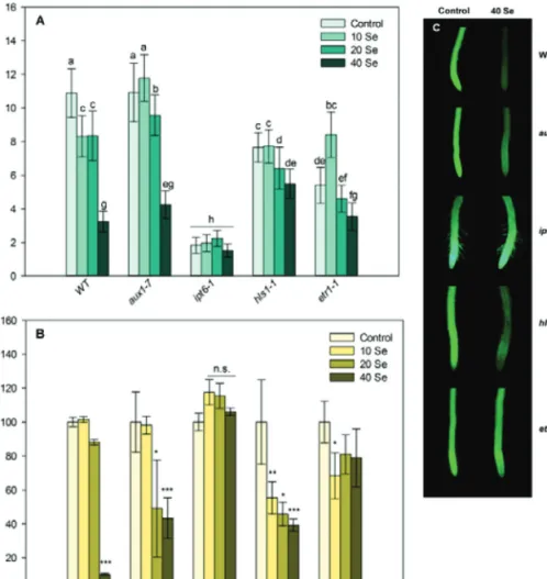
to the WT, auxin-resistant plants possessed increased sensitiv- ity, since 40 μM and 20 μM selenite reduced the viability of the root tip cells. The ethylene-deficient mutant (hls1-1) proved to be the most sensitive, since all selenite concentrations signifi- cantly reduced the cell viability in it. In root meristems of the other ethylene mutant (ethylene resistant etr1-1), practically no loss in cell viability was detected (Fig. 3B and 3C). Based on these findings, it can be stated that, in the course of auxin and ethylene deficiencies, selenite is able to exert its strong effects on PR shortening and meristem cell death, which reflects selen- ium sensitivity. However, differences were found between the ethylene-deficient plants in Se tolerance, since hls1-1 showing low ethylene concentrations possesses heavy selenite sensitiv- ity, whereas etr1-1 Arabidopsis lacking normal ethylene sig- nalling (and having WT-like ethylene levels) was resistant to selenite exposure. This indicates that changes in ethylene con- centration within the Arabidopsis root tissues determine rather the tolerance than ethylene sensitivity under selenite excess.
These results are partly confirmed by the work of Tamaoki et al.
(2008), who found that endogenous ethylene concentrations were significantly increased by 15 μM selenite and that ethylene
proved to be necessary for the induction of sulphur assimilation genes as an effect of selenite excess. Interestingly, the cytoki- nin (zeatin)-overproducing ipt6-1 plants treated with selenite showed no reduction in PR length and cell viability compared to the WT, suggesting the involvement of this hormone in the large-scale Se tolerance. High levels of cytokinin induced the transcription of the adenosine-phosphosulphate-reductase 1 gene (APR1) gene, which promotes selenium metabolism (Ohkama et al., 2002), contributing to tolerance. NR is also cytokinin- inducible (Samuelson et al., 1995) and this may lead to a more efficient nitrogen metabolism and selenium endurance of the plant. It is worth mentioning that NR is one of the major enzymic NO sources in the roots (Xu and Zhao, 2003), therefore, its acti- vation by cytokinin may result in NO production, as well, which can also induce defence mechanisms against selenite.
Selenium alters NO and H2O2 status of WT Arabidopsis roots
As far as is known, this is the first report investigating NO metabolism during selenium exposure in higher plants. NO
Fig. 3. Primary root length (A) and meristem cell viability (B) of control and Se-treated WT, aux1-7 (auxin-resistant), ipt6-1
(cytokinin-overproducing), hls1-1 (ethylene-deficient) and etr1-1 (ethylene-resistant) Arabidopsis 4 days after germination. Different letters indicate significant difference according to Duncan’s test (n = 10, P ≤ 0.05). Asterisks indicate significant difference to control according to Student’s t-test (n = 10, *P ≤ 0.05, **P ≤ 0.01, ***P ≤ 0.001). (C) Representative fluorescence microscopy images of control and 40 μM selenite-treated wild-type and mutant root tips stained with fluorescein diacetate. Bar, 0.5 mm.
by guest on September 20, 2012http://jxb.oxfordjournals.org/Downloaded from
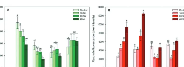
concentration of control PR meristems was very high during the early growth, whereas later on the level of NO decreased and remained at the base level. This suggests the involve- ment of NO in the normal, early seedling development (Gniazdowska et al., 2010). Interestingly, 2 and 4 DAG, sel- enite decreased NO level in a concentration-dependent man- ner; however, during the later plant growth phases, NO levels increased in response to Se, especially at 14 DAG (Fig. 4A).
Being a functional signal molecule, the actual NO concen- tration of a tissue has to be strictly regulated by its synthe- sis and removal. Possibly, during the early development NO can be removed by its reaction with oxygen, glutathione, plant haemoglobins, or different ROS forms (H2O2 and/
or O−2) (Misra et al., 2011). In the present study, the last possi- bility seems to be confirmed by the high H2O2 levels detected in selenium-treated young root tips (Fig. 4B). The selenite- induced NO generation in older roots may be the result of, for example, enzymic NO generation by NR. Selenite exposure was found to intensify NR activity in lettuce (Rios et al., 2010), either directly or indirectly via a molybdenum increase induced by sulphur deficiency (Shinmachi et al., 2010; Yu et al., 2010).
However, other possible mechanisms can contribute to NO level changes in this experimental system.
The H2O2-dependent resorufin fluorescence intensity was low in control plants, and selenite excess enhanced it in a concen- tration-dependent way during the early development (2 and 4 DAG) directly or indirectly as a result of Se-induced glutathione depletion (Grant et al., 2011). In the present experimental sys- tem, high H2O2 levels were not obviously connected to cell death in the young PR meristems (Fig. 1C and Fig. 4B). In the second developmental phase, H2O2 concentrations decreased or did not change in response to selenite, which may be the result of the activation of antioxidant systems. The selenium-induced antago- nism between NO and H2O2 observed during Arabidopsis PR development, can originate from chemical reactions (e.g. H2O2
or O−2 + NO → ONOO–) and enzymic or non-enzymic back- ground mechanisms (e.g. NR, antioxidants), which need to be further analysed in the future.
Arabidopsis mutants possess altered NO and H2O2 homeostasis and differences in selenium tolerance During studies on the GSNO reductase-deficient gsnor3-1 mutant, the NO-lacking double mutant nia1nia2, and the ascor- bate-deficient vtc2-1 and ascorbate-overproducing (via myo-ino- sitol oxygenase) miox4 plants, this study observed a significantly higher NO concentration in PRs of gsnor1-3, while the nia1nia2 roots showed lower NO levels than the WT. In the PR of the ascorbic acid-deficient vtc2-1, lower, but in the miox4 mutant higher, H2O2 and total intracellular ROS contents were detected, although these differences were not significant compared to the WT (data not shown).
Similarly to the WT, all selenium treatments inhibited root elongation in the case of NO excess (gsnor1-3) but the viability of meristem cells was not affected (Fig. 5), which suggests the con- tribution of this molecule to the Se-induced PR shortening and simultaneous Se tolerance. The high GSNO levels of gsnor1-3 were demonstrated to be important also during disease resist- ance or thermotolerance (Feechan et al., 2005; Lee et al., 2008).
Recently, an NO-overproducing tomato mutant (shr) was isolated in which the observed short root phenotype and the disease resist- ance were associated with the enhanced NO production (Negi et al., 2010). In nia1nia2 plants, the reduced NO level resulted in Se sensitivity showing the possible involvement of NO produced by the root NR (Fig. 5B). The reduced NO level also helped to maintain a better root growth under suboptimal conditions (Fig. 5A). This double mutant proved to be less tolerant to other stressors such as water deficit (Lozano-Juste and León, 2010).
The high H2O2 content resulted from the ascorbic acid deficiency in vtc2-1 roots contributed to selenite sensitivity, but the inhibited
Fig. 4. Nitric oxide-dependent fluorescence (4-amino-5-methylamino-2’,7’-difluorofluorescein diacetate, DAF-FM, A) and hydrogen peroxide-dependent fluorescence (resorufin, B) in root meristems of control and selenite-treated wild-type Arabidopsis at 2, 4, 7, and 14 days after germination. Different letters indicate significant differences according to Duncan’s test (n = 10, P ≤ 0.05).
by guest on September 20, 2012http://jxb.oxfordjournals.org/Downloaded from
root growth was alleviated by H2O2 (Fig. 5). This indicates that plants, which can decrease their (root) growth processes signifi- cantly, are able to rearrange their means from development to defence mechanisms, resulting in a better survival. Nitrate reduc- tase-dependent NO seems to be a relevant molecule to coordinate this acclimation process, at least in terms of PR growth.
The relationship between hormonal and signal regulatory components during selenite excess
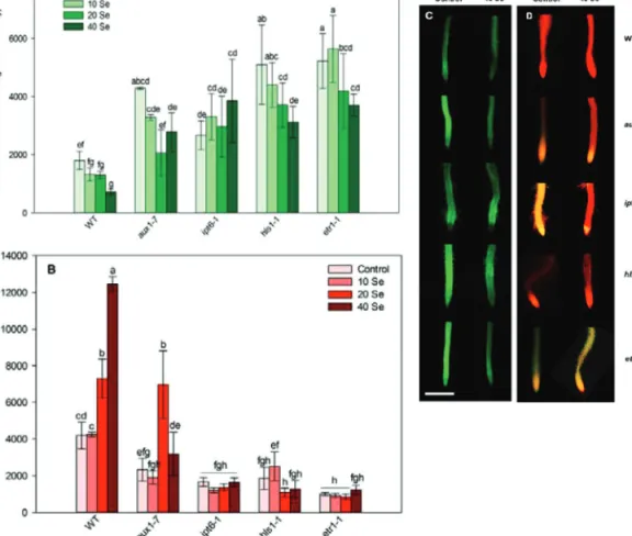
High NO levels were detected in control aux1-7 plants (Fig. 6A and 6C), which reflects a negative correlation between auxin and NO in the PR. During the growth of copper-treated Arabidopsis, a negative regulatory link was discovered between auxin and NO (Pető et al., 2011). Recently, Fernández-Marcos et al. (2011) pub- lished that high levels of NO reduced DR5::GUS expression and PIN1-mediated auxin transport in Arabidopsis PRs. In aux1-7, the low H2O2 levels could explain the slight sensitivity of this mutant to oxidative stress (Blomster et al., 2011) and a putative positive regu- lation between them (Fig. 6B and 6D). ROS can act downstream of auxin signalling in processes such as gravitropism, and auxins are able to modulate H2O2 production in guard cells (Potters et al., 2009). Similarly to the WT, 40 μM Se reduced NO and enhanced H2O2 generation in aux1-7, but these effects were not statistically significant (Fig. 6). This study’s hypothesis is that, during the early seedling development, the H2O2-dependent mitogen-activated pro- tein kinase cascade negatively affects auxin sensitivity by down- regulating the auxin-inducible gene expression (Nakagami et al., 2006), resulting in growth inhibition. In the later growth phase (14 DAG), the Se-induced NO reduces PIN1-mediated auxin transport, resulting in low auxin levels and PR growth inhibition.
In the case of control ipt6-1 Arabidopsis possessing high cyto- kinin content, more NO was produced than in the WT (Fig. 6A and 6C), a finding which is supported by the work of Tun et al.
(2001), where exogenously applied cytokinin rapidly induced NO production. Moreover, in some physiological processes,
such as hypocotyl elongation, NO exerts cytokinin-like effects supporting the positive regulatory relationship between this hor- mone and the signal molecule (Beligni and Lamattina, 2001).
In response to Se, NO levels were not reduced, but showed a slight, nonsignificant increase. The ipt6-1 mutation also resulted in lower ROS production (Fig. 6B and 6D), which supports a possibly negative regulation between these components.
In control hls1-1 and etr1-1 mutants showing lower ethylene concentrations and deficiency in signalling, the level of NO was extremely high compared to the WT (Fig. 6A and 6C). This sug- gests the relevant antagonism between this plant morphogen and NO during Arabidopsis root growth. Another evidence for NO–
ethylene antagonism is provided by the review of Besson-Bard et al. (2009), where the downregulation of the ethylene biosynthetic ACC oxidase gene (ACO4) by NO was reported. However, selenite induced significant decreases in NO contents such as in the WT, which suggests that there is no regulatory relationship between these molecules during PR growth under Se excess. Hydrogen peroxide is a possible downstream element of ethylene signalling, since the level of it was low in control hls1-1 and etr1-1, and Se was not able to increase its content in the mutant roots (Fig. 6B and 6D). These results are supported by the findings that histidine kinases are strongly H2O2-responsive and they also modulate cellu- lar responses to, for example, ethylene (Desikan et al., 2001).
Taken together, higher Se concentrations (20 and 40 μM) reduces PR development, which can be considered an adaptation process of the plant, since the reorientation of means from development for protection mechanisms ensures better survival. Selenium exposure disturbs protein synthesis through the formation of selenomethio- nine and selenocystein directly leading to cell death in the PR meri- stem and growth inhibition. The hormonal balance of the PR is also affected by selenium. During the early development, Se-induced H2O2 can reduce auxin-responsive gene expression, while NO inhib- its auxin transport in older roots and the decrease of root auxin level results in growth inhibition. Selenium enhances cytokinin-responsive gene expression (consequently cytokinin levels), which leads to PR Fig. 5. Primary root length (A) and cell viability (B) in root meristems of control and selenite-treated wild-type, gsnor1-3
(GSNOR-deficient), nia1nia2 (NR-deficient), vtc2-1 (ascorbic acid-deficient), and miox4 (ascorbic acid-overproducing) Arabidopsis at 4 days after germination. Different letters indicate significant difference according to Duncan’s test (n = 10, P ≤ 0.05). Asterisks indicate significant difference to control according to Student’s t-test (n = 10, *P ≤ 0.05, **P ≤ 0.01, ***P ≤ 0.001).
by guest on September 20, 2012http://jxb.oxfordjournals.org/Downloaded from
growth inhibition possibly through NR-dependent NO synthesis and/
or through the reduction of H2O2 level. The selenite-induced enhance- ment of ethylene biosynthesis may cause cell death resulting growth hindrance and H2O2 is a downstream element of its signalling, while there is no regulatory link between ethylene and NO under Se excess.
The optimal level of H2O2 is necessary for Se tolerance and NO over- production in Arabidopsis roots ensures Se tolerance.
Acknowledgements
DR5::GUS transgenic Arabidopsis seeds were obtained from Prof. Tom Guilfoyle (University of Missouri, USA). The NR double mutant nia1nia2 seeds were kindly provided by Prof. Dr.
G. F. E. Scherer (University of Hannover, Germany), the gsnor1- 3 seeds were donated by Dr. Christian Lyndermayr (Helmholtz Zentrum München, Germany) and the seeds of vtc2-1 and miox4 were a kind gift from Dr. Laura Zsigmond (University of Szeged, Hungary). This work was supported by the Hungarian Scientific Research Fund (grant no. OTKA PD100504) and was carried out in the frame of COST Action FA 0905. This study was carried out during a 1-year stay by Devanand Kumar at the Department of Plant Biology, University of Szeged, supported by the C2 type Hungarian Balassi Fellowship. Special thanks
to Dr. Gábor Laskay for the proofreading part. The instrumen- tal background was partly ensured by HURO/0901/147/2.2.2 SZETISA1. project. Publication is supported by the European Union and cofunded by the European Social Fund (project num- ber TÁMOP-4.2.2/B-10/1-2010-0012).
References
Beligni MV, Lamattina L. 2001. Nitric oxide stimulates seed germination and de-etiolation, and inhibits hypocotyl elongation, three light-inducible responses in plants. Planta 210, 215–221.
Besson-Bard A, Astier J, Rasul S, Wawer I, Dubreuil-Maurizi C, Jeandroz S, Wendehenne D. 2009. Current view of nitric oxide-responsive genes in plants. Plant Science 177, 302–309.
Blomster T, Salojärvi J, Sipari N, Brosché M, Ahlfors R, Keinänen M, Overmyer K, Kangasjärvi J. 2011. Apoplastic reactive oxygen species transiently decrease auxin signaling and cause stress-induced morphogenic response in Arabidopsis. Plant Physiology 157, 1866–1883.
Chang C, Kwok SF, Bleecker AB, Meyerowitz EM. 1993.
Arabidopsis ethylene-response gene ETR1: similarity of product to two-component regulators. Science 262, 539–544.
Fig. 6. Nitric oxide-dependent fluorescence (4-amino-5-methylamino-2’,7’-difluorofluorescein diacetate, DAF-FM, A) and hydrogen peroxide-dependent fluorescence (resorufin, B) in root meristems of 0, 10, 20, and 40 μM selenite-treated wild-type, aux1-7 (auxin-resistant), ipt6-1 (cytokinin-overproducing), hls1-1 (ethylene-deficient), and etr1-1 (ethylene-resistant) plants at 4 days after germination. Different letters indicate significant differences according to Duncan’s test (n = 10, P ≤ 0.05). Representative microscopic
images of control and 40 μM selenite-treated wild-type and mutant root tips stained with DAF-FM (C) or Ampiflu (D). Bar, 0.5 mm. by guest on September 20, 2012http://jxb.oxfordjournals.org/Downloaded from
Conklin PL, Pallanca JE, Last RL, Smirnoff N. 2000. Identification of ascorbic acid-deficient Arabidopsis thaliana mutants. Genetics 154, 847–856.
Correa-Aragunde N, Graziano M, Chevalier Ch, Lamattina L.
2006. Nitric oxide modulates the expression of cell cycle regulatory genes during lateral root formation in tomato. Journal of Experimental Botany 57, 581–588.
D’Agostino IB, Deruère J, Kieber JJ. 2000. Characterization of the response of the Arabidopsis response regulator gene family to cytokinin. Plant Physiology 124, 1706–1717.
Desikan R, A-H-Mackerness S, Hancock JT, Neill SJ. 2001.
Regulation of the Arabidopsis transcriptome by oxidative stress. Plant Physiology 127, 159–172.
Feechan A, Kwon E, Yun B-W, Wang Y, Pallas JA, Loake GJ. 2005.
A central role for S-nitrosothiols in plant disease resistance. Proceedings of the National Academy of Sciences, USA 102, 8054–8059.
Fernández-Marcos M, Sanza L, Lewis DR, Muday GK, Lorenzo O. 2011. Nitric oxide causes root apical meristem defects and growth inhibition while reducing PIN-FORMED 1 (PIN1)-dependent acropetal auxin transport. Proceedings of the National Academy of Sciences, USA 108, 18506–18511.
Gniazdowska A, Krasuska U, Czajkowska K, Bogatek R. 2010.
Nitric oxide, hydrogen cyanide and ethylene are required in the control of germination and undisturbed development of young apple seedlings. Plant Growth Regulation 61, 75–84.
Gomes A, Fernandes E, Lima JLFC. 2005. Fluorescence probes used for detection of reactive oxygen species. Journal of Biochemical and Biophysical Methods 65, 45–80.
Grant K, Carey NM, Mendoza M, Schulze J, Pilon M, Pilon-Smits EAH, Van Hoewyk D. 2011. Adenosine 5’-phosphosulfate reductase (APR2) mutation in Arabidopsis implicates glutathione deficiency in selenate toxicity. The Biochemical Journal 438, 325–335.
Guzmán P, Ecker JR. 1990. Exploiting the triple response of Arabidopsis to identify ethylene-related mutants. The Plant Cell 2, 513–523.
Hartikainen H, Pietola L, Simojoki A. 2001. Quantification of fine root responses to selenium toxicity. Agricultural and Food Science in Finland 10, 53–58.
Jefferson RA, Kavanagh TA, Bevan MW. 1987. GUS fusions:
beta-glucuronidase as a sensitive and versatile gene fusion marker in higher plants. The EMBO Journal 6, 3901–3907.
Kolbert Zs, Ortega L, Erdei L. 2010. Involvement of nitrate reductase (NR) in osmotic stress-induced NO generation of
Arabidopsis thaliana L. roots. Journal of Plant Physiology 167, 77–80.
Konze JR, Schilling N, Kende H. 1978. Enhancement of ethylene formation by selenoamino acids. Plant Physiology 62, 397–401.
Kuderová A, Urbánková I, Válkoká M, Malbeck J, Brzobohatỳ B, Némethová D, Hejátko J. 2008. Effects of conditional IPT-dependent cytokinin overproduction on root architecture of Arabidopsis seedlings. Plant Cell Physiology 49, 570–582.
Lee U, Wie C, Fernandez BO, Feelisch M, Vierling E. 2008.
Modulation of nitrosative stress by S-nitrosoglutathione reductase is critical for thermotolerance and plant growth in Arabidopsis. The Plant Cell 20, 786–802.
Lehotai N, Pető A, Bajkán Sz, Erdei L, Tari I, Kolbert Zs. 2011. In vivo and in situ visualization of early physiological events induced by heavy metals in pea root meristem. Acta Physiologiae Plantarum 33, 2199–2207.
Lequeux H, Hermans C, Lutts S, Verbruggen N. 2010. Response to copper excess in Arabidopsis thaliana: impact on the root system architecture, hormone distribution, lignin accumulation and mineral profile. Plant Physiology and Biochemistry 48, 673–682.
Lorence A, Chevone BI, Mendes P, Nessler CL. 2004.
myo-Inositol oxygenase offers a possible entry point into plant ascorbate biosynthesis. Plant Physiology 134, 1200–1205.
Lozano-Juste J, León J. 2010. Enhanced abscisic acid-mediated responses in nia1nia2noa1–2 triple mutant impaired in nia/NR- and Atnoa1-dependent nitric oxide biosynthesis in Arabidopsis. Plant Physiology 152, 891–903.
Maher EP, Martindale SJB. 1980. Mutants of Arabidopsis thaliana with altered responses to auxins and gravity. Biochemical Genetics 18, 1041–1053.
Maksymiec W. 2011. Effects of jasmonate and some other signalling factors on bean and onion growth during the initial phase of cadmium action. Biologia Plantarum 55, 112–118.
Malamy JE, Benfey PN. 1997. Organization and cell differentiation in lateral roots of Arabidopsis thaliana. Development 124, 33–44.
Mazid M, Khan TA, Mohammad F. 2011. Role of nitric oxide in regulation of H2O2 mediating tolerance of plants to abiotic stress:
a synergistic signalling approach. Journal of Stress Physiology and Biochemistry 7, 34–74.
Medford JI, Horgan R, El-Sawi Z, Klee HJ. 1989. Alterations of endogenous cytokinins in transgenic plants using a chimeric isopentenyl transferase gene. The Plant Cell 1, 403–413.
Misra AN, Misra M, Singh R. 2011. Nitric oxide: a ubiquitous signaling molecule with diverse role in plants. African Journal of Plant Science 5, 57–74.
Nakagami H, Soukupová H, Schikora A, Žárskỳ V, Hirt H. 2006.
A mitogen-activated protein kinase kinase kinase mediates reactive oxygen species homeostasis in Arabidopsis. The Journal of Biological Chemistry 281, 38697–38704.
Negi S, Santisree P, Kharshiing EV, Sharma R. 2010. Inhibition of the ubiquitin–proteasome pathway alters cellular levels of nitric oxide in tomato seedlings. Molecular Plant 3, 854–869.
Ohkama N, Takei K, Sakakibara H, Hayashi H, Yoneyama T, Fujiwara T. 2002. Regulation of sulfur-responsive gene expression by exogenously applied cytokinins in Arabidopsis thaliana. Plant Cell Physiology 43, 1493–1501.
Pasternak T, Rudas V, Potters G, Jansen MAK. 2005.
Morphogenic effects of abiotic stress: reorientation of growth in Arabidopsis thaliana seedlings. Environmental and Experimental Botany 53, 299–314.
Peer WA, Blakeslee JJ, Yang H, Murphy AS. 2011. Seven things we think we know about auxin transport. Molecular Plant 4, 487–504.
Peng A, Xu Y, Liu JH, Wang ZJ. 2000. Study on the dose–effect relationship of selenite with the growth of wheat. Biological Trace Element Research 76, 175–181.
by guest on September 20, 2012http://jxb.oxfordjournals.org/Downloaded from
Pető A, Lehotai N, Lozano-Juste J, León J, Tari I, Erdei L, Kolbert Zs. 2011. Involvement of nitric oxide (NO) in signal transduction of copper-induced morphological responses in Arabidopsis seedlings. Annals of Botany 108, 449–457.
Pickett FB, Wilson AK, Estelle M. 1990. The aux1 mutation of Arabidopsis confers both auxin and ethylene resistance. Plant Physiology 94, 1462–1466.
Potters G, Pasternak TP, Guisez Y, Jansen MAK. 2009. Different stresses, similar morphogenetic responses: integrating a plethora of pathways. Plant, Cell and Environment 32, 158–169.
Ribeiro DM, Mapeli AM, Antunes WC, Barros RS. 2011. A dual role of selenium in the growth control of seedlings of Stylosanthes humilis. Agricultural Sciences 2, 78–85.
Rios JJ, Blasco B, Rosales MA, Sanchez-Rodriguez E, Leyva R, Cervilla LM, Romero L, Ruiz JM. 2010. Response of nitrogen metabolism in lettuce plants subjected to different doses and forms of selenium. Journal of the Science of Food and Agriculture 90, 1914–1919.
Riou-Khamlichi C, Huntley R, Jacqmard A, Murray JAH. 1999.
Cytokinin activation of Arabidopsis cell division through a D-type cyclin. Science 283, 1541–1544.
Růžička K, Ljung K, Vanneste S, Podhorská R, Beeckman T, Friml J, Benková E. 2007. Ethylene regulates root growth through effects on auxin biosynthesis and transport-dependent auxin distribution. Plant Cell Online 19, 2197–2212.
Samuelson ME, Campbell WH, Larsson C-M. 1995. The influence of cytokinins in nitrate regulation of nitrate reductase activity and expression in barley. Physiologia Plantarum 93, 533–539.
Shinmachi F, Buchner P, Stroud JL, Parmar S, Zhao F-J, McGrath SP, Hawkesford MJ. 2010. Influence of sulfur deficiency on the expression of specific sulfate transporters and the distribution of sulfur, selenium, and molybdenum in wheat. Plant Physiology 153, 327–336.
Simojoki A. 2003. Allocation of added selenium in lettuce and its impact on roots. Agricultural and Food Science in Finland 12, 155–164.
Sors TG, Ellis DR, Salt, DE. 2005. Selenium uptake, translocation, assimilation and metabolic fate in plants. Photosynthesis Research 86, 373–389.
Suzuki KT. 2005. Metabolomics of selenium: Se metabolites based on speciation studies. Journal of Health Science 51, 107–114.
Tamaoki M, Freeman JL, Pilon-Smits EAH. 2008. Cooperative ethylene and jasmonic acid signaling regulates selenite resistance in Arabidopsis. Plant Physiology 146, 1219–1230.
Tsuchisaka A, Theologis A. 2004. Unique and overlapping expression patterns among the Arabidopsis
1-amino-cyclopropane-1-carboxylate synthase gene family members.
Plant Physiology 136, 2982–3000.
Tun NN, Holk A, Scherer GFE. 2001. Rapid increase of NO release in plant cell cultures induced by cytokinin. FEBS Letters 509, 174–176.
Ulmasov T, Murfett J, Hagen G, Guilfoyle TJ. 1997. Aux/IAA proteins repress expression of reporter genes containing natural and highly active synthetic auxin response elements. The Plant Cell 9, 1963–1971.
van der Graaff EE, Hooykaas PJJ, Auer CA. 2001. Altered development of Arabidopsis thaliana carrying the Agrobacterium tumefaciens ipt gene is partially due to ethylene effects. Plant Growth Regulation 34, 305–315.
Van Hoewyk D, Takahashi H, Inoue E, Hess A, Tamaoki M, Pilon-Smits EAH. 2008. Transcriptome analyses give insights into selenium-stress responses and selenium tolerance mechanisms in Arabidopsis. Physiologia Plantarum 132, 236–253.
Wang N, Ruqian L, Liangji Z, Zhaoda T. 1992. The relationship between the effect of sodium selenite on the growth of Nicotiana tabacum crown gall tissue and the level of endogenous hormones.
Journal of Plant Physiology and Molecular Biology 18, 160–166.
Wang Y, Li K, Li X. 2009. Auxin redistribution modulates plastic development of root system architecture under salt stress in Arabidopsis thaliana. Journal of Plant Physiology 166, 1637–1645.
Werner T, Nehnevajova E, Köllmer I, Novák O, Strnad M, Krämer U, Schmülling, T. 2010. Root-specific reduction of cytokinin causes enhanced root growth, drought tolerance, and leaf mineral enrichment in Arabidopsis and tobacco. Plant Cell Online 22, 3905–3920.
Wilkinson JQ, Crawford NM. 1993. Identification and
characterization of a chlorate resistant mutant of Arabidopsis with mutations in both NIA1 and NIA2 nitrate reductase structural genes.
Molecular and General Genetics 239, 289–297.
Xu YC, Zhao BL. 2003. The main origin of endogenous NO in higher non-leguminous plants. Plant Physiology and Biochemistry 41, 833–838.
Yu MB, Hu C-X, Sun X-C, Wang Y-H. 2010. Influences of Mo on nitrate reductase, glutamine synthetase and nitrogen accumulation and utilization in Mo-efficient and Mo-inefficient winter wheat cultivars.
Agricultural Sciences in China 9, 355–361.
Zhang L, Ackley AR, Pilon-Smits EAH. 2007. Variation in selenium tolerance and accumulation among 19 Arabidopsis thaliana accessions. Journal of Plant Physiology 164, 327–336.
by guest on September 20, 2012http://jxb.oxfordjournals.org/Downloaded from