ALTERATIONS OF ARTERIOLAR REACTIVITY IN A RAT POLYCYSTIC OVARY SYNDROME MODEL
EFFECTS OF PARALLEL VITAMIN D
3ADMINISTRATION
PhD thesis
Levente Sára MD
Basic Medicine Doctoral School Semmelweis University Budapest
Supervisor: Szabolcs Várbíró MD, PhD Official reviewers:
Attila Szabó MD, PhD, Dsc Sándor Alföldi MD, PhD
Head of the Final Examination Committee:
János Rigó Jr. MD, PhD, Dsc
Members of the Final Examination Committee:
Ádám László MD, PhD Péter Studinger MD, PhD
Budapest 2012
Contents
ABBREVIATIONS 5
1. BACKGROUND 8
1.1. PCOS epidemiology 9
1.2. Definition of PCOS 10
1.3. Diagnosis of PCOS 12
1.4. Aethiology of PCOS 13
1.5. Pathophysiology of PCOS 16
1.5.1. Hypothalamic-pituitary-ovarian axis 16
1.5.2. Hyperandrogenemia (HA) 17
1.5.3. Metabolic syndrome 20
1.5.3.1. Insulin Resistance (IR) 21
1.5.3.2. Dyslipidemia 28
1.5.3.3. Hypertension 31
1.5.4. Conection between Hyperandrogenism and IR 31
1.5.5. Vascular alterations in PCOS 35
1.6. Animal models of PCOS 37
1.6.1. Pre- and postnatal models in mammals 37
1.6.2. Hormonally induced rodent models 38
1.6.3. Genetically modified rodent models of PCOS 40
1.7. The possible role of vitamin D3 in PCOS 41
1.8. Animal models related to 1,25-dihydroxyvitamin D3 endocrine system 42
1.8.1. Vitamin D3 deficient models 43
1.8.2. Atherosclerotic model 43
1.8.3. Models of low dose chronic vitamin D3 treatment 44
2. AIMS OF THE STUDY 45
3. METHODS AND MATERIALS 46
3.1. Chemicals 46
3.2. Animals 47
3.3. Oral Glucose Tolerance Test (OGTT) 47
3.4. In vivo blood pressure measurement 49
3.5. Biomechanics of a Musculocutanous Arteriole (Pressure Arteriography)
49
3.6. Biomechanical calculations 52
3.7. Histology 53
3.8. Statistical analysis 53
4. RESULTS 55
4.1. Physiological parameters 55
4.2. Ovarian morphology 55
4.3. Glucose metabolism 55
4.4. Biomechanical parameters of gracilis arterioles 56
4.4.1. Arteriole geometry 56
4.4.2. Arteriole elasticity 58
4.5. Pharmacological properties of gracilis arterioles 61
4.5.1. Arteriolar contractility 61
4.5.2. Endothelial dilation 63
4.6. Insulin-induced vascular relaxation of gracilis arterioles 64
5. DISCUSSION 67
5.1. Basic physiological and metabolic changes after chronic DHT treatment with or without vitamin D3 administration
68
5.2. Vascular effects of DHT treatment 70
5.3. The metabolic and vascular effects of vitamin D3 71 5.4. Pharmacological effects on gracilis arterioles 73
5.4.1. Pharmacological effects of DHT treatment 73 5.4.2. Pharmacological alterations of arterioles in PCOS 74 5.4.3. Pharmacological effects of vitamin D3 on gracilis arterioles 74 5.4.4. The pharmacological effects of metformin and oral contraceptives as
medical treatment in PCOS
75
5.5. Biomechanical and pharmacological changes of gracilis arterioles after DHT treatment and parallel vitamin D3 administration
77
5.6. Vascular effects of insulin 78
5.6.1. Effects on arterioles 78
5.6.2. Altered insulin relaxation of aortic rings 80
6. CONCLUSIONS 82
7. ACKNOWLEDGEMENTS 84
SUMMARY 85
ÖSSZEFOGLALÁS ( Abstract in Hungarian) 87
8. REFERENCES 89
9. LIST OF PUBLICATIONS 122
Abbreviations
ACh Acetylcholine
ACTH Adrenocorticotropic hormone
ANOVA Analysis of Variance
Apo Apolipoprotein
BMI Body Mass Index
C Control
CAH Congenital Adrenal Hyperplasia
cAMP Cyclic Adenosine Monophosphate
COX Cyclooxigenase
CRP, hs-CRP C-reactive protein, high sensitive C-reactive protein
CVD Cardiovascular disease
DHEA, DHEAS Dehydroepiandrosterone, Dehydroepiandrosterone Sulfate
DHT Dihydrotestosterone
DHT+D3 Dihydrotestosterone + Vitamin D3
E1, E2 Oestrone, Oestradiol
FSH Follicle Stimulating Hormone
GLUT 4 Glucosetransporter Type 4
GnRH Gonadotropic Releasing Hormone
Grb Growth factor receptor - bound receptor
GSK3 Glycogen synthase kinase 3
HA Hyperandrogenemia
HAF rat Hyperandrogenic female rat
HAIRAN-syndrome Hyperandrogenism, insulin resistance and Acanthosis nigricans syndrome
HCG Human Choriogonadotropin
HDL, HDL-C High Density Lipoprotein / -Cholesterol
HOMA-IR Homeostasis Model Assesment of Insuline Resistance
IGF Insulin-like Growth Factor
IGT Impaired Glucose Tolerance
IL-6 Interleukin 6
INR, INSR Insulin Receptor, Insulin Receptor Subunit
IR Insulin Resistance
IRS Insulin Receptor Substrate
L-NAME Nitro-L-Arginine Methyl Esther
LDL Low Density Lipoprotein
LH Luteotropic Hormone
LOD Laparoscopic Ovarian Diathermy
LPL Lipoprotein lipase
MAPK Mitogen-activated protein kinase
NADPH Nicotinamide Adenine Dinucleotide Phosphate
NE Norepinephrine
NIH National Institute of Health
nKR normal Krebs-Ringer
NO Nitric Oxide
NOS Nitric Oxide Synthase
OC Oral Contraceptive
OGTT Oral Glucose Tolerance Test
PC Membran glycoprotein
PCOS, PCO Polycystic Ovary Syndrome
PGE Prostaglandin E
PI3K Phosphatidylinositol 3-kinase
PKA, PKC Protein kinase A, C
PPAR Proliferator-Activator Receptor
SHBG Sex Hormone Binding Globulin
Shc Homology domain transforming protein
SHHF Spontaneously Hypertensive, Heart Failure-Prone rat SHR rat Spontaneously Hypertensive rat
T1DM, T2DM Type 2 Diabetes Mellitus
TA Thoracic Aorta
TC Total cholesterol
TG Triglyceride
TGF Transforming Growth Factor
TNF, TNFα Tumour Necrosis Factor α
TZDs Thiazolidinediones
VDR Vitamin D Receptor
VLDL Very-Low Density Lipoprotein
WKY rats Wistar-Kyoto rats
1. Background
Polycystic ovary syndrome (PCOS) is one of the most common endocrine diseases and the most frequent disorder in women of reproductive age. Nowadays, PCOS is in the focus of research because of its increasing prevalence. The aetiology of this complex and heterogenous disorder is still uncertain. Environmental factors such as physical inactivity, malnutrition, obesity and insulin resistance (IR) have crucial role in development of the disorder [Baranova et al. 2011] PCOS might be a multifactorial and polygenic disorder, but gene variants of the CYP11A, the insulin gene, and the follistatin gene are suspected to contribute to PCOS. There is no gene identified as a substantial cause of PCOS. [Legro et al. 2002, Diamanti-Kandarakis et al. 2005]. The inheritance of PCOS is suggested to be autosomal dominant, which is transmitted to male and female offsprings , but the phenotype occurs only in women [Lunde et al.
1989]. The most common features of PCOS, called the “Rotterdam criteria”, are menstrual disorders (amenorrhoea) such as oligo- or anovulatory menstrual cycles;
polycystic, large ovaries as detected by ultrasound; and clinical or laboratory signs of excess androgen. It often associates with obesity, acne, hirsutism, cardiovascular disorders (CVD) and obstructive sleep apnea [Nitsche et al. 2010]. There is a wide variability in phenotypes; symptoms and severity vary among affected women. PCOS may present at one end of the spectrum with a single diagnosis of polycystic ovarian morphology and at the other end with obesity, menstrual disorders, hyperandrogenism and hypertension. PCOS is the leading cause of female infertility [Boomsma et al. 2008].
A reduced oocyte quality and response after stimulation was found in patients with PCOS; additionally, there is an increase in the failure rate of implantation after in vitro fertilisation [Dumesic et al. 2005, Homburg et al. 1988]. Women with PCOS have a higher prevalence of early-onset atherosclerosis, metabolic syndrome, IR and type 2 diabetes
mellitus (T2DM), and they may develop hypertension during their reproductive years.
PCOS also alters cardiovascular function through various mechanisms [Dokras A. 2008].
The mechanisms underlying this increased risk and its possible therapeutic approaches are still under investigation. PCOS has been reported to affect behaviour and social activity, resulting in masculinisation and defeminised behaviour [Abbott et al. 2008].
1.1. PCOS epidemiology
PCOS affects approximately 4-10% of fertile women around the world [Hart et al. 2011, Knochenhauer et al. 1998], but some researchers estimate the overall rate to be between 4-25% [Homberg R. 2002]. The difference in the above mentioned rates can be attributed to the different criteria used for the diagnosis of the syndrome and the different prevalence rates among races and ethnicities. The incidence of PCOS is significantly greater among South-Asian and Mexican women [Rodin et al. 1998] than Caucasian women. The highest rate was found in South Asian immigrants in Great Britain. The PCOS prevalence was 52%, and approximately 49% of those women had menstrual irregularity [Rodin et al. 1998]. Balen A. et al. reported that patients of South Asian origin tend to present with more severe symptoms at an earlier age with higher incidence of IR than their Caucasian British counterparts [Balen A. et al. 2004]. The severity and features of PCOS largely differ among different populations. Kauffman et al. reported that Mexican American women suffering from PCOS have a higher prevalence of IR than white American women [Kauffman et al. 2002]. Asian populations have a higher risk for PCOS and IR, however the metabolic syndrome is less frequent among Chinese patients compared to patients of other ethnicities [Ni et al. 2009].
Environmental factors such as sufficient or insufficient nutrition and lifestyle management such as physical exercise or domesticity might influence the expression of PCOS. Individuals who have tendency to be obese can preserve their ability to be fertile
by maintaining a threshold weight; however they have a greater survival potential during starvation. A better nutrition and more comfortable environment and lifestyle can contribute to the development of PCOS [Balen et al. 2002]. The prevalence of PCOS in Hungary is approximately 5-7% among fertile women ( up to half million women might be affected); however, cohort studies investigating the associated symptoms and risks of complications, especially cardiovascular problems, are still lacking [Speer G. 2009] .
1.2. Definition of PCOS
The polycystic ovarian morphology was described by Chereau and Rokitanszky in 1844. PCOS was first described in 1935 by Irving Stein and Michael Leventhal [Stein IF., Leventhal ML. 1935]. However, the specific phenotype of the disorder was known since the XVIII century, when Stein and Leventhal first discovered the relationship between ovarian morphology and amenorrhoea [Speer G. 2009].
The first international consensus on the definition of PCOS was established by the National Institute of Health (NIH) in 1990. At the conference held in the United States, three important criteria for PCOS were nominated and accepted: 1. Oligoovulation 2.
Hyperandrogenism (excess androgen activity - clinical or biochemical alterations) and 3. Exclusion of related disorders (i.e., hypothalamic amenorrhoea, hyperprolactinaemia, hyperandrogenism-insulin resistance-acanthosis nigricans (HAIRAN)-syndrome, primary ovarian failure, congenital adrenal hyperplasia (CAH), cushing syndrome, androgen-secreting tumours, exogenous androgen overdose, hypo/hyperthyreosis). The diagnosis of PCOS should be made when patients meet all three criteria. [Azziz et al.
2006].
In 2003, the Rotterdam consensus introduced a new concept for the definition of PCOS.
Polycystic ovarian morphology detected by ultrasound was accepted by experts as a new criterion. [Rotterdam ESHRE/ASRM Sponsored PCOS CWG. 2004., Merino et al. 2011].
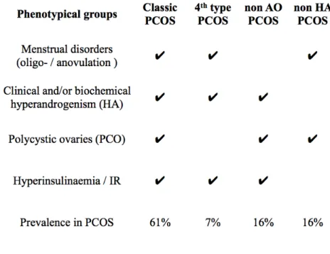
The consensus established the four different phenotypes of the disorder (Table 1.). This consensus was organised by the European Society of Human Reproduction and Embryology (ESHRE) and the American Society for Reproductive Medicine (ASRM).
Based on the consensus, PCOS should be defined by the presence of two from the previous three criteria: 1. Oligo- and/or anovulation 2. Excess androgen activity and 3.
Polycystic ovarian morphology (by ultrasound). The Rotterdam definition was much border than that of the NIH Consensus, which included patients without androgen excess. The diagnosis of polycystic ovaries using an ultrasound remains controversial [Porter MB. 2008].
Table 1.: The different phenotypes of PCOS in terms of the Rotterdam criteria. AO: anovulation.
HA: clinical and/or biochemical hyperandrogenism. IR: insulin resistance. PCO: polycystic ovaries.
PCOS: polycystic ovary syndrome. The phenotypes defined by NIH are highlighted in black, and the ones determined later by the Rotterdam Consensus are highlighted in blue. Based on Lakatos et al. In: Lakatos P, Speer G. Polycystic Ovary Syndrome. Budapest. Semmelweis Kiadó, 2009. p.56.
The two consensuses mentioned above are commonly used in clinical practice [Azziz et al. 2006]. In 2006, the Androgen Excess Society suggested an unofficial modification to the diagnosis. Based on these recommendations, the following three criteria are required
for the diagnosis of PCOS: 1. Hyperandrogenism (clinical e.g. hirsutism and/or hyperandrogenaemia), 2. Ovarian dysfunction (oligo- and/or anovulation and/or polycystic ovarian morphology) and 3. Exclusion of other entities that would cause excess androgen activity [Lakatos et al. 2009]. The Androgen Excess and PCOS Society published the latest guideline for PCOS definition in 2009 as follows: 1.
Hyperandrogenism (clinical and/or biochemical), 2. Ovarian dysfunction (oligo - anovulation and/or polycystic ovaries), and 3. Exclusion of related disorders [Azziz et al.
2009, Wild et al. 2010].
1.3. Diagnosis of PCOS
A history of menstrual disorder, hirsutism, obesity, and acne are strong predictors for PCOS [Pedersen et al. 2007]. These four criteria can be used to diagnose PCOS with a sensitivity of almost 80% and specificity of 93%. The anamnestic data and observation of clinical signs are the primary indicators for identifying the disorder, while an ultrasound and laboratory tests should be used to confirm the exact diagnosis. The role of ultrasound as a sole diagnostic method is controversial [Porter MB. 2008] because of the high rate of false positives. The latest guideline recommends stringent criteria, such as the presence of numerous small follicles (11-19 or more) and a large ovary with increased stroma ( >10 cm3). The follicles are oriented at the periphery of the ovary, and their diameter is not greater than 9 mm (2-9 mm). [Balen AH. et al. 2003, Dewailly et al.
2011]. In the future, a three-dimensional ultrasound might confer a technological advantage, thus providing better chances for a correct diagnosis [Porter MB. 2008]. More than 80% of PCOS women have androgen excess during or before adrenarche. Another 20-30% of patients present with adrenal androgen excess, which manifests as elevated dehydroepiandrosterone (DHEA), dehydroepiandrosterone-sulfate (DHEAS), androstenedione levels and specific hyperandrogenic responses to adrenocorticotropic hormone (ACTH) [Abbott et al. 2008]. An elevated androgen hormone level, especially that of free testosterone, is a strong predictor for PCOS [Huang et al. 2010]. Additionally,
luteotropic hormone (LH) hypersecretion has a low prognostic prevalence for PCOS, while serum anti-Mullerian factor (>5 ng/ml) has a higher prognostic prevalence for the syndrome [Dewailly et al. 2011]. Although a laparoscopy procedure is not routinely used for PCOS diagnosis, it may be used for providing incidental findings of PCOS during an operation.
It is necessary to exclude the related diagnoses of hypophysis disorders, adrenocortical diseases, ovarian and thyroid problems. An irregular menstruation cycle can be caused by hypogonadotropic hypogonadism, cushing syndrome, hyperprolactinaemia and hypothyreosis. A hyperandrogenemia (HA) can be caused by CAH, cushing syndrome, androgen-secreting adrenocortical, ovarian tumours, persisting follicle, and anti- epileptic drugs (i.e., Valproat) [Lakatos et al. 2009].
1.4. Aetiology of PCOS
The aetiology of PCOS appears to be both polygenic and multifactorial, thus explaining the multiple phenotypes, which seem to vary from patient to patient. The exact mechanism of PCOS is still debated. Genetic studies have identified a relationship between insulin resistance and PCOS. It suggests that PCOS might be a phenotypic expression of a complex genetic trait disorder [Franks et al. 2001]; however, it is challenging to determine the genotype due to its multiple variations. This is because different symptoms of PCOS may link to different genetic variants [Balen A. 2004].
Others have hypothesised that androgen excess during the intrauterine life or during early adolescence is a predisposing factor for developing PCOS [Abbott et al. 2008, Motta AB. 2010]. Although external or environmental effects are possibly factors, prior androgen excess seems to be essential. Prepubertal HA was found to increase the number of T lymphocyte cells that can infiltrate ovarian tissue, retroperitoneal and
axillar lymph nodes [Luchetti et al. 2004]. Additionally, the ratio of CD8+ T lymphocytes to CD4+ T cells increases locally in ovaries in hyperandrogenism. The elevated production of cytotoxic T lymphocytes in HA might contribute to oxidative stress in the ovarian tissue [Luchetti et al. 2004]. Prepubertal hyperandrogenism is also reported to increase serum tumour necrosis factor alpha (TNFα) levels; additionally, a mutation of the TNF receptor in association with hyperandrogenism has been demonstrated.
[Luchetti et al. 2004., Peral et al. 2002].
Luchetti et al. reported increased ovarian lipid peroxidation, decreased glutathione content and catalase activity in hyperandrogenism. This means that there is a higher risk for the accumulation of reactive oxygen species and nitrogen-derived products, thus leading to impaired ovarian function [Luchetti et al. 2004]. This vicious cycle might be caused by androgen excess rather than hyperinsulinaemia. Corbould et al. did not find any difference in vitro in basal glycogen synthesis or glucose transport between PCOS and controls in a preadipocyte culture, therefore demonstrating that other factors are involved and not just the intrinsic defects in the insulin sensitivity of the adipocytes. In vivo, they proposed that external factors, such as high circulating androgen levels, are a linkage of the association between hypertrophic adipocytes and insulin resistance [Corbould et al. 2007✲]. Corbould also showed in vitro that androgens can induce insulin resistance in subcutaneous adipose cells [Corbould A. 2007✲✲] In humans as well as in primates, it is suggested that a prenatal androgen excess might contribute to PCOS programming in the foetus [Abbott et al. 2008]. The different timing of this androgen excess during pregnancy may cause different phenotypes and outcomes: androgen exposure during implantation resulted in embryo resorption [Sander et al. 2005]. The various phenotypes of PCOS were determined after prenatal androgen exposure in rhesus monkeys, according to their gestational age. Late-treated (second half of the pregnancy) prenatally androgenised Rhesus females presented with anovulatory and behavioural traits resembling those of human PCOS, while early-treated animals exhibited not only PCOS traits but virilised genitalia [Eisner et al. 2000, Dumesic et al.
2002].
There are several mechanism that may lead to hyperandrogenism during pregnancy, such as: placental aromatase deficiency [Simpson et al. 1997]; elevated free testosterone levels due to maternal PCOS [Sir Petermann et al. 2002]; adrenal 21-hydroxylase deficiency [Barnes et al. 1994] or 17,20 lyase excess [Azziz et al. 1998]; maternal IR [Sir Petermann et al. 2002]; foetal hyperandrogenism due to foetal ovarian hyperplasia; or genetic factors. Barbieri et al. reported that maternal hyperinsulinemia induces excessive placental human choriogonadotropin (HCG) secretion, which contributes to foetal ovarian hyperplasia and hyperandrogenism. [Barbieri et al. 1986] In conclusion, the pathophysiological processes of PCOS seem to start prenatally or in adolescence due to androgen excess; however, concomitant HA is not required for the development of PCOS.
The most important feature of PCOS is menstrual disturbance amongst all the other symptoms such as HA, oligo-amenorrhoea and obesity/metabolic syndrome. A hyperandrogenic state (clinical and/or biochemical) and menstrual disturbance are essential to the diagnosis of PCOS. However, elevated levels of serum androgen were only detected in a third to a half of the women with PCOS. [Legro et al. 1998, Balen A.
2004]. The IR is associated with the syndrome in 20-40% of patients [Speer G. 2009]. An elevated serum LH concentration can be detected in 40-60% of women with PCOS [Balen AH.et al. 1995]. An increased LH concentration is found to be associated with a high risk of miscarriage and infertility [Balen AH. et al. 1993], which is independent of HA or IR. The roles of the hypothalamic-pituitary-ovarian axis and increased LH secretion are still unclear. A correlation between the decrease of circulating LH concentration after laparoscopic bilateral ovarian diathermy or drilling (LOD) and the ovarian response by ovulation was reported (Balen A. 2004]. Although the mechanism of ovulation induction by LOD is unclear, it suggests that injury to the ovaries contributes to the induction of a local cascade of factors. A LOD procedure is commonly used as therapy in Hungary after unsuccessful attempts to stimulate the ovaries hormonally.
1.5. Pathophysiology of PCOS
1.5.1. Hypothalamic-pituitary-ovarian axis
LH excess has been considered to play an essential role in development of ovarian HA and PCOS. LH hyper-secretion independently or along with insulin was demonstrated to contribute to ovarian hyperthecosis and elevated androgen levels [Dumesic et al 2007, Lima et al. 2006]. Elevated LH pulse amplitude and increased LH pulse frequency can be observed in two third of PCOS patients due to altered GnRh pulsatility, which causes a three-fold elevation in circulating LH versus FSH levels [Dumesic et al. 2007]. It was demonstrated in the 1990s that the ovaries played a primary role in the development of HA, instead of hypothalamic-pituitary system, in PCOS [Balen A. 2004]. These findings contradict the initial role of LH in the production of excess androgen in PCOS. LH secretion of PCOS patients shows a reduced hypothalamic sensitivity to progesterone negative feedback [Marshall et al. 1999], which can be restored by flutamide ( androgen receptor blocker) [Eagleson et al.2000]. Exogenous dopamine has been reported to influence increases in LH levels through GnRh pulsatility, while naloxone, β-endorphin, and metoclopramide did not alter the circulating levels of LH. This suggests a deficiency in endogenous dopaminergic inhibition and an underlying hypothalamic defect in opioid control [Cumming et al. 1984]. The plasma inhibin and androstenedione concentrations are shown to be correlated, and women with PCOS have elevated levels of serum inhibin-B. Inhibin stimulates androgen production, and in response, androgen stimulates inhibin secretion. This explains the low concentration of follicle-stimulating hormone (FSH) compared to that of LH in anovulatory PCOS women [Anderson et al.
1998]. However, central hypothalamic-pituitary disturbances were determined to be secondary to peripheral ovarian factors [Balen A. 2004] and prepubertal hyperandrogenism seems to have initial role in reduced hypothalamic negative feedback with rapid GnRh pulsatility [Chhabra et al. 2005].
1.5.2. Hyperandrogenemia
HA is one of the main features of PCOS, and the classical phenotype involves an increase in androgen production by ovarian cells. The definition of PCOS involves clinical and/or biochemical signs of HA. The primary clinical sign is hirsutism;
however, the exact diagnosis is highly dependent on a screening method [Tehrani et al.
2011] because the assessment is often subjective and standardised scores (for different races) are rarely used [Balen A. 2004]. It is controversial to use the presence of acne and androgenic alopecia as indicators of HA [Balen A. 2004]. HA can be a biochemical marker for determining PCOS. Balen A. et al. reported that a third of patients with PCOS had elevated levels of serum testosterone in a study with over 1700 women [Balen A. 2004]. The measurements of androstenedione, DHEA, DHEA-S, sex hormone- binding globulin (SHBG), free testosterone or free androgen index (FAI) are also used.
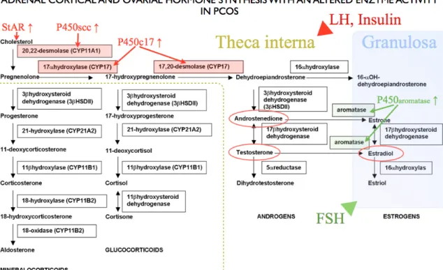
The theca interna cells of the ovaries and zona fasciculata of the adrenal cortex synthesise androgens from cholesterol under the control of LH in the ovaries and ACTH in the adrenal cortex (Fig. 1.). Both glands secrete androstenedione in significantly greater quantities than testosterone. The rate-limiting factor in androstenedione production is the gene expression of P450c17, which is dependent on trophic hormones.
Normally, elevation in the level of LH causes a down-regulation of the LH receptors;
reduces cholesterol side-chain cleavage activity and 17,20 lyase activity; reduces the activity of 17-hydroxylase; and decreases androgen levels. The effects of trophic hormones can be modified by small peptides such as insulin and insulin-like growth factors (IGF) [White DW. et al. 1995]. In theca cells, the elevated expression of the LH receptor, insulin receptor (INR), lipoprotein receptor (HDL and LDL), steroidogenic acute regulatory protein (StAR), P450 side-chain cleavage (P450scc), 3β.hydroxysteroid dehydrogenase (3β-HSD), and cytochrome P450c17 leads to excess production of progesterone, 17α-hydroxyprogesterone, androstenedione and testosterone [Diamanti-Kandarakis et al. 2006✲✲,2008].
Figure 1.: Steroid biosynthesis and altered enzyme activity of the adrenal cortex and the ovary in PCOS. Cholesterol enters the mitochondria with the assistance of steroidogenic acute regulatory protein (StAR). In the mitochondria cholesterol is converted to pregnenolone by the cholesterol side chain cleavage enzyme (P450scc). Pregnenolone and Hydroxypregnenolone are converted to DHEA by microsomal P450c17. The increased expressions of these enzymes lead to higher levels of androstenedion and testosterone in hyperandrogenic PCOS patients. Oestradiol (Estradiol) level is also elevated due to increased expression of P450 aromatase enzyme. Over-expressions of enzymes highlighted in red contribute to androgen excess. Increased activity of aromatase enzymes highlighted in green tries to compensate hyperandrogenism. Applied and modified from: MRCOG Facts. http://img.medscape.com/pi/
emed/ckb/pediatrics_surgery/933425-940347-2132.jpg
The level of oestradiol (E2) increases in PCOS due to the availability of androgens for aromatase enzyme and hyper-reaction to FSH. The granulosa cells have been reported to lose FSH responsiveness and produce low quantities of progesterone [Mason et al.
1994]. Insulin acts as a co-gonadotropin and enhances the effects of testosterone by decreasing serum SHBG concentrations. The suppression of insulin secretion by a somatostatin analogue was demonstrated to lower both serum LH and androgen levels in women with PCOS [Prelevic et al. 1990].
Hyperandrogenemia has multiple effects on different tissues. HA leads to local pro- inflammatory stages in the ovaries and the endometrium. In contrast with the increase of the CD8+/CD4+ T lymphocyte cell ratio in the ovaries, an elevation in the number of CD4+ T lymphocytes and a parallel increase of tissue apoptosis was described in the endometrium [Motta AB. 2010]. In the ovarian pro-inflammatory stage, increased PGF2α levels, enhanced expression of cyclooxygenase-2 (COX2), nitric oxide synthase (NOS), and decreased levels of PGE, catalase and superoxide dismutase were described [Elia et al. 2009]. HA was found to induce morphological changes similar to precancerous endometrial structures [Motta AB. 2010]. These alterations of endometrial tissue are likely linked with infertility and recurrent miscarriage in PCOS.
Adipocyte cells pretreated with testosterone showed significantly impaired glucose uptake in response to insulin. This decrease was associated with a decreased insulin- stimulated phosphorylation of protein kinase C (PKC), which was reversible with anti- androgens such as cyproterone and partly reversible with flutamide [Corbould A.
2007✲✲].
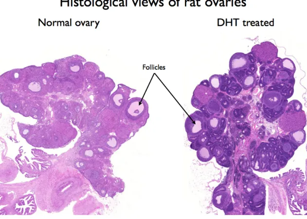
An impairment of the muscle glycogen synthase activity and minor changes in the muscle fibre composition were described in skeletal muscle cells after chronic testosterone treatment in rat model [Corbould et al. 2008]. Manneras et al. developed an animal model for human PCOS by administrating DHT to adolescent female rats. After ninety days, this treatment altered ovarian morphology (Fig. 2.) and vital parameters;
early impairment of glucose metabolism was also found. They have suggested that the DHT model is suitable for studies of metabolic and ovarian features of the syndrome [Manneras et al. 2007, Yanes et al. 2011].
Figure 2.: Histological views of whole rat ovaries. A normal ovary is shown on the left side, and a DHT treated for ten weeks with PCO morphology (DHT 6. animal) is shown on the right side. Numerous premature follicles can be seen side by side peripherally in the ovary of the DHT-treated animal. From the experimental histological sections of the study.
1.5.3. Metabolic syndrome
Metabolic syndrome affects approximately 6% of women in the 20-29 year age group and 15% of 30-39 year age group. Among these women the prevalence of PCOS is twice to four times that of the normal population [Moran et al. 2010]. PCOS is often accompanied by metabolic syndrome, and the possibility of developing metabolic syndrome increases with age. The prevalence of metabolic syndrome was reported in 33-46% of women with PCOS [Ehrmann et al. 2006] The World Health Organization (WHO) criteria for metabolic syndrome were presented in 1999. The definition of metabolic syndrome requires the presence of one of the following: diabetes mellitus,
impaired glucose tolerance, impaired fasting glucose or IR, and two of the following:
blood pressure ≥ 140/90 mmHg; dyslipidaemia (triglycerides ≥ 1.695 mmol/L and high- density lipoprotein cholesterol ≤ 1.0 mmol/L); either central obesity (waist-hip ratio >
0.85, or body mass index > 30 kg/m2); and either microalbuminuria (urinary albumin excretion ratio ≥ 20 µg/min or albumin: creatinine ratio ≥ 30 mg/g) [Meigs JB. 2002].
A prenatal exposure to androgen was reported to produce metabolic syndrome in adult female rats, which is similar to the PCOS model described earlier [Demissie et al. 2008].
Mehrabian et al. presented results on studies of Iranian women with PCOS and demonstrated a strong relationship between hyperandrogenism, IR and metabolic syndrome [Mehrabian et al. 2011]. The presence of central obesity in PCOS seems to be one of the most important independent factors leading to an approximately 14-fold higher risk for metabolic syndrome [Ehrmann et al. 2006]. Obesity associated with PCOS can be characterised by an increase in the size of fat cells (hypertrophic obesity) rather than an increase in the number of fat cells (hyperplastic obesity) [Villa et al. 2011]. In PCOS, adipocyte enlargement is strongly associated with IR [Manneras et al. 2011]. The lipolytic and storage capacity of the adipocyte cells is altered due to cellular hypertrophy. An increase in lipolysis and expression of the transmembrane proteins of fatty acid transport were detected [Villa et al. 2011, Seow et al. 2009, Ek et al. 2002] A significant decrease in lipoprotein lipase activity was demonstrated in subcutaneous adipocyte cells [Manneras et al. 2011].
1.5.3.1. Insulin resistance
Approximately 20-40% of women with PCOS, including obese and non-obese patients, have impaired glucose metabolism [Speer G. 2009]. IR also occurs in metabolic syndrome, T2DM, and PCOS. There are two types of IR syndrome that may develop independently in females. The type A is caused by an insulin signal defect; it affects
young women and is characterised by hyperinsulinemia, hyperandrogenism and obesity.
Type B affects middle-aged women; it is characterised by signs of hyperandrogenism, autoimmune disorder and hyperinsulinemia. In Type B, the autoimmune antibodies block insulin binding or stimulate insulin receptors, leading to intermittent hypoglycaemia [Powers AC. 2008]. IR observed in PCOS is defined as a distinct entity of ovarian dysfunction but is similar to the type A IR syndrome. Although obesity is a common feature associated with IR, in PCOS, decreased insulin sensitivity is an independent component of body weight. The IR in PCOS is present at an incidence 50-70%, independent of obesity [Mukherjee et al. 2010]. The obese individuals with PCOS are more insulin resistant than their BMI counterparts without PCOS.
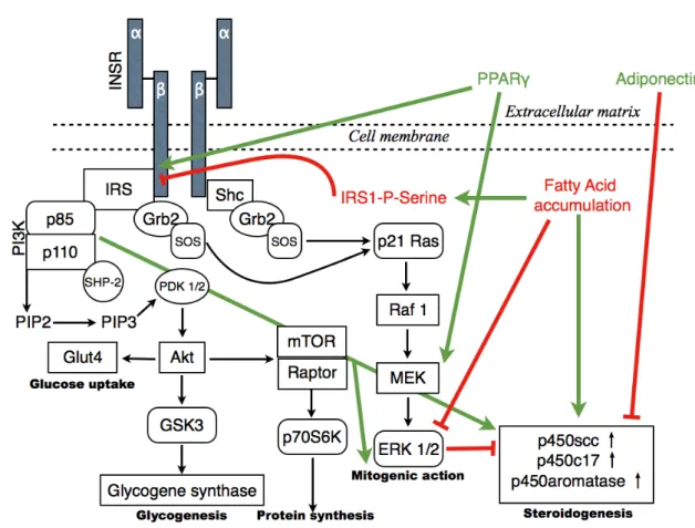
Insulin has a wide range of pleiotropic actions on the target tissue via different signalling pathways. Insulin affects the cellular metabolism, differentiation and growth of the target cells. After the binding of insulin to the α subunit of insulin receptor (INSR), the intrinsic tyrosine kinase is activated and its β subunits are auto- phosphorylated. The activated INR phosphorylates a number of substrates such as the insulin receptor substrate family (IRS1-4), Gab-1, Cb1, APS, homology domain transforming protein (Shc) isoforms and signal regulatory protein (SIRP) family members, which can bind to INSR [Pessin et al. 2000]. The phosphorylated IRS protein acts through several docking proteins such as the growth factor receptor-bound receptor 2 (Grb2), NcK and p85 subunit of phosphatidylinositol 3-kinase (PI3K). PI3K mediates metabolic actions such as glucose transport, glycogen synthesis, protein synthesis, and GLUT 4 translocation (helps in the rapid entry of glucose into cells); additionally, it activates Akt (AS160 substrate involved in GLUT4 translocation) and the atypical protein kinase C (PKC) isoforms [Pessin et al. 2000, Hojlund et al. 2008]. Insulin also has mitogenic actions mediated through the binding of phosphorylated IRS1/2 or Shc with the Grb-2/SOS complex, leading to p21Ras and Raf-1 activation of the mitogen- activated protein kinase pathway (MAPK). PI3K also facilitates the mitogenic response (Fig. 3.) [Venkatesan et al. 2001].
Figure 3.: Illustration of major signalling pathways of insulin action and its modifying factors in PCOS: Binding of insulin to its receptor (INSR) results in autophosphorylation and tyrosine kinase activation of the receptor. It phosphorylates other downstream mediators [insulin receptor substrate (IRS) and Src homology domain-containing transforming protein 2 (Shc)]. Various downstream signalling proteins are activated by these mediators. Phosphatidylinositol 3-Kinase (PI3K) regulates glucose transport, glycogenesis and protein synthesis. The Grb2/SOS (growth factor receptor-bound receptor 2/
Son of sevenless) complex activates the mitogen-activated protein kinase pathway (MAPK), playing a crucial role in cell proliferation. Another pathway that is not described yet may regulate steroidogenesis.
Factors in red and green alter these pathways in PCOS. Red arrows show the inhibiting effects and green arrows the activation of cascades. Abbreviations: Akt-serine/threonine-specific protein kinase, PIP2- phosphatidylinositol 4,5-bisphosphate, PIP3- phosphatidylinositol 3,4,5-triphosphate, mTOR- mammalian target of rapamycin, Raptor-regulatory associated protein of mTOR, Glut4- glucose transporter type 4, GSK3-glycogen synthase kinase 3, PDK1/2-phosphoinositide dependent kinase1/2, p70S6K- ribosomal protein S6 kinase, MEK- mitogen-activated protein kinase, ERK1/2- extracellular signal regulated kinase, SHP-2- SH2 domain containing protein tyrosine phosphatase. On the basis of Mukherjee et al. Indian J Med Res. 2010;131:743-60.
A post-receptor binding defect in the insulin signalling pathway appears to play the primary role in the aetiology of selective IR. There is evidence that the tyrosine kinase auto-phosphorylation of the insulin receptor β subunit decreases and the serine- phosphorylation increases in PCOS. The β subunit can phosphorylate IRS-1 protein, which can activate PI3K and GLUT4 [Pessin et al. 2000, Mukherjee et al. 2010, Speer G.
2009]. Decreased auto-phosphorylation, tyrosine activation and the parallel increase in
serine-phosphorylation influence the intracellular signal transduction of insulin and diminish glucose uptake. The serine-phosphorylation of the β subunit can stimulate the lyase activity of the citochrom-P450c17α enzyme [Bremer et al. 2008](Fig. 3.).
The reduced insulin-receptor level and the increased levels of biologic markers (e.g., free fatty acid, leptin, TNFα, retinol-binding protein) secreted by adipocytes may play a secondary role in development of IR in PCOS, as well as impaired fatty acid oxidation, which generates reactive oxygen species such as lipid peroxides. The elevated levels of free fatty acids and reactive oxygen species have a lipotoxic effect on beta cells in the pancreas [Powers AC. 2008]. Both TNFα and PC1 membrane glycoprotein were reported to decrease tyrosine kinase activity [Spaczynski et al. 1999, Speer G. 2009]. The accumulation of lipids in myocytes impairs mitochondrial oxidative phosphorylation and reduces the insulin-stimulated production of mitochondrial ATP [Powers AC. 2008].
Other researchers have found a correlation between lipin 1β protein and IR [Mlinar et al.
2008]. Lipin 1 plays a key role in triglyceride (TG) and phospholipid synthesis and acts as a transcription co-activator in fatty acid oxidation. Lipin 1 was shown to have an insulin sensitising effect on tissues [Peterfy et al. 2001]. Lipin 1β is the main secreted isoform of mature adipocytes and increases the expression of genes involved in TG and fatty acid synthesis [Peterfy et al. 2005]. The lipin 1 concentration in the adipose tissue and the liver is inversely related to obesity and IR [Croce et al. 2007]. Mlinar et al.
demonstrated that lipin 1β level are inversely correlated with BMI, waist circumference and HOMA-IR [Mlinar et al. 2008] in visceral adipose tissue. A positive correlation was demonstrated between lipin 1β level and HDL-cholesterol.
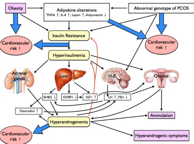
A low level of adiponectin was detected in patients with PCOS (Fig. 5.). This decrease was independent of BMI, but showed a strong correlation with IR [Toulis et al. 2009].The messenger molecule (adipokine) has a direct insulin-sensitising effect, and lower levels of adiponectin can contribute to IR. These pathways can elucidate the increased severity of IR in obesity (Figs. 4. and 5.). Adipocyte production and decreased levels of adipokines contribute to an inflammatory state, which explains the elevated levels of
hsCRP and IL-6 often seen with IR and T2DM [Powers AC. 2008]. Antilla et al. studied obese and non-obese women with PCOS and found that the non-obese women had elevated serum concentrations of bioactive LH related to hyperinsulinaemia. These changes were independent of androgen concentrations and ovarian androgen production. Antilla proposed that the degree of hyperinsulinemia and the severity of IR directly affected the glycosylation of LH (modifying the bioactivity of LH isoforms) [Antilla et al. 1991].
The altered response of insulin to different tissue cells was investigated in PCOS. In adipocytes, basal autophosphorylation of the IR β-subunit was normal and insulin- dependent autophosphorylation was significantly diminished, but receptor-kinase activity was normal [Futterweit W. 1999]. Decreases in levels of IRS-1 and IRS-2 tyrosine phosphorylation as well as in the expression of GLUT4 were also demonstrated [Diamanti-Kandarakis et al. 2006✲✲]. These changes show that there is no defect in the binding of insulin molecules to INR. The diminished insulin sensitivity in PCOS cannot be explained by defective binding at the receptor level. Thus, a step downstream of receptor binding is supposed to act in the insulin signal-transduction pathway, that is defective in IR [Diamanti-Kandarakis et al. 2006✲✲]. In fibroblast cells, basal autophosphorylation was increased in approximately 50% of the cultures obtained from PCOS patients [Dunaif et al. 1995]. A serine kinase associated with a PKA-regulated pathway is related to IR in cultured fibroblasts of PCOS women [Diamanti-Kandarakis et al. 2006✲✲]. In these cultured cells, IRS-1 tyrosine phosphorylation and insulin- dependent glycogen synthesis were decreased [Diamanti-Kandarakis et al. 2006✲✲], while IRS-1 expression and PI3K levels were normal. The decrease in the glycogen synthesis might have been caused by the reduced GSK-3 phosphorylation [Venkatesan et al. 2001].
In adipocytes, similar pathways have been observed. In skeletal muscle cells, a decreased IRS-1-associated PI3K activity was demonstrated in vivo, which was not completely compensated by elevated IRS-2 expression; therefore, the insulin-mediated glucose uptake remained decreased [Dunaif et al. 2001]. In contrast, a higher expression of IRS-1 and GLUT1 and reduced activity of the IRS-1-PI3K and IRS-2-PI3K complex
were detected in vitro [Corbould et al. 2005].
An increase in the prevalence of PCOS and its phenotypic features has been demonstrated in twin and family studies [Diamanti-Kandarakis et al. 2005]. The mode of inheritance is still not clear, but several gene variants are proposed to interact with each other and the environment in the manifestation of the syndrome. A strong linkage has been observed between the susceptibility to the disease and a dinucleotide marker on chromosome 19p13 [Urbanek et al. 2005]. The possible gene variants related to PCOS are depicted in Table 2.
IR increases the risk for developing glucose intolerance, T2DM, hypertension, dyslipidaemia and cardiovascular abnormalities in PCOS patients [Lambrinoudaki I.
2011, Agarwal et al. 2010, Nakka et al. 2011]. The severity of obstructive sleep disorders, which is often observed in PCOS, presents a strong correlation with IR than with the BMI [Nitsche et al. 2010]. IR can be reversed by lifestyle modifications as a first line of therapy [Karakas et al. 2010]. Other agents such as insulin sensitisers are recommended when cardiovascular risk factors persist in spite of lifestyle modification [Katsiki et al.
2010]. The drug metformin is a biguanide (N,N-dimethyl-biguanide) and can improve insulin sensitivity by increasing glucose uptake in peripheral tissues, stimulating glycolysis, decreasing liver glucose production and inhibiting intestinal glucose absorption [Katsiki et al. 2009]. Its effects on lipid metabolism, atherosclerosis and inflammatory markers of metformin are controversial [Katsiki et al. 2010]; however, it is reported to increase adiponectin level [Agarwal et al. 2010]. The use of metformin treatment alone or in combination with clomiphen-citrate improves menstrual irregularities and infertility. The positive effect of metformin on the cardiovascular system of patients with PCOS was also reported [Agarwal et al. 2010, Palomba et al. 2010, Nakka et al. 2011]. The drug significantly decreases the level of total testosterone, free testosterone and androstendione, while it increases the level of SHBG without affecting the levels of FSH, LH or DHEAS.
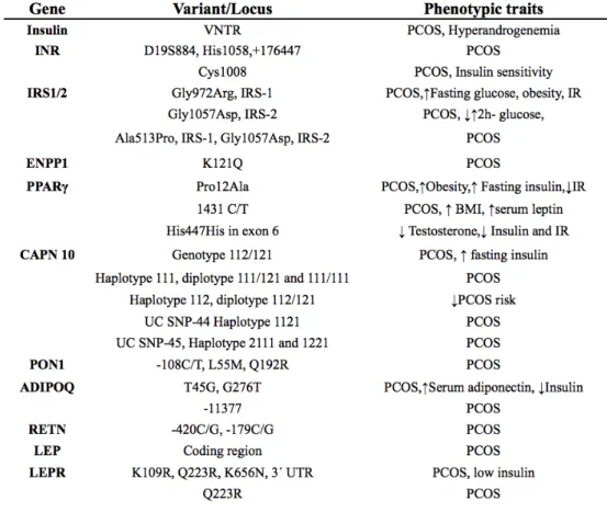
Table 2.: Gene variants related to insulin resistance in PCOS. INR: insulin receptor, IRS1/2: insulin receptor substrate 1-2, ENPP1: Ectoenzyme nucleotide pyrophosphate phosphodiesterase(PC-1) gene, PPARγ: Peroxisome proliferator activated receptor gamma gene, CAPN10: Caplain-10(cystein protease) gene, PON1: Paraoxonase-1 gene, ADIPOQ: adiponectin gene, RETN: resistin gene, LEP and LEPR:
leptin and leptin receptor genes, VNTR: variable number of tandem repeats. From: Mukherjee S.
Molecular & genetic factors contributing to insulin resistance in polycystic ovary syndrome. Applied and modified from: Mukherjee et al. Indian J Med Res. 2010;131:743-60.
Metformin treatment in combination with oral contraceptives (OCs), pioglitazone and flutamide has a stronger modulatory effect on hormone levels [Katsiki et al. 2010].
However, metformin is used worldwide without a full understanding of the mechanism involved. The thiazolidinediones (TZDs) are ligands of the peroxisome proliferator- activator receptor-gamma (PPARγ), a nuclear transcription factor, which is mainly expressed in adipose tissue, intestinal, pancreatic beta cells, macrophages, vascular endothelium and skeletal muscle [Perry et al. 2002] The two best-known agents in this group, rosiglitasone and pioglitasone, were reported to improve IR (HOMA-IR and insulin levels); restore menstrual cycles; induce ovulation; increase adiponectine, HDL- C and SHBG levels; decrease androgen, TC, TG, LDL and hs-CRP levels; and improve
hirsutism [Ortega-Gonzalez et al. 2005, Glintborg et al. 2005]. TZDs compared with OCs have superior effects in improving IR but inferior effects in reducing hyperandrogenism [Katsiki et al. 2010]. The role of TZDs, as a regulatory mechanism of ovarian function, indicates a relationship between insulin signalling, steroidogenic productions and PPARγ [Mukherjee et al. 2010]. The possible action of PPARγ on insulin signalling and the mitogenic cascade is depicted in Fig. 5. A new insulin sensitiser, BGP-15 (O -[3- piperidino-2-hydroxy-1-propyl]nicotinic-amidoxime-dihydrochloride), has been reported to have a non-PPAR agonist mechanism of action[Literati-Nagy et al.2009]. This new molecule, a co-inducer of heat shock proteins, might be a better agent for PCOS therapy. It has been suggested that vitamin D therapy also has positive effects on carbohydrate metabolism [Yiu et al. 2011, Kotsa et al. 2009, Pittas et al. 2006, Hypponen et al. 2001] and can prevent cardiovascular complications [Wong et al. 2010, Przybylsky et al.
2010].
1.5.3.2. Dyslipidemia
Based on ethnicity, genetic factors and lifestyle, 30 to 70% of PCOS patients present with abnormal levels of serum lipids [NCEP 2002]. PCOS patients with IR show a higher prevalence of dyslipidemia than their counterparts without IR (81 to 65%, respectively). The possible pathways of altered adipose tissue function due to IR were mentioned above. IR contributed to elevated triglyceride (TG), increased low-density lipoprotein-C (LDL-C) and very low-density lipoprotein (VLDL); decreased high- density lipoprotein-C (HDL-C) levels due to the impaired functions of the adipose tissue [Brunzell et al. 2003, Wild et al. 1985]. The changes in lipoprotein levels are strongly accompanied with an altered predominance of LDL-III and LDL-IV (small and very-small subspecies of LDL) and decreased ratios of the anti-atherogenic HDL2
subtype in PCOS. Androgens also affect lipoprotein metabolism. Testosterone lowers HDL levels by increasing the expression of the genes involved in HDL catabolism;
scavenger receptor B1 (SR-B1) mediates the selective uptake of HDL to hepatocytes,
and hepatic lipase increases the clearance of HDL by catalysing HDL2 conversion to HDL3 [Diamanti-Kandarakis et al. 2007](Fig. 4.).
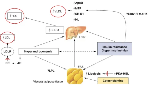
Figure 4.: Pathophysiology of dyslipidaemia in PCOS and its possible mechanisms. The main serum lipid abnormalities in PCOS are indicated in red circles. Broken arrows represent potential interaction.↑activation; ↓deactivation; ≠ inhibition. Abbreviations: ApoB, apolipoprotein b; AR, androgen receptor; ER, estrogen receptor; ERK1/2 MAPK, extracellular signal-regulated kinase 1/2 mitogen-activated protein kinase; FFA, free fatty acids; HL, hepatic lipase; LDLR, low density lipoprotein receptor; LPL, lipoprotein lipase; MTP, microsomal triglyceride protein; PKA-HSL, protein kinase a-hormone sensitive lipase complex; SR-B1, scavenger receptor-b1; VLDL, very low density lipoprotein. Adopted from Diamanti-Kandarakis E. et al. Endocrinol Metab. 2007;18:280-5.
IR increases the expression of microsomal triglyceride protein (MTP), which in combination with excess TG contributes to the overproduction of apolipoprotein B (ApoB) and a subsequent increase in VLDL production [Diamanti-Kandarakis et al. 2007, Taghibiglou et al. 2000](Fig. 5.). Apolipoproteins act on the surface of the circulating particles and helps direct these particles in metabolic and clearing patterns. An elevated ApoCIII/ApoCII ratio was described in PCOS [Wild et al 2012]. ApoCIII/CII regulates
lipoprotein lipase activity and responsible for VLDL metabolism. Additionally, VLDL has a primary role in TG metabolism. ApoCIII can block CII, ApoB and E (delay VLDL lipolysis), which results in greater amounts of circulating LDL in the plasma [Wild et al. 2012]. Serum levels of ApoB might be a better atherogenic predictor than non-HDL cholesterol levels [Wild et al. 2012].
A decrease in lipolysis and a reduction in chatecholamine-stimulated PKA/hormone- sensitive lipase complex activation were observed in subcutaneous fat tissue but not in visceral fat adipocytes after chronic testosterone treatment [Dicker et al. 2004, Villa et al.
2011]. These adipocyte lipolytic function impairments can be attributed as a secondary cause of hyperandrogenism [Villa et al. 2011]. Androgens regulate lipoprotein lipase activity. which was detected to be positively correlated with plasma free testosterone levels [Iverius et al. 1988]. Dihydrotestosterone was demonstrated to increase Lipoprotein lipase (LPL) levels [Anderson et al. 2002]. These patterns of dyslipidemia and IR might play a primary role in atherosclerosis in PCOS.
The adipose tissue secretes different types of signalling molecules. In PCOS, the production of leptin is controversial. The overexpression of this messenger protein can induce atherosclerosis and endothelial dysfunction [Reilly et al. 2004, Knudson et al. 2005] and influence hypothalamic GnRH secretion [Walters et al. 2012]. Elevated levels of plasma leptin were reported to positively correlate with Retinol binding protein 4 (RBP4) and asymmetric dimethylarginine in young women with PCOS, however these factors have a poor predictive value in PCOS. [Yildizhan et al. 2011]. The association between elevated levels of leptin and IR or obesity is still debated. Adiponectin was demonstrated to be an insulin sensitising and anti-inflammatory messenger molecule [Weyer et al. 2001]. Adiponectin levels are known to be decreased in obesity. Adiponectin produced by adipocytes is considered a useable marker for metabolic syndrome in PCOS because its level inversely related to adipocyte mass [Groth SW. 2010]. Low adiponectin levels measured in the morning and evening are usually associated with obstructive sleep apnea [Nitsche et al. 2010] in PCOS. Lipin 1 is a nuclear protein that is
essential for adipocyte metabolism. Mlinar et al. studied lipin 1β levels in subcutaneous and visceral fat adipocytes from patients with PCOS and controls patients and found that lipin 1β was negatively correlated with BMI, waist circumference and IR [Mlinar et al. 2008]. The level of lipin 1 was significantly lower in visceral adipose tissue than in subcutaneous adipose tissue after adjusting for BMI. Lipin 1 polymorphisms might also determine the development of PCOS. Lipin 1 gene variations were shown as possible factors in cardiometabolic complications of PCOS [Mlinar et al. 2011].
1.5.3.3. Hypertension
Hypertension is not sine qua non but is a very common symptom in PCOS, often due to developing metabolic syndrome, which starts mainly in the fourth - fifth decade of life.
Increased arterial stiffness in PCOS accompanying hemodynamic changes with ageing contributes to high systolic blood pressure and pulse pressure and an increased ventricular load. The relationship between IR/hyperinsulinemia, hyperandrogensim and hypertonia is still under research. Some reports explained that hypertension seen in PCOS is a consequence of obesity and increased sympathetic activation [Luque-Ramirez et al. 2007✲]. Hypertension was reported to develop in 2-years-old female sheep, which had been prenatally treated with Testosterone-propionate for 60 days [King et al. 2007].
Manneras and Yanes have presented a rat PCOS model in which hypertonia also evolved after chronic dihydrotestosterone treatment (12 weeks) [Manneras et al. 2007, Yanes et al. 2011]. These animal models can prove that prenatally or postnatally induced PCOS correlates with hypertension, however human cohort studies are controversial.
1.5.4. Connection between Hyperandrogenism and Insulin Resistance
Glucose metabolism is not altered in insulin-independent tissues or insulin-dependent
pathways, which might sometimes impair mitogenic-activated protein kinase signals that control cell growth and differentiation. The acceleration of these hyperinsulinaemia pathways may cause hyperandrogenism and atherosclerosis [Powers AC. 2008]. In recent studies, both hyperandrogenemia and hyperinsulinemia were established as the principal factors of PCOS. The cause and effect relationship between these features is still debated [Dunaif A. 1997, Bremer et al. 2008]. There is evidence that insulin can augment steroidogenesis in the ovaries and may reduce the liver production of sex hormone- binding globulin (SHBG) directly or through elevated levels of LH [Poretsky et al. 1999, Bremer et al. 2008]. There are two hypothesis regarding androgen synthesis in ovaries by insulin. It was demonstrated that insulin stimulates testosterone biosynthesis and acts directly via its receptor at physiological concentrations in cultured polycystic ovary theca [Nestler et al. 1998] and granulosa cells [Willis et al. 1995]. Insulin-induced androgen excess occurs via IGF1 receptor activation because insulin acts through IGF1 only when the circulating levels of insulin are extremely high [Poretsky et al. 1999].
Steroidogenesis is mainly regulated by LH in theca cells of ovaries via the cAMP- dependent protein kinase (PKA) pathway. Insulin alone was not found to increase cAMP, but it enhanced LH-induced cAMP accumulation in porcine theca cells [Zhang et al. 2000]. This accumulation might activate PI3K, MAPK or an alternate pathway for insulin signalling. In human theca cells, insulin requires concomitant cAMP signalling by forskolin to enhance 17α-hydroxylase activity [Munir et al. 2004]. This activity is mediated via the PI3K pathway and not via the MAPK pathway [Munir et al. 2004], indicating one of the differences between steroidogenic and metabolic pathways because the glucose metabolic pathway does not require concomitant cAMP signalling. The Akt (serine / threonine-specific protein kinase, protein kinase B) activation pathway may be a different insulin-dependent pathway for steroidogenesis [Diamanti-Kandarakis et al.
2006✲✲]. The altered MAPK pathway with reduced MAPK1/2, extracellular signal- regulated kinase 1-2 (ERK1/2) activity and a parallel increase of CYP17 mRNA expression, and DHT activity might play insulin-independent roles in androgen excess and hyperandrogenism [Nelson-Degrave et al. 2005]. Another hypothesis suggests that serine phosphorylation of the β subunits of INR and P450c17 by a “hypothetic” kinase
can contribute to both hyperandrogenemia and hyperinsulinemia [Bremer et al. 2008].
The above mentioned serine kinase has not been identified yet. The debate about the hypersensitivity of insulin or the state of the preserved ovarian sensitivity still continues. Poretsky pointed at the high androgen excess of ovarian cells independent of hyperinsulinemia [Poretsky L. 2006]. However, cultured theca cells after stimulation by insulin exhibited enhanced androgen production [Mukherjee et al. 2010]. Insulin sensitisers were found to decrease androgen levels. Insulin can also facilitate ACTH- stimulated androgen secretion in the adrenal cortex [Speer G. 2009]. This activation is also responsible for the hyperandrogenemia observed in IR [Speer G. 2009, Bremer et al.
2008]. The higher intraovarian insulin level increases the intraovarian androgen level.
This may halt the maturation of the follicles, increase FSH sensitivity to the granulosa cells and stimulate follicle genesis, contributing to typical polycystic ovarian morphology [Speer G. 2009]. Hyperinsulinemia and IR are responsible for the decreased sensitivity and metabolic effectiveness of insulin in different tissues, including in the ovaries. The ovaries may otherwise remain sensitive to insulin and produce androgen [Mukherjee et al. 2010]. The pharmacological reduction of insulin levels has been found to improve both hyperinsulinemia and hyperandrogenemia [Dunaif A. 1997 ,Poretsky et al.
1999, Bremer et al 2008]. However, the reduction of androgen levels by bilateral oophorectomy and administration of GnRH agonist [Bremer et al. 2008] or antiandrogenic compounds [Dunaif A. 1997] was reported to be ineffective for IR or hyperinsulinemia in PCOS patients.
Insulin can affect steroid production in granulosa cells, as well. Insulin alone or with FSH augments P450 aromatase activity, stimulates progesterone, and affects only E2
production in anovulatory PCOS [Mukherjee et al. 2010]. In granulosa cells, neither MAPK nor PI3K seemed to be involved in the insulin-mediated pathway of steroidogenesis [Poretsky et al. 2001]. Both insulin and LH synergistically up-regulate the transcription of the LDL receptor to facilitate cholesterol uptake [Sekar et al. 2001].
Rice and colleagues reported that an abnormal glucose metabolism, with significantly
impaired insulin, stimulated lactate production in granulosa-lutein cells obtained from women with anovulatory PCOS [Rice et al. 2005]. These granulosa cells exhibited normal steroid production in response to physiological doses of insulin. These findings suggest that insulin activity is not only present at the organ level but also in the microenvironment of the ovary [Diamanti-Kandarakis et al. 2006✲✲✲]. Granulosa cells seem to be resistant to insulin-mediated glucose metabolism while maintaining the insulin regulation of ovarian steroidogenesis.
Figure 5.: The pathophysiology of PCOS. The black and red arrows show direct effects on altered processes and relationships between biochemical/laboratory changes. Blue arrows indicate the increased cardiometabolic risks. TNFα: Tumour Necrosis Factor α, IL-6: Interleukin-6, SHBG: Sexual Hormone Binding Globulin, IGFBP1: Insulin-like Growth Factor Binding Protein 1, LH: Luteotropic Hormone, FSH: Folliculotropic Hormone, HP: Hypothalamus-pituitary system. Adopted and modified from Speer G. In: Lakatos P, Speer G. Polycystic Ovary Syndrome. Budapest. Semmelweis Kiadó, 2009. p.31.
1.5.5. Vascular alterations in PCOS
In earlier sections, it was mentioned that women with PCOS have a higher risk for CVDs. Obesity, IR or T2DM, hypertension, and dyslipidemia associated with PCOS increase the risk for CVDs. IR and obesity were proposed to have a central role in the development of CVDs [Meyer et al. 2005,Cussons et al. 2009]. Although there is a higher incidence of cardiovascular risk factors among PCOS patients, the existence of a significantly higher CVD incidence compared to control patients is debated [Iftikhar et al. 2012]. In research yet little is known about cardiovascular alterations of PCOS.
In PCOS, increased arterial stiffness and pulse wave velocity were demonstrated by ultrasound assessments [Bots et al. 2002]. The mechanism of these alterations is unclear, but endothelial dysfunction and altered collagen metabolism of the vessel walls may be involved. This is similar to the patterns seen in IR and metabolic syndrome [Cussons et al. 2009, Orio et al. 2005]. Human studies have shown both macro- and microvascular dysfunction measured by ultrasound in PCOS, as indicated by the reduction in acetylcholine(ACh)-dependent vasodilation. This impaired response to ACh is similar to that observed in non-insulin-dependent diabetes mellitus. It is proposed to be related to metabolic alterations, especially insulin resistance in PCOS [Lakhani et al. 2005, Kravarati et al. 2005, Diamanti-Kandarakis et al. 2006✲]. Lakhani and Hardiman showed that those women who have a decreased internal carotid artery pulsatility index in PCOS, have a higher cardiovascular risk as well [Lakhani et al. 2000]. Women with PCOS have a high prevalence of early-onset atherosclerosis, metabolic syndrome and insulin resistance, and they might develop hypertension during their reproductive period [Dokras et al. 2008, Wild et al. 2012]. These abnormalities are partly explained by the pharmacological and biomechanical remodelling of resistance arteries. The intima- media thickness of the carotid arteries was confirmed to be greater in PCOS than in age- and BMI-matched controls [Talbott et al. 2000]. Similarly, coronary artery calcification was found to be greater in PCOS patients [Talbott el al. 1995, Christian et al. 2003]. An endothelial dysfunction is the first step in the formation of atherosclerosis. Uncoupling