The potential use of the culture filtrate of an Aspergillus niger strain in the management of fungal diseases of grapevine
Xénia PÁLFI1, Zoltán KARÁCSONY1 (✉), Anett CSIKÓS2, Ottó BENCSIK3, András SZEKERES3, Zsolt ZSÓFI4, Kálmán Zoltán VÁCZY1
Egy Aspergillus niger törzs fermentlevének lehetséges felhasználása a szőlő gombás betegségeinek megelőzésére
1 Eszterházy Károly University, Food and Wine Research Institute, H-3300 Eger, Leányka utca 6., Hungary
2 University of Pannonia, Georgikon Faculty, Institute of Plant Protection, H-8360 Keszthely, Deák Ferenc utca 16., Hungary
3 University of Szeged, Faculty of Science and Informatics, Department of Microbiology, H-6726 Szeged, Közép fasor 52., Hungary
4 Eszterházy Károly University, Department of Viticulture and Oenology, H-3300 Eger, Leányka utca 6., Hungary
✉ Corresponding author: karacsony.zoltan@uni-eszterhazy.hu
ABSTRACT
The present study was aimed to examine the use of culture filtrates of defensin-producing fungal strains against the most important fungal and pseudofungal infections of grapevine as an environmentally sound alternative of chemical control. Twenty-seven isolates of five defensin-producing fungal species were tested for antifungal activity. Four isolates (one Aspergillus giganteus and three Aspergillus niger) decreased the viability of the test organism successfully. The isolate with the highest antifungal efficacy (Aspergillus niger SZMC2759) was subjected to detailed investigation. The culture filtrate of the strain was effective against epicuticular mycelia and conidia of Botrytis cinerea, Guignardia bidwellii and Erysiphe necator, but not against subcuticular mycelia and Plasmopara viticola cells. The culture filtrate also prevented the development of powdery mildew symptoms on grapevine cuttings. Several modes of action were detected: inhibition of germination, membrane permeabilization, plasmolysis, fragmentation of nuclei. Certain parameters (pH, stress, aeration) of the fermentation were optimized for the highest antifungal activity. According to the results, culture filtrate of Aspergillus niger can be a promising agent in the management of fungal diseases of grapevine.
Keywords: Vitis vinifera, fungal pathogens, disease control, antimicrobial peptide
ÖSSZEFOGLALÓ
Jelen tanulmány célja defenzint termelő gombafajok fermentleveinek a szőlő legfontosabb gombás betegségei ellen történő felhasználhatóságának vizsgálata, amely környezetbarát alternatívát nyújthat a vegyszeres növényvédelemmel szemben. Öt defenzint termelő gombafaj 27 izolátuma képezte a vizsgálatok tárgyát. Ezek közül négy (egy Aspergillus giganteus és három Aspergillus niger) izolátum volt képes hatékonyan csökkenteni a tesztorganizmus életképességét.
A leghatékonyabb izolátumot (Aspergillus niger SZMC2759) részletes vizsgálatoknak vetettük alá. A törzs fermentleve fungicid hatást mutatott az epikutikuláris gombafonalakkal és spórákkal szemben a Botrytis cinerea, Giugnardia bidwellii és Erysiphe necator kórokozók esetén, de hatástalan volt a szubkutikuláris gombafonalak, valamint a Plasmopara viticola kórokozóval szemben. A vizsgált fermentlé képes volt tesztfertőzésekben a szőlő lisztharmat tüneteinek kialakulását Received: June 22, 2020; accepted: August 5, 2020
is meggátolni mesterségesen fertőzött dugványokon. A fermentlé számos gombaellenes hatásmechanizmusát sikerült igazolni: spórák csírázásának gátlása, plazmamembrán permeabilizálása, plazmolízis indukálása és sejtmag feldarabolódása. A fermentáció egyes körülményei (pH, stressz, oxigénellátottság) optimalizálásra kerültek a legnagyobb hatékonyság elérése érdekében. Eredményeink szerint az Aspergillus niger gombafaj fermentleve ígéretes gombaellenes szer lehet a szőlő növényvédelmében.
Kulcsszavak: Vitis vinifera, kórokozó gombák, növényvédelem, antimikrobiális peptidek
INTRODUCTION
The increasing demand of agricultural products for the growing global human population is a great challenge for agricultural companies. The widespread use of fertilizers and chemicals drastically increased the productivity of cultivation techniques in the last century (Popp et al., 2013). As a result of the climate change, the growing pressure of diseases on agriculture may increase the consumption of microbicidal sprays, especially in areas with mild climate (Pautasso et al., 2012). The ecological impact of these chemicals and the unpredictable effects of their accumulation in the food chain (Aktar et al., 2009) raised the demand of ecologically and medically safe alternatives. As a part of integrated pest and disease management, biological control has an increasing significance. For this purpose, living microorganisms (Harper, 2013) or microbial products (Stockwell and Duffy, 2012) are used to protect plantations from pests and pathogens.
Grape is one of the most spray-consuming agricultural product, primarily because of the fungal (e.g. black and grey rot, powdery mildew) and pseudofungal (downy mildew) diseases of grapevine. Black rot of grapes is caused by the ascomycetous fungus Guignardia bidwellii.
This fungus is a hemibiotrophic pathogen, which grows as subcuticular mycelia after the penetration of germ tubes into the host epidermis. The pathogen causes significant problems mainly in North America, which may result in as high as 80% yield loss without management (Amsdell and Milholland, 1988). Grey rot of grape is caused by Botrytis cinerea, a necrotrophic fungal pathogen which also belongs to phylum Ascomycota. This fungus is a widespread disease agent which can infect more than 200 different crops (Williamson et al., 2007). Grey rot causes about 2 billion $US per year losses for winegrowers
by reducing grape yields and quality (Elad et al., 2004).
Botrytis cinerea can infect all parts of grapevines, but it mostly occurs on berries and colonizes the subcuticular tissues. Pseudofungal species Plasmopara viticola, the causative agent of downy mildew belongs to Oomycetes and it is an obligate biotrophic parasite of different species of the Vitaceae family. This pathogen grows mainly under the epidermis and only the reproductive structures form on the surface of the leaves. Downy mildew is a very severe disease of grapevines. With permissive weather conditions and without chemical control it can cause 70% yield losses (Gessler et al., 2011). Powdery mildew is caused by the ascomycetous fungus Erysiphe necator.
This biotrophic pathogen can highly decrease grapevine productivity, and it negatively alters the quality of the affected musts (Calonnec et al., 2004). This fungus grows on the surface of the leaves and berries, only its haustoria, specialized structures for the plant-fungus interaction, are localized under the epidermis of the host.
Several low-price and effective chemicals are used for the control of the above pathogenic organisms, but the ecological and medical risks are also concern to these agents. Moreover, the non-specific management of grape microflora may impair spontaneous fermentation of musts. This way of fermentation is preferred more than the use of commercial yeasts by some producers, because of the enhanced aroma complexity and the area- specific characteristics of the produced wine (Medina et al., 2013).
Defensins are antimicrobial peptides widely distributed in the entire eukaryotic living world. They can be found in fungi (Wu et al., 2014), plants (Stotz et al., 2009), insects (Hoffman and Hetru, 1992) and vertebrates (Ganz 2004), for example as a part of the
innate immune system (Oppenheim et al., 2003). These peptides are very effective against a wide range of pathogens including viruses, bacteria and fungi. They can act in several different ways, which decreases the risk of the development of resistance against them.
Defensins possess low molecular weight, alkaline character and contain 6-10 cysteine residues, which form disulphide bonds (Sahl et al., 2005). The compact structure and the high number of disulphide bonds result in high structural and functional stability. Defensins can withstand immoderate pH and temperature conditions or proteolytic cleavage (Batta et al., 2009; Lacadena et al., 1995; Theis et al., 2005). These properties make them promising agents for both healthcare and agriculture.
The fungal defensins are the most suitable for industrial production due to the simple handling of the producing organisms. Cytotoxicity experiments have shown that fungal defensins have no harmful effects on plant or animal cells (Meyer, 2008; Szappanos et al., 2006). The applicability of fungal defensins against plant pathogens have been tested in numerous studies (Coca et al., 2003;
Girgi et al., 2006; Moreno et al., 2003; Moreno et al., 2005; Oldach et al., 2001; Vila et al., 2001) by using defensins as sprays or expressing these antimicrobials in plants. However, none of these studies focused on the applicability of these peptides against various fungal pathogens of the grapevine. The purpose of the investigation was to examine the potential use of some culture filtrates of defensin-producing fungal species for the biological control of fungal grapevine diseases.
MATERIALS AND METHODS Strains and growth conditions
For antifungal activity tests, strains of defensin- producing fungal species were obtained from the Szeged Microbiology Collection, Department of Microbiology, University of Szeged (Szeged, Hungary, http://szmc.
hu) and listed in Table 1. Botrytis cinerea and G. bidwellii strains were isolated from the vineyard of Eszterházy Károly University, Research Institute for Viticulture and Enology (Eger, Hungary) and identified according to morphological characteristics. All strains were maintained
by cultivation on PDA medium (0.4 w/v% potato extract, 2 w/v% glucose, 1.5 w/v% bacteriological agar in distilled water) at 25 °C. The obligate biotrophic E. necator and P.
viticola samples were collected freshly from Vitis vinifera cv. Kékfrankos plants without disease control.
Table 1. List of fungal strains used in screening for antifungal activity
Strain identifier Species
SZMC0145 Aspergillus niger
SZMC0514 Penicillium chrysogenum
SZMC0918 Aspergillus clavatus
SZMC12629 Penicillium chrysogenum
SZMC12641 Penicillium chrysogenum
SZMC12669 Penicillium chrysogenum
SZMC12676 Penicillium chrysogenum
SZMC13619 Rhizopus microsporus
SZMC13622 Rhizopus microsporus
SZMC1389N Neosartorya fischeri
SZMC2028A Aspergillus clavatus
SZMC2042 Aspergillus clavatus
SZMC2061N Neosartorya fischeri
SZMC2071 Aspergillus clavatus
SZMC20728 Aspergillus clavatus
SZMC2161 Aspergillus niger
SZMC2170 Penicillium chrysogenum
SZMC2176 Aspergillus niger
SZMC2182 Aspergillus niger
SZMC2192 Aspergillus niger
SZMC2197 Aspergillus niger
SZMC2207 Aspergillus niger
SZMC2356 Neosartorya fischeri
SZMC2402 Aspergillus niger
SZMC2660 Aspergillus niger
SZMC2719 Aspergillus clavatus
SZMC2759 Aspergillus niger
Preparation of fermentation broths
Fungi to be tested for antifungal activity were grown in liquid YG medium (0.5 w/v% yeast extract, 2 w/v%
glucose in distilled water) under static conditions. All fermentations were run at 25 °C for 7 days. The fungal strain, selected for the highest antifungal activity, was cultivated under altered conditions. To examine the effect of the initial pH on the antifungal efficiency of the fermentation broth, medium was adjusted to pH 4, 5, 6, 7 and 8 with HCl and NaOH. For the investigation of the effects of stress factors, the medium was supplemented with 1 M NaCl (osmotic stress) or 0.3 v/v% Tween 80 (membrane stress). In order to examine the influence of aeration, fermentations with dynamic conditions (180 rpm stirring) were also carried out. All fermentation broths were filtered through a 0.2 µm pore size membrane to obtain a cell-free filtrate.
Germination ability tests
To test the effect of fungal culture filtrates on spore germination, conidial suspensions of G. bidwelli and B.
cinerea (106 cell/ml) were prepared using 14-day old colonies cultivated on PDA medium. 50 µl aliquots of the suspensions were mixed with 50 µl of fungal culture filtrate or YG medium (control) and spread on the surface of cellophane placed on PDA medium. Conidia were incubated at 25 °C for 8 hours then examined by microscope. The percentage of germinating spores was calculated. The assays were performed in triplicates.
Viability tests
Thiazoyl blue tetrazolium bromide reduction assay For the quantification of antifungal activity of fungal culture filtrates by thiazolyl blue tetrazolium bromide (MTT) reduction assay three agar plugs with young mycelia were cut by cork borer (3 mm in diameter) from the edge of 7 day old G. bidwellii colonies on PDA medium and incubated in 500 µl fungal culture filtrate or YG medium (control) for 12 h at 25 °C. Thereafter broths were discarded and replaced with 500 µl of 5 mM MTT (Sigma Aldrich) in distilled water. To allow the living cells
to convert yellow MTT to purple formazan the samples were incubated for 3 h at 25 °C. The liquid phase was discarded and the samples were incubated with 1 ml extraction solution (0.04 M HCl in 2-propanol) for 2 h at 60 °C to dissolve the produced formazan. Formazan contents were determined through spectrophotometric measurement (SmartSpec Plus, Bio-Rad) at 575 nm wavelength. The viability of mycelial cells was expressed as percentage relative to the untreated control. The investigations were carried out in triplicates.
Fluorescein diacetate hydrolysis assay
For the quantification of biocidal activity of fungal culture filtrates by fluorescein diacetate (FDA) hydrolysis assay suspensions of conidia were prepared from B. cinerea, E. necator, G. bidwellii, while from P. viticola sporangial suspension was made. All suspensions were adjusted to 106/ml by sterile distilled water. A 100 µl aliquot of suspensions were mixed with the same volume of the serial dilutions of fungal culture filtrate and incubated for 4 h at 25 °C. Thereafter the mixtures were supplemented with 10 µM FDA (Sigma Aldrich) and incubated for additional 30 min. For calibration, the cell suspensions were serially diluted and treated as described previously, except that YG medium was used instead of culture filtrate.
Fluorescence was detected and documented by Gel Doc XR System (Bio-Rad). Quantification of fluorescence was done by imageJ software (Schneider et al., 2012). The percental viability of treated cells was calculated by the aid of the calibration.
Characterization of the active fungicidal agent of culture filtrate
Fungicidal activity test of protein fraction of culture filtrate
The antifungal activity of the protein fraction of culture filtrate was tested against G. bidwellii conidia after precipitation by trichloroacetic acid (TCA). Briefly, 25 µl of 100 w/v% TCA solution was added to 100 µl of culture filtrate and incubated for 1 h on ice. Proteins were pelleted by centrifugation at 12000 g for 10 min and washed twice with 200 µl ice-cold acetone. The pellet was dried and
resuspended in 100 µl distilled water. Protein fractions were also prepared in reducing conditions. Reduced samples are obtained as described above, with a previous 2-mercaptoethanol (2-ME) treatment (incubation with 100 mM 2-ME for 5 h, at 30 °C) to examine the possible role of disulphide bonds in the fungicidal agent. The viability of treated and control conidia of G. biwellii was measured by fluorescein diacetate hydrolysis assay as described above. All experiments were conducted in triplicates.
Estimation of the molecular weight of fungicidal agent by gel-electrophoresis and bioautography
Bioautography tests were done by sodium dodecyl- sulphate acrylamide gel electrophoresis (SDS-PAGE) with non-reducing conditions and the subsequent detection of fungicide activity of culture filtrate. Culture filtrate was supplemented with 5 v/v% glycerol, 2 w/v% SDS, traces of bromophenol blue and 50 mM Tris (pH 8). Samples were incubated at room temperature for 30 min prior to electrophoresis. Electrophoresis was carried out in a discontinuous (5% T stacking and 15% T resolving gel) polyacrylamide gel (Laemmli, 1970), at 30 mA current at 4 ºC until bromophenol blue reached the bottom of the gel. SDS was removed from the gel by washing twice with 20 v/v% 2-propanol for 20 min. The fungicide activity was detected by agar overlay method. Briefly, solid YG medium at 50 °C temperature was supplemented with 105/ml Saccharomyces cerevisiae cells (CGC 62, Uvaferm) and poured on top of the acrylamide gel. The gel was incubated at 30 ºC for 8 hours after solidification. Viable cells were detected by staining the medium with 5 mM MTT at 30 °C for 3 hours.
Microscopic examinations
For the microscopic examination of the effects of culture filtrates on fungal cells, small sections of young mycelia were cut from the edge of 7-day old G. bidwellii or B. cinerea colonies growing on PDA medium. The mycelia were immersed in 100 µl of the tested culture filtrate or YG medium at 25 °C for 4 hours. Samples were stained with 0.4 w/v% Crystal Violet for 20 min and washed with
distilled water to visualize cytoplasm. For the staining of nuclei mycelia were stained with 10 µg/ml SYBR GREEN I (Sigma Aldrich) for 15 min. For the assessment of cell membrane permeability mycelial samples were stained with 30 µg/ml ethidium bromide (EtBr) for 30 min then washed with distilled water. Viability stainings were done with asexual spores of B. cinerea, E. necator, G. bidwellii, P. viticola or leaf discs expressing the symptoms of E.
necator or G. bidwellii infections. Samples were treated with culture filtrate or YG medium (control) at 25 °C for 4 hours. Staining was done by the use of FDA as described previously in FDA fluorescence assay. Leaf discs with G.
bidwellii infections were treated with 1 unit/ml A. niger pectinase (Fluka) in citrate-phosphate buffer (pH 4) at 25
°C for 2 hours prior to staining. All examinations were carried out by Alpha BIO-5F microscope (Alpha). Images were taken by the use of Artcam-500MI camera (Artray) and Quickphoto Camera 2.3 software (PROMICRA).
Testing of antifungal activity of culture filtrate on grapevines cuttings
Examination of in planta antifungal effect of culture filtrates was done by the use of one-year old V. vinifera cv.
Kékfrankos cuttings. Cuttings were obtained by planting two-bud cane sections in soil:perlite (1:1) mixtures placed in plastic pots. Plants were grown in a greenhouse under natural light cycle, without artificial lighting and humidity control. The air temperature of the greenhouse was half- controlled by an automatic system, which regulated the opening of the upper windows during summer. Leaves were inoculated by spraying conidial suspension of E.
necator (105 conidia/ml, prepared in distilled water) on the leaves. Artificially infected cuttings were incubated in the greenhouse under the conditions described above, except that the plants were covered with plastic bags for the first three days of the experiment (27.-29. of July, 2016) to provide increased humidity. After one day, the grapevines were sprayed with YG medium (control) or fungal culture filtrate. Plants were incubated for an additional four weeks, and the spray treatments were repeated weekly.
Symptoms of powdery mildew were examined visually.
All experiments were done in triplicates.
Statistical analysis
Statistical analysis was done by two-way ANOVA analysis using GraphPad Prism 5 software demo version (GraphPad Software, San Diego California USA, www.
graphpad.com). Diagrams were generated with the same software and the layout was edited by Adobe Photoshop CS5 demo version.
RESULTS
Screening of fungal culture filtrates for antifungal activity
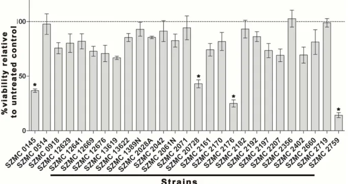
Culture filtrates of 27 isolates from five species were tested for antifungal activity against G. bidwellii mycelia by MTT assay (Figure 1). One A. clavatus (SZMC20728) and 3 A. niger (SZMC0145, SZMC2176, SZMC2759) isolates showed high antifungal activity (more than 50%
loss of viability). Repeated experiments with dilutions of culture filtrates showed that the culture filtrate of SZMC2759 (AnCF) had the highest antifungal potential.
Further works were focused on the study of this strain.
Figure 1. Antifungal activity of fungal culture filtrates on G. bidwellii mycelia assessed by thiazolyl tetrazolium bromide assay. The indicated values represent percental viability of treated mycelia relative to control sample, which treated with YG medium. Culture filtrates of fungal strains that caused more than 50% decrease in the viability (SZMC0145, SZMC20728, SZMC2176, SZMC2759) are marked with asterisk. Indicated values represent the average of 3 measurements, error bars show standard deviances
Determination of fungal and pseudofungal pathogens of grapevine susceptible to AnCF
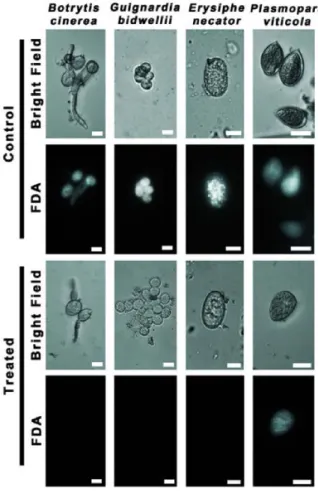
The biocidal activity of AnCF was tested by FDA assay against the asexual spores of the most important fungal and pseudofungal pathogens of grapevine: B. cinerea (grey rot), E. necator (powdery mildew), G. bidwellii (black rot) and P. viticola (downy mildew). The results are presented in Figure 2. The control samples (treated with YG medium) of all species showed bright green fluorescence as a result of the hydrolysis of FDA by living cells. The treatment by AnCF completely eliminated the fluorescence in the case of B. cinerea, E. necator and G. bidwellii conidia indicating the absence of hydrolytic activity, while P. viticola sporangia were not affected.
The effects of AnCF on conidia and mycelia
For the investigation of mode of action of AnCF against fungal cells, B. cinerea and G. bidwellii were chosen as test organisms, since they can be easily maintained by in vitro cultivation in contrast with the biotrophic species. Germination tests were carried out with spores treated with AnCF or YG medium as a negative control.
The presence of AnCF drastically decreased the rate of
Figure 2. Effect of AnCF on the viability of asexual spores of B.
cinerea, G. bidwellii, E. necator and P. viticola examined by fluo- rescein diacetate staining and fluorescent microscopy. The vi- ability of cells treated with YG medium (Control) or with AnCF (Treated) are indicated by the fluorescence of the hydrolysed dye. Scalebars represent 10 µm
germinating conidia. In case of the control conidia of G.
bidwellii 82±9.8% of the spores were able to germinate, while only 5±4.6% of the treated conidia developed germ tubes. The same phenomenon can be observed in the case of B. cinerea conidia. As the result of the treatment with AnCF 0% of conidia produced germ tubes, while this value was 78±2.5% in the case of YG-treated control.
To reveal the mechanisms behind the antifungal effect of AnCF, several microscopic examinations were carried out with AnCF-treated and untreated G. bidwellii and B.
cinerea mycelia (Figure 3). Plasmolysis was frequently detected in treated mycelial cells (Figure 3A, panel f;
Figure 3B, panel f) by crystal violet staining, while the untreated cells were able to maintain their intracellular pressure (Figure 3A, panel a; Figure 3B panel a).
SYBR GREEN I staining of untreated (Figure 3A, b and c panels; Figure 3B, b and c panels) and AnCF-treated mycelia (Figure 3A, g and h panels; Figure 3B, g and h panels) revealed that as a result of the treatment with AnCF, the mycelial nuclei were frequently fragmented.
Cell membrane permeability of control and AnCF- treated mycelia was investigated by EtBr exclusion and microscopic observations. Mycelia treated with AnCF (Figure 3A, i and j panels; Figure 3B, i and j panels) could uptake EtBr, while the untreated control showed no fluorescence (Figure 3A, d and e panels; Figure 3B, d and e panels) in the case of both G. bidwellii and B. cinerea.
Figure 3. A: Microscopic observation of G. bidwellii myce- lia treated with YG medium (Control) or with AnCF (Treated).
Mycelia were stained for cytoplasm (Crystal violet; a, f), nuclei (SYBR Green I staining; b, c, g, h) and for membrane permeabil- ity (Ethidium bromide staining; d, e, I, j). B: Microscopic obser- vation of B. cinerea mycelia treated with YG medium (Control) or with AnCF (Treated). Mycelia were stained for cytoplasm (Crystal violet; a, f), nuclei (SYBR Green I staining; b, c, g, h) and for membrane permeability (Ethidium bromide staining; d, e, I, j). Arrowheads mark plasmolysed cells, asterisks mark nuclei.
Scalebars represent 10 µm
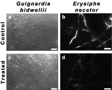
Antifungal effect of AnCF on infected leaf disks
Viability staining with FDA and subsequent microscopic studies were carried out with leaf discs showing symptoms of black rot (G. bidwellii) or powdery mildew (E. necator) infections. Results are presented in Figure 4. Both untreated mycelia of E. necator (Figure 4b) and G. bidwellii (Figure 4a) showed green fluorescence, indicating viable cells. As a result of the AnCF treatment, the fluorescein signal disappeared from E. necator mycelia, only a weak autofluorescence could be detected (Figure 4d). However, the treatment did not affect the viability of G. bidwelii cells (Figure 4c).
Chemical properties of the active fungicide agent of AnCF
The antifungal activity of AnCF is localized in the solid fraction after TCA precipitation and mostly restored its activity after re-solubilization (Figure 5a). The activity of the antifungal agent was decreased significantly by the application of 2-ME treatment (Figure 5a). The
Figure 4. Fluorescent microscopic examination of mycelia treat- ed with YG medium (Control) or AnCF (Treated) in case of G.
bidwellii (a, c) or E. necator (b, d) on leaf discs. Samples were stained with fluorescein diacetate (FDA). The fluorescence of hydrolysed FDA indicates viability. Scalebars represent 100 µm approximate molecular weight of the active agent of AnCF was investigated by non-reducing SDS-PAGE and bioautography (Figure 5b). The molecular weight of the antifungal agent fell into the range between 3 and 6 kDa.
Figure 5. Characterization of the antifungal component of AnCF. A: Effect of trichloro acetic acid (TCA) precipitation and 2-mercap- toethanol (2ME) treatment on the antifungal activity of AnCF against G. bidwellii conidia, measured by fluorescein diacetate hydro- lysis assay. Indicated values represent the average of 3 measurements, error bars show standard deviance values. Asterisks mark the significance of differences (** p<0,005, *** p<0,0005). B: Results of non-reducing gel electrophoresis of AnCF (lane 1: 40 µl, lane 2:
10 µl, lane 3: 5 µl) and subsequent bioautography with S. cerevisiae. The positions of molecular weight marker proteins are labelled on the left. Arrowhead marks the zone of inhibition detected by thiazolyl tetrazolium bromide staining
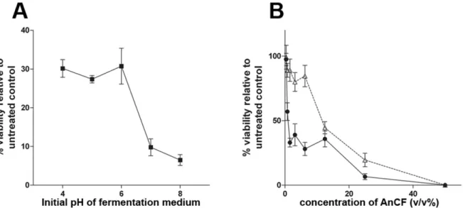
Optimization of fermentation conditions for AnCF The effect of certain parameters of cultivation on the antifungal activity of AnCF was tested by FDA assay on G. bidwellii conidia. Fermentations were carried out with different initial pH of the fermentation medium (Figure 6a).
Between pH 4-6 AnCF caused ~70% loss in the viability of conidia. Above pH 6 increased antifungal activity was observed (up to 93% at pH 8). The effect of aeration was also significant (Figure 6b). AnCF prepared with dynamic conditions had a MIC50 (minimal concentration for 50%
inhibition) value of 1 v/v%, while this value for AnCF prepared with static condition was 12.5 v/v% (values represent the volumetric rate of AnCF in the sample mixtures). AnCF prepared under the optimized conditions (YG medium, pH 8, 180 rpm stirring) was tested also against B. cinerea and E. necator by FDA-hydrolysis assay.
Figure 6. The effect of initial pH of the fermentation medium (A) and aeration (B) on the antifungal activity of AnCF against G. bid- wellii conidia by quantitative fluorescein diacetate hydrolysis assay. On panel B, open triangles represent fermentation with static condition, closed circles represent dynamic condition (stirring with 180 rpm). Indicated values represent the average of 3 measure- ments, error bars show standard deviances
The MIC50 values were 2.4 v/v% for B. cinerea and 2.9 v/v% for E. necator. In the presence of osmotic (1 M NaCl) or membrane stressors (0.3 v/v% Tween 80) in the fermentation medium the antifungal activity of AnCF did not altered significantly (data not shown).
Antifungal effect of AnCF on grapevine cuttings
Grapevine cuttings were artificially infected with E.
necator conidia and were sprayed regularly with AnCF prepared with optimal fermentation conditions or with YG medium (control). After four weeks of incubation all of the control plants showed the symptoms of powdery mildew (white spots of mycelia on leaves) with varying severity (Figure 7a) The AnCF-treated plants did not show the symptoms of powdery mildew or any negative effect, which would be caused by AnCF (Figure 7b).
Figure 7. Leaves of Kékfrankos grapevine cuttings artificially infected with E. necator conidia and incubated in greenhouse for four weeks. Plants treated with YG medium (a) or with AnCF (b) weekly
DISCUSSION
In the present study a range of culture filtrates of potential defensin-producing fungal isolates were tested for antifungal activity. One A. niger strain (SZMC2759) out of the four isolates showing the highest antifungal potential (one A. clavatus and three A. niger) was examined in details. The spectrum of susceptible pathogenic species of grapevine, mode of action, optimal fermentation parameters, chemical characteristics of the active agent, in vitro and in planta antifungal effects were investigated.
The active component of AnCF proved to be a low molecular weight, TCA-precipitable compound with sensitivity to 2-ME (Figure 5). The TCA-precipitable nature of the active compound suggest that it is a protein, and the sensitivity to 2-ME indicates that it contains disulphide bonds. These above results suggest that the active antifungal agent of AnCF is the ANAFP defensin (Gun Lee et al., 1999) of A. niger. The measured molecular weight of the active agent was lower than the molecular weight of ANAFP (10 kDa). This latter result may be explained by the combination of the disulphide bonds -which stabilize the structure of ANAFP- and the non- reducing condition of the electrophoresis, which latter was necessary for the recovery of antifungal activity after electrophoresis. The protein without the reduction of the disulphide bonds retained its compact structure, which resulted in a higher electrophoretic mobility.
The susceptibility tests have proven that AnCF is an effective fungicide not only against G. bidwelli but B.
cinerea and E. necator as well. AnCF decreased the viability of the treated cells by 50% in high dilutions (1-3 v/v%), but it was not toxic on sporangia of P. viticola a pseudofungal species. The ineffectiveness of AnCF against P. viticola can be explained by the large taxonomical distance between the oomycetous P. viticola and the tested ascomycetous fungi.
The present study on the mode of action of AnCF have revealed mechanisms already known concerning different defensins but have not been investigated yet in the case of ANAFP, the proposed active agent of AnCF.
Plasmolysis frequently occurred in the treated G. bidwellii
and B. cinerea cells (Figure 3A, panel f; Figure 3B, panel f).
This phenomenon is not lethal on its own, but it decreases the fitness of fungal cells and prevents the proliferation at the mycelial tips (Bitsikas et al., 2011). This result would be the first observation of defensin-induced plasmolysis in fungi. However, this theory needs further verification by the use of purified ANAFP. A similar observation was only reported in the case of Escherichia coli cells treated by human β-defensin 2 (Estrela et al., 2013). Treatment with AnCF caused the permeabilization of the fungal cell membrane as well (Figure 3A, i and j panels; Figure 3B, i and j panels). Cells with impaired plasma membrane cannot maintain the optimal intracellular environment and inevitably die. The same phenomenon was detected in the case of A. giganteus AFP and P. chyrsogenum PAF defensins (Theis et al., 2003; Leiter et al., 2005). Fragmented nuclei were observed in a significant proportion of mycelial cells treated with AnCF (Figure 3A, g and h panels; Figure 3B, g and h panels). These particles are looked similar to apoptotic bodies (Cao et al., 2012). These results suggest the presence of an apoptosis-like mechanism, which was previously observed in Aspergillus nidulans when treated with P. chrysogenum PAF defensin (Leiter et al., 2005). The several mechanisms of action suggest that AnCF could be applied without the risk of the development of resistance in the targeted fungi.
The fungicide tests carried out on E. necator and G.
bidwellii-infected leaf discs showed a limitation of the AnCF as a spray (Figure 4). The agent is not able to penetrate the leaf cuticle, therefore cannot be applied as a curative fungicide against subcuticular mycelia.
However, its high efficiency against unprotected fungal cells (epicuticular mycelia, fruiting bodies and spores) can be useful in the prevention of subcuticular infections.
Preliminary, laboratory-scale experiments were carried out for the optimization of fermentation conditions for high antifungal efficacy (Figure 6). A simple complete medium (YG) with a slightly alkaline pH (pH 8) and aerated conditions were sufficient for the preparation of a culture filtrate, which was effective in high dilutions. However, further industrial-scale experiments are needed for the
optimization of the cost-effective production of this fungicide.
In vitro experiments cannot show the actual applicability of a fungicide. The unpredictable interactions between the host, the pathogen, the environment and the fungicide can lead to loss in the antifungal effect.
To investigate the possible effects of the grapevine on the antifungal effect of AnCF, in vivo experiments were carried out on grapevine cuttings artificially infected with E. necator. The application of AnCF prevented the formation of foliar symptoms of powdery mildew, while all the control plants (sprayed with YG medium) showed these symptoms (Figure 7). These results suggest that the active fungicide component of AnCF is not inhibited by the action of the plant (e.g. by proteolytic cleavage).
Moreover, the treated plants did not show any negative effect of AnCF on grapevines. These results support that AnCF would be used in field conditions with high reliability.
CONCLUSIONS
In the present study it is demonstrated that the culture filtrate of A. niger could be used for the control of some important fungal pathogens of grapevine. The agent exhibited high antifungal activity against important pathogens of grapes such as B. cinerea, E. necator and G. bidwellii. However, the results also pointed out some limitations of AnCF as a spray against fungal and pseudofungal infections of grapevine. The active agent of AnCF cannot penetrate in the leaf cuticle and did not affect P. viticola cells. Optimization of some parameters of fermentation was done, however further examinations should be carried out. Nevertheless, the widespread use of A. niger in the fermentation industry, suggests the cost- effective production of the antifungal agent as a side product of widely used fermentation processes.
ACKNOWLEDGEMENTS
We would like to offer special thanks Dr. Zsuzsanna Hamari and Dr. Judit Ámon from the Department of Microbiology, University of Szeged (Szeged, Hungary) for their help in the execution of bioautographic experiments.
We also thank Elvira Nacsa-Farkas and Dr. Tamás Papp from the same department for providing fungal strains for the examinations. This work was supported by the Széchenyi 2020 program, the European Regional Development Fund and the Hungarian Government under Grant GINOP-2.3.2-15-2016-00061.
REFERENCES
Amsdell, D. C., Milholland, R. D. (1988) Black rot. In: Pearson R.C., Goheen A.C. (ed.) Compendium of Grape Diseases, APS Press, Saint Paul, USA, pp. 15–7.
Aktar, M. W., Sengupta, D., Chowdhury, A. (2009) Impact of pesticides use in agriculture: their benefits and hazards. Interdisciplinary Toxicology, 2 (1), 1-12.
DOI: https://doi.org/10.2478/v10102-009-0001-7
Batta, G., Barna, T., Gáspári, Z., Sándor, S., Kövér, K. E., Binder, U., Sarg, B., Kaiserer, L., Chhillar, A. K., Eigentler, A., Leiter, É., Hegedűs, N., Pócsi, I., Lindner, H., Marx F. (2009) Functional aspects of the solution structure and dynamics of PAF a highly stable antifungal protein from Penicillium chrysogenum. FEBS Journal, 276 (10), 2875- 2890. DOI: https://doi.org/10.1111/j.1742-4658.2009.07011.x Bitsikas, V., Karachaliou, M., Gournas, C., Diallinas, G. (2011) Hypertonic
conditions trigger transient plasmolysis, growth arrest and blockage of transporter endocytosis in Aspergillus nidulans and Saccharomyces cerevisiae. Molecular Membrane Biology, 28 (1), 54-68. DOI: https://
doi.org/10.3109/09687688.2010.510484
Calonnec, A., Cartolaro, P., Poupot, C., Dubourdieu, D., Darriet, P. (2004) Effects of Uncinula necator on the yield and quality of grapes (Vitis vinifera) and wine. Plant Pathology, 53 (4), 434-445.
DOI: https://doi.org/10.1111/j.0032-0862.2004.01016.x
Cao, S., Xu, W., Zhang, N., Wang, Y., Luo, Y., He, X., Huang, K. (2012) A mitochondria-dependent pathway mediates the apoptosis of GSE- induced yeast. PLoS One, 7 (3), e32943
DOI: https://doi.org/10.1371/journal.pone.0032943
Coca, M., Bortolotti, C., Rufat, M., Penas, G., Eritja, R., Tharreau, D., Del Pozo, A. M., Messeguer, J., San Segudo, B. (2004) Transgenic rice plants expressing the antifungal AFP protein from Aspergillus giganteus show enhanced resistance to the rice blast fungus Magnaporthe grisea. Plant Molecular Biology, 54 (2), 245-259.
DOI: https://doi.org/10.1023/B:PLAN.0000028791.34706.80 Elad, Y., Williamson, B., Tudzynski, P., Delen, N. (2004) Botrytis: Biology,
Pathology and Control. Kluwer Academic Publishers, Dordrecht, The Netherlands
DOI: https://doi.org/10.1007/978-1-4020-2626-3
Estrela, A. B., Rohde, M., Gutierrez, M. G., Molinari, G., Abraham, W.
R. (2013) Human β-defensin 2 induces extracellular accumulation of adenosine in Escherichia coli. Antimicrobial Agents and Chemotherapy, 57 (9), 4387-4393.
DOI: https://doi.org/10.1128/AAC.00820-13
Ganz, T. 2004 Defensins: antimicrobial peptides of vertebrates. Comptes Rendus Biologies, 327 (6), 539-549.
DOI: https://doi.org/10.1016/j.crvi.2003.12.007
Gessler, C., Pertot, I., Perazzolli, M. (2011) Plasmopara viticola: a review of knowledge on downy mildew of grapevine and effective disease management. Phytopathologia Mediterranea, 50 (1), 3-44.
Girgi, M., Breese, W. A., Lörz, H., Oldach, K. H. (2006) Rust and downy mildew resistance in pearl millet (Pennisetum glaucum) mediated by heterologous expression of the afp gene from Aspergillus giganteus.
Transgenic Research, 15 (3), 313-324.
DOI: https://doi.org/10.1007/s11248-006-0001-8
Gun Lee, D., Shin, S. Y., Maeng, C. Y., Jin, Z. Z., Kim, K. L., Hahm, K. S.
(1999) Isolation and characterization of a novel antifungal peptide from Aspergillus niger. Biochemical and Biophysical Research Communications, 263 (3), 646-651.
DOI: https://doi.org/10.1006/bbrc.1999.1428
Harper, D. R. (2013) Biological Control by Microorganisms. John Wiley
& Sons Ltd., Chichester, UK
Hoffman, J. A., Hetru, C. (1992) Insect defensins: inducible antibacterial peptides. Immunology Today, 13 (10), 411-415.
DOI: https://doi.org/10.1016/0167-5699(92)90092-L
Lacadena, J., Del Pozo, A. M., Gasset, M., Patino, B., Campos Olivas, R., Vazquez, C., Martinez Ruiz, A., Mancheno, J. M., Onaderra, M., Gavilanes, J. G. (1995) Characterization of the antifungal protein secreted by the mould Aspergillus giganteus. Archives of Biochemistry and Biophysics, 324 (2), 273-281.
DOI: https://doi.org/10.1006/abbi.1995.0040
Laemmli, U. K. (1970) Cleavage of structural proteins during the assembly of the head of bacteriophage T4. Nature, 227 (5259), 680- 685. DOI: https://doi.org/10.1038/227680a0
Leiter, É., Szappanos, H., Oberparleiter, C., Kaiserer, L., Csernoh, L., Pusztahelyi, T., Emri, T., Pócsi, I., Salvenmoser, W., Marx F. (2005) Antifungal protein PAF severely affects the integrity of the plasma membrane of Aspergillus nidulans and induces an apoptosis-like phenotype. Antimicrobial Agents and Chemotherapy, 49 (6), 2445- 2453. DOI: https://doi.org/10.1128/AAC.49.6.2445-2453.2005 Medina, K., Boido, E., Farina, L., Gioia, O., Gomez, M. E., Barquet, M.,
Gaggero, C., Dellacassa, E., Carrau, F. (2013) Increased flavour diversity of Chardonnay wines by spontaneous fermentation and co-fermentation with Hanseniaspora vinae. Food Chemistry, 141 (3), 2513-2521 DOI: https://doi.org/10.1016/j.foodchem.2013.04.056 Meyer, V. (2008) A small protein that fights fungi: AFP as a new promising antifungal agent of biotechnological value. Applied Microbiology and Biotechnology, 78 (1), 17-28.
DOI: https://doi.org/10.1007/s00253-007-1291-3
Moreno, A. B., Del Pozo, A. M., Borja, M., Segundo, B. S. (2003) Activity of the antifungal protein from Aspergillus giganteus against Botrytis cinerea. Phytopathology, 93 (11), 1344-1353.
DOI: https://doi.org/10.1094/PHYTO.2003.93.11.1344
Moreno, A. B., Peñas, G., Rufat, M., Bravo, J. M., Estopa, M., Messeguer, J., Segundo, B. S. (2005) Pathogen-induced production of the antifungal AFP protein from Aspergillus giganteus confers resistance to the blast fungus Magnaporthe grisea in transgenic rice. Molecular Plant-Microbe Interactions, 18 (9), 960-972.
DOI: https://doi.org/10.1094/MPMI-18-0960
Oldach, K. H., Becker, D., Lörz, H. (2001) Heterologous expression of genes mediating enhanced fungal resistance in transgenic wheat.
Molecular Plant-Microbe Interactions, 14 (7), 832-838.
DOI: https://doi.org/10.1094/MPMI.2001.14.7.832
Oppenheim, J. J., Biragyn, A., Kwak, L. W., Yang, D. (2003) Roles of antimicrobial peptides such as defensins in innate and adptive immunity. Annals of Rheumatic Diseaes, 62 (Suppl 2), 1117-1121.
DOI: http://dx.doi.org/10.1136/ard.62.suppl_2.ii17
Pautasso, M., Döring, T. F., Garbelotto, M., Pellis, L., Jeger, M. J. (2012) Impacts of climate change on plant diseases— opinions and trends.
European Journal of Plant Pathology, 133 (1), 295-313.
DOI: https://doi.org/10.1007/s10658-012-9936-1
Popp, J., Pető, K., Nagy, J. (2013) Pesticide productivity and food security. A review. Agronomy for Sustainable Development, 33 (1), 243-255. DOI: https://doi.org/10.1007/s13593-012-0105-x Sahl, H. G., Pag, U., Bonness, S., Wagner, S., Antcheva, N., Tossi, A. (2005)
Mammalian defensins: structures and mechanism of antibiotic activity. Journal of Leukocyte Biology, 77 (4), 466-475.
DOI: https://doi.org/10.1189/jlb.0804452
Schneider, C. A., Rasband, W. S., Eliceiri, K. W. (2012) NIH Image to ImageJ: 25 years of image analysis. Nature Methods, 9 (7), 671.
DOI: https://doi.org/10.1038/nmeth.2089
Stocwell, V. O., Duffy, B. (2012) Use of antibiotics in plant agriculture.
Revue scientifique et technique, 31 (1), 199-210.VII.
DOI: http://dx.doi.org/10.20506/rst.31.1.2104
Stotz, H. U., Thomson, J. G., Wang, Y. (2009) Plant defensins: defense, development and application. Plant Signaling and Behavior, 4 (11), 1010-1012. DOI: https://doi.org/10.4161/psb.4.11.9755
Szappanos, H., Szigeti, G. P., Pál, B., Rusznák, Z., Szűcs, G., Rajnavölgyi, É., Balla, J., Balla, G., Nagy, E., Leiter, É., Pócsi, I., Hagen, S., Meyer, V., Csernoch, L. (2006) The antifungal protein AFP secreted by Aspergillus giganteus does not cause detrimental effects on certain mammalian cells. Peptides, 27 (7), 1717-1725.
DOI: https://doi.org/10.1016/j.peptides.2006.01.009
Theis, T., Wedde, M., Meyer, V., Stahl, U. (2003) The antifungal protein from Aspergillus giganteus causes membrane permeabilization.
Antimicrobial Agents and Chemotherapy, 47 (2), 588-593.
DOI: https://doi.org/10.1128/AAC.47.2.588-593.2003
Theis, T., Marx, F., Salvenmoser, W., Stahl, U., Meyer, V. (2005) New insights into the target site and mode of action of the antifungal protein of Aspergillus giganteus. Research in Microbiology, 156 (1), 47-56. DOI: https://doi.org/10.1016/j.resmic.2004.08.006 Vila, L., Lacadena, V., Fontanet, P., Del Pozo, A. M., Segundo, B. S. (2001)
A protein from the mold Aspergillus giganteus is a potent inhibitor of fungal plant pathogens. Molecular Plant-Microbe Interactions, 14 (11), 1327-1331.
DOI: https://doi.org/10.1094/MPMI.2001.14.11.1327
Williamson, B., Tudzynski, B., Tudzynski, P., Van Kan, J. A. (2007) Botrytis cinerea: the cause of grey mould disease. Molecular Plant Pathology, 8 (5), 561-580.
DOI: https://doi.org/10.1111/j.1364-3703.2007.00417.x
Wu, J., Gao, B., Zhu, S. (2014) The fungal defensin family enlarged.
Pharmaceuticals, 7 (8), 866-880.
DOI: https://doi.org/10.3390/ph7080866
850