ORIGINAL PAPER
Partial mummification and extraordinary context observed
in perinate burials: a complex osteoarcheological study applying ICP-AES, μ XRF, and macromorphological methods
János Balázs1 &Zsolt Bereczki1&Attila Bencsik2&György V. Székely3&László Paja1&
Erika Molnár1&Ágnes Fogl1&Gábor Galbács4&György Pálfi1
Received: 18 February 2016 / Accepted: 12 September 2016
#Springer-Verlag Berlin Heidelberg 2016
Abstract Very small, green colored, partially mummified remains of a perinate individual were found buried in a ceramic pot with a copper coin in the Late Medieval cem- etery of Nyárlőrinc-Hangár út (BNyárlőrinc 3. lelőhely^) in southern Hungary. The remains must date back to the second half of the nineteenth century AD. In this paper;
we present data gathered in a series of multidisciplinary investigations targeted to the partially mummified remains (ind. no. 14426) and two other non-mummified perinates (ind. no. 10662 and no. 14336) possibly buried under similar circumstances in the cemetery. Besides standard macromorphological and stereomicroscopic examinations, we compared Cu concentrations in the remains using ICP- AES and mapped Cu concentration changes usingμXRF.
The partially mummified perinate showed the highest Cu concentrations, while the individual buried without a pos- sible Cu source showed the lowest. Body parts in the closer proximity of the copper coins always showed higher concentration. The mummified individual showed 497 times higher Cu values than average, and even the
perinate buried without copper coin measured higher than the otherwise normal Cu content of the soil. Extremely high Cu values may be related to the corrosion of the coins included in the burials. Mummification must have been facilitated by copper deriving from the coins.
Uneven Cu concentrations and only partial mummifica- tion of one of the individuals refer to the importance of other environmental factors involved in a possible quasi- natural mummification process. However, the Nyárlőrinc perinate burial no. 14426 may be the first solely copper- driven mummification case ever reported, and hopefully, more cases are to appear in the future.
Keywords Partial mummification . Copper . ICP-AES . μXRF . Perinate . Pot burial
Introduction
During data collection in a Late Medieval osteological series of Nyárlőrinc-Hangár út, we have come across very small, green colored remains of a perinate individual that seemed to be partially mummified. Preservation of these minute remains was so good that we decided to invite a specialist of other scientific fields to conduct a series of multidisciplinary investigations on the bones in order to come to a common conclusion concerning these rare find- ings unparalleled in the bioarcheological record.
Mummified human remains are a rarity and the custom of perinate pot burials have never been documented in osteoarcheological series before.
The term Bmummy^ covers all dead bodies with well- preserved soft tissues (Cockburn et al. 1998; Quigley 1998; Goffer 2007; Lynnerup 2007), while the process resulting in the preservation of a dead animal’s or a Gábor Galbács and György Pálfi contributed equally to this work as last
authors.
* János Balázs
janos.balazs@gmail.com
1 Department of Biological Anthropology, University of Szeged, Hungary, 6726 SzegedKözép fasor 52., Hungary
2 Department of Mineralogy, Geochemistry and Petrology, University of Szeged, Hungary, SzegedEgyetem u. 2., 6722, Hungary
3 Katona József Museum, KecskemétRákóczi út 1., 6000, Hungary
4 Department of Inorganic and Analytical Chemistry, University of Szeged, Hungary, SzegedDóm tér 7., 6720, Hungary
DOI 10.1007/s12520-016-0391-3
person’s body is called mummification. The process may be spontaneous (natural), anthropogenic (artificial), or a combination of the two (Aufderheide 2003; Lynnerup 2007). The majority of natural mummies are found in dry places such as the sandy soil of deserts or dry caves, where desiccation takes place rapidly (Cockburn et al.
1998; Aufderheide 2003; Józsa 2006; Lynnerup 2007).
Natural and artificial mummified archeological findings are equally scarce in Hungary (Susa and Józsa 1995;
Aufderheide2003; Józsa 2006). Most of these cases are intentional anthropogenic mummifications (Susa and Józsa1995; Józsa 2006) except for the Vác series (Susa and Józsa1995; Pap et al.1997). Very few spontaneously mummified corpses are known in the country (Susa and Józsa 1995; Józsa 2006). The most important partially mummified relic is The Holy Right Hand, the mummified right hand of first Hungarian king St. Stephen from the early eleventh cetury AD (Bochkor 1960; Rácz 2013;
Kristóf 2015). Perinate individuals have never been de- scribed in the Hungarian material.
Copper compounds often cause superficial green colora- tion on human remains. Soluble copper may also penetrate soft tissues and bones, as confirmed by X-ray fluorescence analysis of the BCopper Man^ (New York’s American Museum of Natural History) (Cockburn et al.1998). There are relatively few reliable references in the literature. Data from copper measurements in different bones (Szpunar et al.
1978; Lambert et al.1979; Shafer et al.2008) and mummified tissues (Nunnelley et al.1976; Reyman et al.1976; Kłys et al.
1999) are summarized in Table1. While copper is essential for the functioning of some human enzymes (Shafer et al.2008), it is also known to have antimicrobial character (Cockburn et al.1998) and its compounds have been used as fungicides and bactericides for many years (e.g., CuSO4, Bordeaux mix- ture) (Adriano2001).
Copper and other elements from hair samples are frequent- ly measured in mummy studies to provide nutrition data (Benfer et al. 1978; Sanford et al. 1983; Sandford and Kissling1994). The determination of trace metals in ancient bones, teeth, and soft tissues is possible and have been de- scribed in the literature using several alternative atomic spec- troscopy methods, including flame atomic emission spectrom- etry (Farkas 1972), atomic absorption spectrometry (AAS) (Kłys et al.1999), synchrotron microprobe and X-ray fluores- cence spectrometry (XRF) (Carvalho et al.2000), inductively coupled plasma atomic emission spectrometry (ICP-AES) (Zlateva et al.2003), inductively coupled plasma mass spec- trometry (ICP-MS) (Degryse et al.2004), and others.
In this paper, we present data gathered in a series of multi- disciplinary investigations targeted to perinate burials with coin offerings possibly deriving from the second half of the nineteenth century AD found in the Late Medieval cemetery of Nyárlőrinc-Hangár út (BNyárlőrinc 3. lelőhely^) in
southern Hungary, with special emphasis on partially mum- mified remains of a perinate buried in a ceramic pot.
Material and methods
The partly mummified individual no. 14426 was excavated in Nyárlőrinc (Fig.1), southern Hungary from the twelfth to sixteenth century CE cemetery of Nyárlőrinc-Hangár út (Nyárlőrinc 3. lelőhely, id. no. 27955) situated around an twelfth to sixteenth century CE (Árpádian Age) church in a series of excavations between 1982 and 1992 supervised by György V. Székely (V Székely1987; Balázs et al.2005).
The excavations yielded 541 graves mainly from the twelfth to sixteenth centuries, but occasional solitary burials took place in the site until the mid nineteenth century when the walls of the ruined church were still standing (Balázs et al.
2005).
Some of the excavated graves contained no human remains while others housed multiple burials (Balázs et al. 2005).
Osteoarcheological investigation was carried out involving the remains of altogether 483 individuals (Balázs 2005;
Bölkei 2005). The Nyárlőrinc series has already furnished important paleopathological (e.g., syphilis, skeletal tuberculo- sis) and paleomicrobial data (Pálfi et al.1997,2009; Molnár et al.1998; Balázs et al.2005,2015; Marcsik et al.2006).
The mummified remains were buried in a ceramic pot (Fig.2) near the edge of the cemetery. The date of this unique funeral can be approximated on the basis of a copper coin (BKreuzer or krajcár^) found right next to the remains. This particular type of coin was in circulation between 1858 and 1862 (Unger1997). The pot burial took place at least 150 years after the cemetery has been abandoned (Balázs et al.2005);
thus, the remains of this individual are not part of the Late Medieval series. Preliminary results of the investigations targeted to the mummified remians were presented in 2007 (Balázs and Bölkei2007).
The partially mummified remains (no. 14426) were ana- lyzed together with the bone remains of two other non- mummified individuals (no. 1062 and no. 14336) possibly buried under similar circumstances in the cemetery.
Thorough macromorphological and stereomicroscopic exam- ination was performed on all three individuals. Age at death was determined using the method and definitions of Kósa (1989).
Non-mummified status of the other two individuals implies different chemical conditions not favoring preservation of soft tissues. To test this assumption, we applied state-of-the-art measuring techniques to determine the copper content of the remains. In case of liquid sampling analytical methods, the samples first need to be subjected to acid digestion in order to bring bone samples into solution; thus, these methods are highly invasive. Even though AAS would be a possible tool to
estimate the concentration of copper from bone and soil sam- ples, because of the minute size of the mummified remains,
neither AAS nor some non-invasive examinations (e.g., CT) were possible to perform.
Table 1 Cu concentrations from the literature measured in different tissues, bone, resin and soil in dry weight %
Cu concentration in dry weight %
Reference
Tested specimen—mummified
PUM I. soft tissue 0.00019 Reyman et al.1976
PUM I. long bone 0.00023
PUM II. muscle 0.00041 ± 0.00003 Nunnelley et al.1976
PUM II. skin <0.0002
PUM II. tendon 0.00034 ± 0.00003
PUM II. resin 0.00058 ± 0.00004
hair from Sudan 0.00124–0.00166 Sandford and Kissling1994
Iset Iri Hetes teeth 0.00128 Kłys et al.1999
Iset Iri Hetes nails 0.00081
Iset Iri Hetes bones 0.00238
Tested specimen—non mummified
20 year or older individuals from Gibson 0.00075 ± 0.00016 Szpunar et al.1978
Rib from Gibson 0.00106 ± 0.00075 Lambert et al.1979
Rib from Ledders 0.00105 ± 0.00023
Males from Gibson 0.00090 ± 0.00028
Females from Gibson 0.00082 ± 0.00025
Males from Ledders 0.00103 ± 0.00012
Females from Ledders 0.00100 ± 0.00023
0–3 years children from Gibson 0.00162 ± 0.00120
Exhumate bones 0.00013–0.00020 Kłys et al.1999
Iron Age bones (soil-corrected) 0.000595 Shafer et al.2008
Tested specimen—other
Modern muscle 0.00018 ± 0.00008 Nunnelley et al.1976
Mammalian muscle 0.00031
Mound from Gibson 0.00077–0.00127 Lambert et al.1979
Soil 0.0002–0.0250
(average value 0.0030)
Adriano2001
Burial soils 0.00255–0.00268 Shafer et al.2008
Fig. 1 Map of Hungary showing the location of the site
Instead of these, XRF and ICP-AES techniques were applied to measure Cu concentrations in the remains.
μXRF was used for measuring and mapping copper with- in the remains. The elemental composition was measured Fig. 2 The ceramic pot where the
Nyárlőrinc, ind. no. 14426, was buried in
Table 2 Measurements, calculated age, length, and weight of the Nyárlőrinc perinates (using Fazekas and Kósa1978and Kósa1989) Measurements Calculated data
Length (m) Lunar month Body length (m) Body weight (kg)
14426 right humerus 0.041 6.5 0.310 0.520–0.770
14426 left humerus 0.041 6.5 0.310 0.520–0.770
14426 right radius – – –
14426 left radius 0.034 6.5 0.358 0.770–0.910
14426 right ulna – – –
14426 left ulna 0.038 6.5–7 0.313 0.520–0.770
14426 right femur 0.044 6.5–7 0.287 0.400–0.510
14426 left femur 0.044 6.5–7 0.287 0.400–0.510
14426 right tibia 0.039 6.5–7 0.287 0.400–0.510
14426 left tibia – – – –
14426 right fibula 0.038 6.5–7 0.293 0.510–0.560
14426 left fibula 0.038 6.5–7 0.293 0.510–0.560
14426 maxilla – – – –
14426 mandible 0.029 5.5–6 0.310 0.520–0.770
10662 right humerus 0.038 5.5–6 0.288 0.380–0.510
10662 left humerus 0.037 5.5–6 0.280 0.380–0.510
10662 right radius 0.031 5.5–6 0.326 0.520–0.620
10662 left radius 0.030 5.5–6 0.316 0.520–0.620
10662 right ulna 0.034 5.5–6 0.281 0.380–0.510
10662 left ulna 0.034 5.5–6 0.281 0.380–0.510
10662 right femur – – – –
10662 left femur 0.042 6.5 0.274 0.320–0.450
10662 right tibia – – – –
10662 left tibia 0.036 6 0.265 0.320–0.340
10662 right fibula – – – –
10662 left fibula 0.035 6 0.262 0.320–0.340
10662 maxilla 0.014 6–6.5 0.287 0.380–0.510
10662 mandible 0.031 6–6.5 0.316 0.520–0.620
14334 right humerus 0.050 8 0.378 0.820–0.980
14334 left humerus 0.050 8 0.378 0.820–0.980
14334 right radius 0.040 8 0.422 1.510–1.940
14334 left radius – – – –
14334 right ulna 0.047 8–8.5 0.387 0.950–1.150
14334 left ulna 0.047 8–8.5 0.387 0.950–1.150
14334 right femur 0.054 7.5–8 0.352 0.770–0.910
14334 left femur 0.054 7.5–8 0.352 0.770–0.910
14334 right tibia 0.049 8–8.5 0.359 0.770–0.910
14334 left tibia – – – –
14334 right fibula – – – –
14334 left fibula – – – –
14334 maxilla 0.018 7.5–8 0.370 0.820–0.980
14334 mandible 0.039 8 0.403 1.200–1.460
with a Horiba Jobin Yvon XGT-5000 Micro X-ray fluo- rescent spectrometer (μXRF), equipped with a Rh X-ray source. TheμXRF technique is suitable for the qualitative and quantitative determination of major and minor ele- ments from sodium to uranium (Beckhoff et al. 2006).
The instrument is able to identify the lateral distribution of chosen elements with 10-μm spatial resolution.
Measurements were made at 30-kV excitation voltage, 0.5-mA anode current, and 1800-s measuring time.
ICP-AES measurements were carried out with liquid sample introduction; thus, ca. 0.1 g of each bone, careful- ly weighed on an analytical balance, was acid-digested by employing 4 mL 1:1 diluted HNO3(Suprapur grade trace analytical quality, Merck) in acid-cleaned, dry polypropyl- ene vessels (VITLAB No. 130394, GL40, 60 mL) prior to analysis. The digestion took an hour at 70–80 °C in a heating block (Kutesz 660 type). The digestion was com- plete—only a very small amount of suspended matter were left in the solution. The solution was then quantita- tively transferred to acid-cleaned polymethylpentene (PMP) volumetric flasks. All rinsing and dilutions were carried out by trace analytical quality deionized labwater
(Millipore MilliQ). ICP-AES analysis was performed using an all-argon sequential ICP-AES spectrometer (Jobin-Yvon 24), equipped with a sample introduction system consisting of a Teflon V-groove nebulizer, a Scott double-pass spray chamber, and a Gilson MiniPuls III multi-roller peristaltic pump. The quantitative copper determination was performed at two wavelengths (Cu I 324.75 nm and Cu I 327.39 nm) using four-point calibra- tion plots and two-sided background correction, also employing Gauss fitting for drift-correction. Cu calibrat- i n g s o l u t i o n s w e r e d i l u t e d f r o m a 1 0 0 0 m g / L monoelement stock solution (Merck Certipur).
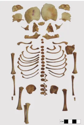
Fig. 3 Nyárlőrinc, ind. no. 14426, skeletal elements, mummified right forearm, and dorsum
Fig. 4 Nyárlőrinc, ind. no. 14426, right forearm, from two angles. The mummified parts and the bones of the forearm showgreen coloration from the coin offering
Fig. 5 Nyárlőrinc, ind. no. 14426, mummified skin from the dorsal region with five embedded vertebral bodies shown from two angles.
The mummified soft tissue inseparably sticks to the remains of the textile that the body was wrapped in
Results
Osteometrical data, calculated body lengths, and ages at death are presented in Table2.
Among the remains of no. 14426 (Fig.3), the right forearm (Fig.4) and mainly some skin from the dorsal area were mummified (Fig.5). No cranial elements were found except for the right side of the mandible. Big green-colored patches can be seen on some vertebrae, both radii, ulnae, ilia, femora, tibiae, and elements of the feet on both sides. The green color must derive from the strongly corroded copper coin found next to the remains.
On the basis of the bone dimensions, the age at death of the mummified pre-term individual was 6.5–7 lunar months. The body might have been approx. 28–35 cm long and weighed 0.4–0.91 kg. Our examination revealed no disease-related symptoms in the remains.
The ceramic pot that the individual was buried in (Fig.2) was 18.9 cm in height and 53.6 cm in circumference (maximum) with a volume of 2700 cm3. These dimensions correspond to the ethnographical definition of a pot (Bfazék^) by Igaz and Kresz (1965). Similar vessels have been typically used since the eighteenth century CE (Lajkó2015). No traces of glazing or other coating was present on the inner surface of the pot as an otherwise possible source of copper. According to ourμXRF measurement, the strongly corroded copper coin (Bkrajcár^, Fig.6) contained 99.6 % Cu, with Zn (0.207 %), Ni (0.171 %), and Fe (0.030 %) contaminants. Compared to the nominal weight of theBkrajcár,^this particular coin lost 0.7633 g due to the corrosion, 26.37 % of its original weight.
Fig. 7 Nyárlőrinc, ind. no. 10662, cranial and postcranial elements.
Some bones showgreen colorationfrom the coin offering
Fig. 8 Nyárlőrinc, ind. no. 14334, cranial and postcranial elements. No green colorationis visible; coin offering was not recovered
Fig. 6 An example of a well-preservedBKreuzer or krajcár^(a) recov- ered elsewhere, and the two coins recovered from our burials (b Nyárlőrinc, burial no. 14426;cNyárlőrinc, burial no. 10662)
Table 3 Cu concentration of bone in dry weight % (ICP-AES) Tested
specimen
Nyárlőrinc 14426
Nyárlőrinc 10662
Nyárlőrinc 14336
Scapula 0.066 0.930 0.00517
Femur 3.140 1.740 0.00435
Ileum 3.994 3.331 0.00669
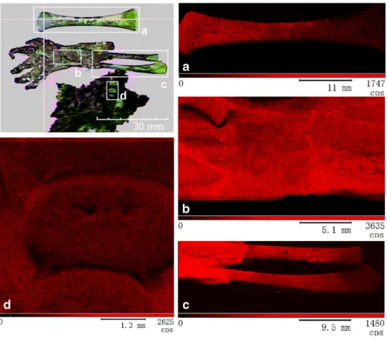
Fig. 9 The picture in thetop left cornerdesignates the areas (ato d) investigated byμXRF in the Nyárlőrinc, ind. no. 14426 item.
Insertsa–dshow the found distribution of Cu in the respective areas, such asathe left femur,bthe right palm,cthe right forearm (radius, ulna), anddthree lumbar vertebrae. The intensity of red colorin theμXRF images correlates with the Cu concentration on the surface of the sample
Fig. 10 A visual image of (top) and Cu distributions measured byμXRF (bottom) in the longitudinal and cross sections of the left femur of the Nyárlőrinc, ind. no. 14426 item. The intensity ofblue colorin theμXRF
images correlates with the Cu concentration. Please note that the Cu distribution found byμXRF shows the same pattern as thegreenCu decoloration in the visual image of the bone sections
The remains of ind. no. 10662 are skeletonized showing green coloration on the vertebrae, costae, and left side of the body (temporale, mandible, ilium, femur, tibia, fibula) (Fig.7). Age at death of the pre-term individual was 5.5–6.5 lunar months, the body might have been approx. 26–32 cm long and weighed 0.32–0.620 kg. The remains were also found in a ceramic pot with a copper coin, but the vessel cannot be reconstructed, and the release date of the coin is also indecipherable (Fig.6) since their state of preservation was very bad.
In the case of individual no. 14334, no green coloration can be seen on the skeletonized bones (Fig.8). Age at death was 7.5–8.5 lunar months; the body might have been approx. 35–
42 cm long and weighed 0.77–1.94 kg. Ceramic pot or copper coin was not recovered in the burial.
The copper concentrations found using ICP-AES in green- colored and normal bones of no. 14426 (mummified + pot + coin) were compared with copper concentrations found in no.
10662 (skeletonized + pot + coin) and no. 14336 (skeleton- ized−no pot−no coin). The mummified forearm was too small and brittle to be sampled, so we only analyzed identifi- able skeletonized parts instead. The measured data are sum- marized in Table3.
Cu concentration in the remains of ind. no. 14426 was mapped usingμXRF analysis in surfaces of mummified parts (Fig. 9), longitudinal and cross-sections (Fig. 10) of green bones.
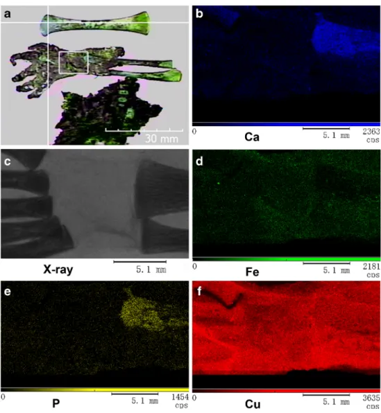
Besides mapping element concentrations, μXRF anal- ysis is also suitable for differentiating between soft and bony tissues of the mummified body parts: P and Ca Fig. 11 The picture in thetop left
cornerdesignates the area investigated by X-ray imaging andμXRF in the Nyárlőrinc, ind.
no. 14426 item.cthe X-ray image of the area, whereasb,d,e,fthe Ca, Fe, P, and Cu distributions as measured byμXRF. The intensity of the artificial coloring, identified at the bottom of eachμXRF im- age, correlates with the surface concentration of the respective element
Fig. 12 Nyárlőrinc, ind. no. 14426, right forearm with the copper coin in the palm
distributions in the X-ray picture clearly outline the shape of the bones within the organic mass of the remains (Fig.11). In our current paper, however, we are only an- alyzing Cu values in detail.
We have sampled the dry soil sediment containing the ce- ramic pot of burial no. 14426 and measured the Cu concen- tration three times ranging from 38.96 to 79.21 ppm with an average of 55.05 ppm. This value corresponds to the usual average Cu concentration in the soil (Adriano2001). This means that high Cu content of the remains is not a result of high background concentration.
Cu concentrations tend to be the highest in the mum- mified remains buried with a copper coin (no. 14426), values measured in the skeletonized remains buried with a coin (no. 10662) tend to be intermediate, and values of the skeletonized remains buried without a copper coin (no. 14336) are the lowest. Cu concentrations within the remains of one individual tend to be the highest in the ilium (possibly in the closest proximity of the coin), lower in the femur and scapula. Within bones of the lower limb, proximal epiphyses show higher concentrations than the distal epiphyses, and external surfaces also contain more Cu than the inside of bones.
Discussion and conclusion
Resources of fetal osteology usually address the issues of the status of these premature individuals (Fazekas and Kósa1978;
Kósa1989; Scheuer and Black2000; Baker et al.2005; White and Folkens2005; Huxley2010; Barfield2011). Depending on the clinical, forensic, or bioarcheological context, these works try to categorize pre-term human remains considering gestational age, developmental phase, measurements, and whether the child was viable or stillborn. But these works usually also acknowledge that the lack of any of these type of information may make it impossible to determine the status of the remains.
In our three cases, not a single bone element of the three children shows an age at death older than 8.5 lunar months.
This means all three children were underdeveloped for a nor- mal birth and they were not buried together with their mothers so we are presumably seeing a pre-term birth in all three cases.
However, neither the remains nor the archeological context provides any sufficient data to unequivocally determine the status of these very young individuals. Even though they were definitely born older than 20 lunar months, we do not know whether they were born dead or alive, stillborn or neonate. We are only sure that they definitely fall in some of the categories for perinatal death described by Barfield (2011). However, on the basis of what we know about the chances of survival as a preterm neonate before the twentieth century, it is very likely
all three of them died a couple days the latest after their pre- term birth.
The fate of stillborn babies and perinate or neonate deaths can follow different paths. According to ethnographical refer- ences (Pintér 1891; Bartucz 1928; Habenstein and Lamers 1960; Selmeczi1983), newborns who died without being bap- tized were rolled up in some sort of textile and buried in a pot (for example a milk-jug) or a small wooden box in abandoned cemeteries usually located close to ruins of medieval churches. Occasionally, low-value coins were put next to the body as offerings; the coins were intended to facilitate the soul’s admission to heaven or served to pay the fee on the way to the underworld or Saint John the Baptist for christen- ing the deceased in heaven (Dömötör1990; Selmeczi1992).
However, prior to this investigation, such traditions have not been documented in the archeological or osteological record in Hungary.
Literature references for Cu concentrations of mummified human remains range from 2.3 to 23.8 ppm (see references in Table1). In contrast with this, the 66.9 ppm value of our no- coin perinate (no. 14336) is almost three times higher than the upper limit of the reference range. In the presence of a copper coin, the skeletonized perinate (no. 10662) showed concentra- tions 495 times higher than those of the no-coin perinate, while the mummified individual (no. 14426) showed 497 times higher values.
Higher-than-background Cu concentration of the individu- al without coin (no. 14336) may simply be explained by the fact that remains of children younger than 3 years of age tend to show multiple times higher Cu values than adults (Lambert et al.1979). Several hundred times higher Cu values than average may be related to the corrosion of the coins included in the burials. Since Cu concentration of the soil sample is normal, and the examinable vessel (that of burial no. 14426) had no glazing or coating possibly containing copper, mum- mification must have been facilitated by copper deriving from theBkrajcár^coin.
Cu distribution in the remains is, however, far from even.
Differences of concentration must be in connection with the actual localization of the coin within the burial. Body parts in the closest proximity will show the highest concentrations and possibly (but not necessarily) a better level of preservation too. This is why the scapula of the non-mummified individual Nyárlőrinc no. 10662 shows a higher concentration than the scapula of the mummified Nyárlőrinc no. 14426. In case of individual no. 10662, the coin might have been located near the scapula. In the case of individual no. 14426, posture of the fingers and very high Cu concentration values around the hip refer to a copper coin having been placed in the right palm.
Nearby soft tissues (right hand and forearm, lower trunk, hips) were possibly exposed to the highest level of copper infiltra- tion in the entire study resulting in quasi-natural mummifica- tion of the affected body parts. As we move further away from
the corroded copper coin, superficial copper concentrations gradually decrease in the remains. The same is observed in the longitudinal and cross sections of bones: the closer prox- imity to the coin, the higher is the copper concentration, and bone surfaces always show higher concentration than the in- side of bones. These phenomena imply that individual no.
14426 was surrounded by a moist microenvironment in the pot, where gradual diffusion of soluble copper originating from the coin was facilitated for a longer period of time, and this relatively isolated setting did not allow complete decay of the little dead body.
Even though our comparative measurements show that high copper concentration in the microenvironment alone is not sufficient for complete mummification processes (consid- er the non-mummified individual no. 10662), a major cause of the mummification of individual no. 14426 was still the cor- rosion of the copper coin being placed next to the corpse in an almost closed ceramic vessel.
Copper as a mummifying agent is also rarely mentioned in the worldwide literature. Even the mummification process of the famousBCopper Man^ found in the hyperarid Atacama Desert is mostly attributed to desiccation, and copper ions had not penetrated soft tissues of the body beyond the depth of the skin (Aufderheide2003).
In contrast to this, our findings show copper concentrations in the inside of the bones several hundred times higher than normal. Despite the well-known antimicrobial and pro- mummification character of this element, copper-driven qua- si-natural mummification has not been documented in the in- ternational osteoarchaeological literature. Our perinate indi- vidual buried with a copper coin in the palm (Fig.12) gives a unique yet expressive example of this process. The Nyárlőrinc perinate burial no. 14426 may be the first copper-driven mummification case ever reported, and hope- fully, more cases are to appear in the future.
The analytical techniques applied in these perinatal cases (ICP-AES,μXRF) are a novelty in the osteoarcheological routine; thus, we hope our data will furnish new information in Central European subadult osteoarcheology.
References
Adriano DC (2001) Trace elements the terrestrial environment. Springer, New York
Aufderheide AC (2003) The scientific study of mummies. Cambridge University Press, Cambridge
Baker BJ, Dupras TL, Tocheri MW (2005) The osteology of infants and children. Texas A&M University Press, USA
Balázs J (2005) Paleopatológiai vizsgálatok egy XII-XVI. századi széria (Nyárlőrinc Hangár út) leletein. Diplomawork, University of Szeged, Department of Biological Anthropology
Balázs J, Bölkei Z (2007) Partly mummified foetus. VI World Congress on Mummy Studies Program and Abstracts, 277
Balázs J, Bölkei Z, V Székely G (2005) A Nyárlőrinc Hangár utcai széria embertani feldolgozásának eredményei. Cumania 21:57–82 Balázs J, Zádori PG, Vandulek C, Molnár E,Ősz B, Bereczki Z, Paja L,
Palkó A, Fogas O, Zink A, Nerlich A, Pálfi G (2015) Morphological and paleoradiological studies of Pott’s disease cases. Acta Biol Szeged 59(2):211–216
Barfield WD (2011) Clinical reports—standard terminology for fetal, infant, and perinatal deaths. Pediatrics 128:177–181
Bartucz L (1928) Köcsögbe temetés a régi palócoknál. Antropológiai Füzetek. Anthropol Hung III(1–3):19–21
Beckhoff B, Kanngießer B, Langhoff N, Wedell R, Wolff H (eds) (2006) Handbook of practical X-ray fluorescence analysis. Springer, Berlin Benfer RA, Typpo JT, Gaff GB, Pickett EE (1978) Mineral analysis of
ancient Peruvian hair. Am J Phys Antropol 48:277–282
Bochkor Á (1960) A Szent Jobb orvosi szemmel. Vigilia 1960(08):492– 494
Bölkei Z (2005) Embertani vizsgálatok egy középkori széria (Nyárlőrinc Hangár út) leletein. Diplomawork, University of Szeged, Department of Biological Anthropology
Carvalho ML, Casaca C, Pinheiro T, Marques JP, Chevallier P, Cunha AS (2000) Analysis of human teeth and bones from the chalcolithic period by X-ray spectometry. Nucl Inst Methods Phys Res B 168:
559–565
Cockburn A, Cockburn E, Reyman TA (1998) Mummies, disease &
ancient cultures, 2nd edn. Cambridge University Press, Cambridge Degryse P, Muchez P, De Cupere B, Van Neer W, Waelkens M (2004) Statistical treatment of trace element data from modern and ancient animal bone: evaluation of roman and byzantine environmental pol- lution. Anal Lett 37(13):2819–2834
Dömötör T (1990) Temetkezési szokások. In: Hoppál M (ed) Magyar Néprajz VII: népszokás, néphit, népi vallásosság. Akadémiai Kiadó, Budapest, pp 7–67
Farkas G (1972) Antropológiai Paraktikum I. Paleoantropológiai metodikák. József Attila Tudományegyetem, Szeged
Fazekas G, Kósa F (1978) Forensic fetal osteology. Akadémiai Kiadó, Budapest
Goffer Z (2007) Archaeological chemistry. Wiley, Hoboken
Habenstein RW, Lamers WM (1960) Funeral customs the world over.
Bulfin Printers, Milwaukee
Huxley AK (2010) Estimation of age from fetal remains. In: Latham KE, Finnegan M (eds) Age estimation of the human skeleton. Charles C Thomas Publisher, Sringfield, pp 147–160
Igaz M, Kresz M (1965) A népi cserépedények szakterminológiája.
Néprajzi Értesítő47:87–131
Józsa L (2006) Paleopathologia. Elődeink betegségei. Semmelweis Kiadó, Budapest
Kłys M, Lech T, Zieba-Palus J, Białka J (1999) A chemical and physico- chemical study of an Egyptian mummy‘Iset Iri Hetes’from the Ptolemaic period III-I B.C. Forensic Sci Int 99(3):217–228 Kósa F (1989) Age estimation from the foetal skeleton. In: Işcan MY (ed)
Age markers in the human skeleton. Charles C. Thomas Publisher, Springfield, pp 21–54
Kristóf LA (2015) Paleoradiológia: non-invazív módszertani lehetőség a történeti antropológiában. Doctoral Thesis, University of Szeged, Doctoral School in Biology
Lajkó O (2015) Cserepén ismerem, mineműfazék volt…Adatok a kora újkori edényművesség és a magyar népi kerámia eredetének kutatásához. Móra Ferenc Múzeum, Szeged
Lambert JB, Szpunar CB, Buikstra JE (1979) Chemical analysis of exca- vated human bone from middle and late Woodland sites.
Arheometry 21(2):115–129
Lynnerup N (2007) Mummies. Yearb. Phys Anthropol 50:162–190 Marcsik A, Molnár E, Szathmáry L (2006) The antiquity of tuberculosis in
Hungary: the skeletal evidence. Mem I Oswaldo Cruz 101(2):67–71
Molnár E, Dutour O, Pálfi G (1998) Diagnostic paléopathologique des tréponématoses: à propos d’un cas bien conservé. Bull Mém Soc Anthrop Paris 1-2(10):17–29
Nunnelley LL, Smythe WR, Trish JHV, Alfrey AC (1976) Trace elements analysis of tissue and resin from Egyptian mummy PUM II.
Paleopathol Newsl 12:12–14
Pálfi G, Panuel M, Molnár E (1997) Paleoradiologic study of a 17th century case of treponematosis (Nyárlőrinc, Hungary). Acta Biol Szeged 42:113–122
Pálfi G, Zádori P, Balázs J, Vandulek C, Kelemen K, Molnár E,Ősz B, Palkó A (2009) Paleoradiological studies of cases of Pott’s disease.
In: Pálfi G, Molnár E, Bereczki Z, Pap I (eds) Des lésions du passé aux diagnostics modernes. University Press, Szeged, pp 102–103 Pap I, Susa É, Józsa L (1997) Mummies from the 18-19th century
Dominican Church of Vác, Hungary. Acta Biol Szeged 42:107–112 Pintér S (1891) A palócz születése, házassága és halálozása.
Ethnographia 2:99
Quigley C (1998) Modern mummies: the preservation of the human body in the twentieth century. McFarland Publishers, Jefferson Rácz P (2013) Szent István ereklyéi. Rubicon 2014(6):21–25
Reyman TA, Barroco RA, Cockburn TA (1976) Histopathological exam- ination of an Egyptian mummy. Bull N Y Acad Med 52:506–516 Sandford MK, Kissling GE (1994) Multivariate analyses of elemental
hair concentrations from a medieval Nubian population. Am J Phys Anthropol 95(1):41–52
Sanford MK, Van Gerven DP, Meglen RR (1983) Elemental hair analysis:
new evidence on the etiology of cribra orbitalia in Sudanese Nubia.
Hum Biol 55:831–844
Scheuer L, Black S (2000) Developmental juvenile osteology. Elsevier Academic Press, London
Selmeczi L (1983) Négyszállási adatok a kereszteletlen gyerek eltemetéséhez. NyJAMÉ 24–26:177–180
Selmeczi L (1992) Régészeti - néprajzi tanulmányok a jászokról és a kunokról. Folkl Etnográfia 64:227–239
Shafer MM, Siker M, Overdier JT, Ramsl PC, Teschler-Nicola M, Farrell PM (2008) Enhanced methods for assessment of the trace element composition of Iron Age bone. Sci Total Environ 401(1–3):144–161 Susa É, Józsa L (1995) A múmiakészítés technikája és eredményei a
kezdetektől napjainkig. Anthrop Közl 37:45–60
Szpunar CB, Lambert JB, Buikstra JE (1978) Analysis of excavated bone by atomic absorption. Am J Phys Anthrop 48:199–202
Unger E (1997) Magyar éremhatározó–Ungarischer Münzbestimmer.
Magyar Éremgyűjtők Egyesülete, Ajtósi Dürer Könyvkiadó, Budapest
V Székely G (1987) Kun eredetűtárgyak és kulturális elemek Nyárlőrinc középkori temetőjében. Kézirat. Katona József Múzeum, Kecskemét
White TD, Folkens PA (2005) The human bone manual. Elsevier Academic Press, San Diego
Zlateva B, Djingova R, Kuleff I (2003) On the possibilities of ICP-AES for analysis of archaeological bones. Cent Eur J Chem 1(3):201–221