Physiology and Molecular Biology of Plants
Morphological, physiological and biochemical aspects of halophyte Petrosimonia triandra grown in natural habitat
--Manuscript Draft--
Manuscript Number: PMBP-D-19-00115
Full Title: Morphological, physiological and biochemical aspects of halophyte Petrosimonia triandra grown in natural habitat
Article Type: Research Article
Corresponding Author: Gyongyi Szekely
Universitatea Babes-Bolyai Cluj-Napoca, ROMANIA Corresponding Author Secondary
Information:
Corresponding Author's Institution: Universitatea Babes-Bolyai Corresponding Author's Secondary
Institution:
First Author: Dorina Podar
First Author Secondary Information:
Order of Authors: Dorina Podar
Kunigunda Macalik Kinga Reti
Ildiko Martonos Rahela Carpa Edina Torok Jolan Csiszar Gyongyi Szekely Order of Authors Secondary Information:
Funding Information: UEFISCDI-PN-II-RU-TE
(2014-4-0831) Dr. Gyongyi Szekely
Abstract: Salt tolerance mechanisms of halophyte Petrosimonia triandra, growing in its natural habitat in Cojocna commune, Cluj county, Romania, were investigated by analysis of biomass, growth parameters, water status, ion content, photosynthetic and
antioxidative system efficiency, proline accumulation and lipid degradation. Two sampling sites with different soil electrical conductivities (EC) were selected, site 1:
3.14 dS m-1 and site 2: 4.45 dS m-1. Higher salinity proved to have a positive effect on growth, the relative water content did not decline severely, the Na+ and K+ content of the roots, stem and leaves was larger, the function of photosynthetic apparatus and photosynthetic pigment content were not altered. The efficiency of antioxidative defence system was found to be assured by coordination of several reactive oxygen species scavengers. The presence of higher salinity, lead to accumulation of the osmolyte proline, while degradation of membrane lipids was reduced. As a whole, P.
triandra evolved different adaptational strategies to counteract soil salinity, which includes morphological and physiological adaptations, preservation of photosynthetic activity, development of an efficient antioxidative system and accumulation of the osmotic compound, proline.
Suggested Reviewers:
Morphological, physiological and biochemical aspects of halophyte Petrosimonia triandra grown in natural habitat
Abstract
Salt tolerance mechanisms of halophyte Petrosimonia triandra, growing in its natural habitat in Cojocna commune, Cluj county, Romania, were investigated by analysis of biomass, growth parameters, water status, ion content, photosynthetic and antioxidative system efficiency, proline accumulation and lipid degradation. Two sampling sites with different soil electrical conductivities (EC) were selected, site 1: 3.14 dS m-1 and site 2: 4.45 dS m-1. Higher salinity proved to have a positive effect on growth, the relative water content did not decline severely, the Na+ and K+ content of the roots, stem and leaves was larger, the function of photosynthetic apparatus and photosynthetic pigment content were not altered. The efficiency of antioxidative defence system was found to be assured by coordination of several reactive oxygen species scavengers. The presence of higher salinity, lead to accumulation of the osmolyte proline, while degradation of membrane lipids was reduced. As a whole, P. triandra evolved different adaptational strategies to counteract soil salinity, which includes morphological and physiological adaptations, preservation of photosynthetic activity, development of an efficient antioxidative system and accumulation of the osmotic compound, proline.
Keywords: Petrosimonia triandra, salinity, biomass, photosynthetic pigments, antioxidant, proline.
Introduction
Salinity is one of the most important abiotic stress factor reducing crop productivity and quality worldwide (Yamaguchi and Blumwald 2005; Okorogbona et al. 2015). Approximately 20% of the world’s cultivated and 33% of the irrigated land is affected by high levels of soil salinity, limiting plant growth and survival. Furthermore, salinized area increases yearly with 10% due to low precipitation, irrigation with saline water and poor agricultural practices. It is estimated that by 2050, 50% of the arable land would be salinized (Jamil et al. 2011; Shrivastava and Kumar 2015). Sodium (Na+) and chloride (Cl-) ions constitute the major ionic components of soils and seawater, but other ions, such as sulphate (SO42-) and calcium (Ca2+) play a role in salinity formation.
Uptake and overaccumulation of Na ions induce salt stress in many plant species disturbing normal growth and development. Concentrations of 150 - 400 mM NaCl in soil can affect the volume of intercellular spaces in leaves, the thickness of the whole leaf and of the palisade parenchyma and the size of the epidermal and spongy parenchyma cells (Molassiotis et al. 2006; Milic et al. 2013; Montero et al. 2018). Photosynthesis is negatively affected, because damaging
Blinded Manuscript Click here to access/download;Blinded Manuscript;Petrosim
manuscr_final_PMBP.docx
1 2 3 4 5 6 7 8 9 10 11 12 13 14 15 16 17 18 19 20 21 22 23 24 25 26 27 28 29 30 31 32 33 34 35 36 37 38 39 40 41 42 43 44 45 46 47 48 49 50 51 52 53 54 55 56 57 58 59 60
concentrations of Na+ and / or Cl- can accumulate in chloroplasts (Boyer 1976; Sudhir and Murthy 2004). Photosynthetic electron transport is relatively insensitive to salt, but the carbon metabolism or photophosphorylation is affected (Sudhir and Murthy 2004). The lipid and protein composition of plasma membranes are severely modified by salt accumulation leading to ion imbalance and hyperosmotic stress (Hayashi and Murata 1998; Parida and Das 2005). Osmotic effect happens when dissolved solutes near the plant root zone generate a low osmotic potential that decreases the soil water potential, which negatively affects the water balance of the plant. Under normal conditions, the cytosol of the plant cells contains 1 to 10 mM Na+ and100 to 200 mM K+ (Binzel et al. 1988; Blumwald et al. 2000), that allow the enzymes to function optimally. A high ratio of Na+ / K+ and high concentrations of total salt inactivate enzymes and inhibit protein synthesis (Blumwald et al. 2000). Osmotic stress induced by high salinity is typically accompanied by oxidative stress caused by the accumulation of reactive oxygen species (ROS) and consequently, an increase in ROS scavenging enzymes: superoxide dismutase (SOD), catalase (CAT), ascorbate peroxidase (APX), glutathione reductase (Parida et al. 2004; Yildiztugay et al. 2014; Kibria et al. 2017).
Halophyte species are considered extremophile plants for they are able to tolerate and complete their life cycle under a large scale of soil salinity. Halophytes present a series of anatomical and physiological adaptation mechanisms allowing them to counter the ion toxicity and water failure. Some halophytes are able to exclude salt from shoot meristems and leaves as they have developed salt glands on the surface of the leaves (Atriplex sp.) or accumulate Na+ and Cl- ions in the vacuole to avoid cytotoxicity (Taiz and Zeiger 1998). Halophytes, such as Suaeda maritima (Flowers 1972; Flowers and Colmer 2015) and Atriplex nummularia (de Araújo et al. 2006) show growth stimulation up to 300 mM Cl- levels, while Salicornia europaea, Atriplex leucoclada and Kochia scoparia show greater growth under increasing salt concentrations up to 500 mM NaCl (Mohammadi and Kardan 2015). Some halophytes are able to scavange cell detrimental ROS, by a series of osmolytes that ensure protection of the subcellular structures. Halophytes usually accumulate one dominant osmolyte, like proline, glycine betaine, sorbitol, β-alanine betaine, choline-O-sulphate or sugar (Tipirdamaz et al. 2006; Arbona et al. 2010; Lugan et al. 2010; Slama et al. 2015), but some halophytes accumulate more than one compatible solute (Gagneul et al. 2007).
Petrosimonia species, members of Chenopodiaceae family, are typical halophytes native to South East of Europe (Edmondson 1993; Grigore et al. 2012; Todorova et al. 2014) and regionally in Asia (Breckle et al. 2011; Zörb et al. 2013).
All Petrosimonia species identified are living in saline soils with 2 - 28 dS m-1 EC (Grigore et al. 2012; Zörb et al. 2013;
Todorova et al. 2014), reaching up to 0.5 m height in arid areas. Although very little information is published about this species, it has been proposed as an antioxidant source for the expanding food crisis (Grigore and Oprica 2015). P. triandra also represents an interesting species with Kranz anatomy pattern in its stem, making it an attractive tool for plant 1
2 3 4 5 6 7 8 9 10 11 12 13 14 15 16 17 18 19 20 21 22 23 24 25 26 27 28 29 30 31 32 33 34 35 36 37 38 39 40 41 42 43 44 45 46 47 48 49 50 51 52 53 54 55 56 57 58 59 60
anatomical studies, since it is the only Romanian Petrosimonia species with this specificity (Grigore et al. 2012). Because of its salty taste, P. triandra is often consumed, by local people, in salads. Like other halophytes, based on its salt accumulation capacity, it could also be used as a biodesalinating plant, for the purpose of expanding the arable lands and generate pastures. Halophyte crop production represents a major trend in the development and use of territories hardly suitable for agriculture due to the shortage or full absence of freshwater. Halophytes are an alternative source of feeds, grains, grass forage, and medicinal raw materials, as well as a means of reconstructing the plant cover and improve the biological productivity of degraded pasture lands (Shamsutdinov et al. 2017). The ecological importance consists in the non-monetary value of the presence of halophytic vegetation, which has a protective role against soil erosion, ensures the habitat of some animal species, and represents an insular living environment in the Transylvanian Plain.
In the current study, we aimed to investigate the adaptation mechanisms of P. triandra to salt stress by comparing morphological, physiological and biochemical parameters of plants grown in their natural habitat, in soils with different salinities in Cojocna commune, Cluj County, Romania. Characterization of P. triandra contributes to the understanding of salinity tolerance mechanism of halophytes and can provide a substantial advantage for the extension of arable land for crop species in Europe and especially in the Carpathian Basin.
Materials and methods
Plant material sampling site
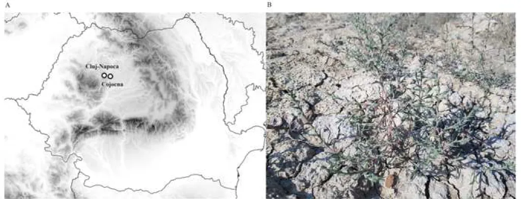
Cojocna commune from Cluj County, Romania (fig. 1A), is located in Fizeş Plain, covers an area of 238 km2, with a population of 4400 inhabitants. The wide and flat meadow, marked by crust and saline efflorescences, from the eastern edge of the Cojocna commune, outlines an important massive salt in the basement. On the surface occupied by medium- sized Miocene deposits, erosion created wide valleys, accompanied by numerous salt springs, sapropelic mud plateaus, salty and halophyte vegetation. The climate is temperate-continental, characterized by warm summers and cold winters.
The average annual temperature is 9.6 oC with a multiannual minimum of -15 oC and a maximum of +30 oC in July / August. Annual average rainfall is 886 mm.
Two sampling sites from Cojocna (Cluj County, Romania, coordinates for 3.14 dS m-1 soil electrical conductivity:
N46.74319 E23.84290 – site 1, and for 4.45 dS m-1 soil electrical conductivity: N46.74328 E23.84295 – site 2) were marked and used for analysis of Petrosimonia triandra (Pall.) Simonk. plants growing there in the summer of 2016 (fig. 1B). The 3.14 dS m-1 and 4.45 dS m-1 values correspond to a moderate saline soil (Marcar and Crawford, 2004). The sampling sites were chosen deliberately to represent different soil salinity levels. The analyzed area and plant species were chosen based 1
2 3 4 5 6 7 8 9 10 11 12 13 14 15 16 17 18 19 20 21 22 23 24 25 26 27 28 29 30 31 32 33 34 35 36 37 38 39 40 41 42 43 44 45 46 47 48 49 50 51 52 53 54 55 56 57 58 59 60
on our earlier results from both soil and plant samples collected in summer of 2014 and 2015. The measured parameters showed the same tendency as in the presented data referring to year of 2016.
Determination of morphological and growth parameters
At each sampling site 10 plants were chosen for the analysis of morphological and growth parameters. The plants were carefully dug out to maintain their root intact. Length measurements were done using a millimetre ruler. Primary root length was measured from the base of the root to its deeper point, shoot length was measured from the base of the shoot to its highest point. For leaf area measurements, the leaves were excised, placed onto millimetre paper and photographed.
ImageJ program was used to quantify the leaf area of the photographed leaves. Analytical scale (G&G, Electronic scale JJ200B) was used to measure the fresh weight of the leaves, stems, and roots right after digging out the plants and immediately after the excision of the plant organs. The samples were carried into the laboratory, oven dried in paper bags at 70oC for 48 h, and then the dry weight of leaves, stems and roots were measured. The ratio of root and shoot (stem+leaf) fresh (FW) and dry (DW) weights were calculated.
Measurement of water relations
For turgid weight measurement, the leaf fresh weight (LFW) was recorded immediately after field sampling, then, the leaves were immersed into distilled water and kept at room temperature. After 24 h of soaking, the leaves were water dried by blotting onto filter paper and leaf turgid weight (LTW) was recorded. Leaf dry weight (LDW) was measured upon incubation at 70oC for 48h. Relative water content was calculated as follows: RWC% = (LFW – LDW / LTW - LDW) x 100.
Determination of plant ion content
The ion-chromatographic analysis was performed at room temperature (21±2°C) using Dionex ICS-1500 system equipped with a temperature compensated conductivity cell (Cataldi et al. 2003). The ion separation was carried out with two different ion-exchange columns: one for cations (Na+, NH4+, K+, Mg2+, Ca2+), another for anions (F-, Cl-, NO2-, Br-, NO3-, PO43-, SO42). Upon harvest, the plant samples were rinsed three times in distilled water, blotted on filter paper (Cataldi et al. 2003), and then the roots were separated from the stems. After separation, the samples were quickly frozen in liquid nitrogen, then ground into powder and stored in airtight vials at room temperature. In order to determine the content of anions and cations, the obtained tissue powder (100 mg) was mixed with ultra-pure water (15 ml), the suspension was shacked for 15 min to facilitate contact between the plant tissue and extracting solvent (ultrapure water) and then 1
2 3 4 5 6 7 8 9 10 11 12 13 14 15 16 17 18 19 20 21 22 23 24 25 26 27 28 29 30 31 32 33 34 35 36 37 38 39 40 41 42 43 44 45 46 47 48 49 50 51 52 53 54 55 56 57 58 59 60
centrifuged at 14000 rpm for 15 min (centrifuge type: Sigma, rotor 12124 PP 399/F). The samples were filtered through single-use MCE 0.45 μm filters to remove any particulate matter and analysed with ion-chromatograph.
In order to determine the NaCl content of the soil samples, we considered the concentration of Na+. The NaCl content of the soil were calculated from the measured IC (Ion Chromatography values) and was converted to mM L-1 using the molecular weight (atomic mass) data. Thus, the soil NaCl in the first sampling site (EC=3.14 dS m-1) was 24.35 mM L-1 while in the second site (EC= 4.45 dS m-1) 41.35 mM L-1, respectively. Therefore, the difference in NaCl content between the two sampling sites is 70%.
Determination of photosystem II efficiency and of photosynthetic pigment content
Quantum yield, a measure of the photosystem II efficiency (PSII) equivalent, in light adapted leaves, to Fv’/Fm’ was determined by a portable FluorPen FP100 (Ptushenko et al. 2014). Leaves were light adapted, the PAR (photosynthetic active radiation) value was 1700-1800 mmol m-2 s-1. Chlorophyll and carotenoid content were determined according to the method described by Lichtenthaler (1987). Leaves (0.1 g) were grinded, then 2 ml 100% acetone was added and kept at 4oC for 24 h. The homogenate was centrifuged at 15000 g for 5 min, and the supernatant was treated with 80% acetone for 24 h. The optical density was measured by a VWR UV-1600PC spectrophotometer at 646.8 and 663.2 nm for Chl a + b determination, and 470 nm for carotenoid determination. The concentration of chlorophyll was calculated according to the equations: Chl a = 12.21 A663.2 - 2.81 A646.8, Chl b = 20.13 A646.8 - 5.03 A663.2 and Chl a + b = 7.15 A663.2 + 18.71 A646.8. The concentration of carotenoids was calculated according to the equation: (1000 A470 ‐ 3.27 Chl a - 104 Chl b) / 227.
Quantification of reactive oxygen species detoxifying enzyme activities
For antioxidative enzyme assays, field collected leaves (1 g) were homogenized in a reaction mixture containing 40 mg polyvinyl pyrrolidone (PVP) and 4 ml of 0.1 M phosphate buffer (pH 7.0) with 0.1 mM EDTA. The homogenate was centrifuged and the supernatant was used for determination of different enzymes activities. Soluble protein concentration was determined by the method of Bradford (1976). The methods used to measure the activity of different enzymes (superoxide dismutase, catalase, ascorbate peroxidase, guaiacol peroxidase, glutathione transferase) were published earlier by Székely et al. (2008).
Measurement of free proline content and of lipid peroxidation
Free proline content was measured in the field collected leaf tissues by colorimetric assay according to the method described by Bates (1973) and modified by Ábrahám et al. (2003). The end product of lipid peroxidation, malondialdehyde (MDA) was measured by thiobarbituric acid (TBA) test (Heath and Parker 1968). Field collected leaf tissues (0.1 g) were 1
2 3 4 5 6 7 8 9 10 11 12 13 14 15 16 17 18 19 20 21 22 23 24 25 26 27 28 29 30 31 32 33 34 35 36 37 38 39 40 41 42 43 44 45 46 47 48 49 50 51 52 53 54 55 56 57 58 59 60
homogenized in 0.5 ml of 20% w/v TCA solution. The homogenate was centrifuged at 15000 g for 5 min at 4oC, and then 0.5 ml of 1% w/v TBA was added to the supernatant. The mixture was incubated at 100oC for 30 min, and then the tubes were placed on ice to stop the reaction. Samples were centrifuged at 10000 g for 5 min, and the absorbance of supernatant (where some shade of pink MDA-TBA complex was formed) was measured at 535 and 730 nm. The intensity of the pink pigment indicates the extent of lipid peroxidation. The amount of MDA–TBA complex was calculated according to the equation: X (%) = 100 x (OD535 - OD730).
Statistical analysis
Statistical analysis was performed using the Welch Two Sample t-test in RStudio statistical software (v1.1.423), where
*p= 0.05-0.01, **p = 0.01-0.001 and ***p < 0.001.
Results
Effect of salinity on growth of Petrosimonia triandra plants
In order to assess morphological responses of P. triandra to salinity, plants grown in their natural environment in Cojocna commune, Cluj County, Romania (fig. 1). Growth parameters of P. triandra plants grown under two different salinities were investigated. The soil EC at the two sampling sites were 3.14 and 4.45 dS m-1, corresponding to approximately 24.35 mM (site 1) and 41.35 mM (site 2) NaCl in soil, respectively. Despite of the higher soil salinity at site 2, P. triandra plants grown there were more vigorous than those grown at site 1. The fresh and dry biomass of the above- and underground organs of P. triandra plants grown in higher salinity were between 21 and 35% significantly larger than of those grown in lower salinity (Table 1). While fresh weight biomass of roots, stem and leaves were by 21-30% larger, the dry biomass differences were 27-35% larger in plants grown in higher salinity compared to those in site 1, with lower soil salinity (Table 1). The enhancement in fresh and dry biomass of leaves was 30% and 35%, the fresh and dry biomass of stems was larger by 27% and 32%, while the root fresh and dry biomass increased with 21 and 33%, respectively (Table 1). Root to shoot fresh or dry weight ratios displayed no statistically significant differences between the two sampling sites (Table 1). The length of the primary root, although 24% larger, was not statistically significant in plants grown at 4.45 dS m-1 EC of soil compared to those grown at 3.14 dS m-1 (Table 2). Nonetheless, plant height and total leaf area were by 29% and 69%, respectively, significantly larger in plants grown in higher salinity (Table 2).
Accumulation of mineral ions in Petrosimonia triandra plants grown in soils with different salinities 1
2 3 4 5 6 7 8 9 10 11 12 13 14 15 16 17 18 19 20 21 22 23 24 25 26 27 28 29 30 31 32 33 34 35 36 37 38 39 40 41 42 43 44 45 46 47 48 49 50 51 52 53 54 55 56 57 58 59 60
The influence of salt on mineral ion distribution in different organs are presented in Table 3. The levels of Na+ and K+ cations were larger in roots (Na+: 51%, K+: 55%), stems (Na+: 5%, K+: 51%) and leaves (Na+ and K+ 30%) of plants grown at higher salinity in comparison to those grown at lower salinity. The levels of Ca2+ and Mg2+ exceeded in roots (Ca2+: 440%, Mg2+: 354%) and stems (Ca2+: 240%, Mg2+: 58%), but decreased in leaves (Ca2+: 160%, Mg2+: 74%) of plants grown at higher soil salinity. The level of Cl- anion increased in roots (28%), stems (6%) and leaves (11%) in presence of higher EC (4.45 dS m-1 S). The Na+ / K+ ratio decreased in stems by 43% and remained unchanged in roots and leaves (Table 3).
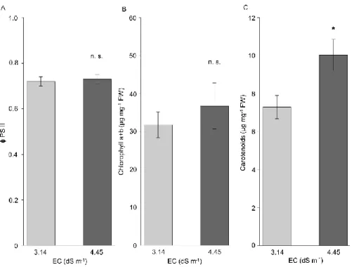
PSII efficiency and photosynthetic pigment content in plants grown under different soil salinity
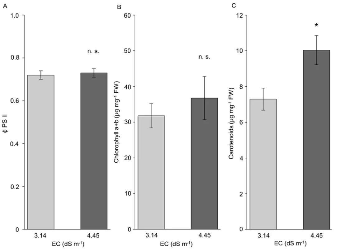
The quantum yield of PSII (Fv’/Fm’) of plants, showed constant values around 72% in light adapted leaves (fig. 2A) in both sites of P. triandra habitat. Regarding the photosynthetic pigments, only carotenoids (fig. 2C), not chlorophyll a and b (fig. 2B) displayed larger accumulation in leaves of plants grown at higher soil salinity. The accumulation of carotenoids in P. triandra was 37% significantly higher in leaves of plants grown in the presence of 4.45 dS m-1 EC compared to those of plants living at 3.14 dS m-1 EC.
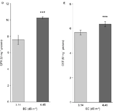
Effect of different salinity on the oxidative defence system
The activity of plant enzymes important for stress tolerance: superoxide dismutase (SOD), catalase (CAT), ascorbate peroxidase (APX), guaiacol peroxidase (GPX) and glutathione transferase (GST) was investigated in leaves of field living plants. The activity of all investigated enzymes was statistically significantly increased in plants grown at higher salinity in comparison to those living at lower soil salinity (fig. 3). SOD and CAT activity levels were 50% and 60% higher, respectively (figs. 3A, B), APX and GPX activity levels were greater by 30-40% (figs. 3C, D), whereas GST activity was 10% increased (fig. 3E) in the presence of elevated soil EC. These findings suggest that P. triandra possesses an efficient antioxidative system, which is activated by elevated salinity and is able to scavenge the harmful ROS during oxidative stress caused by high salinity.
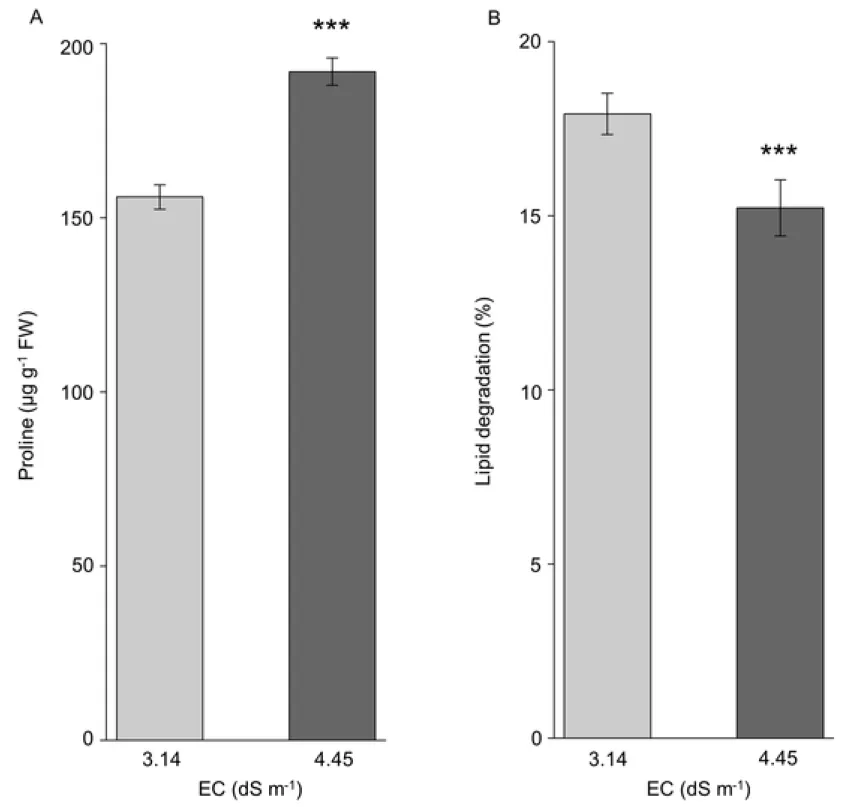
Effect of different salinity on proline and lipid peroxidation levels
The level of free proline in P. triandra leaves was significantly increased by 20% (fig. 4A) in the presence of higher soil salinity. To quantify the lipid degradation, as an effect of oxidative damage, we measured the level of malondialdehyde (MDA), as the end product of lipid peroxidation. MDA was used as an indicator of membrane lipid peroxidation. The level of lipid degradation was 18% significantly lower in plants grown under higher (salinity 4.45 dS m-1 EC) than under lesser soil salinity (3.14 dS m-1) (fig. 4B).
1 2 3 4 5 6 7 8 9 10 11 12 13 14 15 16 17 18 19 20 21 22 23 24 25 26 27 28 29 30 31 32 33 34 35 36 37 38 39 40 41 42 43 44 45 46 47 48 49 50 51 52 53 54 55 56 57 58 59 60
Discussion
Salinization of arable soils is of real concern for it negatively affects cultivated plants, thus posing a risk to food production.
By definition, a saline soil is one in which the electrical conductivity (EC) of the saturation extract in the root zone exceeds 4 dS m-1 (decisiemens per meter), where 4 dS m-1 corresponds to approximately 40 mM NaCl or more at 25oC (Richards 1954; Amacher et al. 2000; Marcar and Crawford 2004; Chinnusamy et al. 2005). Halophyte species could be used in saline soils as they are able to grow and develop under such conditions. In our study, Petrosimonia triandra, a halophyte member of the Chenopodiaceae family, showed enhacement of growth under higher (4.45 dS m-1 EC) in comparison to lower (3.14 dS m-1 EC) soil salinity conditions in its natural environment. The increase in the length and surface area of the aboveground plant organs are in accordance to the increase in their biomass (Table 1), suggesting that the tolerance response of this plant species to high salt is to develop its aerial parts rather than the underground organs. This is not surprising, given that other members of the Chenopodiaceae family like Salsola soda can grow and tolerate even higher soil salinity (EC ∼ 10 dS m-1 or above) (Centofanti and Banuelos 2015; Karakas et al. 2017). Study of six halophyte species (Suaeda maritima, Sesuvium portulacastrum, Clerodendron inerme, Ipomoea pes-caprae, Heliotropium curassavicum and Excoecaria agallocha) which are able to accumulate salt, showed that S. maritima and S. portulacastrum decreased the EC of soil from 4.9 to 1.4 and 2.5 dS m-1, respectively, demonstrating their potential for soil desalinization purposes (Ravindran et al. 2007). Apart from salt stress, halophytes have to be able to cope with water stress, too, for that the presence of high salts in the soil imped water absorbtion. In the current study, the relative water content (RWC) of the leaves of P. triandra was lower only by 17% in plants grown at 4.45 dS m-1 EC, but remained around the value of 70%, showing that the salt stress conditions do not severely affect the plants water status (Table 2). P. triandra is able to maintain a high RWC (~70%) when encounters salt stress, demonstrating that plants possess an efficient mechanism to adjust cell cytosol osmotically, and to absorb enough water from the soil.
The concentrations of Na+ and K+ in plants is an indicator of plant tolerance to salt stress. Na+ accumulation in leaves can repress photosynthetic enzymes and carbohydrate metabolism, process which induce oxidative damage and cell death (Chaves et al. 2009; Iseki et al. 2016), pollen sterility and delayed flowering in Cicer arietinum (Pushpavalli et al.
2016). Plants have developed a series of salt tolerance mechanisms to avoid these damaging effects. They exclude Na+ from roots to reduce uptake and sequester it into vacuoles to protect cytosolic enzymes (Munns and Tester 2008), thus establishing a low cytosolic Na+ / K+ ratio, with important role for physiological activities and growth under salt stress (Yamaguchi and Blumwald 2005). In our study, 30-51% larger concentrations of Na+ in roots and leaves of plants grown under higher soil salinity were accompanied by equally elevated concentrations of K+ ions. Consequently, the Na+ / K+ 1
2 3 4 5 6 7 8 9 10 11 12 13 14 15 16 17 18 19 20 21 22 23 24 25 26 27 28 29 30 31 32 33 34 35 36 37 38 39 40 41 42 43 44 45 46 47 48 49 50 51 52 53 54 55 56 57 58 59 60
ratio was 43% lower in stems, but showed no difference in roots and leaves (Table 3) of P. triandra plants living in higher soil salinity compared to those from lower soil salinity. This suggests that, in order to maintain the Na+ / K+ ratio in leaves, the plants living under higher soil salinity conditions may intensify the K+ transport through the stem. The capacity to reduce Na+ / K+ ratio in the presence of higher soil salt content, indicates the ability to evolve an efficient salt tolerance mechanism. In accordance with the present study, Karakas et al. (2017) observed the accumulation of Na+ in leaves of S.
soda and Portulaca oleracea under increasing soil salinity content. With respect to other cations, such as Mg2+ and Ca2+, in the current study, they accumulated rather in roots than in stem or leaves, which may suggest the presence of an efficient exclusion mechanism at root level developed to avoid excess accumulation of these ions in the leaves.
High EC soil values did not affect significantly the quantum yield (Fv’/Fm’) of plants, showing constant values around 72% in light adapted leaves (fig. 2A). Therefore, oxidative stress induced by salt conditions, did not alter the function of PSII. Significantly increased accumulation of photosynthetic pigments was observed only for carotenoids (fig.
2C). Carotenoids take part in dissipating excessive excitation energy in PSI and PSII, and in reducing the formation of harmful singlet oxygen by scavenging it and quenching the excited triplet state of chlorophyll. They also stabilize chloroplast membranes (Li et al. 2010). Significantly increased concentration of carotenoids in P. triandra leaves (by 37%) in the presence of 4.45 dS m-1 EC compared to plants living at 3.14 dS m-1 EC (fig. 2C), corroborates with their antioxidative role i.e. the protection against photooxidation produced by accumulation of harmful ROS (Das and Roychoudhury 2014).
Furthermore, a series of studies (Rabhi et al. 2012; Zouhaier et al. 2015; Muchate et al. 2016) documented an altering tendency of chlorophylls and carotenoids content depending on plant species and salt content in the growth media. Fiedor and Burda (2014) inferred that a high carotenoid content in the pigment complex may play a role in the protection of chloroplasts from photooxidation and from the deleterious effect of free radicals. Antioxidant role of carotenoids was also documented by Viljanen et al. (2002) and Li et al. (2010), thus, in our study, greater carotenoids concentrations in leaves of P. triandra growing in higher soil salt suggests that the pigments act as an efficient protective photooxidative mechanisms.
Accumulation of ROS in plants causes a set of harmful effects in the cell. To scavenge the ROS, plants possess an effective antioxidative system, which involves also several enzymes. Superoxide dismutases are metal containing enzymes that together with catalases, are extremely important components in catalysing the dismutation of superoxide radicals into molecular oxygen and water, thus protecting the cell from oxidative damage. In response to salinity several halophytes accumulate SOD (Parida et al. 2004; Amor et al. 2005; Benzarti et al. 2012; Amjad et al. 2015), but salinity did not induce SOD accumulation in Salvadora persica (Rangani et al. 2016). In the current study, the largest measured increments of SOD and CAT among ROS enzymes in P. triandra under higher salinity (4.45 dS m-1), suggest that these 1
2 3 4 5 6 7 8 9 10 11 12 13 14 15 16 17 18 19 20 21 22 23 24 25 26 27 28 29 30 31 32 33 34 35 36 37 38 39 40 41 42 43 44 45 46 47 48 49 50 51 52 53 54 55 56 57 58 59 60
two enzymes might play a substantial role in conferring P. triandra resistance to superoxide radicals under salt stress.
Glutathione transferases may protect plants from oxidative injury by functioning as detoxifying enzymes and also have glutathione peroxidase activity thus being important in the prevention of lipid peroxidation (Roxas et al. 1997; Cummins et al. 1999). The ascorbate peroxidases are components of ascorbate-glutathione cycle, where APX enzymes are key component in catalysing the conversion of hydrogen peroxide into water, using ascorbate as electron donor. Enhanced level of SOD and APX activity in P. triandra plants, could be indicators of the elevated ROS levels stimulated by higher salinity. The same tendency was documented by Benzarti et al. (2012) in the halophyte Atriplex portulacoides and Parida et al. (2004) in the mangrove Bruguiera parviflora. NaCl increased the SOD and GPX activity, but decreased the APX activity in Salsola crassa (Yildiztugay et al. 2014) demonstrating that different species choose alternative strategies to adapt to salinity stress. Our results suggest that especially SOD and CAT, but together with APX and GPX play a pivotal protective role against the oxidative stress caused by high salinity, and these enzymes prevent ROS accumulation and thus inhibition or damage of PSII.
Proline accumulation is one of the common physiological responses in higher plants exposed to drought and salinity (Khedr et al. 2003; Székely et al. 2008; Kishor and Sreenivasulu 2014; Per et al. 2017). Proline functions as a possible molecular chaperone in osmotic adjustment and protection of cellular structures, proteins and membranes during osmotic stress in plants (Delauney and Verma 1993; Abid et al. 2018; Zhang and Shi 2018). Proline protects the protein- lipid components of the cell membranes during water stress (Franko and Melo 2000; Szabados and Savoure 2010). It can act as antioxidant, energy metabolism factor and metabolic signal (Anjum et al. 2000; Kartashov 2013). Is able to regulate the metabolite pool and gene expression, and affects plant growth and development (Szabados and Savoure 2010; Eva et al. 2016). Proline is also involved in diminishing photodamage in thylakoidal membranes by scavenging and / or reducing MDA concentration and ROS detoxifying enzymes (Reddy et al. 2004). Proline accumulation in the presence of salinity stress is a well-documented strategy of plants to counteract this adverse environmental condition. Many physiological measurements of different species emphasized an elevated proline level as a response to salt stress (Hong et al. 2000;
Székely et al. 2008; Szabados and Savoure 2010). Our study shows that under higher soil salinity P. triandra plants displayed a significant increase in the accumulation of free proline compared to lower soil salinity conditions (fig 4A).
This result is in accordance with Karakas et al. (2017), who also reported elevation of proline levels in S. soda and P.
oleracea with increasing EC content of soil in greenhouse experiments. The evidence that RWC did not changed significantly in our analysis, shows that accumulation of proline is not a pivotal factor in the control of water status of plants. Moreover, lipid degradation was significantly lower in plants grown under higher than lower soil salinity conditions.
This suggests that there are no evident signs of oxidative damage in the cell membrane of P. triandra leaves (fig. 4B), and 1
2 3 4 5 6 7 8 9 10 11 12 13 14 15 16 17 18 19 20 21 22 23 24 25 26 27 28 29 30 31 32 33 34 35 36 37 38 39 40 41 42 43 44 45 46 47 48 49 50 51 52 53 54 55 56 57 58 59 60
this species possesses an efficient antioxidative defence system in order to scavenge the accumulation of ROS. Same results were obtained by Karakas et al. (2017) and Rangani et al. (2016) in greenhouse grown halophytes S. soda and S. persica.
With respect to glycophytes, several authors documented increase in lipid peroxidation as an effect of salinity stress e.g.
Vigna radiata (Nazar et al. 2011), Oryza sativa (Shobbar et al. 2012), Solanum melongena (Shaheen et al. 2013) and A.
thaliana (Rejeb et al. 2015).
Our future studies on the salt adaptation mechanisms of P. triandra will focus on the expression pattern of salt stress regulated genes, which can bring us closer to the salt tolerance strategies of halophytes.
Conclusions
Based on current results, i.e. biomass enhancement under the effect of elevated soil salinity, we suggest that P. triandra not only tolerates, but needs salt for optimal growth and physiological processes in its natural habitat. P. triandra acts as a real halophyte with diverse adaptation strategies to avoid the harmful effect of high soil salinity. Its adaptation mechanisms include physiological adaptations, maintenance of PSII functional integrity and thus, photosynthetic activity, activation of efficient antioxidative system, accumulation of osmolyte proline and reduce lipid peroxidation. Understanding the mechanisms of salt adaptation of P. triandra, could be of great importance, possibly leading to the extension of the arable area of the crop plants by exploiting the biodesalinating capability of this halophyte in Europe and especially in the Transylvanian Plain.
Figure legends
Table 1 Biomass of Petrosimonia triandra plants grown under different soil salt concentrations. EC: soil electrical conductivity, FW: fresh weight, DW: dry weight. The values are mean ± SD (n = 10), significant values p ≤ 0.05. Significant different means, between the two sampling sites, according to Welch's t test are marked with *p = 0.05-0.01 and **p = 0.01-0.001.
Table 2 Growth parameters (plant height, leaf area, root length) and relative water content (RWC%) of Petrosimonia triandra plants grown in soils with different salinity. EC: soil electrical conductivity. The values are mean ± SD (n = 10).
Significant different means, between the two sampling sites, according to Welch's t test are marked with *p = 0.05-0.01.
Table 3 Accumulation of mineral ions in Petrosimonia triandra plants grown under different soil salt content. EC: soil electrical conductivity.
Figure 1 (A) Location of study area in Cojocna, Cluj County, Romania and (B) Petrosimonia triandra plant.
1 2 3 4 5 6 7 8 9 10 11 12 13 14 15 16 17 18 19 20 21 22 23 24 25 26 27 28 29 30 31 32 33 34 35 36 37 38 39 40 41 42 43 44 45 46 47 48 49 50 51 52 53 54 55 56 57 58 59 60
Figure 2 PSII efficiency and leaf photosynthetic pigment content of Petrosimonia triandra grown in natural habitat, in soils with different salinity. (A) PSII efficiency, (B) chlorophyll a+b, (C) carotenoids. Bars represent mean values ± SD (n
= 10, ns = not significant according to Welch's t test).
Figure 3 Specific activity of ROS detoxifying enzymes in Petrosimonia triandra plants grown in soils with different salinity. (A) Superoxide dismutase (SOD), (B) Catalase (CAT), (C) Ascorbate peroxidase (APX), (D) Guaiacol peroxidase (GPX) and (E) Glutathione transferase (GST). Bars represent mean values ± SD (n = 10). Significant different means according to Welch's t test are marked with **p = 0.01-0.001 and ***p < 0.001.
Figure 4 (A) Proline content and (B) Lipid degradation in Petrosimonia triandra leaves in the presence of two different saline conditions. Bars represent mean values ± SD (n = 10). Significant different means according to Welch's t test are marked with ***p < 0.001.
References
Abid M, Ali S, Qi LK, Zahoor R, Tian Z, Jiang D, Snider JL, Dai T (2018) Physiological and biochemical changes during drought and recovery periods at tillering and jointing stages in wheat (Triticum aestivum L.). Sci. Rep. 8, 4615. https://doi.org/10.1038/s41598-018-21441-7
Ábrahám E, Rigó G, Székely G, Nagy R, Koncz C, Szabados L (2003) Light-dependent induction of proline biosynthesis by abscisic acid and salt stress is inhibited by brassinosteroid in Arabidopsis. Plant Mol. Biol. 51:363-72.
https://doi.org/10.1023/A:1022043000516
Amacher JK, Koenig R, Kitchen B (2000) Salinity and plant tolerance. All archived publications. Paper 43.
https://digitalcommons.usu.edu/extension_histall/43
Amjad M, Akhtar SS, Yang A, Akhtar J, Jacobsen SE. 2015. Antioxidative response of quinoa exposed to iso-osmotic Ionic and Non-Ionic Salt Stress. J. Agro. Crop. Sci. 201, 452–460. http://doi.org/10.1111/jac.12140
Amor NB, Hamed KB, Debez A, Grignon C, Abdelly C (2005) Physiological and antioxidant responses of the perennial
halophyte Crithmum maritimum to salinity. Plant Sci. 168:889–899.
https://doi.org/10.1016/j.plantsci.2004.11.002
Anjum F, Rishi V, Ahmad F (2000) Compatibility of osmolytes with Gibbs energy of stabilization of proteins. Bioch.
Bioph. Acta. 1476:75-84. https://doi.org/10.1016/S0167-4838(99)00215-0 1
2 3 4 5 6 7 8 9 10 11 12 13 14 15 16 17 18 19 20 21 22 23 24 25 26 27 28 29 30 31 32 33 34 35 36 37 38 39 40 41 42 43 44 45 46 47 48 49 50 51 52 53 54 55 56 57 58 59 60
de Araújo SAM, Silveira JAG, Almeida TD, Rocha IMA, Morais DL,Viégas RA (2006) Salinity tolerance of halophyte Atriplex nummularia L. grown under increasing NaCl levels. R. Bras. Eng. Agríc. Ambiental. 10:848-854.
http://dx.doi.org/10.1590/S1415-43662006000400010
Arbona V, Argamasilla R, Gomez-Cadenas A (2010) Common and divergent physiological, hormonal and metabolic responses of Arabidopsis thaliana and Thellungiella halophila to water and salt stress. J. Plant Physiol. 167:1342–
1350. https://doi.org/10.1016/j.jplph.2010.05.012
Bates LS (1973) Rapid determination of free proline content for water-stress studies. Plant Soil. 39:205-207.
https://doi.org/10.1007/BF00018060
Benzarti M, Rejeb KB, Debez A, Messedi D, Abdelly C (2012) Photosynthetic activity and leaf antioxidative responses of Atriplex portulacoides subjected to extreme salinity. Acta Physiol. Plant. 34:1679–1688.
http://doi.org/10.1007/s11738-012-0963-5
Binzel ML, Hess FD, Bressan RA, Hasegawa PM (1998) Intracellular compartmentation of ions in salt adapted tobacco cells.
Plant Physiol. 86:607-614. https://doi.org/10.1104/pp.86.2.607
Blumwald E, Aharon GS, Apse MP (2000) Sodium transport in plant cells. Biochim. Biophys. Acta. 1465:140-151.
https://doi.org/10.1016/S0005-2736(00)00135-8
Boyer JS (1976) Water deficits and photosynthesis. Pages 153-190 in Kozlowsky TT (Ed.) Water deficit and plant growth.
Academic Press, New York – San Francisco – London
Bradford MM (1976) A rapid and sensitive method for the quantitation of microgram quantities of protein utilizing the principle of protein dye binding. Anal. Biochem. 72:248-254. https://doi.org/10.1016/0003-2697(76)90527-3
Breckle SW, Wucherer W, Dimeyeva LA, Ogar NP (2011) Aralkum – a man made desert: the desiccated floor of the Aral Sea (Central Asia). Springer Science & Business Media
Cataldi TRI, Margiotta G, Del Fiore A, Bufo SA (2003) Ionic content in plant extracts determined by ion chromatography with conductivity detection. Phytochem. Anal. 14:176-83. doi:10.1002/pca.700
Centofanti T, Bañuelos G (2015) Evaluation of the halophyte Salsola soda as an alternative crop for saline soils high in selenium and boron. J. Environ. Manage. 157:96-102. https://doi.org/10.1016/j.jenvman.2015. 04.005
Chaves MM, Flexas J, Pinheiro C (2009) Photosynthesis under drought and salt stress: regulation mechanisms from whole plant to cell. Ann. Bot. 103: 551-560. https://doi.org/10.1093/aob/mcn125
1 2 3 4 5 6 7 8 9 10 11 12 13 14 15 16 17 18 19 20 21 22 23 24 25 26 27 28 29 30 31 32 33 34 35 36 37 38 39 40 41 42 43 44 45 46 47 48 49 50 51 52 53 54 55 56 57 58 59 60
Chinnusamy N, Jagendorf A, Zhu J (2005) Understanding and improving salt tolerance in plants. Crop. Sci. 45:437-448.
http://dx.doi.org/10.2135/cropsci2005.0437
Cummins I, Cole DJ, Edwards R (1999) A role for glutathione transferases functioning as glutathione peroxidases in resistance to multiple herbicides in black‐ grass. Plant J. 18:285-292. https://doi.org/10.1046/j.1365- 313X.1999.00452.x
Delauney AJ, Verma DPS (1993) Proline biosynthesis and osmoregulation in plants. Plant J. 4:215-223.
https://doi.org/10.1046/j.1365-313X.1993.04020215.x
Edmondson JR. (1993) Chenopodiaceae, Pages 108-130 in Tutin TG, NA Burges, AO Chater, JR Edmondson, VH Heywood, DM Moore, DH Valentine, SM Walters, DA Webb (Eds) Flora Europaea. Cambridge University Press
Eva C, Gulnara B, Nani K, Luara R, Medea K, Shota C, Lali C (2016) Enzymes activity and content of antioxidants in leaves of halophytes from saline soils of Kumisi lake. Int. J. Agr. Agri. Res. 9:32-46. http://www.innspub. net
Fiedor I, Burda K (2014) Potential role of carotenoids as antioxidants in human health and disease. Nutrients. 6:466-489.
http://doi.org/10.3390/nu6020466
Flowers TJ (1972) Salt Tolerance in Suaeda maritima (L.) Dum: The effect of sodium chloride on growth, respiration and soluble enzymes in a comparative study with Pisum sativum L. J. Exp. Bot. 23:310-321.
https://doi.org/10.1093/jxb/23.2.310
Flowers TJ, Colmer TD (2015) Plant salt tolerance: adaptations in halophytes. Ann. Bot. 115:327–331.
https://doi.org/10.1093/aob/mcu267
Franko OL, Melo FR (2000) Osmoprotectors: plant response to osmotic stress. Russ. J. Plant. Physiol. 147:152-159.
Gagneul D, Ainouche A, Duhazé C, Lugan R, Larher FR, Bouchereau A (2007) A reassessment of the function of the so- called compatible solutes in the halophytic Plumbaginaceae Limonium latifolium. Plant Physiol. 144:1598–1611.
https://doi.org/10.1104/pp.107.099820
Grigore MN, Toma C, Zamfirache MM, Boscaiu M, Olteanu Z, Cojocaru D (2012) Ecological anatomy in halophytes with C4 photosynthesis: discussing adaptative features in endangered ecosystems. Carpath. J. Earth Env. 7:13-21.
Grigore MN, Oprica L (2015) Halophytes as possible source of antioxidant compounds, in a scenario based on threatened agriculture and food crisis. Iran. J. Public. Health. 44:1153-1155.
1 2 3 4 5 6 7 8 9 10 11 12 13 14 15 16 17 18 19 20 21 22 23 24 25 26 27 28 29 30 31 32 33 34 35 36 37 38 39 40 41 42 43 44 45 46 47 48 49 50 51 52 53 54 55 56 57 58 59 60
Hayashi H, Murata N (1998) Genetically engineered enhancement of salt tolerance in higher plant. Pages 133-148 in Sato K, N Murata, (Eds.) Stress Response of Photosynthetic Organisms: Molecular Mechanisms and Molecular Regulation. Elsevier, Amsterdam.
Heath RL, Packer L (1968) Photoperoxidation in isolated chloroplast I. Kinetics and stoichiometry of fatty acid peroxidation. Arch. Biochem. Biophys. 125:189–198. https://doi.org/10.1016/0003-9861(68)90654-1
Hong Z, Lakkineni K, Zhang Z, Verma DPS (2000) Removal of feedback inhibition of Δ1-pyrroline-5-carboxylate synthetase results in increased proline accumulation and protection of plants from osmotic stress. Plant Physiol.
122:1129–1136.
Iseki K, Marubodee R, Ehara H, Tomooka N (2016) A rapid quantification method for tissue Na+and K+ concentrations in salt-tolerant and susceptible accessions in Vigna vexillata (L.) A. Rich. Gen. Res. Eval. 20:144-148.
https://doi.org/10.1080/1343943X.2016.1251826
Jamil A, Riaz M, Ashraf M, Foolad R (2011) Gene expression profiling of plants under salt stress. Crit. Rev. Plant Sci.
30:435-458. https://doi.org/10.1080/07352689.2011.605739
Karakas S, Cullu MA, Dikilitas M (2017) Comparison of two halophyte species (Salsola soda and Portulaca oleracea) for salt removal potential under different soil salinity conditions. Turkish J. Agric. Forest. 41:183-190.
http://doi:10.3906/tar-1611-82
Kartashov AV (2013) Significance of morphop-hysiological peculiarities of Plantago species for the support of water-salt balance under salinization. PhD diss, Moscow 26.
Khedr AHA, Abbas MA, Wahid AAA, Quick WP, Abogadallah GM (2003) Proline induces the expression of salt‐ stress‐
responsive proteins and may improve the adaptation of Pancratium maritimum L. to salt‐ stress. J. Exp. Bot.
54:2553–2562. https://doi.org/10.1093/jxb/erg277
Kibria MG, Hossain M, Murata Y, Hoques A (2017) Antioxidant defense mechanisms of salinity tolerance in rice genotypes. Rice Sci. 24:155-162. https://doi.org/10.1016/j.rsci.2017.05.001
Kishor PBK, Sreenivasulu N (2014) Is proline accumulation per se correlated with stress tolerance or is proline homeostasis a more critical issue? Plant Cell Environ. 37:300–311. https://doi.org/10.1111/pce.12157
1 2 3 4 5 6 7 8 9 10 11 12 13 14 15 16 17 18 19 20 21 22 23 24 25 26 27 28 29 30 31 32 33 34 35 36 37 38 39 40 41 42 43 44 45 46 47 48 49 50 51 52 53 54 55 56 57 58 59 60
Li G, Wan S, Zhou J, Yang Z, Qin P (2010) Leaf chlorophyll fluorescence, hyperspectral reflectance, pigments content, malondialdehyde and proline accumulation responses of castor bean (Ricinus communis L.) seedlings to salt stress levels. Ind. Crop. Prod. 31:13–19. https://doi.org/10.1016/j.indcrop.2009.07.015
Lichtenthaler HM (1987) Chlorophylls and carotenoids: pigments of photosynthetic biomembranes. Meth. Enzimol.
148:350-382. https://doi.org/10.1016/0076-6879(87)48036-1
Lugan R, Niogret MF, Leport L, Guégan JP, Larher FR, Savouré A, Kopka J, Bouchereau A (2010) Metabolome and water homeostasis analysis of Thellungiella salsuginea suggests that dehydration tolerance is a key response to osmotic stress in this halophyte. Plant. J. 64:215–229. https://doi.org/10.1111/j.1365-313X.2010.04323.x
Marcar NE, Crawford DF (2004) Trees for saline landscapes. Rural Industries Research and Development Corporation, Publication Number 03/108, Canberra.
Milic D, Lukovic J, Zoric LN, Merkulov LS (2013) Structural adaptation of Salsola soda L. (Chenopodiaceae) from inland and maritime saline area. Jour. Nat. Sci. Matica Srpska Novi Sad. 125:55-67.
https://doi.org/10.2298/ZMSPN1325055M
Mohammadi H, Kardan J (2015) Morphological and physiological responses of some halophytes to salinity stress. Annales Universitatis Mariae Curie – Sklodowska Lublin Polonia. 70:31-41. https://doi.org/10.17951/c.2016.71.1.31
Molassiotis AN, Sotiropoulos T, Tanou G, Kofidis G, Diamantidis G, Therios I (2006) Antioxidant and anatomical responses in shoot culture of the apple rootstock MM 106 treated with NaCl, KCl, mannitol or sorbitol. Biol.
Plant. 50:61-68.https://doi.org/10.1007/s10535-005-0075-9
Montero E, Francisco AM, Montes E, Herrera A (2018) Salinity induction of recycling Crassulacean acid metabolism and salt tolerance in plants of Talinum triangulare. Ann. Bot. 121:1333-1342. https://doi.org/10.1093/aob/mcy030 Muchate NS, Nikalje GC, Rajurkar NS, Suprasanna P, Nikam TD (2016) Physiological responses of the halophyte
Sesuvium portulacastrum to salt stress and their relevance for saline soil bio-reclamation. Flora. 224:96-105.
https://doi.org/10.1016/j.flora.2016.07.009
Munns R, Tester M (2008) Mechanisms of salinity tolerance. Ann. Rev. Plant Biol. 59:651–681.
https://doi.org/10.1146/annurev.arplant.59.032607.092911
Nazar R, Iqbal N, Syeed A, Khan NA (2011) Salicylic acid alleviates decreases in photosynthesis under salt stress by enhancing nitrogen and sulfur assimilation and antioxidant metabolism differentially in two mungbean cultivars.
J. Plant Physiol. 168:807–815. https://doi.org/10.1016/j.jplph.2010.11.001 1
2 3 4 5 6 7 8 9 10 11 12 13 14 15 16 17 18 19 20 21 22 23 24 25 26 27 28 29 30 31 32 33 34 35 36 37 38 39 40 41 42 43 44 45 46 47 48 49 50 51 52 53 54 55 56 57 58 59 60
Okorogbona AOM, Managa LR, Adebola PO, Ngobeni HM, Khosa TB (2015) Salinity and crop productivity, in:
Lichtfouse, E. (Ed.) Sustainable Agriculture Reviews. Sustainable Agriculture Reviews, vol 17. Springer, Cham.
https://doi.org/10.1007/978-3-319-16742-8_4
Parida AK, Das AB, Mohanty P (2004) Defense potentials to NaCl in a mangrove, Bruguiera parviflora: differential changes of isoforms of some antioxidative enzymes. J. Plant. Physiol. 161, 531–542.
https://doi.org/10.1078/0176-1617-01084
Parida AK, Das AB (2005) Salt tolerance and salinity effects on plants: a review. Ecotox. Environ. Saf. 60:324–349.
https://doi.org/10.1016/j.ecoenv.2004.06.010
Per TS, Khan NA, Reddy PS, Masood A, Hasanuzzaman M, Iqbal M, Khan R, Anjum A (2017) Approaches in modulating proline metabolism in plants for salt and drought stress tolerance: Phytohormones, mineral nutrients and transgenics. Plant Physiol. Biochem. 115:126-140. https://doi.org/10.1016/j.plaphy.2017.03. 018
Pushpavalli R, Quealy J, Colmer TS, Turner ND, Siddique KHM, Rao MV, Vadez V (2016) Salt stress delayed flowering and reduced reproductive success of chickpea (Cicer arietinum L.), a response associated with Na+ accumulation in leaves. J. Agron. Crop. Sci. 202:125-138. https://doi.org/10.1111/jac.12128
Ptushenko VV, Ptushenko OS, Tikhonov AN (2014) Chlorophyll fluorescence induction, chlorophyll content, and chromaticity characteristics of leaves as indicators of photosynthetic apparatus senescence in arboreous plants.
Biochem. Moscow. 79:260-272. https://doi.org/10.1134/S0006297914030122
Rabhi M, Castagna A,Remorini D, Scattino C, Smaoui A, Ranieri A, Abdelly C (2012) Photosynthetic responses to salinity in two obligate halophytes: Sesuvium portulacastrum and Tecticornia indica. S. Afr. J. Bot. 79:39-47.
https://doi.org/10.1016/j.sajb.2011.11.007
Rangani J, Parida AK, Panda A, Kumari A (2016) Coordinated changes in antioxidative enzymes protect the photosynthetic machinery from salinity induced oxidative damage and confer salt tolerance in an extreme halophyte Salvadora persica L. Front. Plant. Sci. 7:50. http://doi.org/10.3389/fpls.2016.00050.
Ravindran KC, Venkatesan K, Balakrishnan V, Chellappan KP, Balasubramani T (2007) Restoration of saline land by halophytes for Indian soils. Soil Biol. Biochem. 39:2661-2664. https://doi.org/10.1016/j.soilbio.2007.02.005
Reddy A, Chaitanya KV, Vivekanandan M (2004) Drought-induced responses of photosynthesis and antioxidant metabolism in higher plant. J. Plant Physiol. 161:1189–1202. https://doi.org/10.1016/j.jplph.2004.01.013 1
2 3 4 5 6 7 8 9 10 11 12 13 14 15 16 17 18 19 20 21 22 23 24 25 26 27 28 29 30 31 32 33 34 35 36 37 38 39 40 41 42 43 44 45 46 47 48 49 50 51 52 53 54 55 56 57 58 59 60
Rejeb KB, Benzartia M, Debez A, Bailly C, Savouré A, Abdelly C (2015) NADPH oxidase-dependent H2O2 production is required for salt-induced antioxidant defense in Arabidopsis thaliana. J. Plant Physiol. 174:5–15.
https://doi.org/10.1016/j.jplph.2014.08.022
Richards LA (1954) Diagnosis and improvement of saline and alkali soils. Soil Sci. 78:154.
Roxas VP, Smith Jr RK, Allen ER, Allen RD (1997) Overexpression of glutathione S-transferase/glutathione peroxidase enhances the growth of transgenic tobacco seedlings during stress. Nat. Biotechnol. 15:988–991.
https://doi.org/10.1038/nbt1097-988
Shaheen S, Naseer S, Ashraf M, Akram NA (2013) Salt stress affects water relations, photosynthesis, and oxidative defense mechanisms in Solanum melongena L. J. Plant. Interact. 8:85–96. https://doi.org/10.1080/17429145.2012.718376
Shamsutdinov NZ (2017) Halophytes: Ecological features, global resources and outlook for multipurpose use, Herald of the Russian Academy of Sciences, pp. 1-11.
Shrivastava P, Kumar R. (2015) Soil salinity: A serious environmental issue and plant growth promoting bacteria as one of the tools for its alleviation. Saudi J. Biol. Sci. 22:123-131. https://doi.org/10.1016/j.sjbs.2014. 12.001
Shobbar M, Azhari O, Shobbar ZS, Niknam V, Askari H, Pessarakli M, Ebrahimzadeh H (2012) Comparative analysis of some physiological responses of rice seedlings to cold, salt, and drought stresses. J. Plant. Nutr. 35:1037–1052.
https://doi.org/10.1080/01904167.2012.671407
Slama I, Abdelly C, Bouchereau A, Flowers T, Savouré A (2015) Diversity, distribution and roles of osmoprotective compounds accumulated in halophytes under abiotic stress. Ann. Bot. 115:433–447.
https://doi.org/10.1093/aob/mcu239
Sudhir P, Murthy SDS (2004) Effects of salt stress on basic processes of photosynthesis. Photosynthetica. 42:481-486.
https://doi.org/10.1007/S11099-005-0001-6
Szabados L, Savouré A (2010) Proline: a multifunctional amino acid. Trends Plant Sci. 15:89-97.
https://doi.org/10.1016/j.tplants.2009.11.009
Székely G, Ábrahám E, Cséplő Á, Rigó G, Zsigmond L, Csiszár J, Ayaydin F, Strizhov N, Jásik J, Schmelzer E, Koncz C, Szabados L (2008) Duplicated P5CS genes of Arabidopsis play distinct roles in stress regulation and developmental control of proline biosynthesis. Plant J. 53:11-28. https://doi.org/10.1111/j.1365- 313X.2007.03318.x
1 2 3 4 5 6 7 8 9 10 11 12 13 14 15 16 17 18 19 20 21 22 23 24 25 26 27 28 29 30 31 32 33 34 35 36 37 38 39 40 41 42 43 44 45 46 47 48 49 50 51 52 53 54 55 56 57 58 59 60
Taiz L, Zeiger T (1998) Plant Physiology. Sinauer Associates Inc, Sunderland, MA, USA.
Tipirdamaz R, Gagneul D, Duhaze C, Aïnouche A, Monnier C, Özkum D, Larher F (2006) Clustering of halophytes from an inland salt marsh in Turkey according to their ability to accumulate sodium and nitrogenous osmolytes.
Environ. Exp. Bot. 57:139–153. https://doi.org/10.1016/j.envexpbot.2005.05.007
Todorova M, Grozeva N, Pleskuza L, Yaneva Z, Gerdgikova M (2014) Relationship between soil salinity and Bassia hirsuta, Salicornia europaea agg. and Petrosimonia brachyata distribution on the territory of Pomorie lake and Atanasovsko lake. Agric. Sci. Technol. 6:465-470.
Viljanen K, Sundberg S, Ohshima T, Heinonen M (2002) Carotenoids as antioxidants to prevent photooxidation. Eur. J.
Lipid Sci. Technol. 104:353-359. https://doi.org/10.1002/1438-9312(200206)104:6<353::AID- EJLT353>3.0.CO;2-5
Yamaguchi T, Blumwald E (2005) Developing salt-tolerant crop plants: challenges and opportunities. Trends Plant Sci.
10:615-620. https://doi.org/10.1016/j.tplants.2005.10.002
Yildiztugay E, Ozfidan-Konakci C, Kucukoduk M (2014) The role of antioxidant responses on the tolerance range of extreme halophyte Salsola crassa grown under toxic salt concentrations. Ecotoxicol. Environ. Saf. 110:21-30.
https://doi.org/10.1016/j.ecoenv.2014.08.013
Zhang C, Shi S (2018) Physiological and proteomic responses of contrasting alfalfa (Medicago sativa L.) varieties to PEG- induced osmotic stress. Front. Plant Sci. 9:242. https://doi.org/10.3389/fpls.2018. 00242
Zörb C, Sümer A, Sungur A, Flowers TJ, Özcan H (2013) Ranking of 11 coastal halophytes from salt marshes in northwest Turkey according their salt tolerance. Turk. J. Bot. 37:1125-1133. doi:10.3906/bot-1205-29
Zouhaier B, Najla T, Abdallah A, Wahbi D, Wided C, Chedly A, Abderrazak S (2015) Salt stress response in the halophyte Limoniastrum guyonianum Boiss. Flora. 217:1-9. https://doi.org/10.1016/j.flora.2015.09.003
1 2 3 4 5 6 7 8 9 10 11 12 13 14 15 16 17 18 19 20 21 22 23 24 25 26 27 28 29 30 31 32 33 34 35 36 37 38 39 40 41 42 43 44 45 46 47 48 49 50 51 52 53 54 55 56 57 58 59 60
Table 1
Biomass of Petrosimonia triandra plants grown under different soil salt concentrations. EC: soil electrical conductivity, FW: fresh weight, DW: dry weight. The values are mean ± SD (n = 10), significant values p ≤ 0.05. Significant different means, between the two sampling sites, according to Welch's t test are marked with *p = 0.05-0.01 and **p = 0.01-0.001.
Site no.
EC (dS m-1)
Leaf weight (g) Stem weight (g) Root weight (g) Root/shoot ratio
FW DW FW DW FW DW FW DW
1. 3.14 0.67 (±0.24)
0.23 (±0.07)
0.62 (±0.575)
0.25 (±0.08)
0.43 (±0.01)
0.12 (±0.01)
0.34 (±0.03)
0.26 (±0.04) 2. 4.45 0.87**
(±0.44)
0.31 (±0.31)
0.79**
(±0.08)
0.33**
(±0.08)
0.52*
(±0.02)
0.16**
(±0.02)
0.31 (±0.04)
0.28 (±0.05)
Table 2
Growth parameters (plant height, leaf area, root length) and relative water content (RWC%) of Petrosimonia triandra plants grown in soils with different salinity. EC: soil electrical conductivity. The values are mean ± SD (n = 10). Significant different means, between the two sampling sites, according to Welch's t test are marked with *p = 0.05-0.01.
Site no.
EC (dS m-1)
Plant height (cm)
Leaf area (cm2)
Root length (cm)
Relative water content (%)
1. 3.14 13.02
(±1.3)
10.98 (±2.64)
8.88 (±0.96)
81.58 (±5.43)
2. 4.45 16.78*
(±2.35)
18.60*
(±2.93)
11.10 (±2.53)
69.79 (±5.53)
Table 3
Accumulation of mineral ions in Petrosimonia triandra plants grown under different soil salt content. EC: soil electrical conductivity.
1 2 3 4 5 6 7 8 9 10 11 12 13 14 15 16 17 18 19 20 21 22 23 24 25 26 27 28 29 30 31 32 33 34 35 36 37 38 39 40 41 42 43 44 45 46 47 48 49 50 51 52 53 54 55 56 57 58 59 60
Plant tissue
EC (dS m-1)
Na+
(mg kg-1 DW) Cl-
(mg kg-1 DW) K+
(mg kg-1 DW) Ca2+
(mg kg-1 DW)
Mg2+
(mg kg-1 DW)
Na+/K
+ ratio
Root 3.14 904 1338 261 24 18 3.46
4.45 1371 1716 406 128 83 3.37
Stem 3.14 1929 2979 248 7 49 7.78
4.45 2025 3182 374 24 77 5.41
Leaf 3.14 1131 3035 473 93 62 2.39
4.45 1481 3368 618 36 35 2.39
Fig. 1 (A) Location of study area in Cojocna, Cluj county, Romania and (B) Petrosimonia triandra plant.
1 2 3 4 5 6 7 8 9 10 11 12 13 14 15 16 17 18 19 20 21 22 23 24 25 26 27 28 29 30 31 32 33 34 35 36 37 38 39 40 41 42 43 44 45 46 47 48 49 50 51 52 53 54 55 56 57 58 59 60
Fig. 2 PSII efficiency and leaf photosynthetic pigment content of Petrosimonia triandra grown in natural habitat, in soils with different salinity. (A) PSII efficiency, (B) chlorophyll a+b, (C) carotenoids. Bars represent mean values ± SD (n = 10, ns = not significant according to Welch's t test).
1 2 3 4 5 6 7 8 9 10 11 12 13 14 15 16 17 18 19 20 21 22 23 24 25 26 27 28 29 30 31 32 33 34 35 36 37 38 39 40 41 42 43 44 45 46 47 48 49 50 51 52 53 54 55 56 57 58 59 60
Fig. 3 Specific activity of ROS detoxifying enzymes in Petrosimonia triandra plants grown in soils with different salinity. (A) Superoxide dismutase (SOD), (B) Catalase (CAT), (C) Ascorbate peroxidase (APX), (D) Guaiacol
peroxidase (GPX) and (E) Glutathione transferase (GST). Bars represent mean values ± SD (n = 10). Significant different means according to Welch's t test are marked with **p = 0.01-0.001 and ***p < 0.001.
1 2 3 4 5 6 7 8 9 10 11 12 13 14 15 16 17 18 19 20 21 22 23 24 25 26 27 28 29 30 31 32 33 34 35 36 37 38 39 40 41 42 43 44 45 46 47 48 49 50 51 52 53 54 55 56 57 58 59 60
Fig. 4 (A) Proline content and (B) lipid degradation in Petrosimonia triandra leaves in the presence of two different saline conditions. Bars represent mean values ± SD (n = 10). Significant different means according to Welch's t test are marked with ***p < 0.001.
1 2 3 4 5 6 7 8 9 10 11 12 13 14 15 16 17 18 19 20 21 22 23 24 25 26 27 28 29 30 31 32 33 34 35 36 37 38 39 40 41 42 43 44 45 46 47 48 49 50 51 52 53 54 55 56 57 58 59 60
Title Page
Click here to access/download
Title Page
Title page.docx
Copyright Transfer Statement
The copyright to this article is transferred to Systems Engineering Society of China and Springer (respective to owner if other than Systems Engineering Society of China and Springer and for U.S. government employees: to the extent transferable) effective if and when the article is accepted for publication. The author warrants that his/her contribution is original and that he/she has full power to make this grant. The author signs for and accepts responsibility for releasing this material on behalf of any and all co‐authors. The copyright transfer covers the exclusive right and license to reproduce, publish, distribute and archive the article in all forms and media of expression now known or developed in the future, including reprints, translations, photographic reproductions, microform, electronic form (offline, online) or any other reproductions of similar nature.
An author may self‐archive an author‐created version of his/her article on his/her own website and or in his/her institutional repository. He/she may also deposit this version on his/her funder’s or funder’s designated repository at the funder’s request or as a result of a legal obligation, provided it is not made publicly available until 12 months after official publication. He/ she may not use the publisher’s PDF version, which is posted on www.springerlink.com, for the purpose of self‐archiving or deposit. Furthermore, the author may only post his/her version provided acknowledgement is given to the original source of publication and a link is inserted to the published article on Springer’s website. The link must be accompanied by the following text: “The final publication is available at www.springerlink.com”.
Prior versions of the article published on non‐commercial pre‐print servers like arXiv.org can remain on these servers and/or can be updated with the author’s accepted version. The final published version (in pdf or html/xml format) cannot be used for this purpose. Acknowledgement needs to be given to the final publication and a link should be inserted to the published article on Springer’s website, accompanied by the text “The final publication is available at springerlink.com”.
The author retains the right to use his/her article for his/her further scientific career by including the final published journal article in other publications such as dissertations and postdoctoral qualifications provided acknowledgement is given to the original source of publication.
Copyright Transfer Click here to access/download;Copyright Transfer;Copyright
Transfer Statement.pdf
The author is requested to use the appropriate DOI for the article. Articles disseminated via www.springerlink.com are indexed, abstracted and referenced by many abstracting and information services, bibliographic networks, subscription agencies, library networks, and consortia.
After submission of the agreement signed by the corresponding author, changes of authorship or in the order of the authors listed will not be accepted by Systems Engineering Society of China and Springer.
Journal: Physiology and Molecular Biology of Plants
Title of article: Morphological, physiological and biochemical aspects of halophyte Petrosimonia triandra grown in natural habitat
Authors name and signature:
Dorina PODAR Kunigunda MACALIK Kinga-Olga RÉTI
Ildikó MARTONOS Rahela CARPA Edina TÖRÖK
Gyöngyi SZÉKELY
Date: 23.02.2019