SINONASAL CYSTS CAUSING DYSPNOEA IN TWO CATTLE – CASE REPORT
Anna STANITZNIG1*, Karsten VELDE2, Thomas WITTEK1 and Sonja FRANZ1 1University Clinic for Ruminants, Department for Farm Animals and Veterinary Public
Health, University of Veterinary Medicine Vienna, Veterinärplatz 1, 1210 Vienna, Austria; 2University Clinic for Horses, Department for Small Animals and Horses,
University of Veterinary Medicine Vienna, Austria (Received 28 June 2018; accepted 20 November 2018)
Two cattle were referred to the University Clinic for Ruminants of the University of Veterinary Medicine in Vienna. The main clinical sign in both cattle was dyspnoea with nasal stridor. Clinical examination of the upper respiratory tract was conducted, supplemented by ultrasonography, endoscopy and radiog- raphy. In addition, histological, bacteriological, and cytological examinations of different specimen materials were performed. The cow of Case 1 suffered from cystic nasal conchae, which was treated successfully by a laser technique. The cow of Case 2 also suffered from cystic nasal conchae. No surgery was performed in this case because the cyst opened spontaneously the day after diagnostic endo- scopic procedures had been performed and the animal did not show any respirato- ry signs anymore. Pathological changes in the upper respiratory tract, such as na- sal obstructions, should be included in the list of differential diagnoses in cattle showing respiratory distress.
Key words: Sinonasal cyst, dyspnoea, cattle, clinical signs, laser treatment, outcome
Nasal obstructions in cattle may be caused by trauma, foreign bodies, si- nonasal neoplasm, deformations, nasal granuloma, nasal polyps, skull fracture, sinusitis or sinonasal cysts (Ross et al., 1986; Saint Jean and Robertson, 1987;
Cohen et al., 1991; Hooper et al., 1997; Eppink et al., 2003; Anderson and Saint Jean, 2008; Schmid et al., 2014). Sinonasal cysts are non-neoplastic fluid-filled structures within the nasal passages, paranasal sinuses and conchae (Tremaine and Dixon, 2001; Freeman, 2003; Woodford and Lane, 2006). The disease seems to occur very rarely. To the authors’ knowledge, from 1986 on, only rather few case reports have described this disease in cattle (Ross et al., 1986; Saint Jean et al., 1987; Cohen et al., 1991; Hooper et al., 1997; Eppink et al., 2003; Schmid et al., 2014). Cattle suffering from these cysts showed different clinical signs, such
*Corresponding author; E-mail: Anna.Stanitznig@vetmeduni.ac.at;
Phone: 0043 (250) 77-5203
as mucopurulent nasal discharge, stridor and facial swelling (Ross et al., 1986;
Cohen et al., 1991; Mundell et al., 1996; Hooper et al., 1997; Eppink et al., 2003). As possible causes of the dyspnoea, an occlusion subsequent to inflamma- tion of the normal communication between the conchae and the nasal cavity or a congenital malformation of the mucociliary sinus clearance was discussed (Ross et al., 1986; Cohen et al., 1991). Different approaches have been described to remove nasal cysts in cattle. One such approach is the application of an obstetric saw wire introduced through the nose or by laryngotomy (Cohen et al., 1991;
Hooper et al., 1997) and a second is the surgical removal of the cyst via the nares with or without an endoscope (Anderson and Saint Jean, 2008). Another possi- bility described in the literature is a dorsolateral nasal bone flap surgery (Ross et al., 1986; Mundell et al., 1996; Eppink et al., 2003), wherein the trephination of the maxillary sinus or sinoscopy is performed (Mundell et al., 1996). Possible complications of using the bone flap technique can be excessive haemorrhage and obstruction due to granulation tissue formation after the surrounding mucosa had been damaged, infections of the bone flap or loss of blood supply (Ross et al., 1986; Hooper et al., 1997). Using an obstetric saw wire, it is likely that the surrounding tissue is severely damaged (Hooper et al., 1997). In equine surgery, the laser resection of cysts in the upper respiratory tract is a commonly applied technique (Cramp et al., 2014). To the best of the authors’ knowledge, this tech- nique has not yet been described in cattle.
These case reports describe the medical history, diagnostic procedures, therapy and clinical outcome of two cattle showing severe dyspnoea due to cysts at the nasal conchae. To the best of our knowledge, Case 1 is the first report of the laser resection of a cyst in the ventral nasal conchae in cattle. In Case 2, laser resection would have been a therapeutic possibility too, but as the cyst had opened spontaneously, no further surgical intervention was required.
Case descriptions
Case 1
A three-year-old Brown Swiss cow was referred to the University Clinic for Ruminants of the University of Veterinary Medicine Vienna. According to the owner, the cow had been showing dyspnoea, nasal discharge and sneezing for five months. It had not been treated with any medication prior to being referred.
At the time of admission to the clinic, the cow had a body temperature of 39.6 °C, the respiratory rate was 28 breaths per minute, the pulse rate was 68 beats per minute, elasticity of the skin was moderately decreased, auscultation of the lungs showed bilateral, slightly harsh lung sounds, the conjunctiva of the left eye was moderately hyperaemic and a moderate mucous eye discharge could be observed bilaterally. Furthermore, the patient showed severe breathing sounds on ausculta-
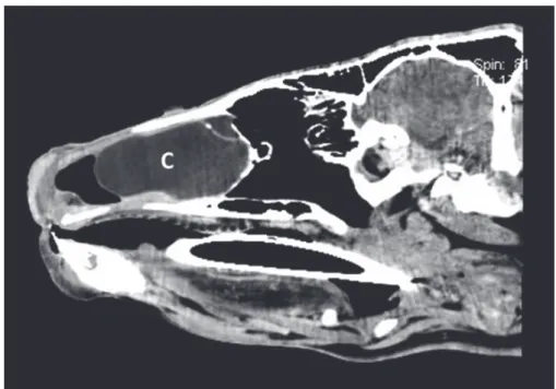
tion and had inspiratory and expiratory dyspnoea and mucopurulent nasal dis- charge on the left side. There was no airflow via the left nostril. For further diag- nosis, a complete blood count was carried out. The packed cell volume (PCV), the mean corpuscular volume (MCV) and the leukocyte count were slightly de- creased, and the mean corpuscular haemoglobin concentration (MCHC) was slightly increased. To assess lung involvement, an ultrasonographic examination of the lungs was performed which did not show any pathological signs. For fur- ther diagnosis, an endoscopic examination of the upper respiratory tract was per- formed, using a flexible endoscope with a diameter of 8 mm (Karl Storz Endos- kop Austria GmbH). The right nostril revealed a normal nasal cavity, pharynx and larynx. On the left side, approximately five centimetres interior to the nostril, a smooth mass of pale pink colour was visible. It was impossible to pass by the mass with the endoscope on any side. On the basis of the endoscopic view, the suspected diagnosis of cystic nasal conchae or abscess was reached. To examine the bony structure/involvement of bones and to determine the real dimensions of the pathological change, an X-ray and computed tomography (CT) were per- formed. Radiography showed a bone proliferation with shading of the soft tissue of the left rostral nasal cavity. For the CT scan, the animal was anaesthetised with 0.2 mg/kg xylazine (Sedaxylan 20 mg/ml®, Eurovet Animal Health B.V., Bladel, Netherlands), 0.2 mg/kg ketamine hydrochloride (Narketan 100 mg/ml®, Ve- toquinol, Ismaning, Germany) and maintained with isoflurane inhalation anaes- thesia (Isoba 1 ml/ml®, Intervet GmbH, Vienna, Austria). The CT scan showed a mass filling the entire rostral area of the left nasal cavity. The lateral bony boundary of the nose (maxillary bone) was displaced by the mass. The nasal sep- tum was also deviated to the right. In the area of the mass, the normal structure of the conchae was not visible. The CT findings confirmed the suspected endo- scopic diagnosis: cystic nasal conchae (Figs 1 and 2). Due to the diagnosis and the preferable prognosis, the decision was made to treat the cyst surgically, using a laser (MedArt® 426, Asah Medico A/S, Hvidovre, Denmark). The cow was se- dated with 0.2 mg/kg xylazine (Sedaxylan 20 mg/ml, Eurovet Animal Health B.V., Bladel, The Netherlands). Using the laser contact application, an opening had been cut into the rostral area of the cyst. As a second step, the non-contact appli- cation of the laser was used to destroy the inner epithelial lining of the cyst. Cy- tological and histological examination from a part of the mass had been carried out. Cytology revealed a benign, mildly inflamed cyst, and the histological ex- amination showed respiratory epithelium with goblet cells as well as focal bony structures but no evidence of a neoplastic process. Postoperatively, the cow was intravenously treated with sodium chloride (10 litres of NaCl 0.9% Fresenius so- lution®, Fresenius Kabi AG, Bad Homburg, Germany) and 500 ml glucose solution (G-40%®, B. Braun Melsungen AG, Melsungen, Germany), ketoprofen (Rifen 100 mg/ml®, Richter Pharma AG, Wels, Austria) i.m. for six days and benzyl- penicillin dihydrostreptomycin (Penistrepto 200 mg/ml®, Virbac, Vienna, Austria)
i.m. for ten days. Ten days after referral to the clinic, the cow left the hospital.
The animal was reported to be doing well with no recurrence of any of the res- piratory signs. Six months after leaving the clinic, she gave birth to a healthy calf and is still showing no clinical signs of recurrent dyspnoea or nasal discharge.
Fig. 1. The CT scan shows a mass filling the entire rostral area of the nasal cavity. C: cyst
Case 2
A four-year-old Brown Swiss cow was referred to the University Clinic for Ruminants of the University of Veterinary Medicine Vienna. According to the owner, the cow showed dyspnoea and mild bilateral nasal discharge. Before referral to the clinic, it had been treated with macrolide antibiotics and cortisone.
That treatment did not result in any improvement of the clinical signs. During the initial clinical examination at the clinic, the cow showed an extended head and neck position, severe inspiratory and expiratory dyspnoea and slightly harsh lung sounds on both sides. Both eyes showed a slightly hyperaemic mucous membrane, mucopurulent nasal discharge was observed on the right side and there was no airflow via the left nostril. The cow was treated with theophylline (Theospirex® 200 mg, Gebro Pharma GmbH, Fieberbrunn, Austria) i.v. and chlorphenamine maleate (Ancesol 10 mg/ml®, Richter Pharma AG, Wels, Austria) i.m.. Ultraso- nographic examination of the lungs was performed to clarify the involvement of the organ, and revealed no pathological changes. For further diagnosis, an en- doscopic examination was conducted with the same instrument and technique as
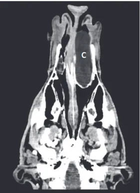
Fig. 2. The CT scan shows a mass filling the entire rostral area of the left nasal cavity. The lateral bony boundary of the nose (maxillary bone) was expanded by the mass. The nasal septum was
deviated to the right. In the area of the mass, the normal structure of the conchae could not be delimited. C: cyst
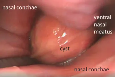
described in Case 1. Endoscopic examination via the left nostril showed a red- dish mucous membrane of the ventral nasal meatus and a smooth pink mass ap- proximately 10 cm caudal to the nostril (Fig. 3). It was possible to pass the mass with the endoscope into the pharynx. At the pharynx, a slight swelling of lymph follicles was diagnosed, and the arytenoid cartilages were in the ‘open’ position showing some slight movements. The trachea displayed no pathological changes, and the right ventral nasal meatus displayed a reddish mucosa with mild muco- purulent secretion. In the endoscopic view there was inflammation of the left and right nasal meatus, and a cystic nasal concha or an abscess in the left ventral na- sal meatus was suspected. It was planned to take a biopsy of the mass the subse- quent day, guided by endoscopy, but by that time the cyst had disappeared (Fig. 4).
Only moderate mucopurulent discharge and some remaining tissue of the wall of the mass could be seen during endoscopy in the left ventral nasal meatus. To ex- amine bony structures, an X-ray was performed, showing circumscribed shading of the left-sided region of the nasal sinuses. Bony structures were not affected. A bacteriological swab sample was taken under endoscopic control from the region of interest, and Staphylococcus haemolyticus was isolated. Due to this finding,
the differential diagnoses were sinusitis and cystic nasal conchae in the left nasal cavity. After the cyst had disappeared, the animal did not show any dyspnoea anymore and airflow via the left nostril was fully restored. The owner was in- formed about the clinical findings and about the possible recurrence of the mass.
Because of late pregnancy status of the cow the owner decided to take the cow home. Four weeks after leaving the clinic, the cow was reported to be doing well, but was experiencing a recurrence of mild inspiratory and expiratory dyspnoea.
Fig. 3. Endoscopic examination via the left nostril shows a smooth pink mass approximately 10 cm caudal to the nostril
Fig. 4. At the second endoscopy via the left nostril on the subsequent day, the cyst had disappeared.
Only a moderate amount of mucopurulent discharge and some of the wall of the mass could be seen by endoscopy in the left ventral nasal meatus
Discussion
Ten days after the cow of Case 1 had been referred to the Clinic for Rumi- nants, it could leave the clinic. About one year after surgery, the cow was report- ed to be doing well with no recurrence of any of the previous respiratory signs.
In Case 1, a surgical diode laser MedArt® 426 (Asah Medico A/S, Hvidovre, Denmark) was used to remove the cyst. It was utilised at 15 watts. Using contact application, an opening was cut in the rostral area of the cyst. After that, the in- ner lining of the cyst was destroyed using a non-contact application. Further- more, precise incision capabilities that were particularly well suited to the treat- ment of lesions were provided, using contact techniques (Blikslager and Tate, 2000). When using non-contact techniques, the laser causes tissue ablation, which is helpful when treating lesions such as ethmoid haematomas or nasal conchae cysts, as described in horses (Blikslager and Tate, 2000). In addition to this, the non-contact technique is useful for treating cysts because the laser ener- gy can be used to heat the fluid contents of the cyst in order to destroy the secre- tory lining of the cyst and thereby preventing recurrence (Blikslager et al., 2000).
Most important is to completely destroy or remove the inner epithelial layer of the cyst, otherwise the cyst is likely to close and fill up with fluid again. This method of therapy is known in horses (Blikslager and Tate, 2000), but to the best of the authors’ knowledge, Case 1 seems to be the first report of the laser resec- tion of a cyst in a ventral nasal concha in cattle. Therapy of the cow was success- ful and neither intraoperative nor postoperative complications occurred.
In Case 2, laser resection would have been a therapeutic option too, but as the mass had opened spontaneously or possibly induced by the endoscopic diag- nostic procedure one day before planned laser surgery and the animal could breathe normally again, that intervention was cancelled. When being informed about this, even the owner did not want any further surgical procedure, arguing that the cow was high pregnant. He was informed that, because of the remaining wall of the cyst at the concha, a recurrence of the cyst refilling with fluid could be possible. About four weeks after leaving the clinic, the cow was reported to be doing well, but a slight recurrence of inspiratory and expiratory dyspnoea was detected. It seems possible that these clinical signs may indicate the recurrence of the cyst.
In cattle showing respiratory distress, it is important to consider pathologi- cal changes in the upper respiratory tract. When patients show clinical signs like both cattle in this report (nasal discharge, breathing sounds, inspiratory and ex- piratory dyspnoea or no airflow via the affected nostril), the lungs are frequently assumed to be the causative problem. Veterinarians should keep in mind differ- ential diagnoses such as foreign bodies, sinonasal neoplasm, deformations, nasal granuloma, nasal polyps, skull fracture, sinusitis, and sinonasal cysts. Clinical signs such as unilateral airflow, nasal discharge, breathing sounds or dyspnoea
may be indications of an obstruction. If, after the clinical examination, the as- sumption of a pathological change in the upper respiratory tract is expressed, fur- ther diagnostic methods have to be used. There are a few options to diagnose pathological changes in the upper respiratory tract, such as endoscopy, X-ray and computed tomography (CT). Moreover, histological and cytological examina- tions from specimens taken under visual control can be performed. By the use of ultrasonography, in addition to a clinical examination, the lungs as the main problem can be ruled out with certainty, although it has to be mentioned that on- ly pathological changes at the surface of the lungs can be visualised by ultraso- nography. Because of the air content and existence of bone structure, ultrasonog- raphy is not an appropriate technique for diagnosing upper respiratory tract dis- eases. By the use of endoscopy, the pathological changes can be visualised and biopsy can be performed during the examination under visual control (Franz and Baumgartner, 2007; Franz, 2011). As shown in our case, a disadvantage of en- doscopy can be that often the full extent of the pathological change and the pos- sible bone involvement cannot be estimated. In such cases, X-ray, or even better, CT scanning should be carried out. Methods such as CT are usually not available for veterinarians in the field, since they typically do not have such equipment.
Also endoscopic examinations are rarely conducted in the field due to the high costs of the instruments. However, such patients should be considered to be re- ferred to a well-equipped specialised clinic. Of course, economic aspects have to be taken into account. As shown in these two cases, sinonasal cysts in cattle can be treated successfully; therefore, diagnosis and treatment should be considered, especially in valuable animals.
References
Anderson, D. E. and Saint Jean, G. (2008): Surgery of the upper respiratory system. Vet. Clin.
North Am. Food Anim. Pract. 24, 319–334.
Blikslager, A. T. and Tate, L. P. (2000): History, instrumentation, and techniques of flexible endo- scopic laser surgery in horses. Vet. Clin. North Am. Equine Pract. 16, 251–268.
Cohen, N. D., Vacek, J. R., Seahorn, T. L. and Schumacher, J. R. (1991): Cystic nasal concha in a calf. J. Am. Vet. Med. Assoc. 198, 1035–1036.
Cramp, P. A., Prange, T. and Nickels, F. A. (2014): Standing equine surgery of the upper respirato- ry tract. Vet. Clin. North Am. Equine Pract. 30, 111–141.
Eppink, L., Bell, G., Dixon, P. M. and Barakzai, S. (2003): Treatment of a cystic nasal concha in a yearling bull. Vet. Rec. 153, 436–437.
Franz, S. (2011): Endoscopy in cattle. Tierärztl. Praxis Ausgabe G, Grosstiere, Nutztiere 39, 281–288.
Franz, S. and Baumgartner, W. (2007): Diagnostischer Einsatz der Endoskopie bei einem Rind mit einem pharyngealen Chondrosarkom. Dtsch. tierärztl. Wschr. 114, 183–187.
Freeman, D. E. (2003): Sinus disease. Vet. Clin. North Am. Equine Pract. 19, 209–243.
Hooper, R. N., Schumacher, J. and Roussel, A. J. (1997): Removal of a cystic nasal concha from a bull. Aust. Vet. J. 75, 641–642.
Mundell, L. D., Smith, B. P. and Hoffman, R. L. (1996): Maxillary sinus cysts in two cattle. J. Am.
Vet. Med. Assoc. 209, 127–129.
Ross, M. W., Richardson, D. W., Hackett, R. P., Tulleners, E. P., Orsini, J. A. and Ohnemus, T. F.
(1986): Nasal obstruction caused by cystic nasal conchae in cattle. J. Am. Vet. Med. Assoc.
188, 857–860.
Saint Jean, G. and Robertson, T. (1987): Cystic nasal conchae as a cause of unilateral nasal ob- struction in a young bull. Can. Vet. J. 28, 251–253.
Schmid, T., Braun, U., Hagen, R. and Hug, S. A. (2014): Clinical signs, treatment, and outcome in 15 cattle with sinonasal cysts. Vet. Surg. 43, 190–198.
Tremaine, W. H. and Dixon, P. M. (2001): A long-term study of 277 cases of equine sinonasal dis- ease. Part 2: treatments and results of treatments. Equine Vet. J. 33, 283–289.
Woodford, N. S. and Lane, J. G. (2006): Long-term retrospective study of 52 horses with sinunasal cysts. Equine Vet. J. 38, 198–202.