III./10.4. Diagnosis
Laboratory tests, electrophysiology, muscle biopsy, genetic testing, imaging techniques
After studying this chapter, you will become familiar with the most commonly used diagnostic methods.
Keywords: creatine kinase (CK), EMG, muscle biopsy, immunohistochemistry, trinucleotide repeat, mutation, MRI
Introduction
In this chapter, an introduction is given about the most important diagnostic methods used in the diagnosis of muscle diseases. In the first part of the chapter, the basic principles and clinical values of these diagnostic methods are discussed. It is important to know what type of diagnostic methods are appropriate for a given clinical question, and the results should be interpreted in the context of the clinical picture. In the second part of the chapter, the typical findings in given disorders are described.
Structure of this chapter:
A.) Laboratory tests
B.) Electrophysiological examination C.) Myopathology
D.) Genetic testing
E.) Diagnosis of muscle diseases F.) Summary
A.) Laboratory tests
The easiest laboratory test is the measurement of serum CK activity. Serum CK levels (in most cases the muscle subunit, and not the MB) may also be elevated in healthy subjects as a result of physical exertion or muscle trauma. Slightly elevated CK levels may indicate myopathies or neurogenic conditions (muscle denervation). CK activity above 1000 IU/L is most likely an indication of muscle dystrophy, inflammatory myopathy or necrotizing myopathy. In addition to CK, serum levels of other muscle enzymes may also be elevated in muscle disorders, such as aspartate aminotransferase, alanine aminotransferase, lactate dehydrogenase (LDH), and aldolase.
For the diagnosis of metabolic disorders, the measurement of serum lactate and pyruvate levels is essential both during rest and after stress (lactate stress test). This test has two types: 1. lactate is measured after physical activity, 2. ischemic lactate test. In healthy individuals, lactate levels are elevated 3-3.5-fold compared to baseline levels after physical activity, and return to baseline after 30 minutes. If aerobic metabolism is abnormal, lactate elevation is much higher and returns slower to baseline, still showing at least a two-fold increase after 30 minutes. This phenomenon is explained by a dominant anaerobic metabolism due to the impairment of the electron transport chain.
Because of the activation of glycolytic processes, the elevation of lactate levels is several fold higher than in healthy individuals. As a consequence, lactate acidosis and ammonemia develop, and the beta hydroxybutyric acid/acetoacetic acid ratio decreases.
Concerning immunoserological tests, the following auto-antibodies are important in the diagnosis of muscle diseases: ANA, RF, AMA, myositis specific autoantibody,
anti-Mi-2, and anti-Jo-1 antibodies.
B.) Electrophysiological examination
Neuromuscular diseases may affect the skeletal muscles (myopathies), the neurons innervating the muscles (neurogenic diseases), and the neuromuscular junction. Based on the clinical picture alone, it is often difficult to differentiate these disorders.
Electrophysiological examination is an important help in the localization of the
pathologic process. Electrophysiological examination allows the assessment of muscles, peripheral nerves, and the neuromuscular junction.
Electromyography (EMG) records bioelectric voltage changes in striated muscles with the help of a concentric needle electrode inserted into the muscle. EMG has four steps:
1, recording of insertional activity 2, recording of spontaneous activity during rest 3, assessment of motor unit potentials during slight innervation, and 4, recording of interference pattern during maximal innervation of the muscle studied.
In addition to these parameters, the response to repetitive stimulation of peripheral nerves can be tested to assess the neuromuscular junction.
Spontaneous activity
No spontaneous activity is recorded in healthy muscles during rest. In certain disorders, fibrillation potentials and positive sharp waves (brief monophasic positive spikes) may occur. Fibrillation potentials are spontaneous discharges and have a similar clinical importance as positive sharp waves. Both are typical in conditions with muscle
denervation, but may also be seen in inflammatory myopathies, metabolic and endocrine myopathies, and in muscle dystrophies. Spontaneous activity is a sensitive sign of motor unit damage, appearing at the earliest 10-14 days following the lesion.
Video 1.: Spontaneous activity and fibrillation potentials recorded with EMG Video 2.: Spontaneous activity recorded with EMG: positive sharp waves Myotonic discharges are high frequency repetitive discharges, in which both the frequency and amplitude show a gradual increase then decrease. They are usually provoked by needle insertion or movement, but may occasionally occur spontaneously.
A crescendo-decrescendo sound effect is heard over the loud-speaker, which is similar to the sound of a dive bomber.
Video 3.: Myotonic discharges detected by EMG Motor Unit Potentials
A motor unit potential (MUP) is the summation of the action potentials of individual muscle fibers belonging to the same motor unit. The following parameters of motor unit potentials are assessed: duration, amplitude, configuration, phases, stability, discharge frequency. In myopathic disorders, amplitude and duration of MUPs are decreased, polyphasia is increased, and the wave shape is stable. In weak or atrophic muscles due to myopathy, MUP recruitment is early and the interference pattern has a low amplitude.
Fig. 1: Motor unit potential, myopathic motor unit
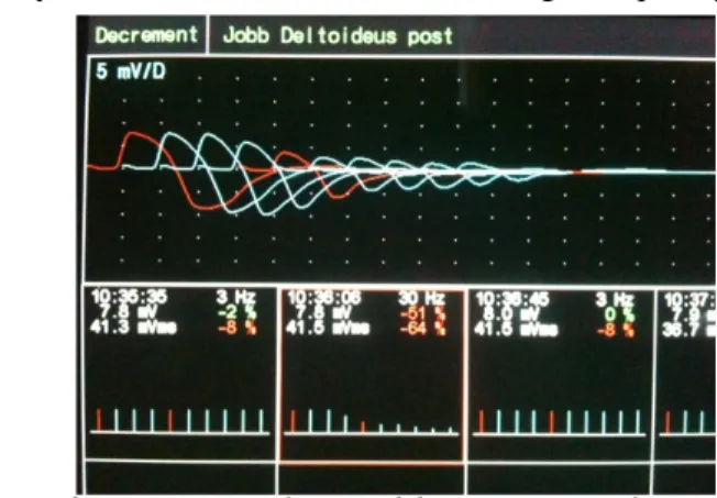
Repetitive stimulation
Dysfunction of the neuromuscular transmission (myasthenia gravis) may be diagnosed with repetitive supramaximal peripheral nerve stimulation: supramaximal repetitive nerve stimulation is done at different frequencies (3 Hz, 10 Hz, 30 Hz). ʽDecrement‘ is diagnosed if the amplitude of the fifth potential decreases by at least 10% compared to the first one, which is a specific sign of the dysfunction of the neuromuscular junction.
In Lambert-Eaton syndrome, increment is seen with high-frequency stimulation.
Fig.2: Repetitive stimulation and decrement in myasthenia gravis
Medical imaging
The radiological examination of skeletal muscles is limited. MRI, MR spectroscopy (MRS) and ultrasound (US) examinations have been used. Imaging techniques are mainly used to show exactly which muscles are involved and to decide which muscle is appropriate for muscle biopsy.
C.) Myopathology
Izombiopszia Finding the right muscle for muscle biopsy
Together with the physical and electrophysiological examination, muscle biopsy is an essential element in the differential diagnosis of neuromuscular diseases. It is critical to choose the right muscle for biopsy. The optimal site of biopsy where specific
morphological alterations are best seen is a muscle which is affected but not severely damaged by the disease. In severely wasted muscles, connective tissue has mostly replaced muscle tissue and muscle fibers are damaged to such an extent that no conclusions can be drawn concerning the specific pathomechism of the disease.
Morphological assessment of muscle biopsy specimens
With histochemical and immunohistochemical analysis of cryostat frozen muscle sections, the morphological differential diagnosis of muscle diseases has improved significantly.
For the preparation of sections for light microscopy, several staining methods are routinely applied, half thin sections are prepared, and occasionally electron microscopy is also carried out. With these techniques, fiber diameter variability, fiber type grouping, fiber necrosis and the proportion of muscle fibers with fissures can be assessed.
Furthermore, the extent of fiber regeneration, myofibrillary anomalies, the presence of vacuoles or tubular aggregates may be seen. The shape, number and the distribution of mitochondria may also be analyzed. The description of muscle fiber nuclei and muscle vessels, and the presence of any inflammatory cells are also very important. Based on these factors, the differentiation of neurogenic and myogenic processes is possible, and valuable information concerning the etiology of the disease is acquired. With the help of immunohistochemical tests, protein deficiencies characteristic for certain genetically determined myopathies may be identified, indicating which gene should be tested.
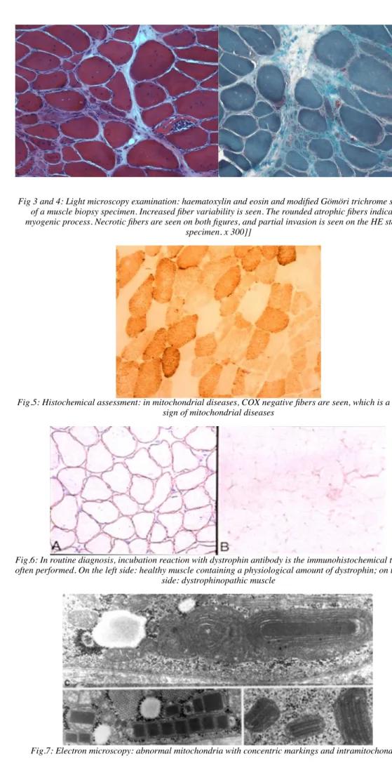
Fig 3 and 4: Light microscopy examination: haematoxylin and eosin and modified Gömöri trichrome staining of a muscle biopsy specimen. Increased fiber variability is seen. The rounded atrophic fibers indicate a myogenic process. Necrotic fibers are seen on both figures, and partial invasion is seen on the HE stained
specimen. x 300]]
Fig.5: Histochemical assessment: in mitochondrial diseases, COX negative fibers are seen, which is a specific sign of mitochondrial diseases
Fig.6: In routine diagnosis, incubation reaction with dystrophin antibody is the immunohistochemical test most often performed. On the left side: healthy muscle containing a physiological amount of dystrophin; on the right
side: dystrophinopathic muscle
Fig.7: Electron microscopy: abnormal mitochondria with concentric markings and intramitochondrial
paracrystalline inclusions, seen in mitochondrial diseases.
D.) Genetic testing
Genetic testing and molecular biologic investigations have contributed significantly to the improvement of the diagnosis of muscle diseases. DNA or mtDNA extracted from blood samples or muscle biopsy specimens can be tested.
After physical and electrophysiological examination, genetic testing is the next step in the diagnostic algorithm if the following muscle disorders are suspected: myotonic dystrophy, facioscapulohumeral muscular dystrophy and Duchenne muscular dystrophy.
In these disorders, the phenotype is so typical that diagnosis can usually be given based on physical examination.
In myotonic dystrophy, the genetic impairment is a trinucleotide repeat in the DMPK gen. In facioscapulohumeral muscular dystrophy, a repeat deletion in D4Z4 locus of the 4th chromosome is found.
For Duchenne muscular dystrophy, dystrophin gene deletions are tested.
If these genetics tests do not provide a diagnosis, muscle biopsy is recommended before further genetic testing is indicated.
It is also possible to do prenatal diagnostics if the gene to be tested in the fetus is known.
Fig.8: Dystrophin gel electrophoresis showing multiple deletions.
E.) Diagnosis of muscle diseases
In the majority of muscle disorders, muscle biopsy is essential to arrive at the final diagnosis. Histological examination often shows non-specific signs of muscle fiber destruction: increased variability of fiber diameters, large number of internal nuclei, increased amount of the endomysial connective tissue, and in some cases the presence of necrotic and regenerating fibers. In metabolic myopathies, absence and imbalance of oxidative enzyme reactions can be seen. With immunohistochemical tests, several proteins are analyzed, such as components of the dystrophin complex: dystrophin, alpha-, beta, gamma-, delta sarcoglycan; other plasma membrane proteins: caveloin-3, dysferlin; proteins of the extracellular matrix: merosin, collagen VI; the inner nuclear proteins: laminA/C, emerin; sarcomeric proteins: telethonin. Calpain-3 deficiency can be diagnosed only by Western blot technique. The absence of alpha-dystroglycan may suggest glycosilation defects (fukutin, FKRP, POMGnT, POMT1, LARGE). These can be diagnosed with both immunohistochemical tests and Western blot.
Genetic testing should always be done in a targeted way, i.e. after a particular protein deficiency has been identified with prior histopathological examination or Western blotting. In Duchenne/Becker muscular dystrophy, the typical clinical picture is usually sufficient to ask for the genetic analysis of the dystrophin gene and invasive muscle biopsy is not necessary. If genetic testing did not confirm a deletion in the dystrophin gene, muscle biopsy is recommended with immunohistochemical analysis of the
specimen. In metabolic myopathies, the measurement of particular enzymes is helpful before genetic testing is indicated.
Muscle biopsy is the crucial element in the diagnosis of inflammatory myopathies.
Lymphocytic infiltration is typically seen in these myopathies. Lymphocytes and macrophages can enter muscle fibers, which have not necrotized yet (partial invasion).
With the help of specific markers, the functional characterization of inflammatory cells (e.g. T or B cells, NK cells, T helper, T suppressor, T cytotoxic cells) is also possible
Fig.9: Presence of inflammatory cells in inflammatory myopathies.
F.) Summary
In addition to detailed medical history and physical examination, the following tests have an important role in the diagnosis of muscle disorders and neuromuscular junction diseases: measurement of serum CK levels, EMG, muscle histology and genetic tests.
With the help of the histopathological analysis of muscles, neurogenic and myogenic processes may be differentiated, inflammatory signs may be seen, and the activity of metabolic enzymes can be checked. Using immunohistochemical tests, protein
deficiencies can also be determined. Genetic tests should always be targeted. The result of any test should be interpreted in context of all pieces of information, and the final diagnosis is determined by the synthesis of all results and data.