TOMUS 7.
NUMERUS 2.
ACTA
BIOLOGICA PLANTARUM AGRIENSIS
REDIGIT
PÉTER SZŰCS
EGER, 2019
(ABPA)
from Acta Academiae Paedagogicae Agriensis Sectio Biologiae
a Journal of Plant Biology
TOMUS 7.
Eger 2019
Editor-in-Chief:
Tamás Pócs (Taxonomy) Senior Editors:
Endre Lehoczky (Biophysics) László Mustárdy (Cell Biology)
Sándor Orbán (Ecology) Editorial Board:
Éva Darkó (Biotechnology)
Sándor Dulai (Physiology, Stress and Ecophysiology) Marianna Marschall (Biochemistry, Stress and Ecophysiology)
István Molnár (Molecular Biology) Márta Molnár-Láng (Genetics)
Mária Papp (Anatomy) Erika Pénzes-Kónya (Ecology) Andrea Sass-Gyarmati (Taxonomy)
Péter Szűcs (Ecology, Taxonomy) Zsolt Zsófi (Grapevine Biology and Physiology)
András Vojtkó (Geobotany) Editor:
Péter Szűcs Managing Editor:
Erika Pénzes-Kónya Technical Editors:
Péter Szűcs, Gabriella Fintha Reviewers of papers:
Éva Darkó, Liliya Dimeeva, Sándor Dulai, József Geml, MáriaHöhn, Zsolt Kotroczó, László Mustárdy, Sándor Orbán, Viktor Papp, Ferenc Pál-Fám, Erika Pénzes-Kónya, Tamás Pócs,
Andrea Sass-Gyarmati, Péter Szűcs, Zsolt Zsófi, András Vojtkó HU ISSN 2061-6716 (Print)
HU ISSN 2063-6725 (Online)
Papers of this volume are available: http://abpa.ektf.hu/
© 2019, Eszterházy Károly University, Hungary, Department of Botany and Plant Physiology A kiadásért felelős az Eszterházy Károly Egyetem rektora
Megjelent az EKE Líceum Kiadó gondozásában/Published by Líceum Publisher EKE Kiadóvezető/Head of publisher: Nagy Andor
Tördelőszerkesztő/Layout editor: Szűcs Péter Megjelent/Year of publication: 2019
Nyomdai munkák: Eszterházy Károly Egyetem nyomdája /Printed by Károly Eszterházy University Press Felelős vezető/Responsible of printing: Kérészy László
© 2019, Eszterházy Károly University, Hungary Department of Botany and Plant Physiology
EFFECTS OF LIGHT INTENSITY AND SPECTRAL COMPOSITION ON THE GROWTH AND METABOLISM
OF SPINACH (SPINACIA OLERACEA L.)
Lőrinc Utasi1,2, István Monostori1, Balázs Végh1, Zoltán Pék2
& Éva Darkó1*
1Hungarian Academy of Sciences, Centre for Agricultural Research, Agricultural Institute, H-2462 Martonvásár, Brunszvik str. 2, Hungary; 2Szent István University, Faculty of Agricultural and Environmental Sciences, H-2100 Gödöllő, Páter Károly
str. 1, Hungary; *E-mail: darko.eva@agrar.mta.hu
Abstract: Spinach rich in proteins, minerals (Fe, K) and antioxidants (vitamin C) is often cultivated in greenhouses under artificial light. In this study, the effects of light intensity and spectral composition provided by light emitting diodes (LEDs) was studied on the yield quality and quantity of spinach. Plants were grown under 3 different light intensities (100/200/300 mol m-2s-1) and 3 spectral compositions (white/white completed with blue or far-red) for 4 weeks. Then plant weight, leaf number and area, photosynthetic activity and pigment composition of leaves were determined. The leaf quality was characterized by measurements of protein, starch, soluble sugar and ascorbate contents of leaves.
Moreover, minerals (K, Fe and NO3̅) were also determined. While the biomass production was mainly determined by the light intensity, the yield quality is influenced significantly by the spectral composition. When the white light was supplemented with blue, high protein and ascorbate content was achieved. When far-red was added to white light, elevated sugar and Fe accumulation was observed in the leaves, while the K content decreased. In order to reach the hight quality and quantity of food production, not only the light fluence, but also the spectral composition should be regulated.
Keywords: LED lighting, spinach, hydroponics, artificial light
INTRODUCTION
Light is one of the most important environmental factors that affect plant growth and development. Through photosynthesis and the operation of photoreceptors, both the light intensity and the spectral composition have broad regulatory effects on the morphogenesis, such as leaf growth and expansion, on many
Acta Biol. Plant. Agriensis 7: 3–18
4
physiological and metabolic processes in plants, which finally determines the main characteristics of plants: the appearance and quality.
With the use of artificial light in phytotrons, greenhouses and in modern plant factories, the light environment of plants inherently changes compared to the natural light situation. This influences multiple aspects of plant functioning, including photosynthesis, photomorphogenesis, water relations, nutritional quality, biomass and yield production and quality. In most cases, the metal halide, high-pressure sodium (HPS) lamps and/or fluorescent lamps are used either as supplementary or sole-source (Kim et al. 2004).
They are often inefficient for plant cultivation due to their fix and rather different spectral distribution than the solar light (Morrow 2008; Darko et al. 2014). The plants grown under these artificial light conditions often shows many symptoms of suboptimal light condition, such as internode elongation, shift in flowering time, decrease of fertility and low quality and quantity of crop production. In addition, these lamps have high energy consumption and high operation temperature make them economically inefficient (Kim et al. 2004, Morrow 2008).
Light emitting diodes (LED) represent an innovative artificial lighting source in plant cultivation. LEDs have low energy consumption, long-lifetime and small size, high fluence and variable spectral characteristics, which make them better suited to crop production than other artificial lighting systems (Morrow 2008).
Therefore, the application of LEDs in agricultural/horticultural practice is increasing continuously. Cultivation of plants under LED lighting in environmentally controlled rooms enables standard vegetable production regardless of the weather conditions. The aim is to achieve high productivity and/or high vegetable quality without excess energy consumption. Changes of spectral composition could provide a solution for improving the yield and quality of crops.
Spinach (Spinacia oleracea) plants are widely cultivated in greenhouses throughout the world. This leafy green is rich in nutrients such as protein, dietary fiber, vitamins and folate and minerals such as K, Ca, Mg, Mn, and Fe, which may play an important role in human nutrition and diet. Most studies on the effect of light intensity and spectral composition delivered by LEDs on the quality of leafy vegetables have focused on biomass
5
production, leaf area and branching, while less is known about how LEDs affect the mineral composition and nutritional quality (Lin et al. 2013). In addition, spinach is less characterized than lettuce (Burattini et al. 2017).
This paper aims to determine whether similar productivity and/or quality can be achieved even under limited light intensity by changing of spectral composition of light. Therefore, spinach plants were cultivated under white light at low (100 mol m-2s-1), medium (200 mol m-2s-1) or high (300 mol m-2s-1) light intensities and at white light (200 mol m-2 s-1) with far-red or blue light supplementation in hydroponic growth conditions. The biomass production, leaf architecture will be compared together with the physiological response of plants , which was determined through monitoring the photosynthetic activity and chlorophyll contents of leaves. The leaf quality was estimated by determination of leaf protein, starch, and soluble sugar contents, by the measurements of nitrate, ascorbate, iron and potassium contents of plants grown under different light environment. We hope that this complex research brings a better understanding of the relationship of light conditions and yield quality and can provide further information for efficient indoor plant cultivation.
MATERIALS AND METHODS Plant cultivation
Spinach (Spinacia oleracea L.) cv. Sparrow RZ F1 hybrid variety was used in the experiments. The seeds were germinated at 5°C for three days, then at 10°C for seven days and finally at 15°C for further 3 days. The plantlets were put into Jiffy coco plugs (Jiffy-7C, 50mm std, Jiffy Products S.L. Ltd, Sri Lanka) and placed in phytotron growth chamber equipped with LED light sources and automatic control system for circulation of nutrient solution in the Agricultural Institute, Centre for Agricultural Research, Hungarian Academy of Sciences. The light characteristics used in the experiments are summarized in Table 1. Briefly, 3 different spectral compositions (White light, blue dominant white light and white light complemented with far-red radiation) at the same light intensity (200 mol m-2s-1) and 3 different light intensities (low:
100 mol m-2s-1, medium: 200 mol m-2s-1 or high: 300 mol m-2s-1)
Acta Biol. Plant. Agriensis 7: 3–18
6
were used for spinach cultivation in hydroponic growth system using diluted Hoagland solution described by Pál et al. (2005). The pH and the ionic strength of the nutrient solution was kept between 6.0-6.5 and EC 1.1-0.9 respectively. Plants (at least 15 plants of each light regimen) were grown for 4 weeks at 20/15°C day/night temperature and 75% of humidity. The growth and development of spinach plants were monitored twice a week, and the measurements were started in the fourth week.
Table 1. The light spectral composition used in the experiment.
Light characteristics White
300 White
200 White
100 White
+ FR 200 White + Blue 200 PAR (400-700nm)
μmol m-2 s-1 310 202 100 205 208
Ratio:
Blue: Red 0.208 0.200 0.184 0.189 4.86
Red: Far-red 8.0 8.8 10.2 1.0 9.2
Blue: Far-red 1.7 1.7 1.8 0.18 44.7
12h/12h photoperiod were used.
Determination of the photosynthetic activity of leaves
The photosynthetic activity of plants was determined on intact leaves with a Ciras 3 Portable Photosynthesis System (Amesbury, USA) instrument using a narrow leaf area (4.5 cm2) chamber equipped with a chlorophyll a fluorescence module. Since the Ciras 3 uses LED modules, the photosynthetic activity of leaves was measured under the same light conditions as was found in growth chambers. The net assimilation rate (A), stomatal conductance (gs), intracellular CO2 concentration (Ci) and transpiration (E), as well as the effective quantum yiled of PS II [Y(II)] were determined at the steady state of photosynthesis by the use of 400 L L-1 CO2
level.
Determination of biomass production, leaf area and sample collection for the analytical investigations
At harvest, the plant weight, leaf number and leaf area were determined in order to characterize the biomass production of plants. 10 plants of each light regimens were measured. The leaf area was determined using an Area Meter 500 from ADC
7
Bioscientific Ltd. Then, samples for biochemical investigations were collected. Thus, 3 leaf discs (with 1.3 cm diameters) were collected from 5 plants (5 x 3 leaf discs) of each light regimen for determination pigment composition of leaves. Also, 5 x 0.2g samples were collected for determination of protein, starch, soluble carbohydrate and ascorbate content of leaves, respectively.
In addition, samples were collected for determination of mineral (K, Fe and nitrate) content of leaves. The leaf samples were frozen immediately and stored at -80 °C until utilization.
Analytical investigations
Chlorophyll a and b, as well as the carotenoid contents of leaf discs, were determined using a Cary-100 UV-Vis spectrophotometer (Varian, Middelburg, Netherlands) after extraction of leaf discs in 80% acetone, according to the method of Lichtenthaler (1987).
The total protein content was determined according to the method of Bradford using bovine serum albumin as standard (Kruger 2009). Total soluble carbohydrates were extracted and measured following the method of Antron as described in Sinay and Karuwal (2014). The starch content was determined from twice-washed and dried pellet remaining after extraction of total soluble sugars according to the method of Thayumanavan and Sadasivam (1984).
Similar isolation protocol was used for determination of K, NO3̅, Fe and ascorbic acid contents. All compounds were diluted with MQ water (1:10). The measurements were performed by the use of RQflex plus 10 (Merck KGaA 64271 Darmstadt, Germany) instrument according to the manufacturer's instructions for K:
1.17945.000; NO3̅: 1.14761.0002; Fe: 1.14761.0002 and Ascorbic acid: 1.16981.000.
Statistical analyses
The experiments were repeated 3 times. In each experiments at least 15 plants were grown under each light regimens and the samples were collected from randomy selected plants. The measuerements were repeated at least in 5 biological replicates.
The data were analysed by SPSS 16.0 statistical program and Tukey’s post hoc test were used to determine differences between treatments. Different letters indicate significant differences at the P
< 0.05 level.
Acta Biol. Plant. Agriensis 7: 3–18
8 RESULTS
Morphological responses of spinach cultivated under different light regimens
To characterize the plants grown under different light environment several morphological parameters, such as leaf number, leaf area, and leaf mass were determined (Figure 1). All leaf mass, leaf area and leaf number increased with increasing of white light intensity (Figure 1A-C). However, the specific leaf area, the ratio of leaf area to leaf mass, decreased with increasing light intensity (Figure 1D).
It is due to the fact that the increase of light intensity increased leaf area of plants with relatively less extent (3.23 and 4.47 fold change) than the leaf mass (they was 4.91 and 8.61 fold change). When the spectral composition of light was compared in grown of spinach plants at 200 μmol m-2 s-1, the supplementation of white light with FR resulted in a slightly higher expansion of leaves and longer stems (data not shown) than without far-red application. Inversely, the blue light decreased the leaf area and created a compact and short plant. While the effect of spectrum in leaf area and weight were not significant, the specific leaf area increased under far-red and decreased when blue light was applied (Figure 1).
Photosynthetic properties of spinach under different light environment
Evidently light regulates the photosynthetic processes. As the light intensity increased, so did the CO2 assimilation rate (Pn), stomatal conductance (gs) and transpiration (E) in spinach leaves, meanwhile the intracellular CO2 level decreased (Figure 2). On the other hand, not only the intensity of the light, but the modification of spectral composition also influenced the CO2 assimilation processes: the blue light reduced and the FR light slightly (but not significantly) stimulated the CO2 assimilation capacity of plants (Figure 2A). However, the stomatal conductance changed inversely, it increased to blue and slightly decreased to far-red, when they were compared to white light having the same light intensity (Figure 2B).
9
Figure 1. Effect of the light intensity and spectral composition on the growth parameters of spinach. A: Leaf number; B: Leaf area: C: Leaf mass; D: Specific leaf area. Values are the mean ± STD of 10 biological replicates per light treatment. The different letters indicate statistically significant differences at P < 0.05, using Tukey's post hoc test.
Figure 2. Comparison of photosynthetic properties of spinach leaves grown under different light regimens. A: Net CO2 assimilation rate (Pn); B: Stomatal conductance (gs); C: Transpiration (E) and D: Intercellular CO2 level. Values represent the means (± STD) of 5 independent measurements per light treatment.
The different letters indicate statistically significant differences at P < 0.05, using Tukey's post hoc test.
Acta Biol. Plant. Agriensis 7: 3–18
10
The photosynthetic electron transport processes was also investigated. The effective quantum yield of PSII, Y(II), determined at steady of photosynthesis indicates the ratio of the number of photons utilized photochemically/total number of quanta absorbed. Typically, the conversion ratio of absorbed light energy to photochemistry in PS II decreased in parallel to the increase of light intensity in spinach leaves, which was indicated by the lower Y(II) values (Figure 3). Lower Y(II) was also measured in leaves grown under the blue light as compared to those plants grown under white light at the same light intensity with or without FR application. In fact, Y(II) values were similar in spinach leaves grown under blue light at 200 μmol m-2s-1 and under white light at 300 μmol m-2s-1.
Figure 3. Photochemical utilization of absorbed light energy determined in spinach leaves grown under different light conditions at steady state level of photosynthesis. Y(II) – effective quantum yield of PSII photochemistry. Values represent the means (± STD) of 5 independent measurements per light treatment.
The different letters indicate statistically significant differences at P < 0.05, using Tukey's post hoc test.
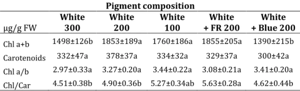
When the pigment composition of leaves grown under different light regimens were compared, two interesting tendency was observed: the pigment composition hardly varied in the function of light intensity, but changed according to the spectral composition (Table 2). The lowest chlorophyll a and b contents were detected in plants grown under blue light, followed by the highest light
11
intensity, while the highest chlorophylls were detected when FR was applied.
These results suggest that both the light fluence and spectral composition determines the photosynthetic properties of plants.
Table 2. Effect of the light intensity and spectral composition on chlorophyll (a+b) and carotenoid contents of leaves.
Pigment composition µg/g FW White
300 White
200 White
100 White
+ FR 200 White + Blue 200 Chl a+b 1498±126b 1853±189a 1760±186a 1855±205a 1390±215b Carotenoids 332±47a 378±37a 334±32a 329±37a 300±42a Chl a/b 2.97±0.33a 3.27±0.20a 3.44±0.22a 3.08±0.21a 3.41±0.20a Chl/Car 4.51±0.38b 4.90±0.36b 5.27±0.34ab 5.63±0.28a 4.62±0.44b
Effect of light intensity and spectral composition on the yield quality of spinach leaves
The leaf quality was estimated by determination of leaf protein, starch, and soluble sugar contents and by the measurements of nitrate, ascorbate, iron and potassium contents of plants grown under different light environment (Figure 4).
The primary metabolites, such as the soluble sugars, starch and proteins increased with increasing light intensity in spinach leaves (Figure 4A-C). It was mainly due to the light-dependent increase of the photosynthetic activity. Blue light stimulated the protein accumulation, while decreased significantly the amount of soluble sugars and starch in spinach leaves as compared to white light used at the same light intensity. The supplementation of white light with far-red resulted in a significant accumulation of soluble sugars, while the proteins level decreased to the similar value found in leaves grown under low (100 μmol m-2s-1) light intensity (Figure 4A).
Spinach is an excellent source of vitamin C. The amount of ascorbate was hardly affected by light intensity and spectral composition (Figure 4D). Significant increase in ascorbate content was detected only when high proportion of blue light was applied.
Acta Biol. Plant. Agriensis 7: 3–18
12
Figure 4. Production of primary and secondary metabolites in spinach leaves grown under different light regimens. A: protein content; B: starch content; C:
soluble sugar content; D: ascorbate content. Values are the mean ± STD of 5 biological replicates per light treatment. The different letters indicate statistically significant differences at P < 0.05, using Tukey's post hoc test.
Figure 5. Mineral content of spinach leaves grown under different light regimens.
A: nitrate content; B: iron content; C: potassium content; Values are the mean ± STD of 5 biological replicates per light treatment. The different letters indicate statistically significant differences at P < 0.05, using Tukey's post hoc test.
13
Among minerals, the K, Fe and NO3 ̅ content of leaves were determined (Figure 5). Surpisingly, only the accumulation of Fe showed ligh intensity dependent changes in spinach (Figure 5B).
Neither the NO3 ̅ nor the K content showed similar tendency (Figure A and C). The spectral composition influenced both the iron and the potassium content of leaves. The blue light reduced the accumulation of Fe, while far-red decreased the K content of leaves (Figure 5B and C). All these modifications affects the yiled quality of spinach.
DISCUSSION
Cultivation of leafy plants under artificial lighting becomes more and more important in the future to produce high quality and quantity of foods throughout year. They are influenced by both light fluence and spectral composition. Providing variable fluence and spectral composition, LEDs give a unique possibility to optimize the growth conditions.
Through activation of photosynthetic processes, the increase of light intensity forces the biomass production which in case of leafy plants, manifests in the increase of leaf number, area and mass. In our experiments it seemed that the biomass production was mainly determined by the light intensity while the spectral composition was less influenced. However, the increase of photosynthetic activity induced leaf expansion less than producing assimilates, resulting in a decrease of sepcific leaf area. This phenomenon (e.g.
sepcific leaf area decreased with increasing light intensity) was also observed by Xiao-Xue et al. (2013) and Urrestarazu et al. (2016). In the opinion of Gommers et al. (2013), this response to light could help plants to increase the efficiency of light capture and maximize carbon gain at low light intensity (Gommers et al. 2013). It can help plants under shade environment. Our results can support it, since the specific leaf area (cm2/g) also increased when the white light was supplemented with far-red light. However, it should be mentioned that the application of far-red light resulted in weak and thin leaves (data not shown). Inversely blue light inhibited the leaf expansion thus decreased the specific leaf area and increased the leaf thickness (it was observed, but not measured), similarly as was found at higher light intensity by us and by Meziane and Shipley (1999).
Acta Biol. Plant. Agriensis 7: 3–18
14
Photosynthetic pigments absorb and convert the light energy into chemical energy via complex photosynthetic machinery. Blue and red light play an active role in photosynthesis and also stimulate chlorophyll and carotenoid biosynthesis (Xiao-Xue et al.
2013, Wang et al. 2015). In the present study, light intensity caused the largest effects on photosynthetic processes, which were manifested in elevated CO2 assimilation capacities, induction of stomata opening and transpiration, and also in the decreased ratio in the conversion of absorbed light energy to photochemistry in PS II, as demonstrated by the low Y(II) value. However, not only the light intensity but also its spectral composition affects CO2
assimilation processes. The blue light decreased the CO2
assimilation, while stimulated the stomatal opening. Kim et al.
(2004) also found that the high proportion of blue light decreased the CO2 assimilate rate, in spite of the fact that blue light stimulated the stomatal opening. The blue light-induced stomatal movement is explained by (Kinoshita and Hayashi 2011), who found that the blue light-induced stomatal opening is mediated by phototropins (activate by blue light) through the activation of the plasma membrane H+-ATPase in guard cells. When the effects of blue light were studied on cucumber plants grown under different combinations (0-100%) of red and blue light, Hogewoning et al.
(2010) was found that the blue light provided ‘sun-type’
characteristics of leaves even at the relatively low growth irradiance. Blue light overexcited the PS II reaction centres resulting in a lower photochemical utilization of absorbed light energy at PS II, similarly as was found under higher light intensity.
We also found the decrease of Y(II) when blue light was applied. A relative overexcitation of PSII can results in an imbalance of photosynthetic electron transport between PS II and PS I, which can decrease the efficiency of the photosynthetic processes. In addition, the blue light-induced ‘sun-type’ characteristics of leaves was manifested also in the decrease of chlorophylls in spinach leaves, similarly as was found by Zhang et al. (2016) and Monostori et al. (2018). To ensure the optimal growth a fine balance between the blue and red ratio is necessary to provide equilibrated and efficient utilization of absorbed light energy between the PS II and PS I.
While it is widely understood that light intensity can positively affect the accumulation of assimilates, the effects of light quality is
15
more complex. In this way, the higher light intensity was used, the more primary assimilates including proteins, starch and sugars were produced in spinach leaves. But, light quality also affected the production of primary and secondary metabolites in spinach leaves. Blue light significantly increased protein and ascorbate contents of leaves, while less sugar and starch were accumulated in these plants. In fact, when the plants were grown under white light supplemented with blue, as high protein and ascorbate content was obtained at 200 μmol m-2s-1 light intensity as plants achieved under higher 300 μmol m-2s-1 light intensity. On the other hand, when the light was completed with far-red light the spinach leaves contained more sugars and Fe than in any other cases. The blue-light induced shift in protein and ascorbate metabolism was also detected in lettuce (Chen et al. 2016). However, the reason of protein accumulation is controversial. As the nitrate provides main N source in proteins of plants, it was suppose that the high protein content is originated from the elevated nitrate uptake. However, when Zhang et al. (2018) compared the effect of different LEDs (white, red and blue in different proportion) on the nitrogen metabolism in lettuce, they found that both red and blue light promoted the N assimilation by improving the activity of the N- metabolism-related enzymes such as nitrate and nitrite reductases, in lettuce. In other studies, it was found that the nitrate reductase activity was stimulated by red light. Samuoliené et al. (2009) used high-intensity red LEDs treatment 3 days prior to harvets to reduce the nitrate content in lettuce. It can be useful, especially in case of leafy plants. However, our results can not prove these findings since the accumulation of nitrates did not change either by light intensity (except at low light, when the low transpiration reduced the nitrate and any kind of ion uptakes) or by spectral composition.
Nevertheless, the physiological significance of LED-induced regulation of nitogen metabolism remained undercharacterized.
CONCLUSION
Although the mechanisms of the changes in spinach growth under LED lighting are not well understood yet, the results showed that LED light can be used to modify the plant growth and metabolism in spinach. Via the variable fluence and spectral composition, the LEDs provide a unique possibility to change both the light intensity
Acta Biol. Plant. Agriensis 7: 3–18
16
and the spectral composition during the lifetime of plants in order to ensure the best light combinations resulting in the highest nutritional values of leafy plants in indoor plant cultivation. On the bases of the results it seems that white light at adequately high intensity (300 μmol m-2s-1) can ensure the optimal growth for spinach to reach the harvest quality in 4 weeks.
At the early developmental stage the application of far-red can induce leaf expansion, while before harvest the increase of the proportion of blue light can stimulate the accumulation of proteins and ascorbate content, which can improve the leaf quality.
However, the appliaction of red or far-red light can increase the sugar content, which imporve the sweetness of leaves. Anyway, although the LEDs are suitable for manipulation the growth and metabolism in plants, it is unlikely that only one spectral combination can ensure the optimal growth and yield quality throughout the life cycle.
Acknowledgements – This work was supported by the TÁMOP 4.2.2.A- 11/1/KONV-2012-0008 project.
REFERENCES
BURATTINI,C.,MATTONI,B.&BISEGNA,F. (2017). The impact of spectral composition of white LEDs on spinach (Spinacia oleracea) growth and development.
Energies 10(9): 1383. https://doi.org/10.3390/en10091383
CHEN, X., XUE, X., GUO, W., WANG, L. & QIAO,X. (2016). Growth and nutritional properties of lettuce affected by mixed irradiation of white and supplemental light provided by ligh-emitting diode. Scientia Horticultrae 200: 111-118.
https://doi.org/10.1016/j.scienta.2016.01.007
DARKO,E.,HEYDARIZADEH,P.,SCHOEFS,B.,&SABZALIAN,M.R. (2014). Photosynthesis under artificial light: the shift in primary and secondary metabolism.
Philosophical Transactions of the Royal Society B: Biological Sciences 369(1640): 20130243. https://doi.org/10.1098/rstb.2013.0243
GOMMERS,C.M.M,VISSER,E.J.W.,ONGE,K.R.S.,VOESENEK,L.A.C.J.&PIERIK,R. (2013).
Shade tolerance: when growing tall is not an option. Trends in Plant Science 18:
65–71. https://doi.org/10.1016/j.tplants.2012.09.008
HOGEWONING,S.W.,DOUWSTRA, P.,TROUWBORST, G.,vanIEPEREN, W.& HARBINSON,J.
(2010). An artificial solar spectrum substantially alters plant development compared with usual climate room irradiance spectra. Journal of Experimental Botany 61: 1267–1276. https://doi.org/10.1093/jxb/erq005
KIM, H.H., GOINS, G.D., WHEELER, R.M. & SAGER, J.C. (2004). Green-light supplementation for enhanced lettuce growth under red-and blue light- emitting diodes. HortScience 39(7): 1617–1622.
https://doi.org/10.21273/HORTSCI.39.7.1617
17
KINOSHITA,T.&HAYASHI,Y. (2011). New Insights into the Regulation of Stomatal Opening by Blue Light and Plasma Membrane H+-ATPase. International Review of Cell and Molecular Biology 289: 89–115.
https://doi.org/10.1016/B978-0-12-386039-2.00003-1
KRUGER,N.J. (2009). The Bradford method for protein quantitation. Humana Press, Totowa, N.J., pp. 17–24.
LICHTENTHALER, H.K. (1987). Chlorophylls and Carotenoids: Pigments of Photosynthetic Biomembranes. In: Methods in Enzymology 148: 350–382.
https://doi.org/10.1016/0076-6879(87)48036-1
LIN,K-H.,MENG-YUAN,H.,WEN-DAR,H.,MING-HUANG,H.,ZHI-WEI Y.& CHI-MING. Y.
(2013). The effects of red, blue and white light-emitting diodes (LEDs) on growth, development and edible quality of hydroponically grown lettuce (Lactuca sativa L. var. capitata). Scientia Horticulturae 150: 86–91.
https://doi.org/10.1016/j.scienta.2012.10.002
MEZIANE,D.&SHIPLEY,B. (1999). Interacting determinants of specific leaf area in 22 herbaceous species: Effects of irradiance and nutrient availability. Plant, Cell and Environment 22: 447–459. https://doi.org/10.1046/j.1365- 3040.1999.00423.x
MONOSTORI,I.,HEILMANN,M.,KOCSY,G.,GALIBA,G.&DARKO,E. (2018). LED Lighting - Modification of growth, metabolism, yield and flour composition in wheat by spectral quality and intensity. Frontiers in Plant Science 9: 605.
https://doi.org/10.3389/fpls.2018.00605
MORROW,R.C. (2008). LED Lighting in Horticulture. HortScience 43: 1947–1950.
https://doi.org/10.21273/HORTSCI.43.7.1947
PÁL,M.,HORVÁTH,E.,JANDA,T.,PÁLDI,E.&SZALAI,G. (2005). Cadmium stimulates the accumulation of salicylic acid and its putative precursors in maize (Zea mays) plants. Physiologia Plantarum 125: 356–364.
https://doi.org/10.1111/j.1399-3054.2005.00545.x
SAMUOLIENĖ,G.,URBONAVICIÜTE A.,DUCHOVSKIS,P.,VITTA,P.&ZUKAUSKAS,A. (2009).
Decrease in nitrate concentration in leafy vegetables under a solid-state illuminator. HortScience 44: 1857–1860.
https://doi.org/10.21273/HORTSCI.44.7.1857
SINAY,H.& KARUWAL,R.L.(2014). Proline and total soluble sugar content at the vegetative phase of six corn cultivars. International Journal of Advance Agricultural Reseach 2: 77-82.
THAYUMANAVAN,B.&SADASVIAM,S. (1984). Estimation of starch by anthrone reagent - Carbohydrates | Laboratory Methodology. [Retrieved: 25.04.2019], from https://biocyclopedia.com/index/plant_protocols/carbohydrates/starch_by_a nthrone_reagent.php
URRESTARAZU,M.,NÁJERA,C.&GEA,M. (2016). Effect of the Spectral Quality and Intensity of Light-emitting Diodes on Several Horticultural Crops. HortScience 51: 268–271. https://doi.org/10.21273/HORTSCI.51.3.268
WANG,Z.,TIAN,J.,YU,B.,YANG,L.,&SUN,Y. (2015). LED Light Spectrum Affects the Photosynthetic Performance of Houttuynia Cordata Seedlings. American Journal of Optics and Photonics 3: 38–42.
https://doi.org/10.11648/j.ajop.20150303.12
XIAO-XUE,F.,ZHI-GANG,X.,XIAO-YING,L.,CAN-MING,T.,LI-WEN W.,&XUE-LIN H. (2013).
Effects of light intensity on the growth and leaf development of young tomato
Acta Biol. Plant. Agriensis 7: 3–18
18
plants grown under a combination of red and blue light. Scientia Horticulturae 153: 50–55. https://doi.org/10.1016/j.scienta.2013.01.017
ZHANG,T.,SHI,Y.,PIAO,F.&SUN,Z. (2018). Effect of different LED sources on the growth and nitrogen metabolism in lettuce. Plant, Cell, Tissue and Organ Culture 134: 231-240. https://doi.org/10.1007/s11240-018-1415-8
(submitted: 15.12.2018, accepted: 07.05.2019)
© 2019, Eszterházy Károly University, Hungary Department of Botany and Plant Physiology
WILD PEARS OF ARMENIA:
DIVERSITY, ENDEMICS AND CONSERVATION Anna Asatryan
Institute of Botany after A.L. Takhtajyan, National Academy of Sciences of the Republic of Armenia, Acharyan str. 1, Yerevan 0040, Armenia;
E-mail: crocus@post.com
Abstract: The paper presents the wild pear diversity and distribution in Armenia with focus on endemism. As result of the fieldwork, literature and herbarium studies six main “hotspots” for pear diversity are identified within the country. The list of pear species for each site are given. The questions and challenges in research and conservation of Pyrus L. in Armenia are discussed - the main difficulties are linked with the proper evaluation of the morphological polymorphism in populations and ongoing hybridization processes within the genus. It is mentioned, that there is strong need for critical taxonomic review of Pyrus sp. in Armenia and so the taxonomic status of some endemic pear species need to be clarified.
Keywords: Pyrus sp. in Armenia, wild pears, flora of Armenia, endemic pears, Pyrus gergerana, Pyrus daralagezi
INTRODUCTION
Armenia is located in the Caucasus Ecoregion – one of the Planet’s biodiversity hotspots. This small mountainous country is remarkable for its rich and diverse flora and vegetation. Flora of Armenia includes about 3800 vascular plant species, 144 of which are local endemics (Anonymus 2014). Rare species, genetic diversity within a number of taxa, including wild relatives of cultivated plants and habitats of regional and global conservation concern are of particular scientific interest and conservation importance. Pyrus L. is one of the most interesting genera in this context: there are 32 pear species in the flora of Armenia 12 of which are endemics of Armenia and 6 are endemics of the Southern
Acta Biol. Plant. Agriensis 7: 19–31
20
Transcaucasia. 18 from all the known pear species were described from Armenia (Akopian 2007).
Pyrus L. represents deciduous tree and shrub species, estimated number of which differs considerably, ranging from 20 to 80 species. They are distributed in temperate Eurasia, reaching the Atlas Mountains in North Africa as well as Japan and South China.
The centers of diversity for Pyrus are in the mountainous regions of East Asia, the Mediterranean, and South-West Asia, including the Caucasus (Korotkova et al. 2018).
Transcaucasia is considered as one of the speciation and evolution centers for pear species (Gabrielyan 1988). Gladkova (1990) mentions the Caucasus to be the main center for diversity of wild pears. Wild pears in the Caucasus form two main ecological groups: mesophytic and xerophytic and, accordingly, found in arid open woodlands and in deciduous forests, where occur mixed with oak or form small groves on forest glades and by the forest edges.
Wild pears actively reproduce on open grasslands left after felling as pear is one of the “pioneers” of the forest vegetation (Sokolov 1954). One can also see sparse pear communities by the lower limit of deciduous forests – result of the trees selective cutting.
Especially remarkable for the wild pear diversity are the southern provinces of Armenia: except the fact, that all 32 pear species of Armenian flora are found there, half of them are characteristic only to this part of Armenia. Particularly interesting is Vayots Dzor province with 25 pear species, 9 of which are national endemics.
In general, 10 pear species are listed in the Red Data Book of Armenia (Tamanyan et al. 2010) and all under threatened categories; 7 of them are Armenian endemics. 9 pear species from Armenian flora are included in the IUCN Red List (http://www.iucnredlist.org/) under threatened categories – all are national endemics.
Here is their list with the IUCN Red List status: Pyrus browiczii Mulk. (CR), P. sosnovskyi Fed. (EN), P. tamamschjanae Fed. (EN), P.
complexa Rubtzov (VU), P. theodorovii Mulk. (EN), P. hajastana Mulk. (EN), P. daralagezi Mulk. (EN), P. voronovii Rubtzov (CR), P.
gergerana Gladkova (CR).
Not only particular species, but wild pear communities of Armenia are of conservation importance (Asatryan and Fayvush 2013). Lack or absence of data on distribution, biology and threats
21
to these unique botanical objects as well as absence of any research on population level arouse difficulties for their effective conservation.
In 2016 and 2017 we carried out work with literature, herbarium studies and field research on some endemic pears of Armenia in order to clarify their distribution and to collect data on the threats to the species. With support from Fauna and Flora International (FFI) in the framework of the Global Trees Campaign (GTC) the following scoping grants have been implemented by
“Nature Rights Protection” NGO: “Herher pear scoping project”,
“Scoping wild pears in southern Armenia” in 2016, “Identification of the pear species and their distribution in the Herher state sanctuary” in 2017–2018. P. gergerana (Figure 1) was chosen as the main target species, and two other rare endemic pear species P.
daralagezi (Figure 2) and P. voronovii were involved too as their distribution areas partially overlap with the area of P. gergerana.
So, the main objectives of the studies were the following:
1) to check the presence of endemic species P. gergerana, P.
darlagezi and P. voronovii in the locations, known for them from previous investigations;
2) to make an assessment of the pear diversity on the territory of Herher state sanctuary.
Only two trees of P. gergerana were found in the area around village Goghtanik, Vayots Dzor province and one – on the sanctuary’s territory. Four trees of P. daralagezi were found on the territory of Herher state sanctuary and this was a new location for the species. The presence of P. daralagezi near Kechut reservoir (its locus classicus) was confirmed. As a result of our research we consider the taxonomic status of P. voronovii doubtful. None of the available herbarium specimens has rhomboid leaves as given in the original description of the species and seen on the type specimen.
Also, we didn’t find any individual which could be identified as P.
voronovii.
Acta Biol. Plant. Agriensis 7: 19–31
22
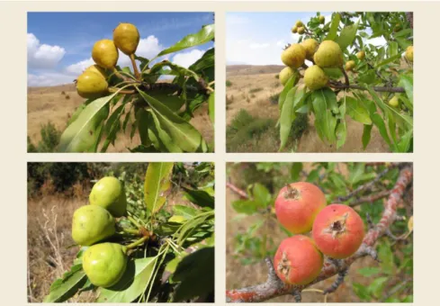
Figure 1. Pyrus gergerana Gladkova. The tree is found by the road to Herher village and the locals consider it a symbol of the village. This is the biggest known individual for the species (photo by A. Asatryan)
Figure 2. Pyrus daralagezi Mulk. (photo by A. Asatryan)
23
Except P. gergerana and P. daralagezi, the following pear species have been identified as occurring on the sanctuary’s territory: P.
salicifolia, P. pseudosyriaca, P. nutans, P. caucasica, P. medvedevii, endemics of Armenia P. elata and P. hajastana. Also, four hybrid forms, possibly between P. salicifolia and P. oxyprion, P.
pseudosyriaca and P. nutans, P. pseudosyriaca and P. daralagezi, P.
pseudosyriaca and P. elata were found (Asatryan 2018).
Sensible intrageneric and intraspecific variability of the taxonomically important traits such as, leaf (Figure 3) and fruit (Figure 4) shape, size, colour and texture often creates difficulties in identification and so, assessment of the species distribution.
Figure 3. Diversity of Armenia’s wild pear leaves (photo by A. Asatryan)
The research let us to identify sites of ‘concentration' of genetic diversity of wild pears in Armenia, to explore the distribution of some endemic species and to outline the difficulties in effective conservation of wild pears in Armenia.
Acta Biol. Plant. Agriensis 7: 19–31
24
Figure 4. Fruit diversity within Pyrus pseudosyriaca Gladkova (photo by A.
Asatryan)
The main aim of the study was to process the collected data to identify the main ‘hotspots’ of the pear diversity in Armenia and give their descriptions. Some questions aroused during the implementation of the above mentioned projects are discussed as well.
MATERIALS AND METHODS
The research has been carried out in 2016–2018 and included field surveys in the south of Armenia (Vayots Dzor and Syunik provinces). The itinerary was designed in accordance with the target species’ (P. gergerana, P. daralagezi and P. voronovii) distribution data, taken from the herbarium of the Institute of Botany after A.L. Takhtajyan of the National Academy of Sciences, Republic of Armenia (ERE).
Almost all pear trees along the roadsides have been studied along the trip (most of the known locations for the target species were by the roads), herbarium samples were taken for further
25
processing and identification. The specimens of special interest were marked with labels.
The literature on pear species and their habitats in Armenia was studied; special attention was paid to the original descriptions of the target species and other endemics in order to be prepared to distinguish them both in the field and during identification of the collected herbarium.
Herbarium material – about 150 sheets, on Pyrus sp. kept in the Institute of Botany NAS RA were studied. For the endemics we analysed the original descriptions of the species and the herbarium samples (including the type specimens) to understand the main diagnostic features for the target species and to make comparisons with the herbarium identifications made by previous researchers.
The research area covered roads to Herher village, then Yeghegis river gorge, way to Jermuk and its surroundings, including the forest near Kechut reservoir, the roads from Kapan to Vachagan and Srashen villages, Geghi river gorge, Tashtun pass, surroundings of Tashtun and Lichq villages. The field research covered also the area of Herher state sanctuary (6139 hectares) in Vayots Dzor province.
The pear diversity hotspots were identified by analysing the data on the species’ distribution, taken from the herbarium of the Institute of Botany of the NAS RA (ERE) and from our field research.
Certain difficulties and problems became evident during the work. We had difficulties trying to identify some samples – they just did not match any of the described taxa. High level of polymorphism in populations, variability of diagnostic features of the species, big number of hybrid forms etc. made the identification process challenging. There is strong need of critical taxonomic review of the group, based on field observations, statistical data, DNA studies etc.
RESULTS
About 120 herbarium samples have been collected during our fieldtrips and processed later.
Six main diversity hotspots for Pyrus sp. in Armenia have been identified (Figure 5). The pear diversity for each of them is represented below – based on herbarium and literature studies and
Acta Biol. Plant. Agriensis 7: 19–31
26
data, collected during our fieldwork. The sites on the map are marked according to their numbers given here.
Figure 5. Wild pear diversity hotspots in Armenia (d-maps.com)
1. Yeghegis river gorge, Vayots Dzor province
The total number of pear species is 25, 8 of them are endemics of Armenia.
According to the herbarium data: P. ketzkhovelii, P. vsevolodii, P.
demetrii, P. hyrcana, P. turcomanica, P. caucasica, P. raddeana, P.
acutiserrata, P. syriaca, P. medvedevii, P. fedorovii, P. georgica, P.
takhtadzhianii, P. communis and following endemics P. browiczii, P.
elata, P. complexa, P. daralagezi, P. sosnovskyi, P. hajastana, P.
tamamschjanae; we added to the list P. salicifolia, P. oxyprion, P.
pseudosyriaca and the endemic P. gergerana.
27
2. Tashtun pass, surroundings of Tashtun and Lichq villages and part of Megri Pass, Syunik province
The total number of pear species is 22, 8 of them are endemics of Armenia. According to the herbarium data: P. grossheimii, P.
hyrcana, P. demetrii, P. raddeana, P. takhtadzhianii, P. acutiserrata, P. syriaca, P. saliciflia, P. medvedevii, endemics P. voronovii, P.
daralagezi, P. complexa, P. gergerana, P. tamamschjanae, P. elata;
we added to this list P. caucasica, P. zangezura, P. nutans, P.
pseudosyriaca, P. georgica and two endemics P. megrica, P.
hajastana.
3. Surroundings of Jermuk town, Vayots Dzor province
The total number of pear species is 19, 5 of them are endemics of Armenia. According to the herbarium data: P. nutans, P. fedorovii, P.
takhtadzhianii, P. medvedevii, P. caucasica, P. ketzkhovelii, P. syriaca, P. pseudosyriaca, P. zangezura, P. salicifolia endemics P. sosnovskyi, P. hajastana, P. daralagezi, P. gergerana, P. megrica we found P.
oxyprion, P. taochia, P. georgica, P. acutiserrata.
4. “Khosrov Forest” state reserve, Ararat province
The total number of pear species is 15, 5 of them are endemics of Armenia. According to the herbarium data: P. communis, P.
caucasica, P. vsevolodii, P. turcomanica, P. syriaca, P. salicifolia, P.
medvedevii, P. oxyprion, P. fedorovii, P. takhtadzhianii and endemics P. tamamschjanae, P. sosnovskyi, P. theodorovii, P. hajastana, P.
chosrovica.
5. Surroundings of Herher village and Herher state sanctuary, Vayots Dzor province
The total number of pear species is 14, 5 of them are endemics of Armenia. According to the herbarium data: P. demetrii, P. fedorovii, P. takhtadzhianii, P. salicifolia and endemics P. gergerana and P.
hajastana; we added to the list P. nutans, P. communis, P.
pseuadosyriaca, P. caucasica, P. medvedevii and endemics P.
sosnovskyi, P. daralagezi, P. elata.
6. Shikahogh state reserve, Syunik province
The total number of pear species is 12, 3 of them are endemics of Armenia. According to the herbarium data: P. communis, P. hyrcana, P. caucasica, P. zangezura, P. raddeana, P. syriaca, P. medvedevii, P.
Acta Biol. Plant. Agriensis 7: 19–31
28
fedorovii, P. takhtadzhianii and endemics P. tamamschjanae, P.
megrica and P. gergerana.
DISCUSSION
The main characteristics of the ‘hotspots’ for pear diversity in Armenia are the following: they located in deep gorges and valleys and include fragments of arid open forest where narrow leaved pear species occur (mainly P. salicifolia and P. oxyprion) and deciduous forest, where broad leaved mesophytic species occur (P.
caucasica, P. syriaca, P. pseudosyriaca, P. daralagezi and others).
The pear diversity areas contain also old settlements (at least one village) and the roads. The ‘intermediate’ leaved pear species and hybrid forms are found mostly by the roadsides and on the glades in the deciduous forests. So, all the pear trees found in the area, including ones in orchards and village gardens have been involved in hybridization process. Certain questions appear in relation to the original descriptions of some rare endemic species, which have been described from just one tree with no data on population and distribution of the particular taxon.
Clarifications of their taxonomic status need to be done.
According to Gladkova (1989) P. sosnovskyi, P. demetrii, P.
tamamschjanae, P. vsevolodii are close and represent garden escapees on the different stages of transformation from P.
communis group. Such a high level of polymorphism in Pyrus, according to her (Gladkova 1990) is caused by two groups of factors: one represents natural evolution, the other is linked with human activity. She thinks that it is here, in the Caucasus region, where the ways of evolution of two ecological groups of species, formed in different ecological conditions, linked. The first group is formed with more or less mesophile species, which ancestors have been part of ancient Tertiary forest flora, remnants of which are still found in relic refugiums in Eastern Asia and Transcaucasia. Our target P. daralagezi belongs to this group. The second group is formed with xerophyte species of P. salicifolia type, which have been formed in later ages – in arid conditions of the Mediterranean area. Intensive hybridisation processes between representatives of these two groups have been the causes of appearance of many more or less stabile forms carrying the intermediate features of
29
both groups. The other target species P. gergerana is from this group.
Gladkova (1990) writes, that morphologically similar forms appear in different spots of the distribution area as a result of hybridisation of the parental forms. Very often they occur by the roadsides and nearby villages. Many of these individual trees of hybrid origin became the only specimens, which have been considered in description of new species.
Armenia is located in the South-Western Asian – one of the Vavilov’s world centers of origin of cultivated plants (Vavilov 1926) and is notable for great diversity of wild relatives of cultivated plants. Caucasus is known as one of the most ancient centres of agriculture and domestication of wild plants. Human activity has been another factor, promoting active hybridisation in populations of wild pears. Main centres for pear diversity are linked with ancient settlements – still existing or abandoned. During many centuries wild forms have been domesticated with further selection activities, at the same time the opposite process of escaping from gardens used to take place. One can still find many ancient pear sorts all over Armenia, which are close to wild forms.
Fruits of wild pears particularly, fruits of Caucasian pear (P.
caucasica), which is more common in the north of the country, also P. salicifolia and P. pseudosyriaca in the southern Armenia are used widely in Armenia by local communities and companies to produce compote, vodka, vinegar. In the southern Armenia, where the diversity of species and forms is much higher locals distinguish particular trees by characteristics of the fruits such as the taste, juiciness and the time when they are perfect for eating: some pear fruits have astringent taste and become edible (soft and brown) some weeks after they fell from a tree. This data, which comes from ages-long observations and practice on site may be very valuable not just for promoting in-situ conservation, but for researchers who work on taxonomy of this group. Wild pear seedlings are used as rootstocks for grafting (Sokolov 1954).
Pyrus L. is the largest among the genera of Armenian flora, represented in the IUCN Red List. 9 of total 71 plant species of Armenian flora, listed there under threatened categories are pear species. As mentioned before, 10 pear species are included in the Red Data Book of Armenia, but only part of the populations of just 5