AGGREGATE CULTURES: A MODEL FOR STUDIES OF BRAIN DEVELOPMENT
Nicholas W. Seeds Galo Ramirez Michael J. Marks
Department of Biophysics and Genetics University of Colorado Medical Center
Denver, Colorado
I. INTRODUCTION
Brain is a very complex and heterogenous tissue whose morpho- genesis reflects extensive cell migration and specific cell as- sociations. During development brain cells must possess both self-recognition properties for interaction with homologous cells and complementary recognition mechanisms for specific cell in- teractions with heterologous cell populations. These cell recog- nition capabilities display temporal alterations during develop- ment suggesting concomitant changes in the cell surface (Gottlieb et al., 1974; Moscona, 1974; Seeds, 1975). Furthermore normal brain function requires that specialized intercellular associa- tions, such as those related to synaptogenesis and myelination, be formed in a very selective manner. Thus these recognition mechanisms regulated by the cell genome (Sidman, 1974) and the cell surface (Moscona, 1974) are necessary developmental events.
23
24 NICHOLAS W. SEEDS et al.
Ideally a culture system for studying the cellular and molecu- lar aspects of brain development should retain the specificity of cell interaction found in situ and display morphological, bio- chemical and electrical differentiation similar to normal brain maturation. ' Furthermore, it should be possible to manipulate the numbers and types of cells in a rigorously controlled culture environment.
II. AGGREGATION
Embryonic cells possess a very selective adhesiveness that allows them to distinguish like from unlike cell types (Moscona, 1973). This property of fetal cells permits dissociated single cells to reaggregate into multicellular structures where specific cell-cell contacts can be reestablished. Formation of these ag- gregates is enhanced by gently rotating suspensions of trypsin dissociated cells to increase the number of collisions between sticky cells while minimizing the shearing forces. Aggregation is influenced by the tissue of origin, the state of differentiation, cell specific aggregation factors, as well as environmental fac- tors including rotation speed, culture medium, temperature and serum proteins (Moscona, 1973; Seeds, 1973). Aggregation is a rapid process being completed in a matter of hours. The initial aggregates are composed of loosely packed and randomly dispersed cells (Fig. l a ) . Numerous small cellular extensions are present by one day of culture and after several days there are extensive cellular outgrowths that serve to stabilize the complex.
During the first week of culture the cells undergo extensive migration and segregate into clusters of similar cell types (Fig.
lb). In addition reaggregates prepared from specific brain re- gions show distinctive patterns of aggregation. Cerebral cortex aggregates display a cell poor peripheral region composed largely of outwardly directed cellular extensions from cells whose psri-
Q) 0 0
*M 4J oj 0 03 Q) ,M Ü
U <+H Ή Μ Η t J • H
•H Q) C 03 * H
u
03 Q) 03Q) * H Q)
0 Q) Es
Q) • 0 JH
to 3
10 G • U c: 0 0 •H 3
• H 4J 0 4J Ü
Ü Q) M H
φ to 0 to to
0 03 3 i
•M M 03 Q< t J
0 0 •M QO 10 to 0 J H Q)
Ü • • U
" H Q) <4H
e 3 -M 4J — ^ - ς 3 V u
Ü
«M to
«M Q)
0 -U
• 03
tO — s b>
Q) QJ
* H S u 4J 3 to b>
3 i 03 3 03 <D
Ο
Q) Q)
4J Q)
4J • th 0) * H to
Q) 0
u
* H 4J 0 Q)
b i »CJ
M + J
03 0
03 Q)
• H - N Q) 03 »Q th
03 V u 0)
• H * H tO En 0) 0
Q) 03 ϋ
Q) •u 03
b» c;
0 c: 03 4J 4J 0
to - Ν * 9
*M 4J Ο Ο
03 03 V u
to 3 i i H Q) <D
Î H + J 3 * H 0
2 4J 0 D M Ü to
Ο 3 •H
H Ü
PM 03 Ü
4J Q)
<D M H
Î H 0
26 NICHOLAS W. SEEDS et al.
karyon is more centrally located (Fig. l c ) , In contrast cerebel- lar aggregates have a cell dense periphery of large cells whose extensions are directed internally or laterally (Fig. I d ) . The identity of these exterior cells in cerebellar aggregates is not complete; however, many of these cells can concentrate 3H-GABA from the medium and fetal cells that undergo their last nuclear division on gestational day 13, namely Purkinje and Golgi II neu- rons, are only found in this region; whereas, later developing cells principally occupy more internal positions. More impressive is the histotypic pattern formation demonstrated by DeLong (1970) for pyramidal neurons in hippocampal aggregates and the lack of this cellular alignment in aggregates of cells from the "reeler"
mutant mouse (DeLong and Sidman, 1970).
III. MORPHOLOGICAL DEVELOPMENT
The ability of dissociated cells to reproduce histotypic cellular patterns suggested that these cultures may show ultra- structural and biochemical differentiation similar to that of de- veloping mouse brain. The two most characteristic features of brain maturation are synaptogenesis and myelination. Both of these events occur late in brain differentiation; most mouse brain synapses form during the third postnatal week and myelination is most active during the fourth week (Aghajanian and Bloom, 1967;
Karlsson, 1967). The tissue we most often use to prepare aggre- gate cultures, 16 day old fetal mouse brain, has very few syn- apses and these are generally restricted to the spinal cord (Crain et al., 1968).
The initial reaggregates are loosely associated and lack any membrane specialization suggestive of synaptic-like structures.
After four days of culture numerous symmetrical intercellular complexes or desmosomes are found (Seeds and Haffke, 1976). The first synapses are seen at 7 to 9 days of culture and they both
AGGREGATE CULTURES 27 TABLE I Synaptogenesis in Aggregate Cultures3
Days in culture Synapses/field
1 0
7 0
9 3.7
15 4.3
21 6.7
25 10
33 11
49 8.3
aAggregate cultures were prepared from dissociated brain cells of 16 day mouse fetuses. Aggregates were fixed, embedded and sectioned for electron microscopy at several day intervals. The sections were observed at a constant magnification {43 μ2 field) and photographs taken of fields containing at least one synapse.
The number of synapses per field was determined from about ten photographs at each time point.
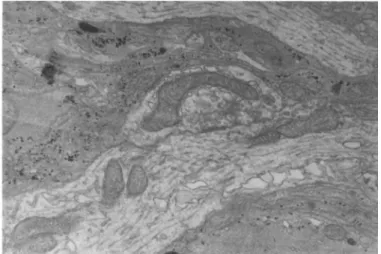
increase in number and mature in appearance during subsequent cul- ture (Seeds and Vatter, 1971). Synaptogenesis reaches a peak at 4-5 weeks of culture (Table I ) . A typical mature synaptic complex containing many clear vesicles and encircled by numerous electron opaque glial cells with abundant glial filaments and glycogen granules is shown in Fig. 2.
During culture the reaggregated cells show increased amounts of rough endoplasmic reticulum and golgi complexes. Myelinated axons are present by the fourth week of culture (Seeds and Vat- ter, 1971). Furthermore, studies in our laboratory by Schmidt (1975) have demonstrated increased sulfatide synthesis during chick brain aggregate development.
28 NICHOLAS W . SEEDS et al.
FIGURE 2 Ultrastructure of a Brain Aggregate. A clear vesicle containing synaptic complex surrounded by more electron dense glial processes is present.
IV. BIOCHEMICAL DEVELOPMENT
A. Neuronal Development
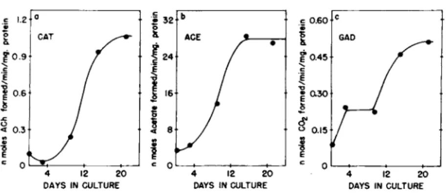
One of the main reasons for choosing the mouse as the source of tissue was the limited development of biochemical activities related to neurotransmission in the fetal mouse brain. The great- est development occurs postnatally and the 16 day fetal brain cells are relatively undifferentiated. However, when cultured as reaggregates these cells show marked increases in specific activi- ties of three neuronal enzymes: choline acetyltransferase (EC 2 . 3 . 1 . 6 ) , acetylcholinesterase (EC 3 . 1 . 1 . 7 ) and glutamate decar- boxylase (EC 4 . 1 . 1 . 1 5 ) (Fig. 3 ) . The greatest increase in these activities occurs during the second week of culture, thus resembl- ing the in situ development in both rate and extent (Seeds, 1 9 7 3 ) . Cultures maintained for as long as three months possess high lev- els of enzyme activity often surpassing the specific activities found in adult mouse brain. Furthermore, these enzymes appear
AGGREGATE CULTURES 29
FIGURE 3 Biochemical Differentiation in Aggregate Culture, The development of choline acetyl transferase (a}, acetylcho- linesterase (bl , and glutamate decarboxylase Qc) are shown as a function of time in culture. The starting undissociated brain tissue has activities of 0.18, 11, and 0.13 respectively (from Seeds, 1971).
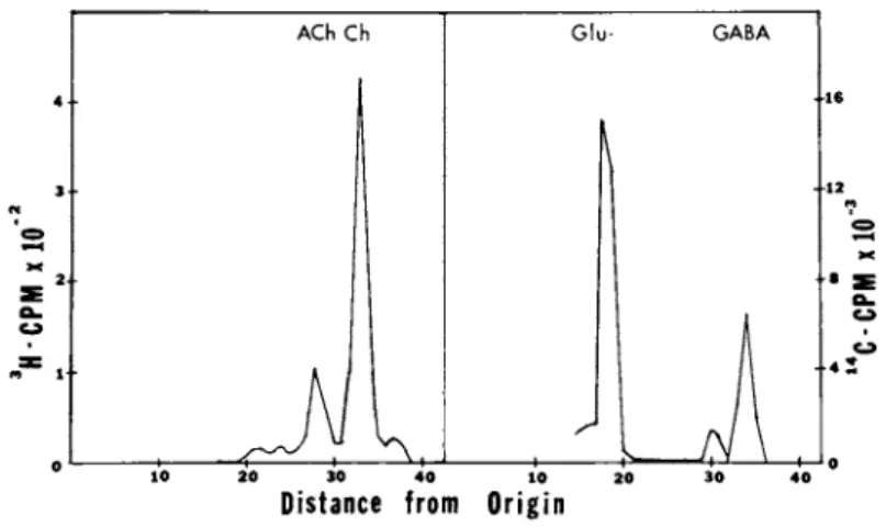
functional in the aggregates, since incubation with radiolabeled choline and glutamate leads to accumulation in the aggregates of acetylcholine and γ-aminobutyrate (Fig. 4 ) .
In addition to the presynaptic cholineacetyltransferase activ- ity, we have examined the postsynaptic component of cholinergic development. Development of the muscarinic receptor in reaggre- gates has been followed by specific binding of ^H-guinuclidinyl benzilate (Yamamura and Synder, 1974) to particulate fractions of reaggregate homogenates. The developmental profile of the mus- carinic receptor is similar to that of cholineacetyltransferase activity but somewhat slower in rate of appearance, reaching a maximum expression of receptor after 24 days in culture (Seeds and Haffke, 1976). Thus cholinergic development in these reaggregate cultures reaches maximum levels prior to synaptogenesis; however, since we cannot at present distinguish microscopically the cho- linergic synapses, it is possible that cholinergic synapses may be formed first and the slower development reflects other types of synapses.
30 NICHOLAS W. SEEDS et al.
20 30 40 10 20
Distance from Origin
FIGURE 4 Neurotransmitter Synthesis in Reaggregate Cultures.
Komogenates from cultures incubated with 3K-choline or 14Q-
glutamate were subjected to high voltage electrophoresis at pH 1.9 to separate acetylcholine (ACh) and y-aminobutyrate (GABA) from their precursors.
B. Brain Development
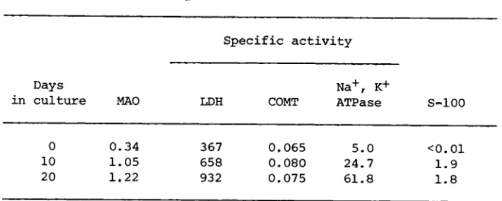
Reaggregated brain cell cultures also acquire enzymatic acti- vities found in cells other than neurons (Seeds, 1975a: Marks and Seeds, 1975). Some activities such as monoamine oxidase (EC 3.6.1.3) increase 3 to 10 fold during brain development; whereas, other enzymes such as catechol-0-methyltransferase (EC 2.1.1.6) show little change. Similar developmental increases are found in reaggregate cultures (Table I I ) . There is a shift in the relative amount of lactate dehydrogenase isoenzymes during brain develop- ment; this transition is also seen in the aggregates (Seeds, 1975a). In addition, the cultures express the brain specific S- 100 protein that is found relatively late in development and is concentrated in glial cells. However, the maximal level of S-100 is only 25% that found in adult brain, and possibly reflects the limited (<5%) division of cells in reaggregate cultures.
AGGREGATE CULTURES 31 TABLE II Development of Biochemical Activities0
Specific activity
Days N a+, K+
in culture MAO LDH COMT ATPase S-100
0 0.34 367 0.065 5.0 <0.01 10 1.05 658 0.080 24.7 1.9 20 1.22 932 0.075 61.8 1.8
aReaggregated brain cell cultures from 16 day fetal mice were maintained in basal Eagle's medium with 10% fetal calf serum. En- zyme specific activities are expressed as η moles product/min/mg protein. S-100 protein is expressed as μg S-100 per mg soluble protein.
TABLE III Biochemical Development versus Aggregate S i z ea
Cells ACE CAT MAO LDH S-100
Dissociated cells (5- 2.5 0.08 0.30 367 <0.01 50 μ)
(
Big aggregates (1050 μ 36.0 0.84 1.05 985 diam)Small aggregates (365 25.0 0.38 1.37 878 μ diam)
!
Big aggregates (1066 μ 25.0 0.54 - - 0.7
diam)
Small aggregates (510 11.0 0.22 - - 0.6 . μ diam)
aT h e biochemical activity of reaggregated brain cells cultured for 15 days in basal Eagle's medium on gyrotary shaker baths is compared to that of the initial dissociated cells. The speed of rotation was 70 or 90 rpm to produce aggregates of two distinct size classes, with the average aggregate diameter given in parenthesis.
32 NICHOLAS W . SEEDS et ai
Specific activity
Days in culture 5mM 25mM
2 11 7
4 10 8
5 17 21
7 19 28
9 25 36
11 29 39
17 51 60
21 59 69
aAggregate brain cell cultures from 16 day fetal mice maintained in basal Eagle's medium (5,4mM K+) or in medium where some of the N a H C 03 had been replaced with KHCO3 to 25mM K+. The enzyme spe- cific activity is expressed as η moles Pi released/min/mg protein and standard deviations were 3-8%.
The appearance of neurohormone receptors on the surface of brain cells is a characteristic feature of development. One such molecule is the ß-adrenergic receptor that is closely linked to adenyl cyclase and appears between postnatal day 4 and 10 (Schmidt et al./ 1970). Binding of norepinephrine to this receptor leads to a rapid 5 fold increase of cyclic AMP levels in brain slices.
Although newly formed aggregates show no response to catechola- mines, one week old aggregate cultures show a similar 4-6 fold response in cellular cAMP levels (Seeds and Gilman, 1971). This response probably represents the formation or activation of cell surface 3-adrenergic receptors.
The biochemical development of these aggregate cultures is sensitive to a variety of environmental factors, including ion levels, drugs and culture medium composition (Marks and Seeds, 1977; Seeds, 1976c). Aggregate size is also an important variable
(Table III). Small aggregates (200-500 μ) show greatly reduced TABLE IV Potassium Induction of N a+, K+- A T P a s ea
AGGREGATE CULTURES 33
levels of choline acetyltransferase and acetylcholinesterase ac- tivity compared to normal (900-1100 μ) size aggregates. Further- more, only the neuronal activities are affected, monoamine oxi- dase, lactate dehydrogenase and S-100 develop normally, thus sug- gesting that a critical cell mass or cell interactions are nec- essary for maximal neuronal differentiation; whereas, glial de- velopment may not be so restrictive.
C. ATPase Induction
One of the major advantages for studying development in cell culture is the opportunity to control the environment. Regula- tion of the ionic balance between intra- and extracellular fluids is critical for functional activity of the nervous system and this regulation is performed by the energy dependent N a+, K+ ATPase in brain, we have examined the influence of extracellular ions on this enzyme during development (Marks and Seeds, 1977).
Chronic exposure to elevated K+ during culture leads to an in- duction of the N a+, K+-ATPase activity. Table IV shows the de- velopment of the enzyme activity in normal medium, 5.4mM K+ and in medium containing 25 mM K+. After a lag of several days there is a 50% increase in the activity by day 7 and this increment is retained during further development. Furthermore, this stimula- tion increases linearly as the K+ is raised.
Additional studies using ouabain to inhibit the N a+, K+-ATPase in the aggregate cultures show a similar induction of enzyme ac- tivity with exponential increases between 1 0 ~5 and 2 χ 10~4M ouabain. Collectively these findings suggest that changes in mem- brane potential may regulate the synthesis of specific cell pro- teins necessary for controlling the membrane potential; thus, in- creased bioelectric activity during brain maturation may promote the observed developmental increases in N a+, K+-ATPase of brain.
34 NICHOLAS W . SEEDS et al.
ι
ο.Ε σ»
Ε
σ ο σ
150
A Tectum
: / :
Rttina
• Β '
: ^ :
/ Retina
/ :
: î S - '
η Ttctum
y -
1 1 1 1 Ι I
/ :
Ι . Ι . Ι
1.2
1.0 ο
0.8 Ε c
ε
0.6 Φ
0.4
ε
ο3 ε
c 0 10 2 0 3 0 0 10 2 0 3 0Days in culture
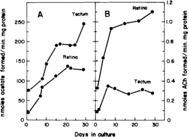
FIGURE 5 Development of Acetylcholinesterase (A) Choline Acetyltransferase (B) Activity in Tectum and Retina Aggregates.
Aggregate cultures were prepared from seven day old chick embryos and enzyme activity was determined at periodic intervals (from Ramirez and Seeds, 1977).
D. Cell Interaction
The specificity of cell recognition displayed by retina and optic tectum cells of the chick is well documented (Barbera et al., 1973; Merrell and Glaser, 1973; Gottlieb et al., 1974).
These tissues show both regional and temporal specificity in their interaction. For these reasons the retino-tectal system is an attractive candidate for studying the relationship of cell recog- nition events and biochemical development (Ramirez and Seeds, 1977). Both chick retina and optic tectum show large increases in choline acetyltransferase and acetylcholinesterase specific activities from fetal day 7 to ten days after hatching. The specific activity of acetylcholinesterase in aggregate cultures prepared from 7 day chick embryo retina and tectum increases 3-6 fold during four weeks of culture (Fig. 5 A ) . The maximal activity is 55-75% of the in situ activity. In retina aggregates the cho- line acetyltransferase activity increases 4 fold to 55% of the
AGGREGATE CULTURES 35
Ο ΙΟ 2 0 3 0 Ο 10 2 0 3 0
Days in culture
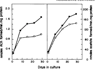
FIGURE 6 Biochemical Development of Retinal-Tectal CoAggre- gates. Seven day old chick embryo retina and tectum cells were mixed and allowed to coaggregate, then assayed for choline acetyl-
transferase and acetylcholinesterase activity. The culture con- tained 87% retina and 13% tectum cells. The dashed line (open circle) represents the expected developmental values for the above cell ratio determined from Figure 5 (from Ramirez and Seeds,
in situ levels; however, optic tectum aggregates show only a slight increase, 7% of the activity in a 10 day old chick tectum
(Fig. 5 B ) .
The influence of cell recognition events on development of these cholinergic activities was examined by preparing retinotec- tal coaggregates from dissociated retina and tectum cells. The development of these coaggregates is shown in Fig. 6. There is a 100% synergistic effect on choline acetyltransferase development and a 30% stimulation of acetylcholinesterase specific activity.
In addition, this synergism requires cell-cell contact since co- culture of retinal and tectal aggregates does not lead to an in- crease in enzyme activity. Furthermore, the synergistic effect shows temporal specificity, requiring the two cell populations to be of identical embryonic age (Ramirez and Seeds, 1977). These results suggest that cell recognition events in aggregate cultures and the resulting cell-cell interaction can lead to specific bio- 1977).
36 NICHOLAS W . SEEDS et al.
chemical changes in the participating cells. The possibility that this synergism reflects the formation of specific retino-tectal synapses in the coaggregates is currently being investigated.
Thus, reaggregates offer a useful cell culture system for studies of development, especially where three dimensional cell- cell interactions may be required. Hopefully these cultures will provide new information on the role of cell recognition and cell- cell interaction in the regulation of morphogenesis and biochemi- cal differentiation.
ACKNOWLEDGMENTS
We gratefully acknowledge the excellent technical assistance of Susan Haffke and Barbara Fonda, and wish to thank Sally Planalp for preparing the manuscript. The studies reported here were sup- ported in part by U.S. Public Health Service Grants NS-09818 and CA-15549 and a Career Development Award K4-GM40170.
REFERENCES
Aghanjanian, G. Κ. , Bloom, F. Ε., (1967), Brain Res. 6, 716-727.
Barbera, A. J., Marchese, R. Β., Roth, S., (1973), Proc. Nat.
Acad. Sei. U.S.A. 70, 2482-2486.
Crain, S. Μ., Peterson, E. R., Bornstein, Μ. Β., (1968), in
"Growth of the Nervous System," Ciba Symposium, pp. 13-40, Little, Brown, Boston.
DeLong, G. R. , (1970), Develop. Biol. 22, 563-583.
DeLong, G. R., Sidman, R. L., (1970), Develop. Biol. 22, 584-600.
Gottlieb, D. Ε., Merrell, R., Glaser, L., (1974), Proc. Nat. Acad.
Sei. U.S.A. 71, 1800-1802.
Karlsson, U., (1967), J. Ultrastruct. Res. 17, 158-175.
Marks, M. J., Seeds, N. W., (1977), J. Biol. Chem. (in press).
AGGREGATE CULTURES 37
Merrell, R., Glaser, L., (1973), Proc. Nat. Acad. Sei. U.S.A. 70, 2794-2798·
Moscona, Α. Α., (1973), in "Cell Biology in Medicine," (E. Bittar, e d . ) , pp. 571-591, J. Wiley, New York.
Moscona, Α. Α., (1974), in "The Cell Surface in Development," (A.
A. Moscona, e d . ) , pp. 67-99, J. Wiley, New York.
Ramirez, G., Seeds, N. W., (1977), Nature (in press).
Schmidt, G. L., (1975), Brain Res. 87, 110-113.
Schmidt, J. J., Palmer, E. C., Dettbarn, W. D., Robison, G. Α., (1970), Develop. Biol. 3, 53-67.
Seeds, N. W., (1971), Proc. Nat. Acad. Sei. U.S.A. 68, 1858-1861.
Seeds, N. W., (1973), in "Tissue Culture of the Nervous System,"
(G. Sato, e d . ) , pp. 35-53, Plenum Press, New York.
Seeds, N. W., (1975a), J. Biol. Chem. 250, 5455-5458.
Seeds, N. W., (1975b), Proc. Nat. Acad. Sei. U.S.A. 72, 4110-4114.
Seeds, N. W., (1976), in "Molecular and Cellular Analysis of Mental Disorders," (H. Matthaei, e d . ) , Springer-Verlag, Berlin,
(in press).
Seeds, N. W., Gilman, A. G., (1971), Science 174, 292.
Seeds, N. W., Haffke, S. C , (1976), submitted for publication.
Seeds, N. W., Vatter, Α. Ε., (1971), Proc. Nat. Acad. Sei. U.S.A.
68, 3219-3222.
Sidman, R. L., (1974), in "The Cell Surface in Development," (A.
Moscona, e d . ) , pp. 221-253, J. Wiley, New York.
Yamamura, Η. I., Synder, S. H. (1974), Proc. Nat. Acad. Sei.
U.S.A. 71, 1725-1729.