CARBOHYDRATES
G. R. NOGGLE 1. PHOTOSYNTHESIS
A. INTRODUCTION
The essential features of the photosynthetic reaction have been known since 1845 when it was recognized that the fundamental transformation of photosynthesis was the conversion of light energy into chemical energy and that the reaction could be written as follows:
Carbon dioxide + water — ^ ^ .
organic matter + oxygen + chemical energy
green plant /^\
Since that time plant physiologists, biochemists, biophysicists, organic chemists, physicists, microbiologists, physical chemists, and others have contributed to the literature of photosynthesis (1).
The early work in photosynthesis was based on the assumption that carbon dioxide fixation was a process unique in green plants, and studies were made on the effect of various external factors such as light intensity, light quality, carbon dioxide concentration, and temperature. Certain internal factors were also recognized as affecting photosynthesis. Many fundamental studies on photosynthesis were concerned with the chemistry of the chlorophyll molecule. Enzymes were studied, and attempts were made to carry out cell-free photosynthesis by conventional biochemical techniques. While these studies contributed a great deal of information regarding the photosynthetic reaction, they did little to elucidate the mechanism of the process.
During the 1930's, however, research in the field of photosynthesis and in a number of allied sciences indicated that the concept of the nature of the problem of photosynthesis could be very profitably altered. The dis- covery in 1935 by Wood and Werkman (#) that propionic acid bacteria
1. A list of some general references on photosynthesis will be found at the end of Chapter XV (Bibliography).
*. H. G. Wood and C. H. Werkman, J. Bacterial. 30, 332 (1935).
733
734 G. R. NOGGLE
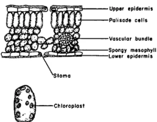
•Upper epidermis -Palisade cells
-Vascular bundle -Spongy mesophyll -Lower epidermis
Chloroplast
FIG. 1. Cross-section of leaf (upper) and enlarged cell from the palisade tissue (lower) showing chloroplasts.
could fix carbon dioxide led to investigations (8) that have shown that practically all living organisms are capable of fixing carbon dioxide. It became possible to view carbon dioxide fixation by plants as a process similar to that occurring in other organisms and to apply to the photo- synthesis problem concepts from bacterial and animal metabolism. The second discovery of major importance was the finding by Hill (4-) that iso- lated chloroplasts, when illuminated, could liberate oxygen and reduce added oxidants. This discovery freed the plant physiologist of the notion that photosynthesis could only be studied in intact living plants.
Since 1940 many new techniques and methods have been applied to photosynthesis research. Isotope studies have been especially valuable in elucidating the path of carbon in photosynthesis. Advantage has been taken of new advances in the field of enzyme chemistry. It is the purpose of this section to outline the essential features of what is presently known about photosynthesis and to review the significance of some of the recent discoveries.
B. STRUCTURAL ASPECTS OF THE PHOTOSYNTHETIC APPARATUS
Photosynthesis in plants is carried on by the chloroplasts of the leaves.
The distribution of the chloroplasts in the leaf of a land plant is shown in Fig. 1. The surface of the leaf is covered by a layer of waxy material called cutin. This waxy layer does not completely block the passage of gases and dissolved materials to and from the interior of the leaf, but the major ex- change of gas and water vapor occurs through openings in the leaf known as stomata. Gases entering the stomata diffuse into the intercellular spaces and then come into contact with the chloroplast-containing cells.
8. M. F. Utter and H. G. Wood, Advances in Enzymol. 12, 41 (1951).
4. R. Hill, Nature 139, 881 (1937).
The leaves of water plants are not covered by cutin and contain no sto- mata. The epidermal cells very often contain chloroplasts as do the other cells in the leaf. Carbon dioxide enters the leaf in the dissolved state and comes into intimate contact with the chloroplast-containing cells.
The chloroplasts are embedded in the cytoplasm of the cell and are one of a number of cytoplasmic particles that are found in plant cells. There is no general agreement as to the nature of these various cytoplasmic par- ticles (5). The term "mitochondria" is commonly used to denote one of these fractions, and a great deal of work has been carried out on their physiological role particularly with respect to the localization of enzymes (6). Comparable work with mammalian tissue has shown the mitochondrial fraction to be the site of action of a number of important enzyme systems
(7).
The chloroplasts are small green bodies that vary somewhat in size and number per cell depending upon the species of plant and the environmental conditions under which the plant is grown. They are found in the higher plants, green algae, red algae, and brown algae, but not in blue-green algae nor the photosynthetic bacteria, where the chlorophyll appears in the form of grana. The structure and morphology of the chloroplasts have been discussed thoroughly in several recent reviews (8, 9). In appearance they are discs or flat ellipsoids 3-10 μ across. The chloroplasts have a membrane, and within this membrane there may be several additional structures. The chlorophyll appears to be concentrated in bodies known as
"grana" which are embedded in a colorless stroma.
The grana have been investigated by means of the electron microscope, and in spinach chloroplasts (10) approximately forty to sixty grana were found. The grana were thin discs about 6,000 Â. in diameter and 800 A.
thick. There is some evidence (11) that the grana themselves are made up of stacks of ten to twenty thin lamallae which are 75 to 100 A. thick. The significance of the ultra-fine structure of the chloroplast is not thoroughly understood. If all of the chlorophyll is concentrated in the chloroplast then each granum, or each lamella in a granum, must contain several chlorophyll molecules. The close packing of the grana may then enable the light energy absorbed by one chlorophyll molecule to be passed along through a
5. E. H. Newcomer, Botan. Rev. 17, 53 (1951).
6. D. R. Goddard and H. A. Stafford, Ann. Rev. Plant Physiol. 5, 115 (1954).
7. A. L. Dounce, in "The Enzymes'' (J. B. Sumner and K. Myrbäck, eds.), Vol. 1, Part 1, p. 187. Academic Press, New York, 1950.
8. S. Granick, in "Photosynthesis in Plants" (J. Franck and W. E. Loomis, eds.), p. 113. Iowa State College Press, Ames, 1949.
9. T. E. Weier and C. R. Stocking, Botan. Rev. 18, 14 (1952).
10. S. Granick and K. R. Porter, Am. J. Botany 34, 545 (1947).
11. S. Granick, Chem. and Eng. News 31, 748 (1953).
736 G. R. NOGGLE
number of chlorophyll molecules until the energy reaches an active center where reduction occurs (11). The structural properties of the chloroplast may be very important in any physical or chemical explanation of the mechanism of photosynthesis.
With the realization that the chloroplast was the site of at least part of the photosynthetic reaction, a great deal of work was done on the isolation of the chloroplasts. Such preparations are generally contaminated with nuclear and cytoplasmic material (12), and attempts to ascribe observed results solely to the chloroplasts should be viewed with caution. Accom- panying the isolation studies have been analyses on the chemical and en- zyme composition of the chloroplasts. On a dry-weight basis, the chloro- plast contains about 45% protein and 25% lipid. The ash content is between 5 and 15 % of the dry weight while the pigments account for 5 to 10 %. The balance of the chloroplast is made up of carbohydrates, enzymes, and other unidentified compounds. The composition of the chloroplast material is quite variable and depends upon the plant species as well as upon the conditions of growth, i.e., nutrition, light, water, etc.
C. KINETIC STUDIES ON PHOTOSYNTHESIS
From a consideration of the photosynthetic equation (1), it is apparent that the rate of photosynthesis may be determined by measuring the disap- pearance of carbon dioxide, the evolution of oxygen, or the increase in dry weight (synthesis of protoplasm). Rate studies, or reaction kinetics, have been widely utilized in studying photosynthesis, and such studies have been recently reviewed by Rabinowitch (18), who points out: "Photo- synthesis is such a complex and heterogeneous process that it is probably impossible to make a complete analysis of its mechanism merely by meas- uring the rate of the over-all process under different conditions. However, this does not mean that kinetic measurements of photosynthesis are useless, but rather that they are most useful when combined with other biochemical and biophysical methods of approach. . . . "
Kinetic studies indicated that photosynthesis is dependent upon a num- ber of external and internal factors. Of the external factors, carbon dioxide concentration, light, and temperature are the most important, whereas the chlorophyll concentration is probably the most important internal factor.
The concentrations of various enzymes as well as certain undefined "proto- plasmic factors" have also been suggested as limiting photosynthesis.
12. T. E. Weier and C. R. Stocking, Am. J. Botany 39, 720 (1952) ; T. E. Weier, Pro- toplasma 42, 260 (1953).
IS. E. Rabinowitch, "Photosynthesis and Related Processes," Vol. II, Part 1, p. 831. Interscience, New York, 1951.
a. Carbon Dioxide Concentration
The average concentration of carbon dioxide in the air is roughly 0.03 % by volume. The rate of photosynthesis increases as the carbon dioxide concentration is increased until the light intensity becomes a limiting fac- tor. If the light intensity is increased, then the rate of photosynthesis is again increased by raising the C02 concentration. Prolonged exposure of plants to high concentrations of CO2 is generally harmful.
b. Light
Two different effects of light on photosynthesis are recognized: the quan- tity or intensity of the light and the quality or spectral distribution of the light. The rate of photosynthesis increases as the light intensity is increased from zero until some other factor becomes limiting. Light-intensity curves are characterized by a linear increase in rate of photosynthesis at low light intensities. As the light intensity increases the curve flattens out, and an increase in light intensity does not bring about a corresponding increase in the rate of photosynthesis—a situation known as light saturation. At very high light intensities photosynthesis may actually be inhibited.
Plants kept in the dark do not carry out photosynthesis but do respire, i.e., evolve carbon dioxide and take in oxygen. As the darkened plants are given increasing amounts of light, photosynthesis will increase, and carbon dioxide will be utilized and oxygen evolved. At some particular light level, called the compensation point, the gas exchange due to respiration and to photosynthesis will exactly balance each other. Above the compensation point, photosynthesis exceeds respiration. In photosynthesis research, it was generally assumed that light had no effect on respiration and that "true photosynthesis" could be calculated by subtracting respiration gas-ex- change values measured on a dark control. There was no direct way of measuring the photoeffect of respiration until Brown (14) used tracer oxygen to measure the respiratory rate. He found no evidence of photo- inhibition or photoenhancement of respiration in several strains of Chlorella commonly used in photosynthesis research. The conditions of the experi- ment were similar to those that are ordinarily used in photosynthesis re- search. The practice of measuring photosynthesis by applying a correction for dark respiration appears to be valid, at least for Chlorella.
Studies on the effect of light of different wavelengths on photosynthesis have been carried out since the essential features of the process were first discovered. In their simplest form, such experiments consisted of measur- ing the rate of photosynthesis in plants placed in light that had passed through different colored filters. From these and more refined measure-
Π. A. H. Brown, Am. J. Botany 40, 719 (1953).
738 G. R. NOGGLE
ments, it has been shown that plants carry out photosynthesis in light varying in wavelengths between 3900 A. and 7600 A.
The study of the wavelength dependence of photosynthesis has been extremely valuable in determining the role of the various pigments in photosynthesis. Such studies have been carried out by comparing the action spectrum of photosynthesis with the absorption spectrum of the plant. The photosynthetic action spectrum is determined by measuring the
Plant absorption Action spectram o </>
JO
<
4 4 0 520 600 680 7 6 0
Wavelength (π\μ)
FIG. 2. Absorption spectrum of the green alga Ulva taeniata. The photosynthetic action spectrum, corrected to relative rates of equal incident quanta, was made to coincide with the absorption spectrum at 675 im*. [From F. T. Haxo and L. R.
Blinks (IS)].
Totol pigments in ether ---Chlorophyll
— o—o-Corotenoids
4 4 0 520 6 0 0 680 7 6 0
FIG.
Wavelength (m/t)
3. Absorption spectra (in ether) of fat-soluble pigments in Ulva taeniata.
The carotenoids were measured at the concentration obtaining in the total pigment extract. The chlorophyll curve was calculated by difference. [From F. T. Haxo and L. R. Blinks (15)].
relative rates of photosynthesis in light of different wavelengths. A compari- son of the action spectrum of photosynthesis with the absorption spectrum of Ulva taeniata {15), a green alga, is shown in Fig. 2. In this figure the ac- tion spectrum is calculated in terms of equal incident quanta. The effect of this correction is to raise progressively the curve toward the blue region of the spectrum. These two curves show clearly that light is absorbed by some pigment which is then active in photosynthesis.
The nature of the fat-soluble pigment system present in Ulva taeniata is shown in Fig. 3. Carotene absorbs light in the blue region which is utilized in photosynthesis. Chlorophyll absorbs light in both the blue and red regions of the spectrum which is utilized in photosynthesis. Such studies with a wide variety of photosynthetic organisms have revealed that a number of different pigments may contribute to the absorption of light energy. Studies with extracted pigment systems are of limited value be- cause the structural relationships of the various pigments within the chloro- plast or grana are destroyed.
c. Temperature
If no other factor is limiting, the rate of photosynthesis increases with rising temperature up to the point at which permanent damage occurs to the plant. The maximum temperature tolerated by plants varies with the species as well as with the growing conditions. The decrease in photosyn- thetic rate at temperatures exceeding the maximum is thought to be con- nected with protoplasmic changes (enzyme denaturation, etc.) that alter the entire metabolism of the plant.
d. Interaction of Factors
From studies on the effect of temperature, carbon dioxide concentration, and light intensity, Blackman (16) showed that at low light intensities and high carbon dioxide concentrations the rate of photosynthesis was not sensi- tive to changes in temperature. Under these conditions the rate-limiting factor is light. At high light intensities and low carbon dioxide concentra- tions, the rate of photosynthesis is extremely sensitive to changes in the temperature. For these conditions, the rate-limiting factor is the carbon dioxide concentration. Blackman concluded that there were at least two distinctly different reactions in photosynthesis: a chemical reaction, re- quiring carbon dioxide and sensitive to temperature, and a photochemical reaction that is insensitive to temperature. The chemical reaction has been called the "dark reaction" or "Blackman reaction."
15. F. T. Haxo and L. R. Blinks, J. Gen. Physiol. 33, 389 (1950).
16. F. F. Blackman, Ann. Botany (London) 19, 281 (1905).
G. R. NOGGLE
COOCH?
< % H t £ = C - C - H
COOC20H39
FIG. 4. Structural formula of chlorophyll-a.
e. Pigments
The fact that chlorophyll is essential for photosynthesis has been known for a long time, but it was not until the work of Willstatter and co-workers (17) in the early 1900's that the chemical nature of chlorophyll was estab- lished. These investigators showed that the chlorophyll in green plants was made up of two components, chlorophyll-a and chlorophyll-b, with the following composition:
Chlorophyll-a Chlorophyll-b
C55H7206N4Mg C55H70OeN4Mg
mol. wt. 893 mol. wt. 907 The structural formula in Fig. 4 indicates the present concept of the chemi- cal structure of chlorophyll-a (the numbering system is that of Hans Fischer). Chlorophyll-b differs from chlorophyll-a with respect to the group at position 3. The methyl group in chlorophyll-a is replaced by an aldehyde group (—CHO) in chlorophyll-b.
In addition to chlorophyll-a and -b, two other pigments are generally present in leaves: carotene, an orange pigment, and xanthophyll, a yellow 17. R. Willstatter and A. Stoll, "Investigations on Chlorophyll" (Translated by F. M. Schertz and A. R. Merz). Science Press, Lancaster, Pa., 1928; "Untersuchungen über die Assimilation der Kohlensäure.'' Springer, Berlin, 1918.
TABLE I
PRINCIPAL PIGMENTS FOUND IN PLANTS, ALGAE, AND PHOTOSYNTHETIC BACTERIA (19)
Pigments Chlorophylls
Chlorophyll-a Chlorophyll-b Chlorophyll-c Chlorophyll-d Protochlorophyll Bacteriochloro-
phyll Bacterioviridin Phycobilins
Phycocyanin Phycoerythrin Carotenoids
Carotene-a,/3
Xanthophyll
Where found
All green plants Green plants, green
algae
Brown algae, diatoms Red algae
Etiolated plants Purple sulfur bacteria Green sulfur bacteria
Blue-green algae, red algae
Red algae, blue-green algae
Principal carotenoids of plants containing chlorophyll-a Variable in most
green plants
Maximum absorption
Red and blue-violet Near red, blue-violet Infra-red,
blue-violet Red and
blue-violet Orange-red Green Blue and
blue-green Blue and
blue-green
Comments
The chlorophylls prob- ably exist in a com- lex with protein and lipide-like material
Exists in several forms
Generally blue pigment Generally red pigment Approximately 6 to 8
different carotenes are known to occur in plants
Approximately 20 dif- ferent xanthophylls are known
pigment. A number of other pigments are found in the algae and photo- synthetic bacteria. These are summarized in Table I. Despite the fact that different chlorophylls are scattered throughout the plant kingdom, all of the organisms that carry out photosynthesis contain chlorophyll-a (bac- teriochlorophyll in the bacteria). The other pigments present are believed to absorb light energy that is eventually utilized in photosynthesis. In a study involving the determination of the absorption and fluorescence spectra of photosynthetic bacteria and algae, Duysens (18) postulated that the vari- ous pigments pass light energy by means of inductive resonance to a pig- ment which is active in photosynthesis. The transfer of light energy is unidirectional and passes the energy with high efficiency. The following scheme of energy transfer was suggested for Chlorella, which contains
18. L. N. M. Duysens, Doctoral thesis, University of Utrecht, Netherlands (1952).
19. R. Hill and C. P. Whittingham, "Photosynthesis," p. 17. Wiley, New York, 1955.
742 G. R. NOGGLE
chlorophyll-a and -b and some carotenoids:
Light Light
1 I
-> Chlorophyll-b —100% ) Chlorophyll-a —> Photosynthesis Fluorescence and
I
other losses
The energy absorbed by the carotenoids was transferred with 40 to 50%
efficiency to chlorophyll-b, which transferred its energy with 100% effi- ciency to chlorophyll-a. In some organisms it was necessary to postulate two types of chlorophyll-a, one that could pass its energy to photosynthesis and an 'inactive" chlorophyll-a which dissipated its energy to heat. The energy that flows into chlorophyll-a is thought to be transferred by inductive resonance to a pigment "reaction center" which participates in the dark reactions of photosynthesis.
Studies on the chemical structure of chlorophyll showed that it was closely related to heme, a pigment found in mammalian tissue. Subsequent work has indicated that a common biosynthetic pathway exists for the synthesis of porphobilinogen, a precursor to these two pigments. The steps leading to the formation of porphobilinogen were worked out by Shemin and co-workers {20) in a series of brilliant tracer experiments.
D. BACTERIAL PHOTOSYNTHESIS
The discovery that certain bacteria could carry out photosynthesis opened up a new field of photosynthetic research. It was found that certain green-, red-, purple-, and brown-colored bacteria could produce organic matter from carbon dioxide upon illumination. The formation of organic matter was not accompanied by oxygen evolution. As a result of work with the green sulfur bacteria, van Niel {21) showed that their C02 assimilation process was in close agreement with the following equation:
C02 + 2 H2S light ) (CH20) + H20 + 2 S (2) The similarity of this process to photosynthesis in green plants is obvious:
CO* + 2 H20 light ) (CH20) + H20 + 02 (3) van Niel then suggested a generalized formulation of photosynthesis as
follows:
C02 + 2 H2A l i g h t ) (CH20) + H20 + 2 A (4) 20. D. Shemin and C. S. Russell, J. Am. Chem. Soc, 75, 4873 (1953).
21. C. B. van Niel, in "Photosynthesis in Plants" (J. Franck and W. E. Loomis, eds.), p. 437. Iowa State College Press, Ames, 1949.
Light Carotenoids
i
Heat and other
I
energy losses
In this equation, H2A represents a hydrogen donor which reduces carbon dioxide with the aid of absorbed radiation, and A is the dehydrogenated donor.
Reaction (4) is a hydrogenation-dehydrogenation reaction which con- forms to the principles of "comparative biochemistry,, set forth by the Dutch microbiologist Kluyver. This concept, as stated by van Niel (21),
"postulates as a fruitful central idea that all metabolic activities are in- trinsically similar, and that each consists of a more or less extended series of inter- or intramolecular hydrogenation-dehydrogenation reactions.,, The comparative biochemistry principle has been extremely valuable in develop- ing theoretical approaches to the photosynthetic reaction.
In addition to the pigmented bacteria, some colorless bacteria are able to fix carbon dioxide in the absence of light. These colorless bacteria, known as "chemosynthetic" or "chemoautotrophic" organisms, obtain energy for assimilating and reducing CO2 by oxidizing N H3, H2S, and H2.
From a consideration of the different types of photosynthesis that are carried on by green plants, photosynthetic bacteria, and adapted algae, van Niel (22) suggested a general mechanism involving three types of reactions common to all photosynthetic organisms. The first reaction is a photochemical step in which water is decomposed to produce a reducing system and an oxidizing system. The second reaction or reactions lead ultimately to the dark reduction of C02 by the photochemically produced reducing system. The third series of reactions leads to the eventual dark reduction of the photochemically produced oxidizing system. These reac- tions involve only one photochemical step, the separation of H and 0 or OH from water. The reduction of carbon dioxide is a dark reaction and can proceed with energy produced from a variety of sources. Photosynthe- tic organisms derive energy for the reduction of C02 from light by way of a pigment system. The chemosynthetic organisms are able to reduce C02 by energy derived from the oxidation of N H3, H2S , H2, or other inorganic substances. Finally some organisms use energy obtained from the oxi- dation of organic material to reduce carbon dioxide.
A consequence of van Niel's formulation of photosynthesis is that the evolved oxygen should originate in the water and not from the carbon dioxide. Tracer studies with 018-labeled water and carbon dioxide have been consistent with this view. In a recent review, however, Brown and Frenkel (23) have pointed out that, although the oxygen probably orig- inates in the water molecule, the experimental evidence on this matter is not adequate to support or deny the theory that water is the sole source of photosynthetic oxygen.
22. C. B. van Niel, in "The Enzymes" (J. B. Sumner and K. Myrbäck, eds.), Vol.
II, Part 2, p. 1074. Academic Press, New York, 1952.
28. A. H. Brown and A. W. Frenkel, Ann. Rev. Plant Physiol. 4, 23 (1953).
744 G. R. NOGGLE
[CH20]n 02
î î î î
C02 2 H20
FIG. 5. Reactions of photosynthesis. (From Rabinowitch.^) E. T H E HILL REACTION
It had been known for a long time that oxygen was evolved during the illumination of ground leaves, isolated chloroplasts, or aqueous suspensions of dried leaf powders. The amounts of oxygen evolved were small, and it remained for Hill (4) to show that illuminated isolated chloroplasts were able to produce considerable quantities of oxygen if an aqueous extract of an acetone powder of yeast or leaves was added. Subsequently it was shown that the yeast or leaf extract could be omitted if certain ferric salts were added. With ferric iron, the reaction proceeds as follows:
4 Fe+++ + 2 H,0 „ J g L . ' 4 Fe++ + 4 H+ + 02 (5) Additional work on this reaction, commonly called the "Hill reaction,"
has led to the conclusion that it probably represents the photochemical phase of photosynthesis. This reaction shares with photosynthesis the con- version of light energy into chemical energy, and the appearance of molecu- lar oxygen.
Comparative studies on the Hill reaction and photosynthesis have led to the view that photosynthesis can be separated into two main groups of reactions as shown in Fig. 5. The right leg of the figure represents the photo- chemical phase which takes place in the chloroplast. The reactions include light absorption, energy transfer, and water photolysis with the concomi- tant production of oxygen and "active hydrogen." The "active hydrogen"
drives the reaction represented on the left side of the figure, the final out- come being reduced carbon dioxide. The key reaction takes place in the chloroplast or grana where the energy from light is converted into chemical energy represented by "active hydrogen."
The nature of the "active hydrogen" or the mechanism of its production is not known. A widely accepted theory {25) of how light energy is trans- formed into chemical energy is that a "biradical" is formed from a mole- cule following light absorption. Such a biradical might have both oxidizing and reducing properties so that the result of such a compound XY absorb-
ai. E. Rabinowitch, Set. American, 179, 24 (1948).
ßß. R. Hill and C. P. Whittingham, "Photosynthesis," p. 131. Wiley, New York, 1955.
ing light energy would be the formation of two radicals, X H and YOH.
The mechanisms for the further reactions of these two radicals are not known. The X H reactions might be similar to other hydrogen transport reactions in metabolic systems.
The transfer of hydrogen or electrons in biological systems is relatively well understood, and it is of interest to examine some of the compounds involved in the transfer mechanism. Table II lists some of the compounds that might serve in the hydrogen transport system of the photosynthetic cell. Since it will be necessary to refer many times to these compounds, their structure is given at this point (see also Chapter VIII). The names of these compounds have not yet been systematized. Diphosphopyridine nucleotide (DPN+) contains nicotinic acid amide, D-ribose, and adenosine 5-phosphate linked through a pyrophosphate group. Triphosphopyridine nucleotide (TPN+, coenzyme II) contains a third phosphate group (Chap- ter VIII).
N H2
N
f\
+ 1
—CONH2
1 1
HC HCOH HCOH
1 o
H C -
o-
• \ / v
N HC
c X II
X
N/ \ c / /
OH N HC HCOH HCOH i
1 ç
HC
CH
H2C — O — P — O — P — 0 — C H2
O O
Diphosphopyridine nucleotide (DPN+), oxidized form
In biological oxidations and reductions the reaction can be represented as follows:
XH2 + Y ^± X + YH2 (6)
The hydrogen donor XH2 is oxidized to X while the hydrogen acceptor Y is reduced to YH2, the reaction being catalyzed by enzymes known as dehydrogenases. In some instances oxygen acts as the hydrogen acceptor, but in many other systems organic hydrogen acceptors or enzymes pass
746 G. R. NOGGLE TABLE II
T H E STANDARD OXIDATION-REDUCTION POTENTIAL E0' OF SOME BIOLOGICAL COMPOUNDS AT P H 7.0 REFERRED TO THE
STANDARD HYDROGEN ELECTRODE
Electron donor Water (H20)
Cytochrome / (reduced) Cytochrome c (reduced)
Flavin nucleotide—bound to protein (reduced)
Ascorbic acid
Diphosphopyridine nucleotide, D P N H | (reduced)
Triphosphopyridine nucleotide, T P N H (reduced)
Lipothiamide pyrophosphate, LTPP (reduced)
Hydrogen (H2)
Electron acceptor Oxygen {% 02)
Cytochrome / (oxidized) Cytochrome c (oxidized) Flavin nucleotide (oxidized) Dehydroascorbic acid DPN+ (oxidized) T P N+ (oxidized) LTPP (oxidized) 2 H +
Potential E0' (volts)
+ 0 . 8 1 + 0 . 3 8 + 0 . 2 8 - 0 . 0 6 - 0 . 0 6 - 0 . 3 2 -0.324 - 0 . 4 2 - 0 . 4 2
the hydrogen from the hydrogen donor to the terminal hydrogen acceptor.
The pyridine nucleotides act in this hydrogen transport role, probably by the following mechanism:
H Hi XH2 +
H \ N / ·
H
CONH2 Hi
^ X +
!H Hi H
-CONH2
+ H+
\ N / H
R R
It is likely that the hydrogen from XH2 dissociates into a hydride ion H~ ( = H+ + 2 e~) and a proton, H+. The oxidized diphosphopyridine nucleotide then combines with the hydride ion leaving the proton in solu- tion as hydrogen ion. These reactions can be summarized as follows:
DPN+ + 2 H+ + 2 e~ ^± D P N H + H+
TPN+ + 2 H+ + 2 e~ ;=± T P N H + H+ (7)
In the rest of the chapter DPN+ (TPN+) will be used to denote the oxi- dized forms of the pyridine nucleotides and DPNH (TPNH) to designate the reduced forms.
Under physiological conditions the reduced pyridine nucleotides are reoxidized by the flavoproteins. One such flavoprotein, flavin adenine
dinucleotide, has the following ί
H2C—0 HOCH HOCH HOCH HCH H 1 C N
structure : 0
— P — 0 -
1 1
OH
/\κ\/\
NH3C—C C C
H3C—C C C
C = 0 NH
1
X/\/\/
C NH C
0
II
. 0
- P — 0 — C H2 1 1
OH CH 1
|HOCH
O 1 ^ HOCH
CH
|
/\/x
N N / C CH HC || |X C N
N C |
NH2
Flavin adenine dinucleotide (FAD)
The flavin component is riboflavin (vitamin B2) or 6,7-dimethyl-9-D-ribityl- isoalloxazine.
Lipothiamide pyrophosphate (LTPP) is a compound postulated by Reed (26) to account for the biological activity of a-lipoic acid. It is not certain that such a compound does exist; however, a-lipoic acid and thia- mine pyrophosphate do act in oxidation-reduction reactions.
H2
C H
HSC C—CH2—CH2—CH2—CH2—C 0 OH S-
-s
α-Lipoic acid (Thioctic acid, 6,8-Dithio-w-octanoic acid)
NH2 CH3 OH O"
C C = ( / \ /
N C—CH2—N+
1 II \
H3C—C CH C—g
\ / H N
>—CH2—CH2—0—P—0—P—OH
II II
0 0
Thiamine pyrophosphate (TPP), Cocarboxylase 26. L. J. Reed, Physiol. Revs. 33, 544 (1953).
748 G. K. NOGGLE
N NH2
C N
\ / C
o
HCH HC C
\ / \
N N
I
HC-
C—N—CH2—CH2—SH CH
I
2I
CH2
I
HN O
HCOH
I
HCOP03H2
HC '· O
I
H2 C O—P—O—P—O
c=o I
HOCH O H3C—C—CH3
-CH2
OH OH Coenzyme A(CoA, CoASH)
Attempts were made {27) to demonstrate that DPN+ or TPN+ could be reduced under conditions found in the Hill reaction. Such a reaction would occur as follows:
H20 + DPN+(TPN+) chloroplasts light 3^02 + DPNH(TPNH) (8) These efforts were not successful until it was shown by Ochoa and Vishniac {28) that TPNH or DPNH could be demonstrated if the reaction was coupled with a second reaction that would remove the reduced pyridine nucleotides from the system. In the presence of the "malic enzyme,"
pyruvate, C 02, and Mn++, the following reactions took place:
light
H20 + TPN+ chloroplasts M02 + TPNH (9) TPNH + pyruvate + C02 "malic enzyme" MI1++ TPN+ + L-malate (10) The reaction was carried out with a suspension of chloroplasts from spinach leaves and "malic enzyme" from a pigeon-liver preparation. The reaction was followed by measuring the production of L-malic acid. In the dark, or in the absence of either the "malic enzyme" or TPN+, no malic acid was observed. These results were confirmed by both Tolmach {29) and Arnon 27. A. S. Holt and C. S. French, Arch. Biochem. 19, 368 (1948) ; A. H. Mehler, Arch.
Biochem. and Biophys. 33, 65 (1951).
28. S. Ochoa and W. Vishniac, Nature 167, 768 (1951).
29. L. J. Tolmach, Nature 167, 946 (1951).
(80). The experiments of Arnon were of interest since the "malic enzyme"
was obtained from the same plant material.that served as the source of the chloroplasts. Subsequent experiments (31) have shown that illuminated chloroplasts are able to carry out a great many reactions involving DPN+
and TPN+. These studies demonstrated that the photochemical system of green leaves could be linked to the glycolytic and respiratory systems of plants, but they did not prove that either DPN+ or TPN+ was directly reduced by light or that carbon dioxide was directly reduced by DPNH or TPNH. Anderson and Vennesland (32) found DPN+ and TPN+ present in green leaves in about equal amounts. The concentrations were such as would give about a 50% activation of the leaf TPN+- or DPN+-linked dehydrogenases.
The possibility that some of the energy derived from light might be stored in the form of "high-energy" phosphate bonds has been considered many times. One such compound of widespread occurrence in biological systems is adenosine triphosphate (ATP). The ATP molecule (see Chapter VIII) contains two "high-energy" bonds (designated by ~), each of which
N = C — N H2
HC C—N
\ CH
-O-
N—C—N- OH
C — C — H H
OH OH OH OH
- C — C — C — 0 — P — 0~P— 0~P=0 H
H H H II II |
O O OH on hydrolysis has a AF value of about —12,000 cal. per mole. The energy released can be used in many different biosynthetic processes. Vishniac and Ochoa (31) demonstrated that the photochemical reduction of DPN+
could be linked to oxidative phosphorylation and the formation of ATP.
When a mitochondrial preparation from mung bean seedlings (33) was in- cubated with a spinach chloroplast preparation, ATP, DPN+, and r e - labeled phosphate, the radioactivity incorporated into ATP was increased fifteenfold upon illumination.
H20 + DPN+ · chloroplasts light
DPNH + y2 02 + 3 ADP + 3 P 0 7 " " - Net reaction: 3 ADP + 3 ΡΟΓ
-> M 02 + DPNH
- > DPN+ + H20 + 3 ATP
light
-► 3 ATP
(11) (12) (13) 80. D. I. Arnon, Nature 167, 1008 (1951).
81. W. Vishniac, and S. Ochoa, in "Phosphorus Metabolism" (W. D. McElroy and B. Glass, eds.), Vol. 2, p. 467. Johns Hopkins Press, Baltimore, 1952.
82. D. G. Anderson and B. Vennesland, J. Biol. Chem. 207, 613 (1954).
88. A. Millerd, J. Bonner, B. Axelrod, and R. S. Bandurski, Proc. Nail. Acad. Sei.
(U.S.) 37,855 (1951).
750 G. R. JNTOGGLE
The ADP in the above reaction indicates adenosine diphosphate, which has the same structure as ATP but which has one less terminal phosphate group. By an entirely different procedure, Strehler (34) has been able to show that the ATP content of illuminated Chlorella is markedly increased following an anaerobic period in the dark. There seems to be little doubt that the production of "high-energy" phosphate bonds is connected to the photochemical phase of photosynthesis, but the mechanism of the reactions involved is not well understood at the present time.
An entirely different sort of mechanism for the photochemical step in photosynthesis was suggested by Calvin and Barltrop (35). It had been observed that when algae in a steady state of photosynthesis were fed radio- active carbon dioxide, the radioactivity could not be found in those prod- ucts characteristic of the tricarboxylic acid cycle (Fig. 11, p. 777). If the algae were allowed to undergo photosynthesis for a short time in the pres- ence of radioactive carbon dioxide and then placed in the dark, the radio- active carbon was found to appear in the members of the tricarboxylic acid cycle. These results were interpreted in terms of the reactions known to be necessary for pyruvic acid to enter into the tricarboxylic acid cycle. The pyruvic acid is oxidatively decarboxylated to yield acetyl-coenzyme A and C 02. Acetyl-coenzyme A then enters the tricarboxylic acid cycle by con- densing with oxalacetic acid.
The decarboxylation of pyruvic acid involves the participation of at least five cofactors, thiamine pyrophosphate (TPP), thioctic acid, coen- zyme A, DPN+, and Mg ions. According to Gunsalus (36) the formation of acetylcoenzyme A from pyruvic acid can be expressed by the reactions (14) to
S
(17). The thioctic acid is designated as R inasmuch as the two sulfurs S
are the reactive sites of the molecule. Calvin and Barltrop interpreted their S
tracer studies as indicating that light reduced the disulfide R to the HS S
dithiol R. In the dithiol form, thioctic acid could not react with \ HS /
84. B. L. Strehler, in "Phosphorus Metabolism" (W. D. McElroy and B. Glass, eds.), Vol. 2, p. 491. Johns Hopkins Press, Baltimore, 1952.
85. M. Calvin and J. A. Barltrop, J. Am. Chem. Soc. 74, 6153 (1952).
86. I. C. Gunsalus, J. Cellular Comp. Physiol. 41, Suppl. 1, 113 (1953); m "Mech-
[CH3CO.TPP] +
CH,COCOO- + TPP+ ^ [CH3CO:TPP] + C02 (14)
S CH3CO:S
R^± R- + TPP+ (15) \
/
S :S
CH3CO:S HS
\ \ R- + CoASH ^± CH3CO:SCoA + R" (16)
/ / :S :S
HS S
\ R- + DPN+ t=;
/
:S S
\
R + DPNH (17)
pyruvic acid to form acetyl-CoA. Such a reaction would prevent the pyruvic acid from entering the tricarboxylic acid cycle. In the dark, the dithiol would not be formed, and the pyruvic acid could enter the tricar- boxylic acid cycle. Calvin and Barltrop also suggested that the thioctic acid might be involved in the primary quantum conversion act of photo- synthesis by the following reaction :
Light + chlorophyll —> excited chlorophyll (18)
S HS
\ \ R + excited chlorophyll —> chlorophyll + R (19)
H20 +
HOS
Additional studies by Calvin and co-workers (37) have given results that are consistent with this concept.
Thioctic acid has been found in green plants (26), but its biological form and distribution are not well established. Using S35-labeled thioctic acid, it was shown (38) that Scenedesmus cells rapidly incorporated the thioctic acid into some bound form. In Chlorella, the thioctic acid became associated with a lipid fraction of the chloroplasts. Wessels (39) found that sulfhy- dryl enzyme inhibitors did not affect the Hill reaction. If thioctic acid is anisms of Enzyme Action" (W. D. McElroy and B. Glass, eds.), P· 545. Johns Hopkins Press, Baltimore, 1954.
37. M. Calvin, Federation Proc. 13, 697 (1954) ; D. F. Bradley and M. Calvin, Arch.
Biochem. and Biophys. 53, 99 (1954); D. F. Bradley and M. Calvin, Proc. Natl. Acad.
Sei. (U. S.) 41,563 (1955).
38. R. C. Fuller, H. Grisebach, and M. Calvin, / . Am. Chem. Soc. 77, 2659 (1955).
39. J. S. C. Wessels, Philips Research Reports 9, 140, 161 (1954).
752 G. R. NOGGLE
involved in photosynthesis, it might be expected that such inhibitors would influence the reaction. However, since little information is available re- garding the distribution and localization of thioctic acid in the plant cell, it is not possible to say much about its role in photosynthesis.
F. T H E PATH OF CARBON IN PHOTOSYNTHESIS
Very little progress was made on this aspect of photosynthesis before isotopic carbon became available. Carbon balance studies had shown that the amount of carbon dioxide that went into a leaf could be almost entirely recovered in the sugars and starch fraction. However, there was no informa- tion regarding the steps involved in these transformations. Early studies
(40) with the short-lived carbon isotope C11 indicated that during photo- synthesis the tracer was fixed principally in the carboxyl group of a complex molecule that contained a number of hydroxyl and carboxyl groups. When the long-lived carbon isotope C14 became available in 1945, these studies were extended by workers in a number of different laboratories.
The method of attack was stated by one group of workers (41) as fol- lows. "First, the plants must be fed labeled carbon dioxide under as wide a variety of conditions as seems feasible, ranging from dark feeding after suitable pretreatments to increasingly long periods of photosynthesis in the presence of radioactive carbon. Included also should be a variation in the dark time following the administration of labeled carbon dioxide in the light. During the courses of these experiments, the kinetics of the total incorporation of the radioactive carbon dioxide should be studied under each set of conditions, following which an analysis of the plant substance is made in order to identify the compounds or substances into which the radioactive carbon has been incorporated. After these have been identified, the distribution of the radioactive atoms within each compound is to be determined."
Following this line of attack, Calvin and co-workers (42) showed that tracer carbon was very rapidly fixed in a large number of different com- pounds. For example, following a 30-second period of photosynthesis by the green alga Scenedesmus in the presence of C1402, radioactivity was de- tected in a glyceric acid phosphate, phosphopyruvic acid, triose phosphate, sugar phosphates, sucrose, malic acid, glycolic acid, succinic acid, fumaric 40. M. D. Kamen, in "Photosynthesis in Plants" (J. Franck and W. E. Loomis, eds.), p. 365. Iowa State College Press, Ames, 1949.
41. A. A. Benson, M. Calvin, V. A. Haas, S. Aronoff, A. G. Hall, J. A. Bassham, and J. W. Weigl, in "Photosynthesis in Plants" (J. Franck and W. E. Loomis, eds.), p. 381. Iowa State College Press, Ames, 1949.
42. M. Calvin, J. A. Bassham, A. A. Benson, V. H. Lynch, C. Ouellet, L. Schou, W. Stepka, and N. E. Tolbert, Symposia Soc. Exptl. Biol. 5, 284 (1951).
acid, citric acid, aspartic acid, alanine, serine, glycine, and lipides. The sep- aration, isolation, and identification of the compounds was carried out by the use of paper chromatography and radioautography (43). These same workers also developed methods for degrading the isolated compounds to determine the distribution of the radioactive carbon within each compound.
Very brief exposures of plants to tracer carbon dioxide in the light re- sulted in the fixation of the tracer in only a few compounds. Of the activity fixed by Scenedesmus during a 5-second period of photosynthesis, 87 % was incorporated into a glyceric acid phosphate, 10 % in phosphopyruvic acid, and 3 % in malic acid. The glyceric acid phosphate was originally believed to be D-glyceric acid 2-phosphate, but recent experiments indicate that it is probably D-glyceric acid 3-phosphate. It was suggested (43) that this com- pound was the major port of entry of carbon dioxide into plant metabolism.
This has been confirmed by other workers (44)- Degradation studies of the D-glyceric acid 3-phosphate established that most of the radioactivity was fixed in the carboxyl group with the a- and ß-carbons being equally labeled.
As a result of the type of labeling found in the D-glyceric acid 3-phos- phate, it was thought that the initial reaction was the carboxylation of some "two-carbon" compound. However, it was not possible to demonstrate the presence of any simple "two-carbon" compound that could be car- boxylated to form the glyceric acid phosphate. Further studies (45) on the alcohol-soluble fraction of plants exposed briefly to tracer carbon dioxide revealed the presence of several additional compounds (Table III). One compound was identified as a D-sedoheptulose (D-aftro-heptulose) phos- phate, while the pentose phosphate was identified as D-ribulose (O-erythro- pentulose) phosphate (46). One of the sugar diphosphates was D-ribulose diphosphate. It was then suggested (47) that the pentose phosphates might be the source of the two-carbon compound that became carboxylated to form D-glyceric acid 3-phosphate. Subsequent research has shown this to be true.
The evidence for the participation of D-ribulose 1,5-diphosphate as a C02-acceptor in photosynthesis came from several sources. First, the data 43. A. A. Benson, J. A. Bassham, M. Calvin, T. C. Goodale, V. A. Haas, and W.
Stepka, / . Am. Chem. Soc. 72, 1710 (1950).
44. H. Gaffron, E. W. Fager, and J. L. Rosenberg, Symposia Soc. Exptl. Biol. 5, 262 (1951).
45. J. G. Buchanan, J. A. Bassham, A. A. Benson, D. F. Bradley, M. Calvin, L. L.
Daus, M. Goodman, P. M. Hayes, V. H. Lynch, L. T. Norris and A. T. Wilson, in
"Phosphorus Metabolism" (W. D. McElroy and B. Glass, eds.), Vol. 2, p. 440. Johns Hopkins Press, Baltimore, 1952.
46. A. A. Benson, J. A. Bassham, M. Calvin, A. G. Hall, H. E. Hirsch, S. Kawagu- chi, V. H. Lynch, and N. E. Tolbert, / . Biol. Chem. 196, 703 (1952).
47. M. Calvin and P. Massini, Experientia 8, 445 (1952),
754 G. R. NOGGLE TABLE III
ALCOHOL-SOLUBLE COMPOUNDS FOUND IN SOYBEAN LEAVES FOLLOWING A 10-SECOND PERIOD OF PHOTOSYNTHESIS
Compound Glyceric acid phosphate — Sedoheptulose phosphate. . . Fructose phosphate Triose phosphate Glucose phosphate Pentose phosphate Sugar diphosphates
Enolpyruvic acid phosphate
of Bassham and co-workers (48) on the distribution of radioactive carbon in the early products of photosynthesis were consistent with such a view.
It was necessary to assume certain enzymic reactions to obtain the observed labeling. The work of Horecker and co-workers (49) and Racker and co- workers (50) provided evidence for the existence of enzymes that catalyzed the reactions proposed by Bassham and co-workers. It was shown (51) that a cell-free sonic-treated Chlorella preparation catalyzed the carboxyla- tion of D-ribulose 1,5-diphosphate to form D-glyeerie acid 3-phosphate.
The carboxylation enzyme was isolated and partially purified from spinach leaves (51a). Tracer experiments showed that in the presence of the car- boxylation enzyme, D-ribulose 1,5-diphosphate was carboxylated to form two moles of D-gly eerie acid 3-phosphate. Fager (52) also found that a cell- free algal extract fixed tracer carbon in the carboxyl group of glyceric acid phosphate but was unable to identify any of the intermediates. Weissbach and co-workers (53) prepared a soluble extract from spinach leaves that catalyzed the fixation of carbon dioxide into glyceric acid phosphate in the presence of D-ribose 5-phosphate. The fixation reaction was stimulated by 48. J. A. Bassham, A. A. Benson, L. D. Kay, A. Z. Harris, A. T. Wilson, and M.
Calvin, / . Am. Chem. Soc. 76, 1760 (1954).
49. B. L. Horecker and P. Z. Smyrniotis J. Am. Chem. Soc. 75, 1009 (1953).
50. E. Racker, G. de la Haba, and I. G. Leder, J. Am. Chem. Soc. 75, 1010 (1953);
E. Racker, G. de la Haba, and I. G. Leder, Arch. Biochem. and Biophys. 48, 238 (1954).
51. J. R. Quayle, R. C. Fuller, A. A. Benson, and M. Calvin, J. Am. Chem. Soc. 76, 3610 (1954).
51a. A. Weissbach, B. L. Horecker, and J. Hurwitz, J. Biol. Chem. 218, 795 (1956) ; W. B. Jakoby, D. O. Brummond, and S. Ochoa, ibid. 218, 811 (1956).
52. E. W. Fager, Biochem. J. 57, 264 (1954).
58. A. Weissbach, P. Z. Smyrniotis, and B. L. Horecker, / . Am. Chem. Soc. 76, 3611 (1954).
Per cent of radioactivity fixed
32 24 19 8 6 5 4 3
TPN+, ATP, and Mg ions. Subsequent work (53a) has shown that D-ribose 5-phosphate is converted to D-ribulose 1,5-diphosphate by the action of ATP and two enzymes, phosphoriboisomerase and phosphoribulokinase.
Both of these enzymes were isolated and purified from spinach extracts.
It was proposed (48) that there was a cyclic process involved in regenerat- ing the carbon dioxide acceptor according to the following scheme :
3 C5 + 3C02 -+ 6 C3 12[H] > 6 C3 + C6
| i — / Y
c5 + c5 < c7 < c4 + c5
I Y l
The details of all of the reactions of this cycle are not completely known but the steps shown in Table IV have been suggested (94). The enzymes neces- sary for these reactions have been found in plant tissues (see reviews by Racker (54) and Vishniac (55).
Racker (56) demonstrated the synthesis of carbohydrates from carbon dioxide and hydrogen in a cell-free system by bringing together many of the enzymes listed in Table IV. A spinach fraction furnished the phospho- pentokinase, carboxydismutase, phosphopentosisomerase, transketolase, transaldolase, and hexose diphosphatase. To this fraction were added the other enzymes, DPN+, ATP, and a hydrogenase preparation. The hydro- genase enzyme furnished DPNH in the presence of hydrogen. When this mixture was incubated at 25° for 60 minutes, the synthesis of fructose 6-phosphate could be demonstrated.
The net result of all of the reactions listed in Table IV is the following:
3 C02 + 9 ATP + 4 H20 + 6 TPNH + 6 H + - 4
1 triose-p + 9 ADP + 6 TPN+ + 8 P O r ~ ( } In order to keep the reaction going it is necessary to generate continuously the TPNH and ATP. As discussed earlier (reaction (9)), it has been shown that illuminated chloroplast fragments are capable of reducing either DPN+ or TPN+. Similarly it has been shown (reactions (11) to (13)) that illuminated chloroplast fragments and mitochondrial preparations are able to bring about the synthesis of ATP.
On the basis of these results, it should be possible to prepare chloroplast 58a. A. Weissbach, P. Z. Smyrniotis, and B. L. Horecker, J. Am. Chem. Soc. 76, 5572 (1954) ; J. Hurwitz, A. Weissbach, B. L. Horecker, and P. Z. Smyrniotis, J. Biol.
Chem. 218, 769 (1956); B. L. Horecker, J. Hurwitz, and A. Weissbach, J. Biol.
Chem. 218, 785 (1956).
ô4. E. Racker, Advances in Enzymol. 15, 141 (1954).
55. W. Vishniac, Ann. Rev. Plant Physiol. 6, 115 (1955).
56. E. Racker, Nature 175, 249 (1955).
756 G. R. NOGGLE T A B L E IV
R E A C T I O N S INVOLVED I N THE CYCLIC R E G E N E R A T I O N OF THE CARBON D I O X I D E ACCEPTOR I N P H O T O S Y N T H E S I S0
Reaction
1. 3 RuMP + 3 ATP — 3 RuDP + 3 ADP 2. 3 RuDP + 3 C02 + 3 H20 -► 6 PGA 3. 6 PGA + 6 ATP -► 6 1,3-PGA + 6 A D P . . . 4. 61,3-PGA + 6TPNH + 6 H+- *
6 Gald-p + 6 TPN+ + 6 Pi 5. 2 Gald-p -► 2 DHAP
6. Gald-p + DHAP -> HDP 7. HDP + H20 -► HMP + Pi 8. HMP + Gald-p -> XuMP + EMP 9. EMP + DHAP -> SDP
10. SDP -» SMP + Pi
11. SMP + Gald-p -> RiMP + XuMP 12. RiMP -► RuMP
13. 2 XuMP -+ 2 RuMP
Enzyme system Phosphopentokinase Carboxydismutase PGA kinase
Triose-p dehydrogenase Triose-p isomerase Aldolase
HDP-ase Transketolase Aldolase SDP-ase Transketolase Phosphoriboisomerase Phosphoketopentosisomerase
0 The following abbreviations are used: PGA, D-glyceric acid 3-phosphate; R u M P , ribulose 5-phosphate; R u D P , ribulose 1,5-diphosphate ; 1,3-PGA, D-glyceric acid 1,3-diphosphate; Gald-p, D-glyceraldehyde 3-phosphate; D H A P , dihydroxyacetone phosphate; H D P , fructose 1,6-diphosphate ; H M P , either glucose 6-phosphate or fructose 6-phosphate; X u M P , xylulose 5-phosphate; E M P , erythrose 4-phosphate;
S D P , sedoheptulose 1,7-diphosphate ; SMP, sedoheptulose 7-phosphate; R i M P , ribose 5-phosphate; Pi, inorganic phosphorus; -p, organically bound p h o s p h a t e .
preparations that would fix carbon dioxide when illuminated. The early experiments indicated that chloroplast preparations capable of carrying out the Hill reaction (photolyze water) could not reduce carbon dioxide.
Recently Arnon and co-workers (57) reported that they were able to pre- pare chloroplast preparations that could photolyze water, fix carbon diox- ide, and carry out photosynthetic phosphorylation (convert light energy into the high-energy phosphate bonds of ATP without the participation of respiration). An increasing order of complexity was observed for the reactions. Water photolysis could be carried out by preparations incapable of photosynthetic phosphorylation and C02 fixation. In turn, photosyn- thetic phosphorylation was found to proceed under conditions of low carbon dioxide concentration. Carbon dioxide fixation, however, occurred only under conditions which were compatible with active photolysis and phos- phorylation.
Additional work on this reaction (58) has shown that the photosynthetic
57. D . I. Arnon, M. B . Allen, and F . R. Whatley, Nature 174, 394 (1954).
58. F . R. Whatley, M. B . Allen, and D . I. Arnon, Biochim. et Biophys. Acta 16, 605 (1955); D . I. Arnon, F . R. Whatley, and M. B . Allen, ibid. 16, 607 (1955).
phosphorylation is an anaerobic process. Vitamin K, ascorbic acid, flavin mononucleotide, and Mg ions are required as cofactors. Menadione has been used as the principal vitamin K compound in these studies. Vishniac (55) suggested that photosynthetic phosphorylation may be the summation of two processes, the first being the reduction in light of the menadione followed by an oxidation coupled with phosphorylation. Arnon suggested the following scheme for the photosynthetic phosphorylation mechanism:
Light energy
2 [H] < [H
i
20] > [O]1 Î
FMN —■> menadione —» ascorbate · · · ?
Between the ascorbic acid system and oxygen the cytochrome system may serve as an electron carrier. There is ample evidence (59) that a number of different cytochromes are associated with the chloroplasts. As the electrons pass through these electron carriers, ATP is generated from inorganic phos- phorus and AMP.
It is inferred in these studies that the chloroplast is the photosynthetic unit and contains all of the enzymes and cofactors essential for reducing carbon dioxide to the carbohydrate level. Because of the known difficulties in obtaining chloroplast preparations free from contamination by mito- chondria and other cytoplasmic particles, it would be well to withhold judgment on this question.
The carboxylation of ribulose diphosphate is not the only pathway for the entry of carbon dioxide into plant metabolism. The finding of C14- labeled malic acid in short-term photosynthesis suggests another pathway.
This may be related to the reaction described by Bandurski and Greiner (60) where "phosphoryl-enolpyruvate" was carboxylated to yield oxal- acetate. Pathways of carbon dioxide fixation known to occur in other or- ganisms (3) may be present in plants under special conditions. The relative importance of the various pathways depends on a number of factors:
species of plant, age of plant, part of plant examined, growth conditions, experimental conditions, and others.
2. T H E BIOSYNTHESIS OF CARBOHYDRATES BY PLANTS
The early workers in plant chemistry recognized that, as the result of photosynthesis, starch, sucrose, fructose, and glucose were formed. It was known that plants also synthesized fats, amino acids, proteins, organic 59. R. Hill, Symposia Soc. Exptl. Biol. 5, 222 (1951) ; R. Hill, Advances in Enzymol.
12, 1 (1951); L. P. Vernon and M. D. Kamen, J. Biol. Chem. 211, 643 (1954); M. D.
Kamen and L. P. Vernon, ibid. 211, 663 (1954).
60. R. S. Bandurski and C. M. Greiner, J. Biol. Chem. 204, 781 (1953).


![FIG. 9. The hexose monophosphate shunt scheme of gly- gly-colysis. [After Gunsalus, Horecker, and Wood (97).]](https://thumb-eu.123doks.com/thumbv2/9dokorg/1109404.77317/41.648.155.498.85.453/hexose-monophosphate-shunt-scheme-colysis-gunsalus-horecker-wood.webp)


