O R I G I N A L A R T I C L E
Different administration schedules of darbepoetin alfa affect oxidized and reduced glutathione levels to a similar extent in 5/6 nephrectomized rats
Pe´ter Monostori• Gabriella F. Kocsis•Zsuzsanna O¨ kro¨s•Pe´ter Bencsik •Orsolya Cze´te´nyi•Zolta´n Kiss• Bala´zs Gelle´n•Csaba Bereczki•Imre Ocsovszki•Judit Pipis•Ja´nos Pa´lo´czi •Ma´rta Sa´rko¨zy•Szilvia To¨ro¨k • Ilona S. Varga•Istva´n Kiss•Eszter Fodor•Tama´s Csont •Pe´ter Ferdinandy• Sa´ndor Tu´ri
Received: 29 August 2012 / Accepted: 21 November 2012 / Published online: 6 December 2012 ÓJapanese Society of Nephrology 2012
Abstract
Background The development of erythropoiesis-stimulat- ing agents (ESAs) with extended serum half-lives has allowed marked prolongation of the administration intervals.
The level of oxidative stress is increased in chronic kidney disease, and is reportedly decreased after long-term ESA treatment. However, the effect of different dosing regimens of ESAs on oxidative stress has not been elucidated.
Methods Five-sixths nephrectomized (NX) rats received either 0.4lg/kg darbepoetin alfa (DA) weekly or 0.8lg/kg DA fortnightly between weeks 4 and 10. NX animals receiving saline and a sham-operated (SHAM) group served as controls. The levels of oxidized and reduced
glutathione (GSSG, GSH) were followed from blood samples drawn fortnightly.
Results During the follow-up, the ratios GSSG/GSH showed similar trends in both DA groups, levels being sig- nificantly lower than those in the SHAM group at weeks 8 and 10. GSSG levels were lower than the baseline throughout the study in all groups except for NX controls. The GSH levels were increased in all three NX groups (weeks 6–10) compared with both the baseline and the SHAM group Conclusion Our results suggest that the extent of oxida- tive stress is similar in response to different dosing regi- mens of DA in 5/6 NX rats when comparable hemoglobin levels are maintained. These findings remain to be con- firmed in chronic kidney disease patients.
Keywords Chronic kidney diseaseErythropoiesis- stimulating agent (ESA) ErythropoietinGlutathione Oxidative stress Subtotal nephrectomy
Introduction
Erythropoiesis-stimulating agent (ESA) therapy plays a fundamental role in the treatment of anemia in patients with chronic kidney disease (CKD) [1, 2]. The develop- ment of ESAs with longer serum half-lives relative to recombinant human erythropoietin (EPO) has allowed an extension of the intervals between administrations with an unchanged or even decreased overall dose [3, 4]. For example, darbepoetin alfa (DA) has been reported to maintain stable hemoglobin (Hb) levels in hemodialysis patients when given at fortnightly or weekly intervals without changing the total dose [5].
Besides the erythropoietic properties of ESAs, their non- hematopoietic effects are also extensively studied [6], P. Monostori (&)Z. O¨ kro¨sO. Cze´te´nyiZ. Kiss
B. Gelle´nC. BereczkiS. Tu´ri
Department of Pediatrics, Albert Szent-Gyo¨rgyi Clinical Center, University of Szeged, Kora´nyi fasor 14-15,
Szeged 6720, Hungary
e-mail: monostoripeter@gmail.com
G. F. KocsisP. BencsikI. OcsovszkiJ. PipisJ. Pa´lo´czi M. Sa´rko¨zyS. To¨ro¨kT. CsontP. Ferdinandy
Cardiovascular Research Group, Department of Biochemistry, University of Szeged, Do´m te´r 9, Szeged 6720, Hungary G. F. KocsisP. BencsikJ. PipisM. Sa´rko¨zyE. Fodor T. CsontP. Ferdinandy
Pharma Hungary Group, Do´m te´r 9, Szeged, Szeged 6720, Hungary
I. S. Varga
Department of Biochemistry and Molecular Biology, University of Szeged, Ko¨ze´p fasor 52, Szeged 6726, Hungary
I. Kiss
Department of Nephrology-Hypertension, St Imre Teaching Hospital, Te´te´nyi u´t 12-16, Budapest 1115, Hungary DOI 10.1007/s10157-012-0749-5
including their roles in protection against ischemic injury of various organs, and effects on the levels of oxidative markers [7]. There are no data regarding the comparison of the levels of oxidative stress during ESA therapy with dif- ferent administration frequencies despite the clinical impor- tance of this issue [3–5,8]. To study this question, stable and similar Hb levels are suggested in the groups with different administration intervals, based on European [1] and American [2] anemia treatment guidelines and the findings of our earlier studies [9,10]. We therefore induced comparable Hb levels with two different frequencies of administration (but equal overall doses) of DA in 5/6 nephrectomized (NX) rats, and followed the changes in whole blood levels of oxidized and reduced glutathione (GSSG and GSH, respectively).
Materials and methods
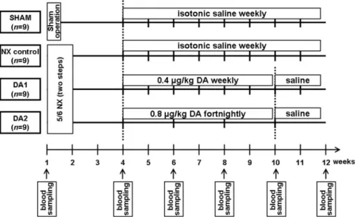
This study conformed to theGuide for the Care and Use of Laboratory Animalspublished by the National Institutes of Health (NIH Pub. No. 85-23, Revised 1996), and was approved by the local ethics committee. Male Wistar rats (n =36, average body weight 300–320 g) were randomly assigned to one or other of the following groups (n=9–9):
a sham-operated group (SHAM); an NX control group; and NX receiving DA (Amgen Inc., Thousand Oaks, CA, USA) weekly or fortnightly (DA1 and DA2, respectively) (Fig.1).
The animals were housed in individually ventilated cages (Sealsafe IVC System, Buguggiate, Italy) with a 12:12 h light:dark cycle, and had free access to standard rat chow and water (containing 10 mg/kg body weight iron(II) sul- fate). Five-sixths NX was performed in two steps by ligation followed by surgical excision of both poles (1/3 on each end) of the left kidney in the first step (baseline, week 1),
followed by removal of the right kidney 1 week later [11].
The rats were anesthetized with 60 mg/kg pentobarbital sodium (Euthanyl, Bimeda-MTC Animal Health, Cam- bridge, ON, Canada) during surgery. On the basis of the results of a pilot study, uremia develops by week 4 in this NX model (as shown by histological examination of the remnant kidney and significant increases in plasma creati- nine levels), and subsequent administrations of DA with doses of 0.4lg/kg weekly or 0.8lg/kg fortnightly can maintain comparable Hb levels for 6 weeks. Accordingly, the subcutaneous administration of DA (DA1 group: 0.4lg/
kg weekly; DA2 group: 0.8lg/kg every other week) or isotonic saline (weekly in both the SHAM and NX control groups) was initiated at week 4 in the present study. At week 10, DA was withdrawn and all animals received saline (Fig.1). The numbers of animals surviving at the end of the 12-week study were 9, 8, 7 and 8 in the SHAM, NX control, DA1 and DA2 groups, respectively.
Blood samples (150–200ll; anticoagulated with EDTA) were obtained from the saphenous vein [12] (weeks 1, 4, 6, 8 and 10), or from the inferior caval vein (week 12), prior to the administration of DA or saline. The whole blood Hb levels were measured spectrophotometrically after reaction with Drabkin’s reagent [13]. The whole blood GSSG and GSH contents were determined essentially as published earlier [14], with the modification that 1-methyl-2-vinylpy- ridinium trifluoromethane sulfonate was used as thiol- masking reagent instead ofN-ethylmaleimide.
Statistical comparisons were performed by using repe- ated-measures two-way analysis of variance (ANOVA), followed by Bonferroni’s post-hoc test. Results are reported as percentage changes in the basal values (mean±SEM;
baseline =100 %). p values \0.05 were considered significant.
Fig. 1 Study protocol.NX nephrectomized,DA darbepoetin alfa
Results
The Hb levels were increased relative to the baseline at weeks 4 and 10 in the SHAM group (p\0.05), and were decreased in the NX controls from week 8 compared with both the baseline and the SHAM group (both p\0.05).
Apart from an elevation at week 10 in the DA1 group (p\0.05), the Hb levels did not change significantly in the DA-treated animals during the follow-up (Fig.2).
Compared with the baseline, the ratios GSSG/GSH were decreased between weeks 6–12 in all groups (weeks 6 and 12: p\0.05; weeks 8–10: p\0.01). The ratios in both DA-treated groups were lower than those in the SHAM
group at weeks 8 and 10 (p\0.05, Fig.3). The levels of GSSG in the SHAM, DA1 and DA2 animals were lower than the respective basal levels throughout the study. The significance levels for the respective groups are as follows:
SHAM: p\0.05 (week 4) and p\0.001 (weeks 6–12);
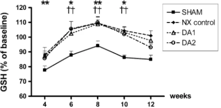
DA1 and DA2: p\0.05 (weeks 4–6), p\0.001 (weeks 8–10) andp\0.01 (week 12). In the NX controls, the GSSG concentrations were not significantly different from the baseline, except for week 4 (p\0.05), and were higher than those in the SHAM group between weeks 6–12 (p\0.01, Fig.4). The GSH concentrations were decreased at week 4 in all groups (p\0.01) compared with the baseline, with elevations in all three NX groups between weeks 6–10 relative both to the baseline (p\0.05 at weeks 6 and 10; andp\0.01 at week 8) and to the SHAM group (p\0.01 at weeks 6–10) (Fig.5). The baseline val- ues for the above parameters are summarized in Table1.
Fig. 2 Hemoglobin levels in the study groups. Darbepoetin alfa (DA1 and DA2 groups) or isotonic saline (SHAM and NX control groups) administration was initiated at week 4. All animals received saline from week 10. Data are presented as percentage changes in the basal values (mean±SEM; baseline=100 %). Repeated-measures two-way ANOVA and Bonferroni’s post-hoc test;n=9, 8, 7 and 8 in the SHAM, NX control, DA1 and DA2 groups, respectively. SHAM group:filled squaresand continuous lines; NX control group:filled rhombusesanddashed lines; DA1 group:empty trianglesanddotted lines; DA2 group: empty circles and dashed-dotted lines. NX nephrectomized. *p\0.05 versus the baseline; p\0.05 versus the SHAM group
Fig. 3 Ratios of oxidized and reduced glutathione (GSSG/GSH) in the study groups. Darbepoetin alfa (DA1 and DA2 groups) or isotonic saline (SHAM and NX control groups) administration was initiated at week 4. All animals received saline from week 10. Data are presented as percentage changes in the basal values (mean±SEM; base- line=100 %). **p\0.01 and *p\0.05 versus the baseline;
p\0.05 versus the SHAM group. Data on the statistical analyses and symbols are the same as for Fig.2
Fig. 4 Levels of oxidized glutathione (GSSG) in the study groups.
Darbepoetin alfa (DA1 and DA2 groups) or isotonic saline (SHAM and NX control groups) administration was initiated at week 4. All animals received saline from week 10. Data are presented as percentage changes in the basal values (mean±SEM; base- line=100 %). ***p\0.001, **p\0.01 and *p\0.05 versus the baseline; p\0.01 versus the SHAM group. Data on the statistical analyses and symbols are the same as for Fig.2
Fig. 5 Levels of reduced glutathione (GSH) in the study groups.
Darbepoetin alfa (DA1 and DA2 groups) or isotonic saline (SHAM and NX control groups) administration was initiated at week 4. All animals received saline from week 10. Data are presented as percentage changes in the basal values (mean±SEM; base- line=100 %). **p\0.01 and *p\0.05 versus the baseline;
p\0.01 versus the SHAM group. Data on the statistical analyses and symbols are the same as for Fig.2
Discussion
To the best of our knowledge, the present study is the first to follow changes in oxidative stress markers during DA therapy with two different administration frequencies in a rat model of CKD. Similar trends in the levels of GSSG/
GSH and GSSG were found throughout the 12-week study with weekly or fortnightly administrations of DA (Figs.3, 4) when comparable Hb levels were maintained in the groups. Further studies with larger cohorts and a parallel- group or cross-over design, comparative studies using ESAs other than DA, as well as assessment of other oxi- dative stress markers are needed to confirm these findings in patients with CKD.
Subtotal (mainly 5/6) NX is the most widely used model of CKD in the rat [11]. The method of renal mass reduction (i.e. surgical excision, or renal ablation by infarction) has a substantial influence on the reproducibility of the proce- dure, the surgical technique giving more reproducible results [11]. In the present study, 2/3 NX of the left kidney was performed in the first step, followed by removal of the right kidney 1 week later [11, 15, 16]. Although the two steps can be reversed [17], we opted for the above protocol due to substantially longer survival of the animals in the pilot study.
The production of reactive oxygen species and the levels of oxidatively modified proteins were earlier reported to be increased in subtotal NX rats [18], in accordance with the present results of markedly higher levels of GSSG in the NX controls than the SHAM group (Fig.4). However, levels of GSSG in the NX controls were not higher than the respective baseline values. One possible explanation is the finding of significant elevations in the GSH concentrations in this group (Fig.5), which could have counterbalanced the pro-oxidant effect associated with NX, resulting in similar ratios GSSG/GSH in both the NX control and SHAM groups (Fig.3). In a recent study, increased levels of GSH after subtotal NX were related with an elevated activity of glutamate-cysteine ligase (the rate-limiting enzyme in GSH synthesis) [19]. Secondly, scavenger activities of thiol moieties other than glutathione may also
play roles in the protection of oxidative stress in rats and other species [20, 21], the assay of which, however, was not performed in the present study. Therefore, it cannot be excluded that such thiols played roles in the change in oxidative stress.
In contrast, the GSSG levels in the two DA-treated groups remained as low as in the SHAM group (Fig.3), resulting in significantly lower GSSG/GSH values at weeks 8 and 10 compared with both the SHAM group and the NX controls (Fig.4). This result is in line with the previously reported antioxidant effect of long-term ESA treatment in CKD [7]. The mechanism of this effect is not well understood [7]; further research is therefore needed in this field. One of the proposed mechanisms is that EPO stimulates cJun-N-terminal kinases (JNKs) in erythroid cells [22], activating heme oxygenase-1 [23], thereby contributing to the reduction of iron-dependent oxidative injury [24]. One limitation of the present study is that levels of iron-related serum parameters were not assayed.
Secondly, JNKs also stimulate Forkhead box class O (FOXO) transcription factor FOXO3a [25], paralleled by increases in survival and antioxidant capacity of the erythrocytes [26,27]. Finally, the proportions of reticulo- cytes and young erythrocytes with high antioxidant capacities and low levels of GSSG/GSH [28] are increased during ESA therapy [29]. However, the actual proportions of these cells were not determined in our study.
As concerns the withdrawal of DA at week 10 in the present study, the ratios GSSG/GSH in the DA-treated groups at week 12 tended to increase but the change did not reach the level of significance (Fig.3). In contrast, a 14-day withdrawal of epoetin beta was associated with a marked increase in GSSG/GSH in hemodialysis patients in our previous study [9]. Besides the different species involved, the difference is probably also connected with the fact that DA has a 3-fold longer serum half-life than that of epoetin beta, allowing exposure of the erythroid cells to DA for an extended period of time [29].
It may be speculated that the repeated blood sampling might have affected the erythropoiesis to an extent which could have compromised the results. It has been reported Table 1 Baseline values of levels of hemoglobin, ratio of oxidized and reduced glutathione (GSSG/GSH), and oxidized and reduced glutathione levels (GSSG and GSH, respectively) in the study groups
SHAM group NX controls DA1 group DA2 group
Hemoglobin (g/l) 139.6±1.5 143.9±2.5 146.4±5.4 140.7±2.0
GSSG/GSH (%) 0.38±0.01 0.42±0.02 0.39±0.01 0.42±0.02
GSSG (lmol/g Hb) 0.041±0.001 0.043±0.002 0.042±0.001 0.046±0.002
GSH (lmol/g Hb) 10.8±0.3 10.2±0.3 10.8±0.4 10.9±0.2
Data are presented as mean±SEM.n=9, 8, 7 and 8 in the SHAM, NX control, DA1 and DA2 groups, respectively NXnephrectomized,DAdarbepoetin alfa,Hbhemoglobin
that a blood volume equivalent to about 0.5 % of the ani- mal’s body weight can be drawn safely at fortnightly intervals without disturbing the animal’s hematological status [12]. In our study, the volume of 150–200ll drawn every other week corresponds to a maximum of about 0.05–0.07 % of the body weight (further decreasing with time as animals grew). Thus, it is not likely that the erythropoiesis was affected negatively by the blood sam- pling protocol.
In summary, the changes in the levels of GSSG and GSH during DA therapy administered at weekly or fort- nightly intervals were followed in 5/6 NX rats for the first time. The levels of GSSG/GSH and GSSG in the DA- treated groups were lower than those in the NX controls, in accordance with the reported antioxidant effects of ESA treatment. When comparable Hb levels were maintained, the level of oxidative stress did not differ in the groups receiving different dosing regimens of DA, as revealed by the similar trends in the levels of GSSG/GSH and GSSG.
These results remain to be confirmed in CKD patients and in comparative studies involving other ESAs and assess- ment of oxidative stress markers other than glutathione.
Acknowledgments This study was supported by Hungarian National Scientific Research Grant OTKA K67895 and by a Research
& Development Grant of the Hungarian Society of Nephrology.
T. Csont holds a Ja´nos Bolyai Research Fellowship of the Hungarian Academy of Sciences.
Conflict of interest The authors have declared that no conflict of interest exists.
References
1. Locatelli F, Aljama P, Ba´ra´ny P, Canaud B, Carrera F, Eckardt KU, et al. Revised European best practice guidelines for the management of anaemia in patients with chronic renal failure.
Nephrol Dial Transplant. 2004;19(Suppl 2):ii1–47.
2. KDOQI; National Kidney Foundation. KDOQI clinical practice guidelines and clinical practice recommendations for anemia in chronic kidney disease. Am J Kidney Dis. 2006;47(Suppl 3):S11–145.
3. Carrera F, Disney A, Molina M. Extended dosing intervals with erythropoiesis-stimulating agents in chronic kidney disease: a review of clinical data. Nephrol Dial Transplant. 2007;22(Suppl 4):iv19–30.
4. Kiss Z, Elliott S, Jedynasty K, Tesar V, Szegedi J. Discovery and basic pharmacology of erythropoiesis-stimulating agents (ESAs), including the hyperglycosylated ESA, darbepoetin alfa: an update of the rationale and clinical impact. Eur J Clin Pharmacol.
2010;66:331–40.
5. Carrera F, Oliveira L, Maia P, Mendes T, Ferreira C. The efficacy of intravenous darbepoetin alfa administered once every 2 weeks in chronic kidney disease patients on haemodialysis. Nephrol Dial Transplant. 2006;21:2846–50.
6. Arcasoy MO. The non-haematopoietic biological effects of erythropoietin. Br J Haematol. 2008;141:14–31.
7. Katavetin P, Tungsanga K, Eiam-Ong S, Nangaku M. Antioxi- dative effects of erythropoietin. Kidney Int Suppl. 2007;107:
S10–5.
8. Locatelli F, Canaud B, Eckardt KU, Stenvinkel P, Wanner C, Zoccali C. Oxidative stress in end-stage renal disease: an emerging threat to patient outcome. Nephrol Dial Transplant.
2003;18:1272–80.
9. Monostori P, Hracsko´ Z, Karg E, Varga IS, Kiss Z, Boros T, et al.
Erythropoiesis-stimulating agent withdrawal and oxidative stress in hemodialysis. Clin Nephrol. 2009;71:521–6.
10. Turi S, Nemeth I, Varga I, Bodrogi T, Matkovics B. The effect of erythropoietin on the cellular defence mechanism of red blood cells in children with chronic renal failure. Pediatr Nephrol.
1992;6:536–41.
11. Liu ZC, Chow KM, Chang TM. Evaluation of two protocols of uremic rat model: partial nephrectomy and infarction. Ren Fail.
2003;25:935–43.
12. Hem A, Smith AJ, Solberg P. Saphenous vein puncture for blood sampling of the mouse, rat, hamster, gerbil, guinea pig, ferret and mink. Lab Anim. 1998;32:364–8.
13. Drabkin DL, Austin JH. Spectrophotometric studies. II. Prepa- rations from washed blood cells; nitric oxide hemoglobin and sulfhemoglobin. J Biol Chem. 1935;112:51–65.
14. Nemeth I, Boda D. Blood glutathione redox ratio as a parameter of oxidative stress in premature infants with IRDS. Free Radic Biol Med. 1994;16:347–53.
15. Tain YL, Freshour G, Dikalova A, Griendling K, Baylis C.
Vitamin E reduces glomerulosclerosis, restores renal neuronal NOS, and suppresses oxidative stress in the 5/6 nephrectomized rat. Am J Physiol Renal Physiol. 2007;292:F1404–10.
16. An WS, Kim HJ, Cho KH, Vaziri ND. Omega-3 fatty acid sup- plementation attenuates oxidative stress, inflammation, and tub- ulointerstitial fibrosis in the remnant kidney. Am J Physiol Renal Physiol. 2009;297:F895–903.
17. Mino M, Ihara H, Kozaki S, Kondo T, Takeshita A, Kusakabe KT, et al. Effects of low protein intake on the development of the remaining kidney in subtotally nephrectomized immature rats:
expression of inducible and endothelial NO synthase. Med Mol Morphol. 2010;43:116–22.
18. Fujimoto S, Satoh M, Horike H, Hatta H, Haruna Y, Kobayashi S, et al. Olmesartan ameliorates progressive glomerular injury in subtotal nephrectomized rats through suppression of superoxide production. Hypertens Res. 2008;31:305–13.
19. Benipal B, Lash LH. Influence of renal compensatory hypertro- phy on mitochondrial energetics and redox status. Biochem Pharmacol. 2011;81:295–303.
20. Rossi R, Milzani A, Dalle-Donne I, Giannerini F, Giustarini D, Lusini L, et al. Different metabolizing ability of thiol reactants in human and rat blood: biochemical and pharmacological impli- cations. J Biol Chem. 2001;276:7004–10.
21. Hempe JM, Ory-Ascani J, Hsia D. Genetic variation in mouse beta globin cysteine content modifies glutathione metabolism:
implications for the use of mouse models. Exp Biol Med (Maywood). 2007;232:437–44.
22. Jacobs-Helber SM, Ryan JJ, Sawyer ST. JNK and p38 are acti- vated by erythropoietin (EPO) but are not induced in apoptosis following EPO withdrawal in EPO-dependent HCD57 cells.
Blood. 2000;96:933–40.
23. Calo` LA, Davis PA, Piccoli A, Pessina AC. A role for heme oxygenase-1 in the antioxidant and antiapoptotic effects of erythropoietin: the start of a good news/bad news story? Nephron Physiol. 2006;103:p107–11.
24. Akisu M, Tuzun S, Arslanoglu S, Yalaz M, Kultursay N. Effect of recombinant human erythropoietin administration on lipid per- oxidation and antioxidant enzyme(s) activities in preterm infants.
Acta Med Okayama. 2001;55:357–62.
25. Essers MA, Weijzen S, de Vries-Smits AM, Saarloos I, de Ruiter ND, Bos JL, et al. FOXO transcription factor activation by oxi- dative stress mediated by the small GTPase Ral and JNK. EMBO J. 2004;23:4802–12.
26. Kops GJ, Dansen TB, Polderman PE, Saarloos I, Wirtz KW, Coffer PJ, et al. Forkhead transcription factor FOXO3a protects quiescent cells from oxidative stress. Nature. 2002;419:316–21.
27. Marinkovic D, Zhang X, Yalcin S, Luciano JP, Brugnara C, Huber T, et al. Foxo3 is required for the regulation of oxidative stress in erythropoiesis. J Clin Invest. 2007;117:2133–44.
28. Clark MR. Senescence of red blood cells: progress and problems.
Physiol Rev. 1988;68:503–54.
29. Elliott S, Pham E, Macdougall IC. Erythropoietins: a common mechanism of action. Exp Hematol. 2008;36:1573–84.