Research Article
The Impact of Fermented Wheat Germ Extract on Porcine Epithelial Cell Line Exposed to Deoxynivalenol and T-
2 Mycotoxins
Judit Mercédesz Pomothy ,1Erzsébet Pászti-Gere ,1Réka Fanni Barna ,1,2 Dorottya Prokoly,1and Ákos Jerzsele 1
1Department of Pharmacology and Toxicology, University of Veterinary Medicine, 1078 Budapest, Hungary
2Department of Physiology and Biochemistry, University of Veterinary Medicine, 1078 Budapest, Hungary
Correspondence should be addressed to Judit Mercédesz Pomothy; pomothy.judit.mercedesz@univet.hu Received 21 July 2020; Revised 24 November 2020; Accepted 27 November 2020; Published 8 December 2020
Academic Editor: Marcos R. de Oliveira
Copyright © 2020 Judit Mercédesz Pomothy et al. This is an open access article distributed under the Creative Commons Attribution License, which permits unrestricted use, distribution, and reproduction in any medium, provided the original work is properly cited.
The effect of fermented wheat germ extract (FWGE) (Immunovet®) was evaluated with cotreatments with deoxynivalenol (DON) and T-2 toxin (T-2). These mycotoxins are produced byFusariummold species. The effects of FWGE on IPEC-J2 with DON and T- 2 have not been studied until now. The IPEC-J2 porcine, nontumorigenic cell line was selected to investigate the outcome of the individually and simultaneously added compounds, as it has in vivo-like properties. The cells were treated for 24 h with the selected solutions; then, the IPEC-J2 cells were allowed to regenerate in a culture medium for an additional 24 h. In our results, DON and T-2 significantly increased the adverse impacts on cell viability and integrity of the cell monolayer. To elucidate the extent of oxidative stress, extracellular H2O2 concentrations and intracellular reactive oxygen species (ROS) were measured.
FWGE appeared to be beneficial to IPEC-J2 cells given the separately and significantly decreased ROS levels. 1% and 2% FWGE could significantly reduce mycotoxin-induced oxidative stress. In conclusion, the results demonstrate that FWGE exerted protective effects to counteract the oxidative stress-provoking properties of applied fusariotoxins in the nontumorigenic IPEC-J2 cell line.
1. Introduction
The number of studies involving natural products and die- tary supplements has shown rapid growth recently. Natural products contain extensive chemical diversity, which makes it difficult to replace the collection of naturally occurring molecules with synthetized drugs.
Wheat germ contains several bioactive ingredients, such asflavonoids, dietaryfibres, as well as lignins, oligosaccha- rides, and vitamins [1]. Hidvegi et al. [2] demonstrated that wheat germ is rich in the glycosylated form of 2,6- dimethoxy-p-benzoquinone (DMBQ). The conversion of DMBQ into its biologically more active forms requiresβ-glu- cosidase enzyme [3]. Wheat germ is fermented bySaccharo- myces cerevisiae yeasts [4] or treated with Lactobacillus plantarumdy-1 [5]. Fermented wheat germ extract (FWGE)
is available in both human (Avemar®) and veterinary (Immu- novet®) medicine. These products are aqueous extractions, which are fermented bySaccharomyces cerevisiae, and they contain several biologically active molecules [4, 6]. FWGE is applied as an adjuvant in human cancer therapy, because benzoquinones have antimetastatic [2], antimetabolic [6], antiangiogenic [7], and antiproliferative properties and are able to induce apoptosis [5, 8]. Furthermore, FWGE can enhance the cellular immune response [4, 9] and has an anti- oxidant effect [2].
It is of key importance worldwide to produce good qual- ity feedstufffor livestock with the least amount of mycotoxin contaminants.Fusariumfungi are abundant in temperate cli- mate zones and contaminate wheat and other cereals. This genus is capable of producing a wide variety of mycotoxins.
One of these groups is the trichothecene mycotoxins,
Volume 2020, Article ID 3854247, 9 pages https://doi.org/10.1155/2020/3854247
including deoxynivalenol (DON), nivalenol (NIV), T-2 (T-2) toxin, and HT-2 toxin [10, 11]. In general, these fusariotoxins generate reactive oxygen species (ROS) and interfere with the normal functions of mitochondria, which can lead to apopto- sis. They also inhibit protein synthesis in eukaryotic cells [12], especially in epithelial and immune cells, where the rate of cell replication is high [13, 14]. Among farm animals, swine is the most sensitive species to fusariotoxin contamina- tion; side effects are decreased feed intake, feed refusal, and vomiting [15].
The most frequently used cell line for oxidative stress- related studies are intestinal porcine epithelial cell line-1 (IPEC-1) and intestinal porcine epithelial cell line 2 (IPEC- J2), which are suitable forin vitromodelling of nontumori- genic epithelium. There are only few publications regarding IPEC-J2 cells treated with both DON and T-2 toxin. In these studies, transepithelial electrical resistance (TEER) decreased while cellular permeability was enhanced in parallel [16] by these toxic substances. Both DON and T-2 increase the ROS level intracellularly [17, 18].
The main objective of this study was to describe the ben- eficial effects of fermented wheat germ extract on the IPEC-J2 cell line induced with DON and T-2 mycotoxins. This study was focused on TEER and two oxidative stress markers:
extracellular H2O2and intracellular ROS productions.
2. Materials and Methods
2.1. Reagents. DON and T-2 were purchased from Merck (Darmstadt, Germany). Acetonitrile was obtained from Thermo Fisher Scientific (Waltham, MA, USA). The final concentration of acetonitrile in the cell culture medium was
<0.5% (v/v). FWGE was diluted from a commercial product in powder form (Immunovet Pets, Immunovet Ltd., Hungary).
Prior to the experiments, cell viability studies were per- formed to select the working concentrations to DON, T-2, and FWGE (data not shown). 8μmol/L DON, 5 nmol/L T- 2, and 1% and 2% FWGE concentrations were chosen from these results for further investigations (Figure 1).
2.2. Cell and Culturing Conditions. The porcine intestinal epithelial cell line IPEC-J2 (ACC 701) is nontumorigenic, intestinal columnar epithelial cells, which were isolated from neonatal piglet midjejunum. IPEC-J2 closely mimicsin vivo pig and human physiology, which makes it a good model to study foodborne and plant-derived components.
This cell line form’s polarized monolayers were main- tained in 75 cm2cell cultureflasks withfiltered caps (Orange Scientific, Braine-l’Alleud, Belgium) at 37°C in a humidified atmosphere of 5% CO2. The culture medium contains 50%
Dulbecco’s modified Eagle’s medium (DMEM) and 50%
Ham’s F12 Nutrient Mixture (Merck, Darmstadt, Germany) supplemented with 1.5 mmol/L HEPES, 5% fetal bovine serum (Biocenter, Budapest, Hungary), 1% insulin/transfer- rin/sodium selenite medium supplement, 5 ng/mL epidermal growth factor, and 1% penicillin/streptomycin (all purchased from Invitrogen, Thermo Fisher Scientific, Waltham, MA,
USA). Cells were used between passages 42 and 45. The media were changed every second day.
2.3. Experimental Design and Cell Treatments.To investigate the cell viability, the seeding density for the cells was1 × 104 cells/well of a 96-well plate (Transwell, Sigma-Aldrich, Corn- ing Costar, Merck, Darmstadt, Germany). The cells were treated the next day after reaching a confluent state. When studying TEER, H2O2 production, and intracellular ROS levels, the cell-seeding density was 1:5 × 10⁵ cells/well in a 6-well polyester membrane insert (4.67 cm2) containing plates (Transwell, Sigma-Aldrich, Corning Costar, Merck, Darmstadt, Germany). These inserts were useful for the api- cally and basolaterally added treatments and for transepithe- lial electrical resistance measurements.
The stock solutions were freshly made with phenol red- free DMEM/F12 (Merck, Darmstadt, Germany). The DON and T-2 were diluted with acetonitrile (final concentration:
<0.5% (v/v)); then, the following concentrations were made:
8μmol/L DON, 5 nmol/L T-2, and 1 and 2%final concentra- tions of FWGE. Cell cultures were exposed to the treatments for an incubation time of 24 hours; then, the IPEC-J2 cells were treated only with phenol red-free DMEM:F12 culture medium for an additional 24 h as regeneration. After the treatment and the regeneration, the TEER was measured, the cell-free supernatants were collected for extracellular H2O2 determination, and the DCFH-DA assay was added to the cells.
2.4. Evaluation of Cell Viability. Cytotoxicity was examined with an MTS reagent (CellTiter96 Aqueous One Solution, Promega, Bioscience, Budapest, Hungary) [19]. This test measures the rate of viable cells by determining the soluble tetrazolium salt conversion in the metabolically active cells to a coloured formazan product with the advantage over MTT that the solubilization step is not required for avoiding formazan precipitation in the aqueous medium.
IPEC-J2 cells were seeded in 96-well culture plates at2
× 104 cells/well and allowed 24 hours to reach confluence.
Mycotoxin and FWGE solutions were added to the cells using a multichannel pipette and were incubated for 24 h at 37°C, 5% CO2. After the incubation time, the treatments were removed, and each well received 100μL of fresh phenol red- free medium containing 20μL of MTS solution. After an incubation time of 1 h at 37°C, the absorbance values were measured at 490 nm using an ELISA Plate Reader (EZ Read Biochrom 400, Cambridge, UK).
2.5. Determination of Cell Membrane Integrity.The integrity of the IPEC-J2 cell monolayer can be followed by measur- ing transepithelial electrical resistance (TEER) between the apical and basolateral compartments of the IPEC-J2 cells (Figure 2). Cells were seeded to 6-well Transwell insert containing plates (polyester, 0.4μm pore size, Corning, Merck, Darmstadt, Germany), and the seeding density was 3 × 10⁶ cells/well. After the cells reached a confluent state, the barrier function was evaluated by measuring with an EVOM Epithelial Tissue Volt/Ohmmeter (World Precision Instruments, Berlin, Germany). 10 days after
seeding, the IPEC-J2 monolayer achieved the 600 Ω/well values. The results were calculated as kΩ× cm2 by multi- plying the values by the surface area of the monolayer (4.67 cm2). The high TEER value of IPEC-J2 monolayers grown on Transwell polyester filters demonstrates the functional integrity of the continuous cell association, act- ing as a single-layer tight physical barrier.
2.6. Detection of Changes in the Extracellular H2O2 Concentrations. Extracellular H2O2 production was moni- tored in IPEC-J2 cells by using the Amplex Red Hydrogen Peroxide Assay Kit (Invitrogen, Thermo Fisher Scientific, Waltham, MA, USA). The Amplex Red reagent reacts with H2O2 (in 1 : 1 stoichiometry) to produce a red fluorescent product called resorufin in the presence of horseradish per- oxidase. After 24 and 48 h incubation time, 50μL of the cell-free supernatants was collected from the basolateral compartments and was mixed with the Amplex Red working solution according to the manufacturer’s instructions. The fluorescence intensity was measured at 590 nm with afluo-
rometer using 530 nm excitation wavelength (Victor X2 2030, Perkin Elmer, Waltham, MA, USA).
2.7. Assessing the Changes in Intracellular ROS Levels. Mea- surement of disruptions in the intracellular redox state of IPEC-J2 cells was carried out using DCFH-DA dye (Sigma- Aldrich, Budapest, Hungary). DCFH-DA is oxidized to the highlyfluorescent form of dichlorofluorescein (DCF) by the intracellular ROS [20]. Following a centrifugation process for 10 min at 10 000 rpm at 5°C, 100μL of cell-free superna- tant was collected and pipetted into a 96-well plate. Samples of supernatant were collected at 24 and 48 h after treatments.
The fluorescence intensities of the supernatants were mea- sured at 530 nm with afluorometer using 485 nm excitation wavelength (Victor X2 2030, Perkin Elmer, Waltham, MA, USA).
2.8. Statistical Analysis.The statistical analysis of the results was performed by using R Core Team (version of 2016) [21]. Differences between groups were analyzed by one-way ANOVA coupled with the post hoc Tukey test for multiple
2,6-Dimethoxy-p-benzoquinone (DMBQ) OCH3 OCH3
O
O O
(a)
Deoxynivalenol (DON) OH
HO OH
H O O
(b)
T-2 toxin (T-2) O OH
OAc OAc
O O
(c)
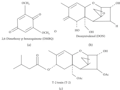
Figure1: The chemical structures of (a) 2,6-dimethoxy-p-benzoquinone (DMBQ) fromImmunovet®and the testedFusariummycotoxins, (b) deoxynivalenol (DON), and (c) T-2 toxin (T-2).
Apical chamber Ohm
Basolateral chamber
Medium IPEC-J2
TEER = Ohm × cm2
Figure2: Schematic representation of TEER measuring method. Cell culture monolayers grown on polyesterfilter separate the apical and basolateral compartments.
comparisons. ∗p< 0:05, ∗∗p< 0:01, and ∗∗∗p< 0:001 were considered to be statistically significant.
3. Results
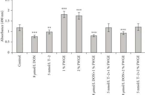
3.1. Cell Viability Assessment Using MTS Assay.IPEC-J2 cell viability was measured with the MTS reagent after a 24 h incubation time with 8μmol/L DON, 5 nmol/L T-2, and 1%
and 2% FWGE. Their combinations were tested also:
8μmol/L DON+1% FWGE, 8μmol/L DON+2% FWGE, 5 nmol/L T-2+1% FWGE, and 5 nmol/L T-2+2% FWGE (Figure 3). According to absorbance values, 8μmol/L DON and 5 nmol/L T-2 significantly decreased cell viability (p< 0:001andp= 0:0039), while 1% and 2% FWGE signifi- cantly increased the values of the treated cells compared to the control (bothp< 0:001). The cytotoxic effect of 8μmol/L DON was not counteracted by simultaneous 1% and 2%
FWGE treatments (p< 0:001). 1% FWGE added with 5 nmol/L T-2 did not change cell viability (p= 1:000) to the control level. The values of 5 nmol/L T-2+2% FWGE- treated cells showed no differences compared to control values (p= 0:999). Comparing the 5 nmol/L T-2 individual treatments with the concurrent 1% FWGE addition, signifi- cant differences in absorbances were observed (p= 0:0038).
The 5 nmol/L T-2+2% FWGE showed significant differences compared to 5 nmol/L T-2 (p< 0:001).
3.2. TEER Measurements of the IPEC-J2 Cell Membrane Integrity. To measure the changes in the integrity of the IPEC-J2 cell monolayer, TEER measurements were carried out prior to the treatments (0 h) after a 24 h treatment and after an additional 24 h (48 h) regenerative treatment (Figure 4). After treatments, 8μmol/L DON (24 h and 48 h) and 5 nmol/L T-2 (48 h) significantly decreased TEER values (bothp< 0:001). In the case of the individually given FWGE (24 h), the TEER increased significantly (bothp< 0:001). The 8μmol/L DON supplemented with 1% and 2% FWGE showed a significant decrease in TEER (bothp< 0:001) com- pared to control values. 5 nmol/L T-2+1% FWGE (24 h) proved significantly reduced TEER values (p< 0:001); the 2% FWGE simultaneous treatment (24 h) indicated a signifi- cant increase (p= 0:018). After an additional 24 h regenera- tion treatment, the TEER values of 1% and 2% FWGE remained at the control levels (1% FWGE: p= 0:348; 2%
FWGE:p= 0:194). In the case of 5 nmol/L T-2 added simul- taneously with 1% FWGE and 2% FWGE, TEER decreased significantly (48 h, bothp< 0:001).
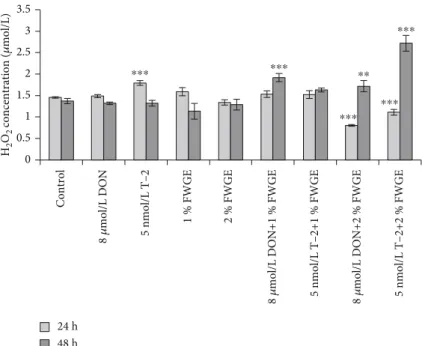
3.3. Evaluation of H2O2 Concentrations from Cell-Free Supernatants with Amplex Red Assay.The changes in extra- cellular H2O2 concentrations were assessed after the 24 h treatment and an additional 24 h regeneration (48 h) in phe- nol red-free DMEM:F12 media (Figure 5). After a 24 h incu- bation with 8μmol/L DON, H2O2concentrations remained unchanged (p= 0:070), while 5 nmol/L T-2 caused a signifi- cant increase (p< 0:001). The 1% and 2% FWGE treatments did not alter the H2O2 level (1% FWGE: p= 0:905; 2%
FWGE: p= 0:705). When these compounds were added simultaneously, 8μmol/L DON+1% FWGE treatment did
not cause a change compared to control treatment (p= 0:844), although 8μmol/L DON given with 2% FWGE significantly decreased H2O2 concentrations (p< 0:001).
5 nmol/L T-2+1% FWGE treatment did not differ signifi- cantly from the control treatment (p= 0:835), while the H2O2 level significantly decreased when 5 nmol/L T-2 was given at the same time with 2% FWGE (p< 0:001). After the regeneration period, the H2O2 concentrations of the mycotoxin-treated cells showed no differences to the control cells (DON:p= 1:00; T-2:p= 1:00). The H2O2production of the priorly 1% and 2% FWGE-treated cells did not change after the regeneration (1% FWGE: p= 0:161; 2% FWGE: p
= 0:996). After a 24 h regeneration period at the 8μmol/L DON+1% FWGE and 2% FWGE, the treated cells signifi- cantly increased the H2O2concentration (p< 0:001andp= 0:009). 5 nmol/L T-2+1% FWGE did not alter the H2O2level compared to control-treated cells (p= 0:097). In contrast, 5 nmol/L T-2+2% FWGE significantly increased the H2O2 production after the regeneration period (p< 0:001).
3.4. Intracellular ROS Determination Using DCFH-DA Assay.
The DCFH-DA assay was used to estimate the intracellular ROS level present after 24 h treatments and additional 24 h regeneration (48 h) (Figure 6). After the treatment with 8μmol/L DON and 5 nmol/L T-2 for 24 h, the intracellular ROS were significantly higher compared to the control (both p< 0:001). The FWGE treatments significantly decrease the ROS levels intracellularly (both p< 0:001). 8μmol/L DON cotreated with 1% FWGE resulted in the samefluorescence intensities as the control-treated cells (p= 1:000), while 2%
FWGE significantly reduced ROS production in cells exposed to DON compared to the control (p< 0:001). The cells given 5 nmol/L T-2 simultaneously with 1% FWGE and 2% FWGE showed significantly decreased values (p< 0:001). After the regeneration period, the prior 8μmol/L DON treatment sig- nificantly increased intracellular ROS (p= 0:043), similarly to the priorly 5 nmol/L T-2 treated cells (p< 0:001). The cells that were given 1% and 2% FWGE did not produce signifi- cantly different amounts of ROS intracellularly compared to the control (both p= 1:000). The ROS levels of the prior 8μmol/L DON+1% FWGE and 2% FWGE-treated cells were significantly higher (p< 0:001). The 1% FWGE cotreated with 5 nmol/L T-2 significantly increasedfluorescence inten- sities (p< 0:001) after a 24 h regeneration, while 5 nmol/L T- 2+2% FWGE-treated cells showed no differences compared to control cells (p= 0:142).
4. Discussion
According to Hernández et al. [22], the wheat germ extract’s main active components include DMBQ, hydroxybenzoic acids, hydroxycinnamic acids, and apigenin. These naturally occurring compounds are glycosylated and physiologically not active [23]. There were only few experiments treating livestock animals with FWGE (Immunovet®). Three-week- old chickens were infected with Mycoplasma gallisepticum and treated with FWGE, and poultry remained clinically healthy [24]. In another research, FWGE was beneficial for the maintenance of general health conditions including
biochemical and physiological parameters, increasing weight gain, and improved immune response to vaccination [25]. In growing pigs, FWGE enhanced weight gain and had a bene- ficial effect on cellular immunity. This effect is instrumental in promoting resistance against facultative pathogens [9].
As reported by Jerzsele et al. [26], 2% FWGE helped broiler chickens to gain greater body weight than the control group.
It was also established that animals infected withSalmonella Typhimurium under controlled conditions and obtained
FWGE treatments were not spreading the pathogens to other chickens.
A prominent issue in feed production is mycotoxin con- tamination. As major contaminants of cereals, DON and T-2 have been implicated in various gastrointestinal problems in farm animals, such as vomiting, feed refusal, diarrhoea [27], and oesophageal perforation as well as malabsorption [28].
DON can also be present in glycosylated form (for example, as DON-3-β-d-glycoside) in plants, which increases the
0
Control 8 𝜇mol/L DON 8 𝜇mol/L DON+2 % FWGE
8 𝜇mol/L DON+1 % FWGE
5 nmol/L T–2 5 nmol/L T–2+1 % FWGE 5 nmol/L T–2+2 % FWGE
1 % FWGE 2 % FWGE
0.5 1 1.5 2 2.5
Absorbance (490 nm)
⁎⁎⁎
⁎⁎
⁎⁎⁎ ⁎⁎⁎
⁎⁎⁎ ⁎⁎⁎
Figure3: Cytotoxicity of 8μmol/L DON, 5 nmol/L T-2, and 1% and 2% FWGE and their combinations on IPEC-J2 cells at exposure of 24 h.
∗∗p< 0:01and∗∗∗p< 0:001compared to the control values. Data are presented asmeans ± SD(n= 8).
0 1 2 3 4 5 6 7 8 9 10
TEER (kOhm × cm2)
0 h 24 h 48 h
Control 8 𝜇mol/L DON 8 𝜇mol/L DON+2 % FWGE
8 𝜇mol/L DON+1 % FWGE
5 nmol/L T–2 5 nmol/L T–2+1 % FWGE 5 nmol/L T–2+2 % FWGE
1 % FWGE 2 % FWGE
⁎⁎⁎
⁎⁎⁎
⁎⁎⁎
⁎⁎⁎ ⁎⁎⁎ ⁎⁎⁎
⁎⁎⁎ ⁎⁎⁎
⁎⁎⁎
⁎⁎⁎
⁎⁎⁎ ⁎⁎⁎
⁎
Figure4: TEER measurements of IPEC-J2 monolayer. Prior to the experiments, TEER was measured (0 h). Cells were incubated with 8μmol/L DON, 5 nmol/L T-2, 1% and 2% FWGE, and the combination of these compounds for 24 and 48 h.∗p< 0:05and∗∗∗p< 0:001 compared to the control values. Data are presented asmeans ± SD(n= 9).
toxicological effect of DON after consumption [29]. Before 2006, low-dose antibiotics were used to help the growth pro- motion of farm animals. These additional antibiotics were particularly not effective against mycotoxins but were benefi- cial to the general health status. Fortunately, numerous recent studies have been conducted in order tofind a natu- rally occurring or plant-based solution to reduce the negative effects of mycotoxins and support the condition of farm ani- mals. FWGE has several beneficial properties, such as its anti-
oxidant effect [2], which can make this extract useful against oxidative stress generated by the two most common myco- toxins. Although mycotoxins negatively affect all farm ani- mals, the swine is particularly sensitive to it.
The gastrointestinal epithelium is the first barrier for mycotoxin-contaminated feed. There are several in vitro studies regarding the impact of mycotoxins on epithelial cell metabolism, toxicity, and barrier integrity. Both DON and T- 2 have demonstrated time- and concentration-dependent
0 0.5 1 1.5 2 2.5 3 3.5
H2O2 concentration (𝜇mol/L)
24 h 48 h
⁎⁎⁎ ⁎⁎⁎
⁎⁎⁎ ⁎⁎⁎
⁎⁎⁎
⁎⁎
Control 8 𝜇mol/L DON 8 𝜇mol/L DON+2 % FWGE
8 𝜇mol/L DON+1 % FWGE
5 nmol/L T–2 5 nmol/L T–2+1 % FWGE 5 nmol/L T–2+2 % FWGE
1 % FWGE 2 % FWGE
Figure5: The changes of H2O2concentrations after incubation of IPEC-J2 cells with 8μmol/L DON, 5 nmol/L T-2, 1% and 2% FWGE, and their combinations for indicated time periods (24 and 48 h).∗∗p< 0:01and∗∗∗p< 0:001compared to the control values. Data are presented as means ± SD(n= 8).
0 5000 10000 15000 20000 25000 30000
Fluorescence intensity (530 nm)
24 h 48 h
Control 8 𝜇mol/L DON 8 𝜇mol/L DON+2 % FWGE
8 𝜇mol/L DON+1 % FWGE
5 nmol/L T–2 5 nmol/L T–2+1 % FWGE 5 nmol/L T–2+2 % FWGE
1 % FWGE 2 % FWGE
⁎⁎⁎
⁎⁎⁎ ⁎⁎⁎ ⁎⁎⁎ ⁎⁎⁎⁎⁎⁎
⁎⁎⁎⁎⁎⁎ ⁎⁎⁎
⁎
⁎⁎⁎
⁎⁎⁎
Figure6: Intracellular ROS levels were measured after 24 and 48 h incubation with 8μmol/L DON, 5 nmol/L T-2, 1% and 2% FWGE, and their combinations.∗p< 0:05and∗∗∗p< 0:001compared to the control values. Data are presented asmeans ± SD(n= 6).
cytotoxicity. Szakács et al. [30] established that FWGE boosted the immune responses compared to T-2-treated weaned pigs. The IC50for DON and T-2 was 23.52μmol/L and 20.4 nmol/L for 72 hours of incubation time on IPEC- J2 [16].
In this study, DON was added at 8μmol/L and T-2 at 5 nmol/L concentrations for 24 hours on IPEC-J2. Both DON and T-2 were shown to decrease the metabolic activity of the cells significantly. This result is in good correlation with that in the study by Sergent et al. [31]. The IC50 of DON was determined on the Caco-2 cell line at 2.22μmol/L, but a 0.67μmol/L concentration of DON inhibited the prolif- eration of cells for 48 hours [31]. In our studies, both 1% and 2% of FWGE increased cell viability compared to control cells. In the literature, FWGE was mostly examined on tumor cell lines where FWGE induced apoptosis and cell death in several cases [32]. 1 or 2% of FWGE did not preserve cell via- bility when 8μmol/L DON was added to the cells. In con- trast, 1% and 2% FWGE enhanced the survival of IPEC-J2 cells treated simultaneously with T-2 compared to cells treated only with T-2.
IPEC-J2 cells can polarize and form a strong barrier through the development of tight junctions between cells [33]. The intercellular tight junction is the rate-limiting bar- rier in the paracellular pathway for permeation by ions and larger solutes. The TEER of cell monolayers can be consid- ered a good indicator of the degree of organization of the tight junctions within the cell monolayer as well as that of epithelial integrity [34].
The authors did notfind earlier results of studies using TEER to detect the effects of FWGE on the integrity of non- tumorigenic intestinal cell monolayers exposed to fusariotox- ins. On the other hand, TEER utilization in mycotoxin research has a more extensive representation in the literature.
As reported by Goossens et al. [16] and Kang et al. [35], DON treatments significantly reduce the TEER values, depending on the dosage. Goossens et al. [16] also found that T-2 up to 210 nmol/L concentration for 72 h significantly lowered the integrity of the IPEC-J2 monolayer. Springler et al. [36]
confirmed that DON reduced TEER significantly at 5– 20μmol/L after 24 h incubation. Our study found that 8μmol/L DON and 5 nmol/L T-2 significantly reduced the TEER values during and after the treatments, while 1% and 2% FWGE alone significantly increased them. Based on our findings, 1% FWGE cotreatment with mycotoxins did not elevate the TEER, while 2% FWGE added for 24 h with 5 nmol/L T-2 helped the cells reach a higher TEER value.
Oxidative stress develops if concentrations of ROS exceed the antioxidant capacity of living entities. ROS are reactive species of radicals with a single unpaired electron, such as superoxide anion radical (O2−) and the hydroxyl radical (OH−), along with nonradical ROS such as hydrogen perox- ide (H2O2). Sharply increasing intracellular ROS can cause oxidative stress with irreversible cell damage [37]. ROS can initiate the process of lipid peroxidation in the lipid mem- brane causing damage to the cell membrane’s phospholipids and lipoproteins, and it can also damage DNA by propagat- ing a chain reaction [38]. Moreover, oxidative stress medi- ated by ROS may increase cell apoptosis [39]. To
counterbalance the prooxidant agents, the cells have intracel- lular nonenzymatic and enzymatic antioxidants, namely, tri- peptide glutathione [40] and catalase, which can protect them from oxidative damage [41].
We examined oxidative stress by measuring extracellular H2O2production and intracellular ROS generation in IPEC- J2 cells. We found that both 8μmol/L DON and 5 nmol/L T- 2 significantly increased intracellular ROS levels during the 24 h treatment and after the 24 h regeneration. This is in agree- ment with thefindings of Kang et al. [35] who published that DON at 6.7μmol/L in IPEC-J2 cells significantly elevated intracellular ROS levels after 24 h of mycotoxin exposure. Both 1% and 2% FWGE significantly decreased the ROS after a 24 h treatment. Thesefindings are in good correlation with Kar- ancsi et al. [42], who elucidated firstly beneficial effects of FWGE in case of LPS-evoked oxidative damage. FWGE could decrease excessive intracellular ROS levels after LPS adminis- tration and exerted protective effect on the integrity of the IPEC-J2 cell monolayer exposed to LPS treatment. Their study also showed that FWGE in different concentrations (1%, 2%, and 4%) did not affect cell death; moreover, FWGE in 2% con- centration improved cell viability significantly after 24 h treat- ment. These results are seemingly contradictory to Otto et al.
[6] who published that 24μmol/L DMBQ from FWGE inhibits cell cycle progress, induce apoptosis, and increase intracellular DCFfluorescence after a 24 h treatment in nine human cancer cell lines. Hidvegi et al. [2] resolved this contra- version by clarifyingfirstly that the effects of FWGE are not solely attributable to benzoquinones and secondly that IPEC-J2 is a nontumorigenic cell line.
When treated simultaneously, both 1% and 2% FWGE significantly decreased the DON- and T-2-mediated ROS levels. An interesting phenomenon was detected in terms of 2% FWGE as the extracellular H2O2 significantly decreased during the 24 h treatment; however, IPEC-J2 cells produced significantly higher H2O2 at the end of the regeneration period when cells were previously exposed to DON or T-2 toxins for 24 h.
5. Conclusions
In conclusion, 1% and 2% FWGE has favourable properties on IPEC-J2 cell lines as FWGE helps the cells to proliferate.
2% FWGE is a beneficial agent against intracellular ROS when treated with DON and T-2 simultaneously. To our knowledge, this is the first published report of FWGE cotreated with DON or T-2 in which TEER was utilized to determine the impact of FWGE on the integrity of the cell monolayer.
Data Availability
The data used to support thefindings of this study are avail- able from the corresponding author upon request.
Conflicts of Interest
The authors declare that there is no conflict of interest regarding the publication of this paper.
Authors’Contributions
Judit Mercédesz Pomothy and Erzsébet Pászti-Gere contrib- uted equally to this work.
Acknowledgments
The research was supported by the Hungarian Scientific Research Fund (grant numbers 115685 and 124522). This work was supported by the European Union and cofinanced by the European Social Fund (grant agreement nos. EFOP- 3.6.1-16-2016-00024, EFOP-3.6.2-16-2017-00012, and EFOP-3.6.3-VEKOP-16-2017-00005). This project was sup- ported by the János Bolyai Research Scholarship of the Hun- garian Academy of Sciences and by the ÚNKP-20-5 New National Excellence Program of the Ministry for Innovation and Technology from the source of the National Research, Development and Innovation Office.
References
[1] A. Fardet,“New hypotheses for the health-protective mecha- nisms of whole-grain cereals: what is beyondfibre?,”Nutrition Research Reviews, vol. 23, no. 1, pp. 65–134, 2010.
[2] M. Hidvegi, E. Raso, R. Tomoskozi-Farkas et al.,“MSC, a new benzoquinone-containing natural product with antimetastatic effect,”Cancer Biotherapy and Radiopharmaceuticals, vol. 14, no. 4, pp. 277–289, 1999.
[3] J.-G. Yoo, D.-H. Kim, E.-H. Park, J.-S. Lee, S.-Y. Kim, and M.- D. Kim, “Nuruk, a traditional korean fermentation starter, contains the bioactive compound 2,6-dimethoxy-1,4-benzo- quinone (2,6-DMBQ),” Journal of the Korean Society for Applied Biological Chemistry, vol. 54, no. 5, pp. 795–798, 2011.
[4] A. Telekes, M. Hegedus, C. H. Chae, and K. Vékey,“Avemar (wheat germ extract) in cancer prevention and treatment,” Nutrition and Cancer, vol. 61, no. 6, pp. 891–899, 2009.
[5] J. Y. Zhang, X. Xiao, Y. Dong, J. Wu, F. Yao, and X. H. Zhou,
“Effect of fermented wheat germ extract with lactobacillus plantarum dy-1 on ht-29 cell proliferation and apoptosis,” Journal of Agricultural and Food Chemistry, vol. 63, no. 9, pp. 2449–2457, 2015.
[6] C. Otto, T. Hahlbrock, K. Eich et al.,“Antiproliferative and antimetabolic effects behind the anticancer property of fer- mented wheat germ extract,”BMC Complementary and Alter- native Medicine, vol. 16, no. 1, 2016.
[7] N. G. Imir, E. Aydemir, and E.Şimşek,“Mechanism of the anti-angiogenic effect of Avemar on tumor cells,”Oncology Letters, vol. 15, pp. 2673–2678, 2017.
[8] T. Mueller and W. Voigt, “Fermented wheat germ extract - nutritional supplement or anticancer drug?,”Nutrition Jour- nal, vol. 10, no. 1, 2011.
[9] P. Rafai, Z. Papp, L. Jakab et al.,“The effect of fermented wheat germ extract on production parameters and immune status of growing pigs,” Journal of Animal and Feed Sciences, vol. 20, no. 1, pp. 36–46, 2011.
[10] C. M. Placinta, J. P. F. D'Mello, and A. M. C. Macdonald,“A review of worldwide contamination of cereal grains and ani- mal feed with Fusarium mycotoxins,” Animal Feed Science and Technology, vol. 78, no. 1-2, pp. 21–37, 1999.
[11] L. Sundheim, G. Brodal, I. Hofgaard, and T. Rafoss,“Temporal variation of mycotoxin producing fungi in Norwegian cereals,”
Microorganisms, vol. 1, no. 1, pp. 188–198, 2013.
[12] S. D. Holladay, B. L. Blaylock, C. E. Comment, J. J. Heindel, and M. I. Luster,“Fetal thymic atrophy after exposure to T-2 toxin: selectivity for lymphoid progenitor cells,” Toxicology and Applied Pharmacology, vol. 121, no. 1, pp. 8–14, 1993.
[13] Y. Li, Z. Wang, R. C. Beier et al.,“T-2 Toxin, a Trichothecene Mycotoxin: Review of Toxicity, Metabolism, and Analytical Methods,” Journal of Agricultural and Food Chemistry, vol. 59, no. 8, pp. 3441–3453, 2011.
[14] P. Pinton, J. P. Nougayrede, J. C. Del Rio et al.,“The food con- taminant deoxynivalenol, decreases intestinal barrier perme- ability and reduces claudin expression,” Toxicology and Applied Pharmacology, vol. 237, no. 1, pp. 41–48, 2009.
[15] M. A. Diekman and M. L. Green,“Mycotoxins and reproduc- tion in domestic livestock,”Journal of Animal Science, vol. 70, no. 5, pp. 1615–1627, 1992.
[16] J. Goossens, F. Pasmans, E. Verbrugghe et al.,“Porcine intesti- nal epithelial barrier disruption by the Fusarium mycotoxins deoxynivalenol and T-2 toxin promotes transepithelial passage of doxycycline and paromomycin,”BMC Veterinary Research, vol. 8, no. 1, p. 245, 2012.
[17] M. Chaudhari, R. Jayaraj, A. S. B. Bhaskar, and P. V. Laksh- mana Rao, “Oxidative stress induction by T-2 toxin causes DNA damage and triggers apoptosis via caspase pathway in human cervical cancer cells,” Toxicology, vol. 262, no. 2, pp. 153–161, 2009.
[18] S. Costa, S. Schwaiger, R. Cervellati, H. Stuppner, E. Speroni, and M. C. Guerra,“In vitro evaluation of the chemoprotective action mechanisms of leontopodic acid against aflatoxin B1 and deoxynivalenol induced cell damage,”Journal of Applied Toxicology, vol. 29, no. 1, pp. 7–14, 2009.
[19] T. M. Buttke, J. A. McCubrey, and T. C. Owen,“Use of an aqueous soluble tetrazolium/formazan assay to measure viabil- ity and proliferation of lymphokine-dependent cell lines,” Journal of Immunological Methods, vol. 157, no. 1-2, pp. 233–240, 1993.
[20] B. Kalyanaraman, V. Darley-Usmar, K. J. A. Davies et al.,
“Measuring reactive oxygen and nitrogen species withfluores- cent probes: challenges and limitations,”Free Radical Biology and Medicine, vol. 52, no. 1, pp. 1–6, 2012.
[21] R Core Team,R: a language and environment for statistical computing, R Foundation for Statistical Computing, Vienna, Austria, 2018, https://www.R-project.org/.
[22] L. Hernández, D. Afonso, E. M. Rodríguez, and C. Díaz,“Phe- nolic compounds in wheat grain cultivars,” Plant Foods for Human Nutrition, vol. 66, no. 4, pp. 408–415, 2011.
[23] C. G. Rizzello, T. Mueller, R. Coda et al., “Synthesis of 2- methoxy benzoquinone and 2,6-dimethoxybenzoquinone by selected lactic acid bacteria during sourdough fermentation of wheat germ,” Microbial Cell Factories, vol. 12, no. 1, p. 105, 2013.
[24] L. Stipkovits, K. Lapis, M. Hidvegi, E. Kósa, R. Glávits, and A. Resetár, “Testing the efficacy of fermented wheat germ extract against Mycoplasma gallisepticum infection of chickens,” Poultry Science, vol. 83, no. 11, pp. 1844–1848, 2004.
[25] H. Ellakany, A. ElSayed, F. Soliman, and A. Elbestawy,“The Effect of Fermented Wheat Germ Extract on Biochemical, Physiological and Performance Parameters of Broiler
Chickens,”Alexandria Journal of Veterinary Sciences, vol. 55, no. 2, p. 91, 2017.
[26] Á. Jerzsele, Z. Somogyi, M. Szalai, and D. Kovács,“Effects of fermented wheat germ extract on artificial Salmonella Typhi- murium infection in broiler chickens,”Magyar Állatorvosok Lapja, vol. 142, pp. 77–85, 2020.
[27] J. J. Pestka, “Mechanisms of deoxynivalenol-induced gene expression and apoptosis,” Food Additives & Contaminants:
Part A, vol. 25, no. 9, pp. 1128–1140, 2008.
[28] W. Awad, K. Ghareeb, J. Böhm, and J. Zentek,“A nutritional approach for the management of deoxynivalenol (DON) toxic- ity in the gastrointestinal tract of growing chickens,”Interna- tional Journal of Molecular Sciences, vol. 9, no. 12, pp. 2505– 2514, 2008.
[29] P. H. Rasmussen, K. F. Nielsen, F. Ghorbani, N. H. Spliid, G. C.
Nielsen, and L. N. Jørgensen,“Occurrence of different tricho- thecenes and deoxynivalenol-3-β-D-glucoside in naturally and artificially contaminated Danish cereal grains and whole maize plants,”Mycotoxin Research, vol. 28, no. 3, pp. 181– 190, 2012.
[30] Á. Szakács, E. Kósa, T. Tuboly et al.,“A T-2 toxin és a fermen- tált búzacsíra kivonatának hatása választott malacok immun- válaszára [effect of T-2 toxin and fermented wheat germ extract on the immune response of weaned pigs],” Magyar Állatorvosok Lapja, vol. 131, no. 5, pp. 276–282, 2009.
[31] T. Sergent, M. Parys, S. Garsou, L. Pussemier, Y. J. Schneider, and Y. Larondelle,“Deoxynivalenol transport across human intestinal Caco-2 cells and its effects on cellular metabolism at realistic intestinal concentrations,” Toxicology Letters, vol. 164, no. 2, pp. 167–176, 2006.
[32] K. Zhurakivska, G. Troiano, V. C. A. Caponio, M. Dioguardi, C. Arena, and L. Lo Muzio,“The effects of adjuvant fermented wheat germ extract on cancer cell lines: a systematic review,” Nutrients, vol. 10, no. 10, p. 1546, 2018.
[33] S. S. Zakrzewski, J. F. Richter, S. M. Krug et al.,“Improved cell line IPEC-J2, characterized as a model for porcine jejunal epi- thelium,”PLoS One, vol. 8, no. 11, p. e79643, 2013.
[34] B. Srinivasan, A. R. Kolli, M. B. Esch, H. E. Abaci, M. L. Shuler, and J. J. Hickman,“TEER measurement techniques for in vitro barrier model systems,” Journal of Laboratory Automation, vol. 20, no. 2, pp. 107–126, 2015.
[35] R. Kang, R. Li, P. Dai, Z. Li, Y. Li, and C. Li,“Deoxynivalenol induced apoptosis and inflammation of IPEC-J2 cells by pro- moting ROS production,”Environmental Pollution, vol. 251, pp. 689–698, 2019.
[36] A. Springler, G.-J. Vrubel, E. Mayer, G. Schatzmayr, and B. Novak,“Effect of Fusarium-Derived Metabolites on the Bar- rier Integrity of Differentiated Intestinal Porcine Epithelial Cells (IPEC-J2),”Toxins, vol. 8, no. 11, p. 345, 2016.
[37] C. C. Winterbourn,“Reconciling the chemistry and biology of reactive oxygen species,”Nature Chemical Biology, vol. 4, no. 5, pp. 278–286, 2008.
[38] B. Chance, H. Sies, and A. Boveris,“Hydroperoxide metabo- lism in mammalian organs,” Physiological Reviews, vol. 59, no. 3, pp. 527–605, 1979.
[39] Z. Chen, Q. Yuan, G. Xu, H. Chen, H. Lei, and J. Su,“Effects of Quercetin on Proliferation and H2O2-Induced Apoptosis of Intestinal Porcine Enterocyte Cells,”Molecules, vol. 23, no. 8, p. 2012, 2018.
[40] M. L. Circu and T. Y. Aw,“Glutathione and modulation of cell apoptosis,” Biochimica et Biophysica Acta, vol. 1823, no. 10, pp. 1767–1777, 2012.
[41] M. Valko, D. Leibfritz, J. Moncol, M. T. D. Cronin, M. Mazur, and J. Telser,“Free radicals and antioxidants in normal phys- iological functions and human disease,” The International Journal of Biochemistry & Cell Biology, vol. 39, no. 1, pp. 44– 84, 2007.
[42] Z. Karancsi, A. V. Móritz, N. Lewin, A. M. Veres, Á. Jerzsele, and O. Farkas,“Beneficial Effect of a Fermented Wheat Germ Extract in Intestinal Epithelial Cells in case of Lipopolysaccharide-Evoked Inflammation,” Oxidative Medi- cine and Cellular Longevity, vol. 2020, Article ID 1482482, 9 pages, 2020.