Assessment of Spinal Cord Injury Strategies for Minimising Risks During Endovascular Thoracoabdominal
Aneurysm Repairs
PhD Thesis Péter Vince Banga Semmelweis University Doctoral School of Basic Medicine
Consultants: László Entz, MD, Ph.D.
Gustavo Oderich, MD.
Official reviewers: Ildikó Vastagh, MD, Ph.D.
Zsolt Palásthy, MD, Ph.D.
Head of the Final Examination Committee: Zoltán Benyó, MD, DSc.
Members of the Final Examination Committee: Viktor Bérczi, MD, DSc.
Gábor Menyhei, MD, Ph.D.
Budapest 2017
1 I N T R O D U C T I O N
Since the start of open thoracoabdominal reconstructions, spinal cord ischemia (SCI) has been one of the most devastating complications. The permanent neurological deficit is a major cause of morbidity and leads to decreased long-term survival of the patients and carries a major burden for patients, families, and society. Although the clamp and sew technique is used successfully in most cases, several studies confirm the need for an additional protective measure. In the past two decades, the neurological outcome of open thoracoabdominal aortic aneurysm (TAAA) repair has improved remarkably with the introduction of several neuroprotective adjuncts as distal perfusion, intercostal arteries reimplantation, segmental aortic clamping and by cerebrospinal fluid (CSF) drainage. In spite of these modalities, the occurrence of SCI is still relevant.
During aortic stent-graft implantation, small branches like the intercostal arteries cannot be incorporated successfully in the long term and they should be covered by stent-grafts. Therefore they are going to become occluded. The general concern was that the coverage of the thoracic aorta without revascularization of the spinal arteries would produce higher rates of spinal cord ischemia than actually observed, challenging traditional anatomical models of spinal cord perfusion.
Most of the protective methods used throughout open surgery are not applicable to endovascular patients. One of the most important discoveries related to the endovascular treatment was the concept of the “collateral network”. This is a new spinal cord blood supply concept which questioned the almost century long, single determining artery (artery of Adamkiewicz) theory. The new concept ‒ based on experimental and clinical data ‒ demonstrates that blood supply to the spinal cord is provided by a rich network of paraspinous arterial collaterals. While preservation of the intercostal arteries is not possible due to the endovascular nature of the
2
repair, the collateral network enables sufficient blood flow after extensive sacrifice of the segmental arteries.
Several other studies support these findings, highlighting the importance of the different collaterals like the iliac, the subclavian and even the lower limb arteries. New endovascular neuroprotective measures are based on enhancing the existing collaterals with sufficient and constant blood pressure, staged repairs, early lower limb revascularization in addition to the adopted CSF drainage and cooling.
Vascular access is also a controversial part of the endovascular therapy.
Optimization of the collateral network includes the early sheath removal from the iliac artery to restore the flow to the lower limb. Beyond that, fast, but secure sheath reinsertion can be necessary for emergency situations.
Percutaneous access gives a less invasive approach for the reconstruction, but it is criticized because a large sheath may mean higher postoperative bleeding risk with hemo-dynamic instabilities and more awkward sheath reinsertion when transfemoral access is needed after the vascular closure.
Intraoperative neuromonitoring (IONM), somatosensory and motor evoked potentials (SSEPs/MEPs) have been extensively used during open surgical repair to help selective intercostal artery reimplantation and intra-operative maneuvers designed to improve spinal cord perfusion. Neuromonitoring during TAAA endovascular re-construction is a novel approach, so there is a lack of experience in neuromonitoring with exclusively fenestrated/branched endo-vascular repair.
3 O B J E C T I V E S
We analyzed our spinal cord protective protocol. We wanted to know how intraoperative neuromonitoring and different femoral accesses work during complex endovascular aortic procedures.
1. The aim of this study was to review the clinical utility and outcomes of a standardized protocol of continuous intra-operative neuromonitoring, CSF drainage, hemodynamic augmentation, and the use of temporary conduits.
2. We wanted to compare the results of the MEP/SSEP monitoring and the postoperative neurological examination to know the sensitivity and the specificity of IONM.
3. Furthermore we wanted to know the success and safety of the percutaneous access with large sheaths.
4 M E T H O D
Study Design
The clinical data and outcomes of patients treated for types I to IV TAAAs, pararenal and paravisceral AAA between November, 2009, and August, 2014, were reviewed in a study approved by the Institutional Review Board of the Mayo Clinic. All patients consented to participation in minimal-risk clinical research protocols. Patients treated by Cook fenestrated/ branched endografts were participants in a prospective physician-sponsored investigational Device Exemption protocol registered at ClinicalTrials.gov (NCT1937949 and NCT02089607).
Data Collection and Definitions
Patient demographics, clinical characteristics, and radiologic and procedural data were reviewed. Clinical risk assessment was determined using standardized scoring systems including the Society of Vascular Surgery cardiac, pulmonary, renal, hypertension, and age scores and the American Society of Anesthesiologists physical status classification.
Technical success for the percutaneous closure was defined by successful arterial closure with no evidence of persistent hemorrhage or arterial ischemia requiring immediate conversion to open femoral artery repair.
Early and late outcomes were noted. Early outcome was defined as occurring in the hospital stay without regard to the number of days after the procedure or within the first 30 days after stent placement. Late outcome was obtained from the medical records, office visits, correspondence with referring physicians, and patient questionnaire or telephone interview.
Follow-up includes clinical examination, computed tomography angiography, and visceral artery duplex ultrasound performed prior to discharge, at 6 to 8 weeks, at 6 months, and annually thereafter.
5 Percutaneous Closure Technique
Percutaneous access was selected whenever the diameter of the femoral and the iliac arteries were larger than 7‒8 cm and the femoral artery anatomy was suitable. Percutaneous access was established under ultrasound guidance with a 0.018-inch micropuncture needle entrance in the anterior common femoral artery wall 1 to 2 cm proximal to the bifurcation. A small oblique incision was made, and the subcutaneous tissue was dilated circumferentially to facilitate placement of the percutaneous vascular closure devices (PVCDs). For each femoral puncture, two Perclose ProGlide closure devices (Abbott Vascular, Santa Clara, CA, USA) were deployed at the 1:30 and 10:30 o’clock positions before the larger-diameter sheath was introduced. At the completion of the procedure, a 0.035-inch stiff glidewire was used to maintain access until adequate closure was confirmed. In patients with inadequate hemostasis, an additional PVCD was used. In patients where closure was not deemed successful using the percutaneous technique, the large sheath was reintroduced or proximal control was obtained over the guidewire with balloon occlusion, and a small groin incision was performed to expose and repair the common femoral artery.
Temporary Conduits
Temporary iliac or femoral artery conduits were used in patients with small iliac arteries. A conduit anastomosed end-to-side to the common iliac or femoral artery was indicated selectively to allow restoration of lower extremity flow in patients with challenging anatomy or severe hypogastric, profunda, and femoral artery disease. Access was established using a puncture into the polyester graft instead of introducing the sheath via the end of the graft which would have led to poor hemostasis and more bleeding.
6 Neuromonitoring
Neuromonitoring with continuous transcortical motor-evoked potential (tc- MEP) and somatosensory-evoked potential (SSEP) has been applied during endovascular TAAA repair at Mayo Clinic. Electrical charges sent to the motor cortex through C3/C4 scalp electrodes evoke MEPs through the motor pathway; values are recorded down the cord over multiple muscles in the upper and lower extremities for at least every 10 to 15 minutes. An upper extremity muscle (extensor digitorum communis) is recorded to help differentiate neurogenic impairment such as spinal and lower limb ischemia from nonspecific changes. Bilateral lower extremity muscles are recorded using subdermal electroencephalographic (EEG) electrodes placed in the hamstring, tibialis anterior, and abductor hallucis muscles. In addition, SSEPs consisting of electrical stimulus generated at the ulnar or tibial nerves that travel from the distal extremity are recorded over the neck and scalp. In patients where the lower extremity tibial SSEPs are not present at the ankle, subdermal EEG electrodes can be positioned behind the knee.
A ≥75% consistent reduction from baseline evoked potential amplitude was considered significant. Cadwell Elite machines and Cascade software were used to collect data (Cadwell Laboratories, Kennewick, WA, USA) by electromyography technologists. In addition, peripheral compound muscle action potential (CMAP) monitoring and tissue (muscle) oxygen saturation (StO2) were added to the original setup to find a correlation between lower limb ischemia and IONM changes. CMAPs were done by stimulating the tibialis anterior and abductor hallucis muscles through the peroneal nerve and posterior tibialis nerve directly (Figure 1).
7 Spinal Cord Ischemia Prevention
To optimize spinal cord perfusion, subclavian and internal iliac arteries were revascularized when it was necessary, and staged endovascular approach was used in all patients with extent type I or II TAAAs. As the first stage, our preference was the coverage of the proximal thoracic aorta from the landing zone to just above the celiac axis, leaving a distal Ib endoleak with completion repair in 6–8 weeks. All procedures were Figure 1. Placement of electrodes and sensors for neuromonitoring during complex endovascular aortic repair.
Placement of electrodes for monitoring of motor-evoked potentials (MEPs) -green, somatosensory evoked potentials (SSEPs) -red and oxygen saturation (StO2) monitoring.
8
performed under total intravenous general endotracheal anesthesia using fixed imaging in a hybrid endovascular suite. The anesthetic consisted of propofol and fentanyl or sufentanil infusion at the discretion of the anesthesiologist with avoidance of nondepolarizing muscle relaxants. As part of the blood pressure management, the mean arterial pressure (MAP) was targeted at ≥80 mmHg intraoperatively and for the first 72 hours after the operation. If there were changes in neuromonitoring detected during the operation or neurological changes observed on postoperative physical examination, MAP goals were incrementally raised up to 100 mmHg. In addition, transfusion of blood products was indicated in the first 48 hours after the procedure to keep a target hemoglobin ≥10 mg/dL and normal coagulation profile prior to removal of the spinal drain. Routine prophylactic CSF drainage was used in all patients with ≥4-cm stent-graft coverage (or 2 sealing stents) above the celiac axis, which was the minimum extent of coverage used for all type IV TAAAs. Spinal fluid pressure was set in a closed, pressure-controlled system at a baseline of 10 mmHg. The spinal drain was opened for 15 minutes every hour with a maximum drainage of 20 mL per hour, after which the drain was clamped for the remainder of the hour. If there were changes in neuromonitoring or the neurological examination, CSF pressure was decreased to 5 or 0 mmHg.
CSF pressure was raised to 10 mmHg once neuro-monitoring or the neurological examination improved. Spinal fluid drainage was continued for 24 hours in patients with type IV TAAA and for 48 to 72 hours in those with types I–III TAAAs. The CSF drain was removed in patients who had stable hemodynamics and neurological examination after a 6-hour clamping trial.
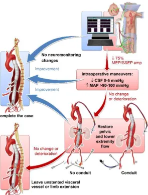
A decrease in IONM (MEP/SSEP) signals triggered the introduction of sequential standardized maneuvers to optimize lower extremity and spinal cord perfusion, including incremental changes in MAP and CSF pressure as previously described. The MAP was raised from 80 up to 100 mmHg along with simultaneous decrease in CSF drain pressure from 10 to 5 or 0
9
mmHg. In patients with improvement after maneuvers, the procedure was completed in standard fashion. In those with no change or deterioration in neuromonitoring, flow was restored to the pelvis and lower extremities as fast as possible by changing the sequence of target vessel stenting. In patients with normalization of MEPs/SSEPs after lower extremity flow was restored, the procedure was completed. If changes persisted, the procedure was left incomplete with flow into the sac via the celiac branch or contralateral iliac limb whenever it was possible (Figure 2).
Figure 2. Standardized maneuvers and protocol.
Protocol steps triggered by changes in motor-evoked potentials (MEPs) and somatosensory-evoked potentials (SSEPs). CSF, cerebrospinal fluid; MAP, mean arterial pressure. By permission of Mayo Foundation for Medical Education and Research.
10 Statistical Analysis
End points were technical success, early and late morbidity and mortality, and freedom from access-related complications. To evaluate the impact of changes in MAP and CSF drainage, the recorded changes in amplitude were analyzed, excluding other potential causes of technical concern such as anesthetic medication or artifact. Motor function, SCI, and stroke were assessed using the American Spinal Injury Association and Modified National Institutes of Health Stroke Scales. The length of the aortic coverage was estimated using centerline of flow measurements (Aquarius iNtuition; TeraRecon, Foster City, CA, USA) to determine the absolute and relative length of the aortic coverage from the left subclavian artery to the aortic bifurcation.
Results are reported as frequencies (percentages) for categorical variables and means ± standard deviations or medians and interquartile ranges (IQRs) for continuous variables. Pearson’s chi-squared test or Fisher’s exact test was used for the analysis of categoric variables. The differences between means were tested with the two-tailed t-test, the Wilcoxon rank-sum test, or the Mann‒Whitney test. Univariate and multivariate logistic models were used to predict MEP and SSEP changes by the extent of coverage, sheath sizes, and times. A value of P < 0.05 was used to determine statistical significance. Data were analyzed by JMP (version 11-pro; SAS Institute Inc, Cary, NC, USA).
11 R E S U L T S
Percutaneous Closure
One-hundred and two (77 male) patients had percutaneous endo-vascular aortic repair (PEVAR), with a mean age of 75 ± 8 years.
A total of 170 common femoral arterial punctures were preclosed using percutaneous vascular closure devices (PVCDs) before placement of the device sheath. The sheath diameter was 20 Fr in 117 femoral arteries (69%), 22 Fr in 40 (24%), 24 Fr in 6 (4%), 18 Fr in 6 (4%), and 16 Fr in 1 (1%).
Technical success of percutaneous closure was obtained in 161 of 170 common femoral arteries (95%) and in 94 of 102 patients (92%). There were no factors that predicted technical failure (P > 0.05), including age, gender, BMI, obesity status, common femoral artery diameter, the presence of femoral artery calcification, prior femoral artery exposure, or sheath diameter. Attempted percutaneous closure failed in 9 common femoral arterial punctures (5%), all with ≥20 Fr sheaths. Although the exact cause of the technical failure was not recorded prospectively, a number of factors contributed to inadequate hemostasis, including high punctures into the inguinal ligament, tears in the arterial wall from calcified plaque, or inability to place the percutaneous suture because of scar or dermis within the percutaneous track. All patients who needed conversion had adequate femoral arterial closure, with no evidence of persistent hemorrhage or ischemia. There were no patients with uncontrolled blood loss associated with systemic hypotension. Patients in whom conversion to open common femoral artery repair was necessary had significantly higher estimated blood loss (median, 500 mL [IQR, 400‒2000 ml] vs. 200 mL [IQR, 100‒
400 mL]; p = 0.05).
12 Neuromonitoring
Forty-nine patients had IONM during endovascular aortic repair at the first stage of this study. The patients’ mean age was 75 ± 8 years (range 47–86;
38 men). There were 5 type C descending thoracic aortic aneurysms (DTAAs) and 44 TAAAs (23 with type IV, 11 with type III, 8 with type II, and 2 with type I). The mean aneurysm diameter was 65 ± 10 mm. Five patients had symptomatic aneurysms and 1 had ruptured acute type B aortic dissection complicated by intramural hematoma and acute aneurysm expansion. Aneurysm etiology was degenerative in 42 (86%) patients and dissection in 7 (14%). Revascularization of the left subclavian artery was performed in 8 patients who required extension of the stent-graft to zone 2.
The total endovascular operating time averaged 175 ± 37 minutes, and the total operating time averaged 290 ± 94 minutes. Median blood loss was estimated to be 450 mL (IQR 200–1151 mL).
A stable MEP/SSEP was achieved in all patients. We did not note any safety issues including bite injuries, seizures, invasive electrode complications, movement-induced injury or arrhythmia. Thirty-one (63%) patients had changes in MEP or SSEP affecting 50 limbs. Of these, 28 (90%) patients had simultaneous decline in MEP and SSEP. Changes in SSEP and MEP occurred at 62 ± 27 and 77 ± 29 minutes, respectively, after vascular access was obtained. The most common pattern of SSEP changes was a signal delay followed by amplitude reduction. All patients with MEP changes had a decrease in the amplitude, first noted in the distal muscle groups. Only 2 (4%) patients had bilateral decline in MEPs at the thigh level. Intra- operative maneuvers restored the neuromonitoring signals in 12 (39%) of the 31 patients. Following restoration of the lower extremity perfusion, MEPs returned to near baseline values within 5 minutes in all but 1 patient who was found to have immediate SCI. The SSEP signals went back to normal at the end of the surgery. His paraplegia gradually improved at a 3- month follow-up, he was able to walk without any support. Two other
13
patients had SCI. One of them had a cardiac arrest and was diagnosed with retrograde type A dissection. The patient was resuscitated and underwent emergent open repair of type A dissection under cardiopulmonary bypass but was noted to be paraplegic after the procedure. One additional patient was readmitted on postoperative day 14 with a complaint of bilateral lower extremity weakness after restarting all his antihypertensive medications.
The patient had complete resolution of symptoms immediately after placement of a CSF drain, intravenous hydration, and discontinuation of all the antihypertensive drugs. The drain was removed in 24 hours with no recurrent symptoms. There were no other patients with lower extremity weakness or SCI. There were 2 (4%) 30-day or in-hospital deaths. The first patient was an 81-year-old high-risk female patient with complicated acute type B dissection, intramural hematoma, and contained ruptured type C DTAA. The patient underwent successful proximal coverage of the dissected thoracic aorta and intramural hematoma extending to the celiac axis but had persistent type Ib endoleak via reentry sites into the area of dissection; she died on postoperative day 2 from aortic rupture. The second patient was a 75-year-old high-risk male patient treated using a 4-vessel, physician-modified, fenestrated/branched stent-graft for rapidly enlarging type II TAAA. The patient’s postoperative course was complicated by myocardial infarction and multiple cerebral and cerebral infarcts, likely from atheroembolization from diseased aortic arch.
In addition, from 2015 of February, as a second stage of the neuromonitoring study, peripheral-evoked compound muscle action potential (CMAP) was performed in 13 patients. From the 13 patients, 9 had StO2 monitoring at the same time. Only 3 patients had IONM changes with more than 75% of drop in the tc-MEP/SSEP amplitude. In every case, CMAP changes were followed by tc-MEP/SSEP changes.
14
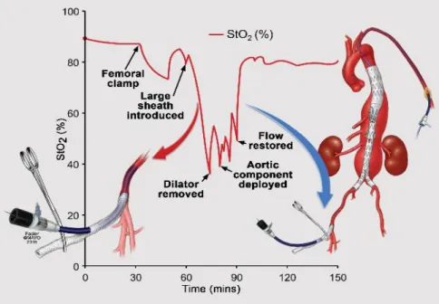
We observed CMAP/tc-MEP/SSEP changes in limbs with significant and long StO2 decrease only. There was a significant difference in iliac artery diameters in patients with changes compared with those who had no changes. The most commonly used sheath size was the 22 Fr sheath (8 limbs). The mean diameter with changes was 7.5 (6.9‒8.2) mm, while 9.6 (8.8‒10.8) without more than 75% signal decrease (P = 0.036) in patients where 22 Fr sheath were used. The use of temporary iliac or femoral conduits was associated with faster recovery and shorter duration of changes in neuromonitoring compared with no use of conduits, but this did not reach statistical significance (38.5 ± 35 vs. 86 ± 81 minutes, respectively; p = 0.272). One patient had StO2 monitoring by use of a temporary conduit. We observed the decline in StO2 with placement of large diameter sheaths in the iliofemoral arteries. After the dilator was retracted to the femoral conduit, StO2 returned to baseline values. In this patient there were no changes observed in neuromonitoring despite the anticipated complex anatomy (Figure 3).
15
Figure 3. This graph illustrates the decline in lower extremity transcutaneous oxygen saturation (StO2) with placement of large diameter sheaths in the iliofemoral arteries. After the dilator is retracted to the femoral conduit, StO2 returns to baseline values. By permission of Mayo Foundation for Medical Education and Research.
C O N C L U S I O N S
1. At the Mayo Clinic, standardized protocol is applied to reduce the risk of spinal cord injury in patients undergoing endovascular TAAA repair. The IONM changes normally would mean spinal cord dysfunction and in our protocol it triggered consistent intraoperative measurements, such as blood pressure elevation and CSF pressure lowering. During our first cases we learnt that changes came from lower limb ischemia rather than SCI. As many
16
studies proved the connection between the lower limb and SCI, we highlighted the importance of the fast lower limb perfusion recovery in our protocol when IONM changes occurred. The one single immediate SCI showed the success of our protective adjuncts. It would be impossible to distinguish between more and less important adjuncts, but oxygen saturation monitoring showed the importance of the stable and elevated blood pressure during the procedure and the use of temporary conduits in certain cases.
2. The lower limb ischemia caused IONM signal loss which disturbed the exact spinal cord monitoring. To differentiate lower limb ischemia from SCI we added the peripheral compound muscle action potential measuring to our IONM, which shows directly lower limb ischemia. It helps to analyze the IONM changes, but when transcranial and peripheral changes occurred at the same time, SCI still could be with lower limb ischaemia. When IONM changes did not improve with blood pressure elevation, we restored the flow to the lower limb and to the pelvis as fast as we could.
With restored lower extremities circulation, the IONM changes could be improved. That time IONM must be specific for the SCI.
This theory partly proved the fact that we had persisting IONM changes only in one case. This patient was who had immediate paraplegia. The IONM can be a helpful tool to monitor spinal cord function during endovascular TAAA repair.
3. Total percutaneous technique can be safely performed with a high technical success rate and a low rate of access complications in patients with thoracic and complex aortic disease requiring large- diameter sheaths. We did not observe uncontrolled bleeding which would lead to hemorrhagic shock and subsequent SCI.
17
B IBLIOGRAP HY OF THE CANDIDATE’S P UBLICAT IONS Publications Related to this PhD Work
Banga PV, Oderich GS, Reis de Souza L, Hofer J, Cazares Gonzalez ML, Pulido JN, Cha S, Gloviczki P. (2016) Neuromonitoring, Cerebrospinal Fluid Drainage, and Selective Use of Iliofemoral Conduits to Minimize Risk of Spinal Cord Injury During Complex Endovascular Aortic Repair. J Endovasc Ther, 23:139‒149. IF: 2,838
de Souza LR, Oderich GS, Banga PV, Hofer JM, Wigham JR, Cha S, Gloviczki P. (2015) Outcomes of total percutaneous endovascular aortic repair for thoracic, fenestrated, and branched endografts. J Vasc Surg, 62:1442‒1449. IF: 3,454
Oderich GS, Baker AC, Banga P. Strategies to Minimize Risk of Spinal Cord Injury During Complex Endovascular Aortic Repair. In: Oderich GS (szerk.) Endovascular Aortic Repair. Rochester: Springer International Publishing, Cham 2017:295‒311.
Entz L, Nemes B, Szeberin Z, Szabó GV, Sótonyi P, Banga P, Csobay- Novák C, Széphelyi K, Hüttl K. (2015) Fenesztrált stent-graft beültetés Magyarországon. Magy Seb, 68:88‒93.
Other Publications
de Souza LR, Oderich GS, Farber MA, Haulon S, Banga PV, Pereira AH, Gloviczki P, Textor SC, Jia F. (2017) Comparison of Renal Outcomes in Patients Treated by Zenith(R) Fenestrated and Zenith(R) Abdominal Aortic Aneurysm Stent grafts in US Prospective Pivotal Trials. Eur J Vasc Endovasc Surg, 53:648‒655. IF: 4,061
18
Jain V, Banga P, Vallabhaneni R, Eagleton M, Oderich G, Farber MA.
Endovascular treatment of aneurysms using fenestrated branched endografts with distal inverted iliac limbs. J Vasc Surg 2016; 64(3): pp.
600‒604. IF: 3,536
Mendes BC, Oderich GS, Reis de Souza L, Banga P, Macedo TA, De Martino RR, Misra S, Gloviczki P. (2016) Implications of renal artery anatomy for endo-vascular repair using fenestrated, branched, or parallel stent graft techniques. J Vasc Surg, 63: 1163‒1169. IF: 3,536
Huang Y, Banga P, De Souza LR, Oderich GS. (2015) Endovascular treatment of visceral artery aneurysms. J Cardiovasc Surg (Torino), 56:
567‒577.
Simó G, Banga P, Darabos G, Mogán I. (2011) Stent-assisted Remote Iliac Artery Endarterectomy: An Alternative Approach to Treating Combined External Iliac and Common Femoral Artery Disease. Eur J Vasc Endovasc Surg, 42: 648‒655. IF: 2,991
Galambos B, Oláh A, Banga P, Csönge L, Almási J, Acsády Gy. (2009) Successful Human Vascular Reconstructions with Long-Term Refrigerated Venous Homografts. Eur Surg Res, 43: 256‒261. IF: 1,500
Galambos B, Banga P, Kövesi Zs, Német J, Jakab L, Czigány T. (2007) Végtagi sérülések ellátása egy regionális centrumban. Magy Seb, 60: 95‒
98.
Galambos B, Banga P, Kövesi Zs, Simon É, Éles Gy, Csönge L, Zsoldos P, Czigány T. (2006) Rekonstruktív érműtétek hosszú ideig hűtve tárolt homografitok felhasználásával. Magy Seb, 59: 388‒392.
19
Olgyai G, Horváth V, Banga P, Kocsis J, Buza N, Oláh A. (2006) Extraskeletal osteosarcoma located to the gallbladder. HPB (Oxford), 8:
65‒66.
Issekutz A, Makay R, Banga P, Németh A, Olgyai G. (2004) Treatment of pancreaticopleural fistulas. Zentralbl Chir, 129: 130‒135. IF: 0,339 Makay R, Banga P, Pohárnok Z, Oláh A. (2004) Invaginatiot okozó malignus jejunum polyp. Egy diagnosztikus kudarc tanulságai. Magy Seb, 57: 290‒292.
Banga P, Vasi I, Hegedüs L, Ladocsi B, Oláh A. (2003) Hogyan változott a colorectalis carcinomák operabilitása az elmúlt két évtized során? Magy Seb, 56: 203‒206.
Makay R, Issekutz A, Banga P, Belágyi T, Oláh A. (2003) Procalcitonin gyorsteszt szerepe az akut necrotizáló pancreatitis steril és inficiált formáinak elkülönítésében. Magy Seb, 56: 31‒33.
Oláh A, Issekutz A, Tóth-G B, Haulik L, Banga P. (2002) Nagyméretű májhaemangiomák sebészi kezelése. Magy Seb 55: 57‒62.
Wellner I, Banga P, Haulik L, Rácz I, Kecskés G. (2001) Distalis duodenum tumorok rezekciójával szerzett tapasztalataink. Magy Seb, 54:
215‒218.