R E S E A R C H Open Access
Exceptional in vivo catabolism of
neurodegeneration-related aggregates
Zsolt Datki1* , Zita Olah1, Tibor Hortobagyi2,3, Lilla Macsai1, Katalin Zsuga4, Livia Fulop5, Zsolt Bozso5, Bence Galik6, Eva Acs7,8, Angela Foldi8, Amanda Szarvas1and Janos Kalman1
Abstract:Neurodegenerative diseases are linked to a systemic enzyme resistance of toxic aggregated molecules and their pathological consequences. This paper presents a unique phenomenon thatPhilodina acuticornis, a bdelloid rotifer, is able to catabolize different types of neurotoxic peptide and protein aggregates (such as beta-amyloids /Aβ/, alpha-synuclein, and prion) without suffering any damage.P. acuticornisis capable of using these aggregates as an exclusive energy source (i.e., as‘food’, identified in the digestive system and body) in a hermetically isolated microdrop environment, increasing their survival. As regards Aβ1–42, five other bdelloid rotifer species were also found to be able to perform this phenomenon. Based on our experiments, the Aβ1–42-treated bdelloid rotifers demonstrate significantly increased survival (e.g. mean lifespan = 51 ± 2.71 days) compared to their untreated controls (e.g. mean lifespan = 14 ± 2.29 days), with similar improvements in a variety of phenotypic characteristics. To our knowledge, no other animal species have so far been reported to have a similar capability. For all other microscopic species tested, including monogonant rotifers and non-rotifers, the treatment with Aβ1–42 aggregates proved to be either toxic or simply ineffective. This paper describes and proves the existence of an unprecedented in vivo catabolic capability of neurotoxic aggregates by bdelloid rotifers, with special focus onP. acuticornis. Our results may provide the basis for a new preclinical perspective on therapeutic research in human neurodegenerative diseases.
Keywords:Alpha-synuclein, Bdelloid rotifer, Beta-amyloid, Catabolism, Lifespan, Prion
Introduction
Neurodegenerative disorders, such as Alzheimer’s disease (AD), Parkinson’s disease (PD), and prion dis- ease, could be regarded as phenotypes secondary to the progressive functional impairment of proteomes.
[29, 52, 53]. The molecular basis of aging in the brain may be described as an accelerated accumulation ac- companied by a decreased clearance and degradation of misfolded proteins [46]. There is a clear correlation between protein aggregation and aged-related patholo- gies. The intramolecular regions arranged in β-sheet conformation are highly resistant to enzymatic deg- radation. [32, 51] The various neurotoxic aggregates, such as those composed of beta-amyloid (Aβ), alpha- synuclein (α-Syn), and prion (normal cellular prion protein /PrPC/ and pathogenic prion protein ‘scrapie’
/PrPSc/), share common features, with their accumu- lation and aggregation facilitating neurodegeneration.
The peptide and protein aggregates in neurodegenera- tive diseases have several characteristics in common;
however, their different molecular structures and pathomechanism may lead to differences in their toxicity [38]. Therefore, investigation of aggregate degradation has emerged from a marginal area of protein chemistry to become a highly relevant field in neuropharmacological science [25]. Although the pathological role of these aggregates has been well established, at present, no universal and satisfactory method exists for their in vivo degradation as a potential therapeutic tool. Misfolded peptide and protein aggregates can be partially digested by several endogenous enzymes, such as insulin-degrading enzyme (IDE) [23], neprilysin (NEP) [16], endothelin- converting enzyme [12], angiotensin-converting en- zyme [14], plasmin [47] and matrix metalloproteinases [1]; however, their presence and function is apparently insufficient in a scenario that leads to neurodegenera- tive disorders.
Amyloids, such as Aβs, are key molecules in aging- associated diseases, representing a starting point in the
* Correspondence:datki.zsolt@med.u-szeged.hu;datkizsolt@gmail.com
1Department of Psychiatry, Faculty of Medicine, University of Szeged, Kalvaria sgt. 57, Szeged H-6725, Hungary
Full list of author information is available at the end of the article
© The Author(s). 2018Open AccessThis article is distributed under the terms of the Creative Commons Attribution 4.0 International License (http://creativecommons.org/licenses/by/4.0/), which permits unrestricted use, distribution, and reproduction in any medium, provided you give appropriate credit to the original author(s) and the source, provide a link to the Creative Commons license, and indicate if changes were made. The Creative Commons Public Domain Dedication waiver (http://creativecommons.org/publicdomain/zero/1.0/) applies to the data made available in this article, unless otherwise stated.
development of dementias. Therefore, their accumula- tion is one of the most important toxic processes during the course of cerebral Aβ-related pathologies, which is potentiated by a reduced clearance and insufficient degradation [30]. The understanding and modulation of Aβ toxicity and its metabolism might provide novel approaches in the treatment of Aβ-related dementias, including AD and cerebral amyloid angiopathy.
Physiologically, two major enzymes are predominantly implicated in the partial degradation of Aβs: NEP and IDE [6, 16]. NEP is a membrane-anchored zinc- dependent endopeptidase, being able to cleave both Aβ monomers and oligomers. The role of NEP in the patho- genesis of AD is indicated by its decreased expression in the AD brain, particularly in vulnerable regions such as the hippocampus and the midtemporal gyrus, a phenomenon associated with increased Aβ-deposition [54]. IDE, a thiol- and zinc-dependent metallopeptidase, appears to participate in the catabolism of insulin and Aβas well, and its decreased expression was reported in the hippocampus of AD patients [55]. Although IDE me- diates these processes in vivo,it still remains a question how this cytoplasmic protein can degrade extracellular Aβaggregates in the brain. Further relevant membrane proteases involved in Aβ degradation include plasmin, cathepsin B, endothelin-converting enzyme, and certain members of matrix metalloproteinase family, which are highly tissue- and brain region-specific [2]. Potential therapeutic approaches to reduce the accumulation of harmful neurotoxic proteins include the facilitation of anti-aggregation processes or the enhancement of their clearance. As an example, β-sheet breakers bind to the central hydrophobic core of Aβ1–42 and attenuate the formation of the β-sheet structures. These molecules could destabilize the senile plaques; however, they do not provide adequate solution to the degradation and catabolism of overexpressed toxic aggregates. [40]
Therefore, an ideal protective strategy against aggregate- induced neuronal damage requires more complex and practical solutions, with dual mechanisms of action targeting both the destabilization and degradation of toxic aggregates.
Treatments with different exogenous Aβ isoforms are widely used models of AD and earlier studies used various in vitro and in vivo systems to reveal their exact effects. Sev- eral studies were performed on human neuroblastoma cells [7, 36], invertebrates, rodents, and primates [13, 20, 42];
however, only a single publication aimed at describing the effects of Aβon bdelloid rotifers, e.g.Philodinaspecies [36].
This unique study by Poeggeler et al. [36] reported the treat- ment of rotifers with Aβ1–42 in order to test the efficacy of an antioxidant molecule (LPBNAH) against the supposed neurotoxicity of the peptide aggregates. In their in vivo studies with rotifers, the authors applied doxorubicin instead
of Aβ1–42, because this toxin gave more consistent results in rotifers. In fact, the neurotoxic effect of Aβ1–42 in this model could not be proven. Our aim was to investigate this intriguing phenomenon that was only slightly touched upon in the paper of Poeggeler.
Bdelloid rotifers, as microinvertebrates, are one of the most commonly used animal models in toxicity-, aging-, and longevity-related research. These organisms are multicellular animals with well-defined anatomical char- acteristics, possessing a ciliated head structure, bilateral ovaries, mastax, ganglia, muscles, digestive, nervous, and secretory systems, and photosensitive, and tactile organs.
[5, 15]. Due to their peculiar anatomy and physiology, these animals have outstanding advantages in terms of culturing and are rather easy to work with [44]. Rotifers are extremely resistant to environmental alterations and successfully adapt to the different types and amounts of nutrients present in their natural habitat. The natural decomposition of organic materials is a process that re- sults in the formation of precipitates and aggregates, which represent potential nutrients for rotifers [50]. The metabolic utilization of all these available organic mater- ial resources is their special property [4].
In a prior publication, we reported the development of a unique and straightforward method [34], which enables the investigation of the effect of several different agents or impacts on various phenotypic parameters of microinver- tebrates. The oil-covered microdrop technology, adopted from human in vitro fertilization, is a well-controllable construction to assess the lifespan and other features of a single isolated animal (one-housed rotifer).
In our present study, we examined the effect of various neurodegeneration-related peptide and protein aggregates under complete dietary restriction, ensuring that the individual rotifers had no other organic source to be used for gluconeogenesis. Observing an intriguing increase in survival upon treatment with aggregates, as a next step, we investigated different types of micro- entities in neurotoxic aggregate-supplemented environ- ment. To our knowledge, this study is the first to address the in vivo catabolism of these molecules as dietary sources in microscopic animals such as rotifers.
Our findings may provide a starting point to understand the possible ways of degradation of abnormally folded neurotoxins despite their aggregated state and conse- quent protease resistance, a subject with high potential relevance in the treatment of neurodegenerative proteinopathies.
Materials and methods Materials
The Aβ1–42, Aβ1–42 [Gln22], Aβ1–40, Aβ25–35, two scrambled isoforms (Aβ1–42 S1: LKAFDIGVEYNKVGEG- FAISHGVAHLDVSMFGEIGRVDVHQA and Aβ1–42 S2:
KVKGLIDGAHIGDLVYEFMDSNSAIFREGVGAGHVHV AQVEF) were prepared in the Department of Medical Chemistry, University of Szeged, Szeged, Hungary. The peptides were synthesized on an Fmoc-Ala-Wang resin using Nα-Fmoc-protected amino acids with a CEM Liberty microwave peptide synthesizer (Matthews, NC, USA). The peptide Aβ11–42 (H-7668.1000) was purchased from Bachem (Torrance, CA, USA), whereas Aβ1–28 (A0184) andα-Syn (type E46K human; S4447) were purchased from Sigma-Aldrich (St. Louis, MO, USA). The mature part (25–
244) of recombinant bovine prion protein (PrPC, AG210) was obtained from Merck Millipore (Darmstadt, Germany).
EZ4U (BI-5000; Biomedica Medizinprodukte, Wien, Austria) and Calcein-AM (17,783; Sigma-Aldrich) cell viability assays were used to measure the toxicity of the aggregates. For in vivo and in vitro investigations of the dif- ferent aggregates, we applied Bis-ANS (4,4′-dianilino-1,1′- binaphthyl-5,5′-disulfonic acid dipotassium salt; D4162) and Congo red (CR; C6277) dyes obtained from Sigma- Aldrich. To detect gold-tagged beta-amyloid (Au-Aβ1–42) inP. acuticorniswith scanning electron microscopy (SEM), we used Gold(III) chloride (AuCl3 x 2H2O; 01216, Reanal, Budapest, Hungary) and Aβ1–42 aggregates. Distilled water (DW) was prepared in our laboratory (Millipore-type, ultra- pure, demineralized DW).
Preparation of aggregating peptides and proteins
The synthesis and characterization of the Aβ peptides were conducted as previously described by Bozso et al. [3] with minor modifications: the concentrations of the stock solutions were 1 mg/mL (DW); the aggrega- tion time was 3 h or 3 days (25 °C, pH 3.5); the neutralization (to pH 7.5) was performed with NaOH (1 N) [17]; after 10-fold dilution with standard medium, the final (working) concentrations were 100 μg/mL. The amount of diluted cations and anions in standard medium (mg/L): Ca2+ 31.05; Mg2+ 17.6;
Na+ 0.9; K+ 0.25; Fe2+ 0.001; HCO3−153.097; SO4−3; Cl
− 0.8; F− 0.02; H2SiO3 3.3 (pH = 7.5) [41]. To prepare the PrPSc form of PrPC, the stock solution of PrPC was aggregated for 24 h at pH 2 [49, 57]. The pH of the prepared prion was also adjusted to pH 7.5 before being used to treat the rotifers.
Collection, isolation, identification and harvesting of different animal species
To collect and isolate different microscopic species, we used the method described by Debortoli et al. [9] with minor modifications. The sampling sites were distributed within two areas of about 500 m2near Szeged (Southern Great Plain, Hungary) and Saint-George (Transylvania, Romania). Fifty patches of highly hydrated moss (with minimal soil) were collected from the northern side of trees (of the genera Acer and Platanus) and from their
close environment. Briefly, the collected samples were hydrated with the standard medium in separate flasks.
After identifying the species by using methods described in the literature [18, 21, 24, 33, 48] we applied species- specific information (e.g., body size in relation to age) to collect relatively young (approximately 3–5 days old) and individuals of the same size. The isolation protocol from the samples was in line with the method of Debor- toli et al. [9]. However, instead of culturing; we collected and isolated these animals (using micropipette) to create monoclonal populations. After washing these groups with standard medium, we harvested as many indivi- duals as needed (n= 30) for the experiments. The spe- cies collected in Hungary comprised Philodina vorax Janson, 1893; Philodina megalotrocha Ehrenberg, 1832;
Lepadella patella Müller, 1773; Lecane arcula Harring, 1914; Lecane agilis Bryce, 1892; Lecane hamata Stokes, 1896; Lecane closterocerca Schmarda, 1859; Brachionus diversicornis Daday, 1883; Brachionus calyciflorus calyci- florus Pallas, 1766; Brachionus forficula Wierzejski, 1891;
Caenorhabditis elegans Maupas, 1900; Typhloplana viri- data Abildgaard, 1789; Vorticella convallaria Linnaeus, 1758; Stylonychia mytilus Müller, 1773. The species col- lected in Romania comprised Mniobia russeola Zelinka, 1891; Adineta steineri Bartoŝ, 1951; Habrotrocha elusa Milne, 1916; Filinia terminalis Plate, 1886; Lindia toru- losa Dujardin, 1841; Hypsibius dujardini Doyère, 1840;
Centropyxis aculeata Ehrenberg, 1832. Processes related to culturing and harvesting Philodina acuticornis Murray, 1906were performed (in artificial environment) as previously described by our group [34]. The animals were photographed (Nikon D5500, Nikon Corp., Kanagawa, Japan) under the light microscope (at 200×
magnification; Leitz Labovert FS, Wetzlar, Germany) by taking serial images at every 5-μm intervals, yielding a total of 20–30 photographic layers per animal, which were subsequently merged into one superimposed pic- ture (by using Photoshop CC software, Adobe Systems Inc., San Jose, CA, USA) to achieve better resolution.
In vitro and in vivo treatment and monitoring
To test the previously described neurotoxic effects of the examined aggregates in vitro,we used a differentiated SH- SY5Y human neuroblastoma cell line (Sigma-Aldrich), with the related culturing and differentiating methods based completely on our previous works [7, 26]. Five-day- old animals (as determined by BSI) were chosen for the experiments, an age that precedes the beginning of the re- productive stage (i.e., egg production) by 2 days. Before treatment (using micropipette) of individual rotifers with aggregated molecules (0.1, 10 and 100μg/mL), the stock solutions (1 mg/mL) were ultrasonicated (Emmi-40 HC, EMAG AG, Mörfelden-Walldorf, Germany) for 10 min at 45 kHz to achieve sterilization and homogenization.
During the in vivo experiments, the viability assay, the assessments of descriptive characteristics (such as norma- lized mean lifespan /NML/, body size index /BSI/, bright light disturbance /BLD/, mastax contraction frequency /MCF/, and cellular reduction capacity /CRC/), and the assembly of the experimental setup (with the oil-covered microdrop) were carried out as presented in detail previously [34].
Detections of exo- and endogenic Aβ1–42 inP.
acuticornis
Optical analysis in live rotifers (Fig. 1a-d): The juvenile (5 days old) animals were identified on the basis of their body size as read from a calibration curve previously de- scribed [34]. For the treatment (in 96-well plate; 3695, Costar, Corning Inc., USA) of preselected and 1-day starved rotifers, we used unlabelled Aβ1–42 (3 h and 3 days aggregated) as ‘food’ source. After 12 days the content of the wells were changed to labelled Aβ1–42, aggregated for 3 h (in vitro marked with 10μM Bis-ANS fluorescent dye for 30 min) or 3 days (in vitro marked with 50 μM CR dye for 1 h). Applying 5 h treatment (‘feeding’), we detected the optical signals (at 200×
magnification) in the digestive system (stomach and in- testine) of individual rotifers by an optical/fluorescence microscope (Olympus IX71, OLYMPUS, Budapest, Hungary). The representative images demonstrate the localization of Aβ1–42 in the body of rotifers compared to untreated, normally fed (600μg/mL yeast suspension) and unfed (starved) controls (Fig. 1a-d). Unfed indivi- duals labelled with these two dyes (control background without Aβ1–42 treatment) have no signal (photo not shown).
Searching for endogenous Aβ1–42 in rotifers: The immunological detection of the presence or absence of natural Aβ1–42 in P. acuticornis species has a central role in interpreting the relations of Aβ1–42 and rotifers.
After harvesting, the animals were put into −75 °C for 30 min in standard medium. The number of rotifers were 104per sample. The thawed animals were ultraso- nicated (at 45 kHz at room temperature for 10 min).
The rotifer homogenate was centrifuged (300 x g for 5 min at 24 °C) to eliminate the exoskeletons of the dead animals. The protein concentration of the supernatant was determined by the Qubit Protein Assay Kit (Q33212; Thermo Fisher Scientific, Wilmington, DE, USA) following the manufacturer’s protocol. The con- centration of the stock suspension was 340.54 μg/mL, which was diluted to the final protein concentrations (μg/mL) of 20, 15, 10, 5 and 1 in standard medium.
Commercially available Aβ1–42-specific sandwich ELISA kits (Innotest Aβ1–42, 81,576, Fujirebio, Gent, Belgium) were used according to the manufacturer’s
protocol for the quantitative detection of Aβ1–42 in the diluted rotifer homogenate.
The Au-Aβ1–42 complex detection with SEM(Fig. 1f, g): The stock solutions of Aβ1–42 (1 mg/mL) and AuCl3
(2.8 mg/mL) were prepared with DW. The 3-day- aggregated Aβ1–42 solution was mixed for 2 h with the AuCl3 solution in a 1:4 M ratio, according to the number of Au-binding sites of monomeric Aβ1–42 (one methionine and three histidine residues). To remove ex- cess Au ions, two rounds of centrifugation (25,000 x g for 10 min at 24 °C) with supernatant replacement were used. NaOH (1 N) was applied to adjust the pH to 7.5.
Peptide content of the final pellet was determined by the Qubit™ Protein Assay Kit (Thermo Fisher Scientific).
The middle-aged (15 days old) harvested rotifers were starved (i.e., complete food deprivation) for 2 days. After washing the rotifers in 6-well plates (containing 104 adherent animals per well), each well was treated with Au-Aβ1–42 complex in a dose of 100 μg/mL for 1 day.
The wells were decanted and washed twice with stan- dard medium and after an incubation period of 6 h (the time to empty the digestive tract of the animals), the wells were washed again and the populations were fixed and dehydrated with 96% EtOH at −75 °C for 5 min, followed by a partial rehydration with 30% EtOH at room temperature for 30 min. After fixation with 1%
paraformaldehyde (for 30 min), the wells were washed twice with DW. The collected animals were transferred with pipette to the centre of a round glass coverslip (diameter: 12 mm, thickness: 0.15 mm; 89,167–106, VWR International, Houston, TX, USA), and were allowed to dry. The samples were not coated with gold.
The structural integrity of the rotifer bodies was con- trolled with digitally recorded photographs (Nikon D5500). The selected bodies (based on their quality) were subjected to SEM (Zeiss EVO MA 10, Carl Zeiss, Oberkochen, Germany) [2]. The sample-carrier coverslip was fixed onto a stub using a double-sided carbon tape.
The fine structure of the rotifers was observed and photo-documented with the SEM (Fig. 1f, g), operating at 10 kV with an 8-mm working distance, using a back- scattered electron detector in variable pressure mode at 30 Pa. The white balance of SEM photographs was normalized.
Congo red aggregation assay
The methods described by Klunk et al. [19] and Datki et al. [8] were used for these experiments. The preparation and aggregation of the peptides and proteins for the CR assay was the same as described in the in vitro and in vivo treatment protocols. A volume of 10 μL CR stock solution (0.5 mM) was added to each aggregate-containing tube (490 μL;
100 μg/mL) and these mixtures were incubated for
20 min at room temperature and shaken every 5 min (at 50 rpm for 10 s), followed by centri- fugation at 25,000 x g for 15 min. The supernatant was carefully removed with a pipette and the sediment (pellet) was resuspended in standard
medium (0.5 mL). The aggregate-bound CR content of the suspensions was measured spectrophotomet- rically at 540 nm with a BMG NOVOStar plate reader (BMG Labtech, Ortenberg, Germany), using a 96-well plate (Costar).
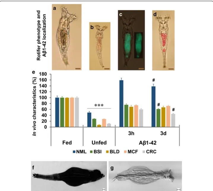
Fig. 1Localization and demonstration of the beneficial effects of aggregated Aβ1–42 onP. acuticornis.Juvenile rotifers were selected in different groups after 1-day starvation: fed (a; 12 days normal feeding), unfed (b; 12 days starvation) and Aβ1–42 treated (for 12 days). The Aβ1–42 in the digestive system (stomach and intestine) of live rotifers were visualized by fluorescent Bis-ANS (c; green colour in the representative photograph; 3 h = aggregated for 3 h) and absorbent Congo red (d; red colour in the representative photograph; 3d = aggregated for 3 days). Untreated individuals labelled with these two dyes showed no signal (not shown).eThe normalized mean lifespan (NML) of the Aβ1–42-treated animals was significantly longer than that of their normally fed and unfed untreated controls, with a significant difference between the 3 h- and 3d–aggregated forms. Besides NML, four viability markers were measured, including body size index (BSI), bright light disturbance (BLD), mastax contraction frequency (MCF), and cellular reduction capacity (CRC). The Aβ1–42-treated rotifers performed significantly better than their unfed untreated controls and there were significant differences between the subgroups treated with two the differently aggregated forms in BSI and CRC. The concentration of Aβ1–42 was 100μg/mL;n= 30 (NML), 50 (BSI), 20 (BLD), and 24 (MCF) one-housed indivi- dual rotifers per group;n= 24, well with normalized absorbance to rotifer number (CRC). The scale bars in the proportional representative photographs repre- sent 20μm (*, significant difference from the fed and Aβ1–42-treated groups; #, significant difference from the group treated with the 3 h aggregated form).
The possible distribution of the exogenous Au-Aβ1–42 complex was studied in vivo with SEM in the body ofP. acuticornis:frotifer without treatment (background);gAu-Aβ1–42-treated rotifer. Homogenously signals of gold can be detected in Au-Aβ1–42-treated entities. Scale bar represents 10μm
Statistics
The error bars represent the standard error of the mean (S.E.M.). For comparative statistical analysis, the one-way ANOVA was used followed by the Bonferroni post hoc test with SPSS 23.0 software for Windows. Ap≤0.05 was regarded as statistically significant, with the different levels of significance indicated as follows: p#≤0.05, p**≤0.01 and p***≤0.001. Kaplan-Meier curves were applied to present the survival of the groups. The GraphPad Prism 7.0b software (GraphPad Software Inc., La Jolla, CA, USA) was used for the illustration and statistical analysis (log-rank; Mantel-Cox) of survival.
Results
To investigate the background of less ‘consistent results’
with Aβ1–42 toxicity in bdelloid rotifers reported by Poeggeler et al. [36], we investigated the effect of diffe- rent neurodegeneration-related protein aggregates on one-housed P. acuticornis in an experimental setup [34]
inspired by that applied human in vitro fertilization (i.e., oil-covered microdrop). This assay system allows the observation of our model organisms at an individual level. First, we examined the effect of Aβ1–42, which we predicted to be toxic toP. acuticornis. Surprisingly; how- ever, treatment of the animals with Aβ1–42 resulted in a significantly longer mean lifespan (51 ± 2.71 days) than in the case of unfed (14 ± 2.29 days) and normally fed (32 ± 2.72 days) controls. To localize and demonstrate the presence of Aβ1–42 aggregates in the body of the rotifers, we used β-sheet-specific fluorescent and absor- bent dyes. Animals in the representative photographs (Fig. 1a-d) are shown in proportional sizes and display the strong differences between the groups. The Fig. 1c, d show the presence of the exogenous Aβ1–42 in the digestive system of the rotifers after ‘feeding’ad libitum (above is the stomach and below is the intestine). To characterize the Aβ1–42-treated P. acuticornis animals, we applied some previously published [34] experimental monitoring assays. The results (Fig. 1e) are evidences to the fact that this bdelloid rotifer can use the Aβ1–42 as food in isolated environment without the presence of any other organic material. The NML of groups treated with either 3 h or 3 days aggregated Aβ1–42 signifi- cantly increased compared to unfed (starved) controls.
The BSI and the BLD indicated phenotypical and physiological changes of the treated animals. These characteristics were increased by 40% and 60%
compared to untreated starved controls, respectively.
The MCF and the CRC suggested intensified energy level as represented by neuromuscular and cellular- redox activities. These two markers were increased by 46% and 42% compared to unfed entities, correspon- dingly. The Aβ1–42-treated one-housed rotifer indivi- duals performed much better in the measured
parameters than their unfed controls, and they do not drastically differ from the normally fed counterparts.
These results suggest that Aβ1–42 is not toxic toP. acu- ticornis and it could be used by them as an exclusive dietary source to live and develop in an hermetically- isolated environment. As the next step, we aimed at de- tecting the possible presence of endogenous Aβ1–42 in P. acuticornisspecies. We applied ELISA for the quanti- tative analysis of Aβ1–42 in the rotifer homogenates.
Interestingly, our findings indicate that endogenous Aβ1–42 is practically absent inP. acuticornisspecies,an observation first reported in the literature. To localize exogenous Aβ1–42 aggregates in the live (Fig. 1a) roti- fers (beyond the digestive system) we applied Au-tagged Aβ1–42 aggregates, detected with SEM (Fig. 1f, g). After fixating and drying the Au-Aβ1–42-treated and un- treated animals, we monitored the possible distribution of the remnants of the potentially catabolized peptide.
We found that in Au-Aβ1–42-treated animals, the signal of gold-ions could be found homogeneously everywhere in their body in contrast to the untreated ones. The only possible source of gold in the samples was the Au-Aβ1–
42 complex taken up during life, as no aspecific gold coating was applied. These SEM photos are only representative.
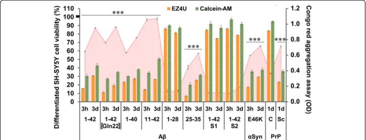
In our study, we used eleven different peptides and proteins, with some of them being accepted as neuro- toxic aggregates in neurodegenerative diseases (Fig. 2).
To test and confirm the toxic effect of these aggregated peptides/proteins, we used a differentiated SH-SY5Y hu- man neuroblastoma cell model, based on our previous works [7, 8]. The CRC-specific EZ4U and cytoplasmic enzyme activity-sensitive Calcein-AM assays were used to test the potential effect of the various aggregates. The time-dependent differences between the respective ag- gregate solutions incubated for 3 h and 3 days were measured with CR spectrophotometric assay [8, 19]. The data demonstrated an inverse correlation between CR- binding property and cellular toxicity of aggregates. The Aβ1–28, the scrambled isoforms Aβ1–42 S1 and S2, and PrPC demonstrated low affinity to CR and were not toxic to SH-SY5Y cells, accordingly.
In our next experiment, we examined whether the P.
acuticornis is capable of catabolizing other neurotoxic aggregates as well (Fig. 3). To investigate the dose- dependency of the effect of Aβ1–42 on rotifers, we used three different treatment concentrations (0.1, 10, and 100μg/mL), comparing the results with those of groups treated with equivalent concentrations of bovine serum albumin (BSA). Interestingly, we observed the highest median survival in the case of 100μg/mL Aβ1–42 treat- ment (Fig. 3a). We found that almost all Aβ peptide forms tested (Aβ1–42, Aβ1–42 [Gln22], Aβ1–40, Aβ11–
42, Aβ1–28, Aβ1–42 S1; Aβ1–42 S2) were favourable
nutrient sources, contrasting with Aβ25–35, which proved to be toxic to the rotifers (Fig. 3 b-c). This‘short’
type of Aβ has a relatively low molecular weight (1060.27 g/mol) compared to the longer Aβ peptides (e.g., Aβ1–42), resulting in higher molar concentration (94.3μM) in a 100-μg/mL dose. We tested this artificial Aβ aggregate in a dose of 10 μg/mL as well, and we found it also significantly toxic (NML 58 ± 5.8%; p≤ 0.01) when analysing the survival of the treated group compared to that of unfed controls. In addition to various Aβ isoforms, P. acuticornis was also able to catabolize one of the most aggregation-prone forms of α-Syn (type E46K) as well as the physiological (PrPC) and pathological forms (PrPSc) of prion protein (Fig. 3d). The Kaplan-Meier survival curves demonstrate significant differences in all treatment groups compared to the respective untreated control curve (Fig. 3a).
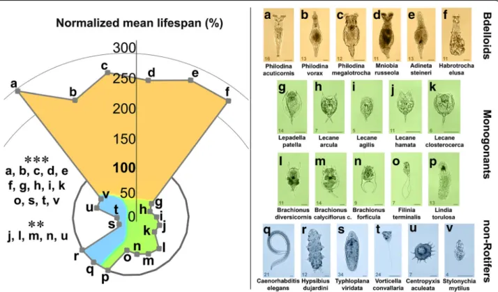
To investigate the universal capability of one-housed microscopic entities to catabolise Aβ1–42 (aggregated for 3 h before treatment; Fig. 4), we examined 22 species with different phylogenetic backgrounds. These species included bdelloid rotifers: P. acuticornis, P. vorax, P.
megalotrocha, M. russeola, A. steineri, H. elusa; monogo- nant rotifers:L. patella, L. arcula, L. agilis, L. hamate, L.
closterocerca, B. diversicornis, B. calyciflorus calyciflorus, B. forficula, F. terminalis, L. torulosa; non-Rotifers: C.
elegans, H. dujardini, T. viridata, V. convallaria, C. acu- leate, S. mytilus. In order to provide adequate basis for comparison, we used an NML where the mean lifespan of the unfed (starved) control group within the respect- ive species were regarded as 100% (contrasting with the comparisons in Fig. 1 where 100% was the mean lifespan of the normally-fed group). From the all examined spe- cies, bdelloids (Fig. 4a-f ) were the only ones that demon- strated significantly longer NMLs (240–290%) compared to their unfed (starved) controls (100%). As regards other species, Aβ1–42 was either toxic (Fig. 4g-o of monogonants and Fig. 4s-v of non-rotifers) or had no ef- fect (Fig. 4p-r of both groups).
The results strongly suggest that P. acuticornis can catabolize various form of toxic and non-toxic Aβs, α-Syn, and prion aggregates and that the investigated bdelloid rotifer species are also able to use the neurotoxic Aβ1–42 aggregates as nutrients (ad libi- tum). This agent is either toxic or has no effect on the lifespan of other microinvertebrates examined, suggesting that the catabolism of aggregates with proteinopathy is an exceptional property of bdelloid rotifers (Fig. 5).
Fig. 2Aggregation-dependent neurotoxicity of different neurodegeneration-related peptides and proteins in cell culture. To test the previously described [10, 11] toxic effect of neurodegeneration-related aggregates, we used a differentiated SH-SY5Y human neuroblastoma cell model. The EZ4U and Calcein-AM cell viability assays were used to detect the NADH- and esterase-activity-dependent cell viability of the cultures (presented in orange and green columns of the chart, respectively). The mean viability of the untreated control wells was regarded as 100% (the S.E.M. of the mean was ±2.8%). The aggregation level of the proteins was measured with Congo red spectrophotometric assay (red line in the background of the chart). The length of incubation and aggregation time (3 h = 3 h and 3d = 3 days) influenced the toxicity of the treatment in most of the peptides and proteins tested. Significant differences (p≤0.01) were detected between the 3 h- and 3d–groups (no markers on columns) in both viability assays. Significant differences could be detected between the 3 h- and 3d–aggregated forms in the Congo red assay for all Aβs, except for Aβ11–42, Aβ1–28, Aβ1–42 S1, and Aβ1–42 S2. Alpha-synuclein (α-Syn, type E46K) and prepared‘scrapie’(PrPSc) form of prion protein also showed significant in vitro toxicity, contrasting with the ineffective normal cellular prion (PrPC). The aggregation time of the prions was 1 day (1d). Each molecule was used in 100μg/mL concentration;n= 8 (for EZ4U and Calcein-AM) and 20 (for Congo red) wells per group. The error bars present the S.E.M. For statistical analysis, one-way ANOVA was used followed by the Bonferroni post hoc test, and the level of significance werep***≤0.001 (*- the significant rate of deviation from the non-toxic and non-aggregated forms /Aβ1–28, 1–42 S1, 1–42 S2, PrPC/ in both viability and Congo red assays)
Discussion
The formation and accumulation of aggregated proteins is a central feature of several neurodegenerative diseases.
The age-related dyshomeostasis promotes the aggrega- tion of misfolded proteins. Neurodegeneration is asso- ciated with the resistance of various aggregated peptides and proteins to enzymatic degradation [39].
In transmissible spongiform encephalopathies, the endogenous PrPC, composed of the pure protein without any presence of nucleic acid components, is converted to the pathogenic and infectious form (PrPSc) [37]. The α-Syn is a presynaptic neuronal protein, which contributes to the pathogenesis of PD via its toxic aggregation [35]. Aβ peptides can accu- mulate as senile plaques, which are regarded as one of the hallmark alterations in AD [2, 43].
Although the degradation of these amino acid- containing organic macromolecules has been in the focus of neuropharmaceutical science for decades, there have been no proper treatments developed or methods identified for the elimination of these toxic aggregates from living systems to date. Moreover, only a fraction of these peptides and proteins can be efficiently digested by proteolytic enzymes either in vitro or in vivo. [22]
In our study, we are the first to describe the existence of the in vivo catabolism of neurotoxic aggregates by bdelloid rotifers (e.g.,Philodina acuticornis), as these mi- croinvertebrates are capable of using even neurotoxic aggregates as exclusive energy and organic material sources, prolonging their lifespan. The one-housed and aggregated Aβ1–42-treated [3] P. acuticornis individuals have significantly longer lifespan and better viability compared to their untreated controls under complete dietary restriction in a fully isolated environment. This capability to improve (NML) or maintain (BSI, BLD, MCF and CRC) viability showed a correlation with the aggregation state (i.e., CR affinity) of Aβ1–42. The administered exogenous peptide aggregates were first lo- calized and distributed within the digestive system of the rotifers, providing evidence of being consumed (Fig. 1).
First in the literature, we demonstrated that this species have no detectable endogenous Aβ1–42 production.
Since the treated P. acuticornis maintained its well- being, function, and redox capacity, we presume that these aggregates may serve as an obligatory energy source for gluconeogenesis in these experimental condi- tions. Prolonged starvation of animals causes significant physiological changes supplying glucose from amino
Fig. 3The effect of different neurodegeneration-related aggregates on the survival ofP. acuticornis.The Kaplan-Meier curves demonstrate the survival of rotifers (n = 24 one-housed individuals per sample type; aggregation time: 3 h = 3 h and 3d = 3 days).aDose dependency: the effect of 3 h–aggregated Aβ1–42 and BSA were tested in three different concentrations. This neurotoxic peptide as well as the non- toxic protein served as nutriments to rotifers. The survival was dose-dependent (0.1, 10, and 100μg/mL) and was significantly higher in both type of treatments compared to the untreated control group (UC;p< 0.001; log-rank test). bNatural Aβisoforms:we tested four natural variants of the Aβpeptide (1–42 [Gln22], 1–40, 11–42 and 1–28) on rotifers in two different aggregated forms (3 h and 3d). All types of natural Aβs had a positive effect on the survival of the treated rotifers compared to the UC (a). cArtificial Aβisoforms:the roti- fers were able to use both scrambled forms of Aβ1–42 (S1 and S2) as nutrients. As a unique exception in the series, Aβ25–35 was toxic to the rotifers, decreasing their survival as compared to the UC (a). All three types of artificial Aβs were measured in 3 h- and 3d–aggre- gated forms.dNon-Aβproteins:the animals treated with alpha-synuclein (α-Syn, type E46K; in 3 h- and 3d–aggregated forms) and the cellular (PrPC) or‘scrapie’(PrPSc) forms of prion protein (1d = aggregated for 1 day) also showed a significantly longer lifespan compared to the UC (a). Each protein was used in 100μg/mL concentration. The significance of log-rank (Mantel-Cox) test wasp< 0.0001 in the statistical analyses in panelsb-d. Median survival (ms) values in days are presented in the graphs. The statistical analysis of survival was performed by GraphPad Prism 7.0b
acids by metabolism, including ketogenesis and gluco- neogenesis. The main function of gluconeogenesis is to maintain the glucose level via its endogenous de novo production from non-carbohydrate substrates, such as glycerol, lactate, or glycogenic amino acids [28]. In rotifers, the resting eggs contain large amounts of non- carbohydrate substrates as obligatory sources for vital anabolic processes during dormancy and hatching via glyoxylate cycle and gluconeogenesis [10]. The bdelloids are extremely resistant to starvation, which has been shown to extend their lifespan [31], suggesting that the enzymatic machinery required for endogenous de novo glucose production may play a crucial role in their me- tabolism. On this basis, their observed survival in the presence of an aggregated peptide suggests its partial catabolism.
In order to assess the toxicity of the examined ag- gregated molecules, the same treatment agents were tested on differentiated SH-SY5Y neuroblastoma cell cultures [7, 26]. The α-Syn and PrPSc, similar to the known toxic Aβs, caused significant reduction of
viability, in correlation with their aggregation state (CR affinity). Our in vitro results (Fig. 2) were in line with our a priori expectations and the academic literature [45, 56].
The catabolic capability of P. acuticornis was not limited to Aβ1–42 (Fig. 3). In the absence of any other potential dietary source, the presence of other neurodegeneration-related aggregates, such as natural (Fig. 3b) or artificial (Fig. 3c) Aβs;α-Syn, PrPC, or PrPSc (Fig. 3d) likewise prolonged the survival of the treated animals compared to the untreated (unfed) controls, ex- cept for Aβ25–35, which resulted in reduced survival.
The median survival showed association with the aggre- gation state. These results together led us to the conclu- sion that this extraordinary catabolic activity is universal as regards almost any type of peptide and protein aggre- gates. In their natural habitat, bdelloids constantly have to cope with extreme fluctuations in nutrient availability [4]. Furthermore, their native food source includes particulate organic detritus, dead bacteria, algae, and protozoa, which offer a large variety of natural
Fig. 4The effect of Aβ1–42 on the normalized mean lifespan of different microscopic species. To investigate the universality of the ability to catabolise 3 h–aggregated Aβ1–42 in one-housed entities, we tested 6 Bdelloids (a-f), 10 Monogonants (g-p), and 6 non-Rotifers (q-v). The data were normalized in percent, where the mean survival of the untreated starved group of the respective species were regarded as 100%. Only bdelloids demonstrated significantly longer lifespan compared to their starved controls after Aβ1–42 treatment. For monogonant rotifers, Aβ1–42 was either toxic (g-o) or ineffective (p) compared to their untreated controls. A similar effect was detected in non-rotifer species, where the aggregated peptide likewise either had no effect (qandr) or was toxic (s-v). The number in the bottom left corner of the images represents the measured real mean survival lifespan (in days) of the untreated starved control individuals. The concentration of Aβ1–42 was 100μg/mL n = 30 one-housed individuals per group. For statistical analysis, one-way ANOVA was used followed by the Bonferroni post hoc test, and the levels of significance were p**≤0.01 and p***≤0.001 (*- significant difference from the untreated starved control). Scale bars represent 50μm
aggregates [50]. Therefore, the capability of these ani- mals to metabolize almost every type of aggregated pep- tides and proteins might be an evolutionary strategy for adaption and survival.
Our aim was to describe the existence of the in vivo catabolism of neurodegeneration-related aggregates by bdelloid rotifers. The results strongly indicate that these animals can use these toxic agents as an exclusive diet- ary source to live and grown in an hermetically isolated environment. The available literature does not describe any other metazoan species with a similar capability. In species widely applied in the animal modelling of AD, such as Caenorhabditis elegans, Drosophila mela- nogaster, Danio rerio, or rodents, the Aβ1–42 are toxic [11, 20, 27, 41]. Based on our experiments (Fig. 4), the Aβ1–42 aggregates were also toxic or ineffective to the other microscopic entities tested (monogonant rotifers and non-rotifers). The universal capability of bdelloid rotifers to catabolise peptide and protein aggregates (in- cluding such that are known to be neurotoxic) implied that the hypothetic catabolic pathway might consist of degrading enzyme(s), their possible cofactors, and/or anti-aggregation compounds. The comprehensive identi- fication of the molecular background of this unique phenomenon requires further analyses.
Conclusion
In summary (Fig. 5), we present findings indicating the exceptional capability of the investigated bdelloids to catabolise neurotoxic aggregates in vivo, which is to our knowledge unprecedented in the literature regarding any multicellular animals. In our experimental system, only amino acids could be used for gluconeogenesis by the rotifers; therefore, the aggregates applied represented the only material to be catabolised in the absence of any other food source in this isolated environment. The one-housed P. acuticornis individuals maintain their normal phenotypic characteristics and demonstrate significant activity during their life, which require constant energy production. The prolonged lifetime in the presence of (almost all types of ) Aβs, α-Syn, PrPC or PrPSc suggests the universal catabolic capability of our model species as regards a wide range of peptide or protein aggregates.
The understanding of the unique catabolic capability of bdelloid rotifers, including P. acuticornis, on neuro- toxic aggregates in an isolated environment without any other energy source available may provide the basis of a novel therapeutic approach (identification of key mole- cules and metabolic pathways) in neurodegenerative proteinopathies.
Fig. 5The general capability of bdelloid rotifers to catabolise neurotoxic aggregates.Neurodegeneration-related peptide and protein aggregates can be utilized as nutrients when given as the only potential energy source for starved bdelloid rotifer species. These agents proved to be either toxic or ineffective for monogonant and non-rotifer species. A currently unidentified mechanism in bdelloid rotifers is hypothesized to enable the utilization of amino acids derived from the degraded aggregates for gluconeogenesis. Compared to the unfed (starved) controls, the bdelloid rotifers treated with aggregates (‘fed’) demonstrate increased survival and viability
Abbreviations
α-Syn:Alpha-synuclein; AD: Alzheimer’s diseaseAu-AβGold-tagged beta- amyloid; Aβ: Beta-amyloid; BLD: Bright light disturbance; BSA: Bovine serum albuminBSIBody size indexCRCongo red; CRC: Cellular reduction capacity;
CSF: Cerebrospinal fluid; DW: Distilled water; EtOH: Ethanol; IDE: Insulin- degrading enzyme; MCF: Mastax contraction frequency;
NEP: NeprilysinNMLNormalized mean lifespanPDParkinson’s
diseasePrPCNormal cellular prion proteinPrPScPathogenic prion protein
‘scrapie’SEMScanning electron microscopeUCUntreated control
Acknowledgements
The authors wish to thank for the contribution of Anna Szentgyorgyi MA (Institute of English and American Studies, University of Szeged, Szeged, Hungary), Ilona Hatvani, and Prof. Botond Penke. We are grateful to Levente Szalardy MD, PhD (Department of Neurology, University of Szeged) for his valuable contribution in proofreading the manuscript.
Consent of publication Not applicable.
Funding
This research was supported by the GINOP-2.3.2–15–2016-00019 grant to EA and AF.
Availability of data and materials
The datasets used and/or analysed during the current study available from the corresponding author on reasonable request.
Authors’contributions
ZsD was the scientific advisor and together with ZO and LM designed and performed the experiments and wrote the manuscript. TH was the medical advisor and contributed with his expertise in neurodegenerative proteinopathies to our experiments. KZs identified the microscopic species.
LF and ZsB synthesized and prepared the Aβpeptides. BG and ASz helped to analyze the data and carried out the statistical analysis. EA and AF took the SEM photographs. JK coordinated and obtained the resources. All authors approved the final manuscript.
Ethics approval and consent to participate
All applicable international, national, and/or institutional guidelines for the care and use of animals were followed. Our experiments were performed on microinvertebrates; therefore, according to the current ethical regulations, no specific ethical permission was needed. The investigations were carried out in accordance with globally accepted norms: Animals (Scientific Procedures) Act, 1986, associated guidelines, EU Directive 2010/63/EU for animal experiments and the National Institutes of Health guide for the care and use of Laboratory animals (NIH Publications No. 8023, revised 1978). Our animal studies comply with the ARRIVE guidelines.
Competing interests
The authors declare that they have no competing interests.
Publisher’s Note
Springer Nature remains neutral with regard to jurisdictional claims in published maps and institutional affiliations.
Author details
1Department of Psychiatry, Faculty of Medicine, University of Szeged, Kalvaria sgt. 57, Szeged H-6725, Hungary.2MTA-DE Cerebrovascular and
Neurodegenerative Research Group, Department of Neuropathology, Institute of Pathology, University of Debrecen, P.O. Box 24, Debrecen H-4012, Hungary.3Department of Old Age Psychiatry, Institute of Psychiatry Psychology & Neuroscience, King’s College London, Box PO70, De Crespigny Park, Denmark Hill, London SE5 8AF, UK.4Agrint Kft., Facan sor 56, Godollo H-2100, Hungary.5Department of Medical Chemistry, Faculty of Medicine, University of Szeged, Semmelweis u. 6, Szeged H-6725, Hungary.
6Bioinformatics & Scientific Computing, Vienna Biocentre Core Facilities, Dr.
Bohr-Gasse 3, 1030 Vienna, Austria.7Danube Research Institute, MTA Centre for Ecological Research, Karolina ut 29-31, Budapest H-1113, Hungary.
8Sustainable Ecosystems Group, MTA Centre for Ecological Research, Klebelsberg Kuno u. 3, Tihany H-8237, Hungary.
Received: 11 January 2018 Accepted: 11 January 2018
References
1. Backstrom JR, Lim GP, Cullen MJ, Tökés ZA (1996) Matrix
metalloproteinase-9 (MMP-9) is synthesized in neurons of the human hippocampus and is capable of degrading the amyloid-beta peptide (1- 40). J Neurosci 16:7910–7919
2. Baranello RJ, Bharani KL, Padmaraju V, Chopra N, Lahiri DK, Greig NH et al (2015) Amyloid-beta protein clearance and degradation (ABCD) pathways and their role in Alzheimer's disease. Curr Alzheimer Res 12:32–46 3. Bozso Z, Penke B, Simon D, Laczkó I, Juhász G, Szegedi V et al (2010)
Controlled in situ preparation of a beta(1-42) oligomers from the isopeptide
“iso-a beta(1-42)”, physicochemical and biological characterization. Peptides 31:248–256. https://doi.org/10.1016/j.peptides.2009.12.001
4. Castro BB, Antunes SC, Pereira R, Soares AMVM, Gonçalves F (2005) Rotifer community structure in three shallow lakes: seasonal fluctuations and explanatory factors. Hydrobiologia 543:221–232. https://doi.org/10.1007/
s10750-004-7453-8
5. Clément P, Amsellem J, Cornillac AM, Luciani A, Ricci C (1980) An ultrastructural approach to feeding behaviour in Philodina Roseola and Brachionus calycyflorus (rotifers). Hydrobiologia 73:137–141. https://doi.org/
10.1007/BF00019437
6. Cook DG, Leverenz JB, McMillan PJ, Kulstad JJ, Ericksen S, Roth RA, Schellenberg GD et al (2003) Reduced hippocampal insulin-degrading enzyme in late-onset Alzheimer's disease is associated with the apolipoprotein E-epsilon4 allele. Am J Pathol 162:313–319
7. Datki Z, Juhász A, Gálfi M, Soós K, Papp R, Zádori D et al (2003) Method for measuring neurotoxicity of aggregating polypeptides with the MTT assay on differentiated neuroblastoma cells. Brain Res Bull 62:223–229 8. Datki Z, Papp R, Zádori D, Soós K, Fülöp L, Juhász A et al (2004) In vitro
model of neurotoxicity of Abeta 1-42 and neuroprotection by a pentapeptide: irreversible events during the first hour. Neurobiol Dis 17:
507–515. https://doi.org/10.1016/j.nbd.2004.08.007
9. Debortoli N, Li X, Eyres I, Fontaneto D, Hespeels B, Tang CQ et al (2016) Genetic exchange among Bdelloid rotifers is more likely due to horizontal gene transfer than to meiotic sex. Curr Biol 26:723–732. https://doi.org/10.
1016/j.cub.2016.01.031
10. Denekamp NY, Thorne MA, Clark MS, Kube M, Reinhardt R, Lubzens E (2009) Discovering genes associated with dormancy in the monogonant rotifer Brachionus Plicatilis. BMC Genomics 10:108. https://doi.org/10.1186/1471- 2164-10-108
11. Donnini S, Solito R, Cetti E, Corti F, Giachetti A, Carra S et al (2010) Abeta peptides accelerate the senescence of endothelial cells in vitro and in vivo, impairing angiogenesis. FASEB J 24:2385–2395. https://doi.org/10.
1096/fj.09-146456
12. Eckman EA, Reed DK, Eckman CB (2001) Degradation of the Alzheimer’s amyloid beta peptide by endothelin-converting enzyme. J Biol Chem 276:
24540–24548. https://doi.org/10.1074/jbc.M007579200
13. Harkany T, Abrahám I, Timmerman W, Laskay G, Tóth B, Sasvári M et al (2000) Beta-amyloid neurotoxicity is mediated by a glutamate-triggered excitotoxic cascade in rat nucleus basalis. Eur J Neurosci 12:2735–2745 14. Hemming ML, Selkoe DJ, Farris W (2007) Effects of prolonged angiotensin-
converting enzyme inhibitor treatment on amyloid beta-protein metabolism in mouse models of Alzheimer disease. Neurobiol Dis 26:273– 281. https://doi.org/10.1016/j.nbd.2007.01.004
15. Hochberg R, Litvaitis MK (2000) Functional morphology of the muscles in Philodina sp. (Rotifera: Bdelloidea). Hydrobiologia 432:57–64. https://doi.org/
10.1023/A:1004003509017
16. Iwata N, Tsubuki S, Takaki Y, Watanabe K, Sekiguchi M, Hosoki E et al (2000) Identification of the major Abeta1-42-degrading catabolic pathway in brain parenchyma: suppression leads to biochemical and pathological deposition.
Nat Med 6:143–150. https://doi.org/10.1038/72237
17. Kalweit AN, Yang H, Colitti-Klausnitzer J, Fülöp L, Bozsó Z, Penke B et al (2015) Acute intracerebral treatment with amyloid-beta (1-42) alters the profile of neuronal oscillations that accompany LTP induction and results in impaired LTP in freely behaving rats. Front Behav Neurosci 9:103. https://doi.
org/10.3389/fnbeh.2015.00103
18. Kertész K (1894) Budapest és környékének Rotatoria-Faunája. Rózsa Kálmán és Neje Print, Budapest
19. Klunk WE, Jacob RF, Mason RP (1999) Quantifying amyloid beta-peptide (Abeta) aggregation using the Congo red-Abeta (CR-abeta) spectrophotometric assay.
Anal Biochem 266:66–76. https://doi.org/10.1006/abio.1998.2933 20. Kong Y, Li K, Fu T, Wan C, Zhang D, Song H et al (2016) Quercetin
ameliorates Aβtoxicity in drosophila AD model by modulating cell cycle- related protein expression. Oncotarget 7:67716–67731. https://doi.org/10.
18632/oncotarget.11963
21. Koste W (1978) Rotatoria - Die Radertiere Mitteleuropas. Ein
Bestimmunsgwerk, begründet von Max Voigt. Überornung Monogononta.
Berlin - Stuttgart. p. 285-289. doi:https://doi.org/10.1002/iroh.19800650226 22. Kuo YM, Webster S, Emmerling MR, De Lima N, Roher AE (1998) Irreversible
dimerization/tetramerization and post-translational modifications inhibit proteolytic degradation of a beta peptides of Alzheimer's disease. Biochim Biophys Acta 1406:291–298
23. Kurochkin IV, Goto S (1994) Alzheimer's beta-amyloid peptide specifically interacts with and is degraded by insulin degrading enzyme. FEBS Lett 345:33–37 24. Kutikova LA (1970) Kolovratki fauna SSSR. Fauna SSSR, vol. 4. Akademia Nauk
Leningrad, pp. 362-367
25. Langer F, Eisele YS, Fritschi SK, Staufenbiel M, Walker LC, Jucker M (2011) Soluble Aβseeds are potent inducers of cerebralβ-amyloid deposition. J Neurosci 31:14488–14495. https://doi.org/10.1523/JNEUROSCI.3088-11.2011 26. Li J, Liu CN, Wei N, Li XD, Liu YY, Yang R et al (2016) Protective effects of
BAY 73-6691, a selective inhibitor of phosphodiesterase 9, on amyloid-β peptides-induced oxidative stress in in-vivo and in-vitro models of Alzheimer's disease. Brain Res 1642:327–335. https://doi.org/10.1016/j.
brainres.2016.04.011
27. Link CD (2006) C. Elegans models of age-associated neurodegenerative diseases: lessons from transgenic worm models of Alzheimer's disease. Exp Gerontol 41:1007–1013. https://doi.org/10.1016/j.exger.2006.06.059 28. Liu P, Wang Y, Du X, Yao L, Li F, Meng Z (2015) Transcriptome analysis of
thermal parthenogenesis of the domesticated silkworm. PLoS One 10:
e0135215. https://doi.org/10.1371/journal.pone.0135215
29. Makin S (2016) Pathology: the prion principle. Nature 538:S13–S16. https://
doi.org/10.1038/538S13a
30. Mawuenyega KG, Sigurdson W, Ovod V, Munsell L, Kasten T, Morris JC, Yarasheski KE, Bateman RJ (2010) Decreased clearance of CNS beta-amyloid in Alzheimer's disease. Science 330:1774. https://doi.org/10.1126/science.
1197623
31. Marotta R, Uggetti A, Ricci C, Leasi F, Melone G (2012) Surviving starvation:
changes accompanying starvation tolerance in a bdelloid rotifer. J Morphol 273:1–7. https://doi.org/10.1002/jmor.11000
32. Morell M, Bravo R, Espargaró A, Sisquella X, Avilés FX, Fernàndez-Busquets X et al (2008) Inclusion bodies: specificity in their aggregation process and amyloid-like structure. Biochim Biophys Acta 1783:1815–1825. https://doi.
org/10.1016/j.bbamcr.2008.06.007
33. Nogrady T, Segers H (2002) Rotifera Vol. 6: Asplanchnidae, Gastropodidae, Lindiidae, Microcodidae, Synchaetidae, Trocoshphaeridae and Filinia. In: H.J.
Dumont ed.: guides to the identification of the microinvertebrates of the continental waters of the world. Leiden. p. 59-79
34. Olah Z, Bush AI, Aleksza D, Galik B, Ivitz E, Macsai L et al (2017) Novel in vivo experimental viability assays with high sensitivity and throughput capacity using a bdelloid rotifer. Ecotoxicol Environ Saf 144:115–122. https://doi.org/
10.1016/j.ecoenv.2017.06.005
35. Peelaerts W, Bousset L, Van der Perren A, Moskalyuk A, Pulizzi R, Giugliano M et al (2015)α-Synuclein strains cause distinct synucleinopathies after local and systemic administration. Nature 522:340–344. https://doi.org/10.1038/
nature14547
36. Poeggeler B, Durand G, Polidori A, Pappolla MA, Vega-Naredo I, Coto- Montes A et al (2005) Mitochondrial medicine: neuroprotection and life extension by the new amphiphilic nitrone LPBNAH acting as a highly potent antioxidant agent. J Neurochem 95:962–973. https://doi.org/10.1111/
j.1471-4159.2005.03425.x
37. Prusiner SB (1991) Molecular biology of prion diseases. Science 252:1515–1522 38. Riek R, Eisenberg DS (2016) The activities of amyloids from a structural
perspective. Nature 539:227–235. https://doi.org/10.1038/nature20416 39. Saido T, Leissring MA (2012) Proteolytic degradation of amyloidβ-protein.
Cold Spring Harb Perspect Med 2:a006379. https://doi.org/10.1101/
cshperspect.a006379
40. Salahuddin P, Fatima MT, Abdelhameed AS, Nusrat S, Khan RH (2016) Structure of amyloid oligomers and their mechanisms of toxicities: targeting
amyloid oligomers using novel therapeutic approaches. Eur J Med Chem 114:41–58. https://doi.org/10.1016/j.ejmech.2016.02.065
41. Sasaguri H, Nilsson P, Hashimoto S, Nagata K, Saito T, De Strooper B et al (2017) APP mouse models for Alzheimer's disease preclinical studies. EMBO J 36:2473–2487. https://doi.org/10.15252/embj.201797397
42. Sharma N, Khurana N, Muthuraman A (2017) Lower vertebrate and invertebrate models of Alzheimer's disease - a review. Eur J Pharmacol S0014-2999:30603-9. https://doi.org/10.1016/j.ejphar.2017.09.017 43. Selkoe DJ, Abraham CR (1986) Isolation of paired helical filaments and
amyloid fibers from human brain. Methods Enzymol 134:388–404 44. Snell TW (2014) Rotifers as models for the biology of aging. Int Rev
Hydrobiol 99:84–95. https://doi.org/10.1080/07924259.2014.925516 45. Song J, Kim BC, Nguyen DT, Samidurai M, Choi SM (2017) Levodopa (L-
DOPA) attenuates endoplasmic reticulum stress response and cell death signaling through DRD2 in SH-SY5Y neuronal cells underα-synuclein- induced toxicity. Neuroscience 358:336–348. https://doi.org/10.1016/j.
neuroscience.2017.06.060
46. Tomita T (2017) Aberrant proteolytic processing and therapeutic strategies in Alzheimer disease. Adv Biol Regul 64:33–38. https://doi.org/10.1016/j.jbior.
2017.01.001
47. Tucker HM, Kihiko M, Caldwell JN, Wright S, Kawarabayashi T, Price D et al (2000) The plasmin system is induced by and degrades amyloid-beta aggregates. J Neurosci 20:3937–3946
48. Varga L (1966) Rotifers I. Hungarian Academy of Sciences, Budapest 49. Vila-Viçosa D, Campos SR, Baptista AM, Machuqueiro M (2012) Reversibility of
prion misfolding: insights from constant-pH molecular dynamics simulations. J Phys Chem B 116:8812–8821. https://doi.org/10.1021/jp3034837
50. Wallace RL, Snell TW (2010) Rotifera, Chapter 8 In: Thorp JH and Covich AP (Eds), Ecology and classification of North American freshwater invertebrates Oxford: Elsevier, p 173–235
51. Wang L, Maji SK, Sawaya MR, Eisenberg D, Riek R (2008) Bacterial inclusion bodies contain amyloid-like structure. PLoS Biol 6:e195. https://doi.org/10.
1371/journal.pbio.0060195
52. Wildburger NC, Esparza TJ, LeDuc RD, Fellers RT, Thomas PM, Cairns NJ et al (2017) Diversity of amyloid-beta Proteoforms in the Alzheimer's disease brain. Sci Rep 7:9520. https://doi.org/10.1038/s41598-017-10422-x 53. Wyss-Coray T (2016) Ageing, neurodegeneration and brain rejuvenation.
Nature 539:180–186. https://doi.org/10.1038/nature20411
54. Yasojima K, Akiyama H, McGeer EG, McGeer PL (2001) Reduced neprilysin in high plaque areas of Alzheimer brain: a possible relationship to deficient degradation of beta-amyloid peptide. Neurosci Lett 297:97–100
55. Zhao Z, Xiang Z, Haroutunian V, Buxbaum JD, Stetka B, Pasinetti GM (2007) Insulin degrading enzyme activity selectively decreases in the hippocampal formation of cases at high risk to develop Alzheimer's disease. Neurobiol Aging 28:824–830. https://doi.org/10.1016/j.neurobiolaging.2006.05.001 56. Zhao C, Wang X, He L, Zhu D, Wang B, Du W (2014) Influence of gold-
bipyridyl derivants on aggregation and disaggregation of the prion neuropeptide PrP106-126. Metallomics 6:2117–2125. https://doi.org/10.1039/c4mt00219a 57. Zhou GP, Huang RB (2013) The pH-triggered conversion of the PrP(c) to
PrP(sc.) Curr Top Med Chem 13:1152–1163
• We accept pre-submission inquiries
• Our selector tool helps you to find the most relevant journal
• We provide round the clock customer support
• Convenient online submission
• Thorough peer review
• Inclusion in PubMed and all major indexing services
• Maximum visibility for your research Submit your manuscript at
www.biomedcentral.com/submit
Submit your next manuscript to BioMed Central and we will help you at every step: