ESC Joint Working Groups on Cardiovascular Surgery and the Cellular Biology of the Heart Position Paper: Peri-operative myocardial injury and infarction in patients undergoing coronary artery bypass graft surgery
Matthias Thielmann
1*
†, Vikram Sharma
2,3†, Nawwar Al-Attar
4,
Heerajnarain Bulluck
3, Gianluigi Bisleri
5, Jeroen JH Bunge
6, Martin Czerny
7, Pe´ter Ferdinandy
8,9, Ulrich H. Frey
10, Gerd Heusch
11, Johannes Holfeld
12, Petra Kleinbongard
11, Gudrun Kunst
13, Irene Lang
14, Salvatore Lentini
15, Rosalinda Madonna
16,17, Patrick Meybohm
18, Claudio Muneretto
19,
Jean-Francois Obadia
20, Cinzia Perrino
21, Fabrice Prunier
22, Joost P.G. Sluijter
23, Linda W. Van Laake
24, Miguel Sousa-Uva
25, and Derek J. Hausenloy
3,26,27,28,29,30*
1Department of Thoracic and Cardiovascular Surgery, West-German Heart and Vascular Center, University Hospital Essen, Hufelandstraße 55, 45122, Essen, Germany;
2Department of Internal Medicine, Cleveland Clinic, 9500 Euclid Avenue, Cleveland, Ohio 44195, USA;3The Hatter Cardiovascular Institute, University College London, 67 Chenies Mews, London WC1E 6HX, UK;4Scottish National Advanced Heart Failure Service, Golden Jubilee National Hospital, Agamemnon Street, G81 4DY, Clydebank, UK;
5Division of Cardiac Surgery, Queen’s University, 99 University Avenue, Kingston, Ontario K7L 3N6, Canada;6Department of Intensive Care, Erasmus Medical Center,’s- Gravendijkwal 230, 3015 CE Rotterdam, Holland;7Department of Cardiac Surgery, University Heart Center Freiburg-Bad Krozingen, Hugstetterstrasse 55, Freiburg, D-79106, Germany;8Department of Pharmacology and Pharmacotherapy, Semmelweis University,Ull€oi} ut 26, H - 1085 Budapest, Hungary;9Pharmahungary Group, Szeged, Graphisoft Park, 7 Zahony street, Budapest, H-1031, Hungary;10Department of Anaesthesia and Intensive Care Medicine, University Hospital Essen, Hufelandstr. 55, 45122 Essen, Germany;
11Institute for Pathophysiology, West German Heart and Vascular Center, University of Essen Medical School, Hufelandstr. 55, 45122 Essen, Germany;12University Clinic of Cardiac Surgery, Innsbruck Medical University, Christoph-Probst-Platz 1, Innrain 52, A-6020 Innsbruck, Austria;13Department of Anaesthetics, King’s College Hospital and King’s College London, Denmark Hill, London, SE5 9RS, UK;14Internal Medicine II, Division of Cardiology, Medical University of Vienna, W€ahringer Gu¨rtel 18-20, 1090, Vienna, Vienna, Austria;15Department of Cardiac Surgery, The Salam Center for Cardiac Surgery, Soba Hilla, Khartoum, Sudan, Italy;16Center of Aging Sciences and Translational Medicine—
CESI-Met and Institute of Cardiology, Department of Neurosciences, Imaging and Clinical Sciences “G. D”’Annunzio University, Via dei Vestini, 66100 Chieti, Italy;17The Center for Cardiovascular Biology and Atherosclerosis Research, Department of Internal Medicine, The University of Texas Medical School at Houston, 6431 Fannin Street, MSB 1.240, Houston, TX 77030, USA;18Department of Anaesthesiology, Intensive Care Medicine and Pain Therapy, University Hospital Frankfurt, Theodor-Stern-Kai 7, 60590 Frankfurt am Main, Germany;19Department of Cardiac Surgery, University of Brescia Medical School. P.le Spedali Civili, 1., Brescia, 25123, Italy;20Department of Cardiothoracic Surgery, Louis Pradel Hospital, 28 Avenue du Doyen Jean Le´pine, 69677 Bron Cedex, Lyon, France;21Division of Cardiology, Department of Advanced Biomedical Sciences, Federico II University, Corso Umberto I 40 - 80138 Naples, Italy;22Department of Cardiology, Institut MITOVASC, University of Angers, University Hospital of Angers, 2 rue Lakanal, 49045 Angers Cedex 01, Angers, France;23Cardiology and UMC Utrecht Regenerative Medicine Center, University Medical Center Utrecht, Heidelberglaan 100, 3584CX, Utrecht, The Netherlands;24Department of Cardiology, Division of Heart and Lungs and Regenerative Medicine Center, University Medical Center Utrecht, Heidelberglaan 100, 3584 CX Utrecht, The Netherlands;25Department of Cardiothoracic Surgery, Hospital da Cruz Vermelha, Lisbon, Portugal;26The National Institute of Health Research University College London Hospitals Biomedical Research Centre, Maple House Suite A 1st floor, 149 Tottenham Court Road, London W1T 7DN, UK;27Cardiovascular and Metabolic Disorder Research Program, Cardiovascular and Metabolic Disorders Program, Duke-National University of Singapore, 8 College Road, Singapore 169857, Singapore;
28National Heart Research Institute Singapore, National Heart Centre Singapore, 5 Hospital Drive, Singapore 169609, Singapore;29Yong Loo Lin School of Medicine, National University Singapore, 1E Kent Ridge Road, Singapore 119228, Singapore; and30Barts Heart Centre, St Bartholomew’s Hospital, West Smithfield, London, EC1A 7BE, UK Received 3 November 2016; revised 30 January 2017; editorial decision 19 June 2017; accepted 20 June 2017; online publish-ahead-of-print 25 July 2017
* Corresponding authors. Tel:þ49-201-723-8-4908, Fax:þ49-201-723-6800, Email:matthias.thielmann@uni-due.de(M.T.); Tel:þ65 66015121/65166719, Fax: +65 6221 2534, Email:derek.hausenloy@duke-nus.edu.sg(D.J.H.)
†The first two authors are the Joint First Authors.
VCThe Author 2017. Published by Oxford University Press on behalf of the European Society of Cardiology.
This is an Open Access article distributed under the terms of the Creative Commons Attribution License (http://creativecommons.org/licenses/by/4.0/), which permits unrestricted reuse, distribution, and reproduction in any medium, provided the original work is properly cited.
.. ..
.. ..
.. ..
.. ..
.. ..
.. ..
.. ..
.. ..
.. ..
.. ..
.. ..
.. ..
.. ..
.. ..
.. ..
.. ..
.. ..
.. ..
.. ..
.. ..
.. ..
.. ..
Introduction ..
Coronary artery disease (CAD) is one of the leading causes of death and disability in Europe and worldwide. For patients with multi-vessel CAD, coronary artery bypass graft (CABG) surgery is a common approach for coronary revascularization, and is of proven sympto- matic and prognostic benefit. Due to an aging population, higher prevalence of co-morbidities (such as diabetes mellitus, heart failure, hypertension, and renal failure), and a growing requirement for con- comitant surgical procedures (such as valve and aortic surgery), higher risk patients are undergoing surgery.1–3This has resulted in an increased risk of peri-operative myocardial injury (PMI)4and Type 5 myocardial infarction (MI), both of which are associated with wors- ened clinical outcomes following CABG surgery. The aetiology and determinants of PMI and Type 5 MI are multi-factorial (seeTables1 and2for summary). Although diagnostic criteria have been proposed for Type 5 MI (based on an elevation in cardiac biomarkers in the 48- h post-operative period and electrocardiogram/angiography/imaging evidence of MI5,13), there is currently no clear definition for prognos- tically significant PMI, in terms of the level of post-operative cardiac
biomarker elevation, which is associated with worsened clinical out- comes following CABG surgery.
Therefore, the aim of this European Society of Cardiology (ESC) Joint Working Groups (WG) Position Paper is to provide a set of recommendations to better define the level of cardiac biomarker ele- vation following CABG surgery at which PMI should be considered prognostically significant, and therefore prompt further clinical evalu- ation. We also provide guidance on how to manage patients with PMI and Type 5 MI.
Defining type 5 myocardial infarction
Type 5 MI has been defined in the Third Universal Definition of MI (2012) as an elevation of cardiac troponin (cTn) values >1099th percentile upper reference limit (URL) during the first 48 h following CABG surgery, in patients with normal baseline cardiac cTn values (<99th percentile URL) together with either: (a) new pathological Q waves or new left bundle branch block (LBBB), or (b) angiographic documented new graft or new native coronary artery occlusion, or (c) imaging evidence of new loss of viable myocardium or new regional wall motion abnormality (RWMA).13In general, Type 5 MI is mainly due to an ischaemic event arising from either a failure in graft function, an acute coronary event involving the native coronary arteries, or inadequate cardioprotection. The incidence of Type 5 MI following CABG surgery varies depending on the diagnostic criteria which are used to define it. When assessed by elevations in cardiac biomarkers and new electrocardiogram (ECG) evidence of Q waves Table 1 Causes of peri-operative myocardial injury in
patients undergoing coronary artery bypass graft surgery
Injury related to primary myocardial ischaemia (mainly graft-related)
Plaque rupture in native coronary artery or graft Thrombus formation in the native coronary artery or graft Acute graft failure due to occlusion, kinking, overstretching, anasto-
motic stenosis or spasm of the grafted blood vessel Arterial graft spasm
Myocardial injury related to unfavourable haemodynamics or oxygen supply
Tachyarrhythmia
Cardiogenic or hypovolaemic shock Severe respiratory failure
Severe anaemia
Left ventricular hypertrophy
Coronary artery or graft micro-embolism Inadequate cardioprotection from cardioplegia
Myocardial injury not related to myocardial ischaemia Cardiac handling during surgery
Direct injury to the myocardium Surgical myectomy
Inflammatory injury due to cardiopulmonary bypass Multifactorial or indeterminate myocardial injury Heart failure
Severe pulmonary embolism Sepsis
Critically ill patients Renal failure Adapted from reference 6.
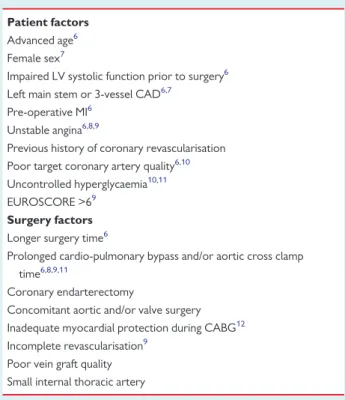
Table 2 Predictors of peri-operative myocardial infarction/graft-failure
Patient factors Advanced age6 Female sex7
Impaired LV systolic function prior to surgery6 Left main stem or 3-vessel CAD6,7
Pre-operative MI6 Unstable angina6,8,9
Previous history of coronary revascularisation Poor target coronary artery quality6,10 Uncontrolled hyperglycaemia10,11 EUROSCORE >69
Surgery factors Longer surgery time6
Prolonged cardio-pulmonary bypass and/or aortic cross clamp time6,8,9,11
Coronary endarterectomy
Concomitant aortic and/or valve surgery Inadequate myocardial protection during CABG12 Incomplete revascularisation9
Poor vein graft quality Small internal thoracic artery
.. ..
.. ..
.. ..
.. ..
.. ..
.. ..
.. ..
.. ..
.. ..
.. ..
.. ..
.. ..
.. ..
.. ..
.. ..
.. ..
.. ..
.. ..
.. ..
.. ..
.. ..
.. ..
.. ..
.. ..
.. ..
.. ..
.. ..
.. ..
.. ..
.. ..
.. ..
.. ..
.. ..
.. ..
.. ..
.. ..
.. ..
.. ..
.. ..
.. ..
.. ..
.. ..
.. ..
or LBBB, the incidence has been reported to range from 5 to 14%,4
.
whereas it ranges from 20 to 30% when using cardiac magnetic reso- nance (CMR) to detect new loss of viable myocardium.14–16
The current definition of Type 5 MI does have several limitations:
(1) The selection of a cTn elevation of 10 URL as a threshold for diagnosing Type 5 MI was arbitrarily chosen. Elevated cTn of 10URL occurs in over 90% of all patients undergoing CABG surgery.8,12
(2) Type 5 MI requires the presence of ECG/angiography/imaging evi- dence of MI, and ignores post-surgical isolated elevations in cardiac biomarkers which may still be prognostically significant (i.e. bio- marker elevations in the absence of ECG/angiographic or other imaging evidence of MI).
(3) The diagnostic criteria for Type 5 MI can also be quite challenging in the setting of CABG surgery for several reasons: (i) In a substantial number of patients, the ECG may not be interpretable and many of the ECG changes following CABG surgery may be non-specific for MI.15–17(ii) Coronary angiography is rarely performed post-surgery to diagnose very early graft failure; and (iii) Echocardiography is the most practical imaging modality for detecting new loss of viable myocardium or new RWMA following CABG surgery, but it may not be diagnostic in many cases.
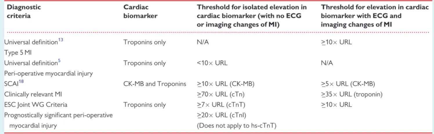
As such, the diagnosis of Type 5 MI in the 48 h post-operative period may be quite challenging, unless it presents with obvious graft failure or a significant ischaemic event. Therefore, in many cases, patients may sustain prognostically significant PMI, but this may be overlooked. The Society for Cardiovascular Angiography and Interventions (SCAI) has proposed a new definition for clinically rele- vant MI, which takes into account isolated elevations in either crea- tine kinase-MB fraction (CK-MB) or cTn within 48 h of CABG surgery.18With respect to CK-MB, these recommendations propose a peak elevation >_10URL in isolation or >_5URL with new patho- logic Q-waves in >_2 contiguous ECG leads or new persistent LBBB.
A substantially higher cut-off for cTn elevation of >_70URL in isola- tion or >_35URL with new pathologic Q-waves in >_2 contiguous ECG leads or new persistent LBBB is also proposed in that paper.18 Again, these threshold levels were arbitrarily chosen, and further studies are required to validate their new definition of clinically rele- vant MI, and explore their relationship to clinical outcomes post- surgery. In addition, these recommendations do not take into consid- eration isolated elevations of cardiac biomarkers below these thresh- olds, which may still be clinically relevant and prognostically significant.
Defining peri-operative myocardial injury
Peri-operative myocardial injury is defined as an isolated elevation in cardiac biomarkers (CK-MB and/or cTn) greater than the upper limit of normal, in the 48-h post-operative period. However, this level of cardiac biomarker elevation occurs in virtually all patients undergoing CABG surgery, and there is no clear consensus on the level of cardiac biomarker elevation above which, it is either clinically relevant or prognostically significant. A recent publication has proposed defining PMI as an isolated elevation in cTn <10 the URL within 48 h of CABG surgery,5but this definition does not include those patients
who have isolated cTn elevations >10URL in the absence of ECG/
angiographic or other imaging evidence of MI. Therefore, in this ESC Joint WG Position Paper we provide recommendations for defining prognostically significant PMI following CABG surgery, which should prompt further clinical evaluation to exclude Type 5 MI. In this paper, we mainly focus on those patients undergoing elective isolated on- pump or off-pump CABG surgery, as the presence of prognostically significant PMI is more challenging to define in patients presenting with an acute coronary syndrome (with elevated pre-operative car- diac biomarkers), and those having concomitant valve or aortic sur- gery. However, patients presenting with an acute coronary syndrome are become increasingly rare since many undergo primar- ily percutaneous intervention.
Isolated elevations in creatine kinase-MB fraction and mortality post-coronary artery bypass graft surgery
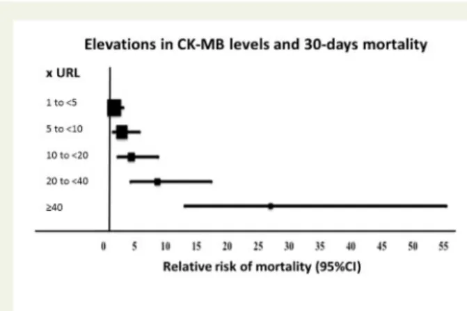
A large number of early studies have assessed the prognostic signifi- cance of isolated elevations in CK-MB following CABG surgery in the absence of ECG/angiographic or other imaging evidence of MI (Table3andFigure1). These studies have demonstrated a graded increase in short, medium, and long-term mortality beginning with an isolated CK-MB elevation >_3 URL within 24 h of CABG surgery.
Above isolated 10URL elevations, there appears to be a progres- sive increase in short-term (30 days) and longer-term mortality (1 year and over), which is independent of other evidence of MI.20,23,29In most centres, CK-MB has now been replaced by the use of cardiac troponins, as the latter are more sensitive and specific for detecting PMI and Type 5 MI following CABG surgery.32,33Hence, we have elected to not use isolated CK-MB elevations post-surgery to define prognostically significant PMI.
Isolated elevations in cTnT and cTnI and mortality post-coronary artery bypass graft surgery
Cardiac troponins have greater sensitivity and specificity for myocar- dial necrosis, when compared to CK-MB, and have been found to be superior to CK-MB in predicting mortality post-CABG sur- gery.30,34–37However, the interpretation of isolated changes in cTn levels in the post-operative period, in the absence of ECG/angio- graphic or other imaging evidence of MI, can be quite challenging given the different cTn assays used, the introduction of high-sensitive assays for cTn, and the presence of renal dysfunction.
As with CK-MB, there appears to be a graded increase in short- term and long-term mortality following CABG surgery, based on the magnitude of post-operative cTnI or cTnT levels (Tables4and5).
Overall, there is a clear association between isolated elevations of cTnT >_7 URL41 and cTnI levels >_20 URL29,41 with significant increases in short-term (30 days) and long-term (one year and over) mortality after CABG surgery (Tables4, 5andFigure2). Importantly, these findings were shown to be independent of ECG/angiography/
imaging evidence of MI, confirming that isolated elevations of cTn fol- lowing CABG surgery can predict mortality. The studies that have been used to define these thresholds used various generations of
‘standard’ cTnT and cTnI assays, and currently there is lack of suffi- cient data to accurately determine these thresholds for the high
...
Table 3 Major recent studies showing elevations in creatine kinase-MB fraction to be associated with mortality post- coronary artery bypass grafting surgery
Study Type of study and surgery
Number of patients
Cardiac biomarker (time)
Time from CABG when biomarker level taken
Major findings
Costaet al.19 (ARTS trial)
Multi-centre prospec- tive study CABG only
496 CK-MB 6,12,18 h <1URL 0.0% 30 d mortality 1.1% 1 yr mortality 1–3URL 0.5% 30 d mortality 0.5% 1 yr mortality
>_3–5URL 5.4% 30 d mortality 5.4% 1 yr mortality
>5URL 7.0% 30 d mortality 10.5% 1 yr mortality Klatteet al.20
(GUARDIAN Trial)
Multi-centre prospec- tive study CABG only
2394 CK-MB
ECG
8, 12, 16, 24 h <5URL 3.4% 6 mth mortality (RR 1.0)
>_5–10URL 5.8% 6 mth mortality (RR 1.69)
>_10–20URL 7.8% 6 mth mortality (RR 2.28)
>_20URL 20.2% 6 mth mortality (RR 5.94
>5URLþnew Q waves worse 6 mth mortality (8.0% vs. 3.1%)
Steueret al.21 Prospective single centre, CABG only
4911 CK-MB 24 h >61 ug/L Relative Hazard 1.3 to 1.4 for late mortal- ity (up to 6 years)
Breneret al.12 Retrospective single centre analysis, CABG only
3812 CK-MB 24 h <_1URL 7.2% 3 yr mortality
1–3URL 7.7% 3 yr mortality 3–5URL 6.3% 3 yr mortality 5–10URL 7.5% 3 yr mortality
>10URL 20.8% 3 yr mortality
>10URL predicted 3 yr mortality (HR 1.3) Marsoet al.22 Single centre registry
post-hoc analysis CABG only
3667 CK-MB Single measurement mean
15.2 h
<_1URL 0.6% 30 d mortality
>1–3URL 1.1% 30 d mortality
>3URL 2.2% 30 d mortality
>4URL associated with increased long-term mor- tality 5.1 yr (RR 1.3)
Ramsayet al.23 Multi-centre prospec- tive randomized trial CABG only
800 CK-MB 4,8, 16, 20,24, 30, 36 h Day 2, 4, 7, 30
0–5URL 0.9% 30 d mortality 5–10URL 0.7% 30 d mortality 10–20URL 0.9% 30 d mortality
>20URL 6.0% 30 d mortality
AUC and peak CK-MB correlated very well.
Engorenet al.24 Retrospective analysis CABG only
1161 CK-MB 10–18 h >8URL HR 1.3 increased 1 yr mortality
Newallet al.7 Observational cohort study
CABG only
2860 CK-MB Single value up to 24 h 3–6URL HR 2.1 for 1 yr mortality
>6URL HR 5.0 for 1 yr mortality
Mahaffeyet al.25 Pooled analysis of four trials
CABG only
1406 CK-MB Single value up to 24 h <3URL 2.5% 30 d mortality; 3.7% 6 mth mortality 3–5URL 2.9% 30 d mortality; 4.7% 6 mth
mortality
5–8URL 3.1% 30 d mortality; 6.1% 6 mth mortality
>_8URL 8.6% 30 d mortality; 9.6% 6 mth mortality Muehlschlegelet al.26Prospective single
centre study CABG only
545 CK-MB Daily from day 1 to 5 24 h 1.23 for each 25 mg/L increase of 5 yr mortality ECG changes alone did not predict 5 year mortality.
Petajaet al.27 Meta-analysis CABG and/or valve
surgery
21 657 CK-MB Variable (peak or absolute value at various time points post-op)
CK-MB >_5URL –RR of short term mortality 3.69% (CI 2.17–6.26); RR of long term (6–60 m) mortality 2.66% (CI 1.95–3.63)
Vikeneset al.28 Prospective single centre study
205 CK-MB 1–3, 4–8, 24, 48 and 72 h CK-MB elevation >_ 5URL was associated with worst long term event free survival (median fol- low-up 92 mths).
Continued
.. ..
.. ..
.. ..
.. ..
.. ..
.. ..
.. ..
.. ..
.. ..
.. ..
.. ..
.. ..
.. ..
.. ..
.. ..
.. ..
sensitivity-cTnT or cTnI assays. Hence, the above threshold for cTnT does not apply to the high-sensitive cTnT assay, and so for this assay, additional ECG and/or imaging evidence of MI appears to be required to identify those CABG patients at a higher risk of mortality when >_10 URL hs-cTnT elevation is measured.8The majority of studies have reported isolated elevations between 24 and 48 h post- surgery as being the most discriminatory for predicting clinical out- comes.27,30,36–38,42
Whether it is necessary to measure the AUC cTn elevation or whether a single time-point measurement of cTn is suffi- cient to predict post-surgical outcomes, is not clear. Recent evidence suggests that the AUC of high-sensitive cTnT may be a good surro- gate for MI size.54
In summary, we recommend, that for patients with a pre-operative cTn <1 URL, isolated elevations of ‘standard’ cTn assays (cTnT >_7URL and cTnI >_20URL) within the 48 h post-operative period (in the absence of ECG/angiographic or other imaging evi- dence of MI), may be indicative of prognostically significant PMI, and require further clinical evaluation to determine whether there is evi- dence for Type 5 MI. This is particularly so if there is additional clinical evidence for MI such as disproportionate chest pain, unusual ECG changes or new regional wall motion abnormalities on
echocardiography in a territory that is dependent on a graft, or dependent on a major ungrafted vessel. However, these threshold values for cTnT and cTnI in defining prognostically significant PMI, may vary from site to site and the actual cTn assay used, and should be established for individual sites. Also, it is important to note that isolated elevations in cTn below these thresholds may still be clini- cally significant, but their impact on post-CABG mortality appears to be small. For patients with additional ECG/angiography/imaging evi- dence of MI, an elevation of cTnT or cTnI >_10URL should be used to define Type 5 MI, as per the 3rd Universal Definition of MI. For the newest generation of high-sensitive cTn assays, the threshold level above which clinical outcomes post-surgery can be predicted remains to be determined.
Other biomarkers for quantifying peri-operative myocardial injury
As mentioned above, cTn elevations between 24 and 48 h have been most clearly shown to correlate with mortality post-CABG surgery.
However, this may be too late to identify prognostically significant PMI or Type 5 MI, as interventions at this stage may fail to salvage a substantial volume of myocardium at risk. Also, cTn elevation in this ...
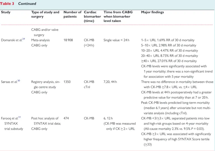
Table 3 Continued
Study Type of study and surgery
Number of patients
Cardiac biomarker (time)
Time from CABG when biomarker level taken
Major findings
CABG and/or valve surgery Domanskiet al.29 Meta-analysis
CABG only
18 908 CK-MB
(<24 h)
Single value < 24 h 1–5URL 1.69% RR of 30 d mortality 5–10URL 2.98% RR of 30 d mortality 10–20URL 4.47% RR of 30 d mortality 20–40URL 8.73% RR of 30 d mortality
>_40URL 27.01% RR of 30 d mortality CK-MB levels were significantly associated with
1 year mortality; there was a non-significant trend for association with 5 year mortality
Søraaset al.30 Registry analysis, sin- gle centre study CABG only
1350 CK-MB
cTnI
7,20, 44 h There was no difference in mortality between those with CK-MB >_7.8URL vs. <_4URL
CK-MB levels at 44 h postoperatively had a greater predictive value for mortality than at 7 or 20 h.
Peak CK-MB levels predicted long-term mortality (median 6.1 years) after univariate but not multi- variate analysis (including cTnI).
Farooqet al.31 SYNTAX trial substudy
Post hoc analysis of SYNTAX trial data;
CABG only
474 CK-MB 6, 12 h
(CK-MB was measured only if CK >_ 2URL
CK-MB <3/>_3URL separated patients into low and high-risk groups based on 4-year mortality (All-cause mortality 2.3% vs. 9.5%P= 0.03).
CK-MB >_3URL was associated with significantly higher frequency of high SYNTAX Score tertile (>_33)
AUC, area under the curve; CABG, coronary artery bypass grafting; CMR, cardiac MRI; CK-MB, creatine kinase-MB fraction; d, day; ECG, electrocardiogram; ECHO, echocar- diocardiogram; HR, hazards ratio; h, hour; LGE, late gadolinium enhancement; LV, left ventricle; MACE, major adverse cardiac events; MI, myocardial infarction; mth, month; ng, nanogram; ONBEAT, on-pump beating heart; CABG ONSTOP, on-pump CABG; OR, odds ratio; post-op, post-operative; PMI, perioperative myocardial injury; RR, relative risk; TEE, transoesophageal echocardiogram; cTnI, Troponin I; cTnT, Troponin T; UA, unstable angina; URL, upper reference limit; yr, year.
.. ..
.. ..
.. ..
.. ..
.. ..
.. ..
.. ..
.. ..
.. ..
.. ..
.. ..
.. ..
.. ..
.. ..
.. ..
.. ..
.. ..
.. ..
.. ..
.. ..
.. ..
.. ..
.. ..
.. ..
.. ..
.. ..
.. ..
.. ..
early time period (<24 h) may be due to non-ischaemic causes, mak- ing it a less reliable marker of regional ischaemia in the first 24 h.
Newer cardiac biomarkers are therefore needed to improve the diagnosis of PMI following CABG surgery with respect to earlier diag- nosis, and improving specificity for regional ischaemia, thereby allow- ing prompt implementation of medical or surgical treatment and to maximise myocardial salvage. Myoglobin, heart-type fatty acid–
binding protein,55,56 copeptin,57 microRNAs (miR-499 and miR-1),58,59and cardiac myosin-binding protein C60have been shown to be associated with PMI following CABG surgery. Some of these are not specific for myocardial necrosis, but they seem to provide additional power in combination with conventional cardiac bio- markers for detecting PMI following CABG surgery. Interestingly, new peptides have been identified via a phage display peptide library screen that might be useful in the future to predict PMI after CABG surgery.49 Although these new biomarkers seem to be extremely sensitive for detecting PMI, technological improvements for early detection, and large validation cohorts are needed to speed-up their clinical application.
Role of electrocardiogram for detecting type 5 myocardial infarction following coronary artery bypass graft surgery
The appearance of new Q waves or LBBB on ECG following CABG surgery remain part of the diagnostic criteria for Type 5 MI.5Using ECG, the incidence of Type 5 MI is in the range of 5 to 14%. New ST- segment elevation or depression may indicate ongoing regional ischaemia, and warrant further diagnostic work-up. However, in many post-surgical patients the ECG may not be interpretable, and ECG changes may be non-specific or transient. A number of clinical studies have found that ECG changes alone are not always predictive of poorer outcomes following CABG surgery,23,26,49although the additional presence of ECG evidence of PMI with an elevation in cTn
appears to be associated with significantly worse outcomes.8,9 Interestingly, a number of studies have shown that many cases of Type 5 MI detected by CMR occur in the absence of new ECG changes (Q waves or LBBB), illustrating the difficulties in relying on ECG changes to detect Type 5 MI.15,61
Role of cardiac imaging for detecting type 5 MI following coronary artery bypass graft surgery
Although several cardiac imaging modalities exist for detecting new loss of viable myocardium or new regional wall motion abnormalities following CABG surgery, only coronary angiography allows for immediate final decision making (conservative, vs. redo CABG vs.
percutaneous coronary intervention).
Echocardiography to detect type 5
myocardial infarction following coronary artery bypass graft surgery
Echocardiography is the most practical imaging modality for detecting new RWMA following surgery.13 However, image quality can be reduced after CABG surgery, due to the presence of pleural or peri- cardial effusions, inflammation or assisted ventilation, and in these cases transoesophageal echocardiography may be preferable.62 Endocardial visualisation might also be enhanced by the use of con- trast agents, especially when 2 or more myocardial segments are not visualised by standard echocardiography.63Moreover, detection of RWMA might be improved by more advanced echocardiography imaging modalities such as tissue Doppler imaging or speckle tracking.64 However, a large retrospective analysis found that RWMA detected by TEE were not able to predict those patients with graft failure as documented by coronary angiography.65 One major limitation of echocardiography is that new RWMA may reflect
Figure 2Relationship between Troponin I elevation post-coro- nary artery bypass graft surgery with relative risk of mortality at 30 days (adapted from meta-analysis by Domanskiet al.29).
Figure 1 Relationship between creatine kinase-MB fraction ele- vation post-coronary artery bypass graft surgery with relative risk of mortality at 30 days (adapted from meta-analysis by Domanski et al.29).
...
Table4MajorrecentstudiesshowingelevationsinTroponinTtobeassociatedwithmortalitypost-coronaryarterybypassgraftingsurgery StudyTypeofstudyand surgeryNumberof patientsCardiac biomarker (time) TimefromCABG when biomarkerlevel taken
Majorfindings Januzzietal.36 Prospectivesinglecentre study CABGonly
224cTnT CK-MB Immediatelypost-op, 6–8h and18–24h
cTnTlevelinthehighestquintile(>_1.58ng/mL;>_15URL)immediatelypost-oporat18–24h predictedin-hospitaldeath. CK-MBlevelsdidnotofferadditionalprognosticbenefittocTnTinmultivariateanalysis Lehrkeetal.38 Prospectivesinglecentre study CABGand/orvalve surgery
204cTnT4,8htheneveryday for7days
cTnT>0.46lg/L(>46URL)at48haftersurgerywastheoptimumdiscriminatorforlong-term cardiacmortality(28mths,OR4.93) Kathiresanetal.37Prospectivesinglecentre study CABGonly
136cTnT CK-MB Immediatelypost-op, 6–8hand 18–24hpost-op
cTnT>1.58lg/Lat18–24hwastheoptimumdiscriminatorfor1yearcardiacmortality(OR5.45) ElevationsinCK-MBwerenotpredictiveofmortality Nesheretal.39Retrospectiveobservatio- nal singlecentrestudy Cardiacsurgery (CABGand/orvalve)
1918cTnTSinglesample<24hcTnTlevel>_0.8lg/L(8URL)wasmostdiscriminatoryforMACE(30daydeath,electrocardio- gram-definedinfarction,andlowoutputsyndrome)(OR2.7) 0–3.9URL0.5%30daymortality 5–5.9URL1.6%30daymortality 6–7.9URL1.0%30daymortality 8–12.9URL1.8%30daymortality >13URL6.8%30daymortality Muehlschlegeletal.26 Retrospectiveanalysis CABGonly
1013cTnTDailyfromday1to524hcTnTrise>110URLHR7.2of5yrmortality cTnTat24hwereindependentpredictorsof5yearmortalityinamultivariatemodel(Noaddi- tionalbenefitofmeasuringcTnbeyond24h). MajorityofpatientshadpeakcTnIandCK-MBlevelsat24h. ECGchangesalonedidnotpredict5yearmortality. Mohammedetal.40Prospectivesinglecentre study, retrospectiveanalysis CABGonly
847cTnT6–8and18–24hAcTnTof<1.60(<160URL)hadgoodnegativepredictivevalueforpoor30dayoutcomes (deathorheartfailure) Petajaetal.41 Meta-analysis CABGand/orvalve surgery
2,547cTnT<48hpostop>_7–16URL:Shorttermmortality3.2%vs.0.5%for<7–16URLelevation(RR4.68–6.4); Longtermmortality(12–28mth)16.1%vs.2.3%(RR5.7–10.09).(PooledRRofmortality couldnotbecalculated) Søraasetal.30 Registryanalysis,single centrestudy CABGonly 1,350cTnT CK-MB
7,20,44hpostopPatientswithpeakcTnT>_5.4URLhadmuchhigherlong-termmortality(median6.1years) thanthosewith<5.4URLcTnTelevation. cTnTlevelsat44hpostoperativelyhadagreaterpredictivevalueforlong-termmortalitythanat 7or20h. PeakTropTlevelspredictedlong-termmortalityaftermultivariateanalysis. Continued
.. ..
.. ..
.. ..
.. ..
.. ..
.. ..
.. ..
.. ..
.. ..
.. ..
.. ..
.. ..
.. ..
.. ..
.. ..
.. ..
.. ..
.. ..
.. ..
.. ..
.. ..
.. ..
.. ..
.. ..
.. ..
.. ..
.. ..
.. ..
.. ..
.. ..
.. ..
.. ..
.. ..
.. ..
.. ..
.. ..
.. ..
.. ..
.. ..
.. ..
.. ..
.. ..
.. ..
.
conditions not necessarily associated with Type 5 MI and include acute ischaemia (without infarction), stunning or hibernation, and non-ischaemic conditions, such as inflammation.Myocardial nuclear imaging and cardiac computed tomography to detect type 5 myocardial infarction following coronary artery bypass graft surgery
Radionuclide single-photon emission computed tomography (SPECT) and positron emission tomography (PET) imaging can allow the direct assessment and quantification of myocardial viability before and after CABG surgery,66,67although given the relatively low spatial resolution of this imaging technique, small areas of non-viable myo- cardium (especially subendocardial MI), which are commonly found with Type 5 MI, may be missed. Other radionuclide imaging approaches are currently under intense investigation, and will likely be tested in the next few years.68
New loss of viable myocardium may be also visualised by cardiac CT.69Multi-slice CT coronary angiography is another useful non- invasive imaging modality that can be utilized to evaluate graft patency following CABG surgery.10,11,70,71
However, the radiation dose and the risks of cumulative ionising radiation need to be weighed against the obvious advantages of an early and accurate diagnosis.72
Cardiac magnetic resonance to detect type 5 myocardial infarction following coronary artery bypass graft surgery
Cardiovascular magnetic resonance (CMR) imaging is a well validated imaging technique with high spatial resolution, for the accurate assessment of both myocardial function and viability, which has pro- ven to be an excellent tool in the diagnosis of Type 5 MI.73The pres- ence of new areas of late gadolinium enhancement (LGE), on CMR performed in the first couple of weeks following CABG surgery can detect the presence of new non-viable myocardial tissue required for diagnosing Type 5 MI (seeTable6). These clinical studies suggest that Type 5 MI occurs in 20–30% of all patients undergoing elective CABG surgery. Interestingly, the pattern of LGE observed on CMR post-CABG surgery reflects the multi-factorial aetiology of Type 5 MI with examples of transmural infarction (suggesting native artery or graft failure), subendocardial infarction (suggesting inadequate cardio- protection), and patchy areas of infarction (suggesting coronary microembolisation or non-ischaemic myocardial necrosis).16,17,77
Overall, there is a good correlation between elevations in cardiac biomarkers post-surgery and new LGE mass quantified by CMR (see Table6). However, in some patients with absence of LGE on CMR, there was still a significant elevation in AUC cTnI, suggesting that not all post-operative cTnI release represents irreversible myocardial injury,15 or that the tissue loss was too small to be detected by CMR.78Therefore, the prognostic significance of post-surgical eleva- tions in cardiac biomarkers in the absence of MI on LGE-CMR remains to be determined. One study has demonstrated that a single cTnI value at 1 h post-surgery accurately predicted new LGE on CMR, increasing the clinical utility of measuring cardiac biomarkers and implementing a change in management to avoid future complications.61
...
Table4Continued StudyTypeofstudyand surgeryNumberof patientsCardiac biomarker (time) TimefromCABG when biomarkerlevel taken
Majorfindings Wangetal.8Retrospectiveanalysis CABGonly
560hs-cTnT ECG/ECHO changes
12–24hafterCABGInamultivariatemodel>10URLriseinhs-TNTþECG/ECHOevidenceofrecentMIor regionalischaemiapredicted30day(HR4.9)andlong-termmortality(medianfollow-up 1.8years)(HR3.4).>10URLriseinhs-cTnTwasseenin90%patients. Goberetal.42Retrospectivestudyfrom registrydata CABGonly
290cTnT CK-MB
8,16hpostopcTnT>0.8ng/mL(>80URL)at6–8hwaspredictiveofinhospitaladverseoutcomesandlong term(4yr)mortality(OR4.0).However,cTnTmeasuredat6–8hwasinferiortocTnTtaken at20hinitsprognosticability. AUC,areaunderthecurve;CABG,coronaryarterybypassgrafting;CMR,cardiacMRI;CK-MB,creatinekinase-MBfraction;d,day;ECG,electrocardiogram;ECHO,echocardiocardiogram;HR,hazardsratio;h,hour;LGE,lategadolinium enhancement;LV,leftventricle;MACE,majoradversecardiacevents;MI,myocardialinfarction;mth,month;ng,nanogram;ONBEAT,on-pumpbeatingheart;CABGONSTOP,on-pumpCABG;OR,oddsratio;post-op,post-operative; PMI,perioperativemyocardialinjury;RR,relativerisk;TEE,transoesophagealechocardiogram;cTnI,TroponinI;cTnT,TroponinT;UA,unstableangina;URL,upperreferencelimit;yr,year.