Severity of Asymptomatic Carotid Stenosis and Risk of Ipsilateral Hemispheric Ischaemic Events: Results from the
ACSRS Study
A.N. Nicolaides,1,4*S.K. Kakkos,1M. Griffin,1M. Sabetai,1S. Dhanjil,1T. Tegos,1 D.J. Thomas,2A. Giannoukas,1G. Geroulakos,1,3N. Georgiou,4S. Francis,1 E. Ioannidou,4C.J. Dore´5and For the Asymptomatic Carotid Stenosis and Risk of
Stroke (ACSRS) Study Group
Departments of1Vascular Surgery, Imperial College,2Neurology, St Mary’s Hospital,3Vascular Surgery, Ealing Hospital, London, UK;4The Cyprus Institute of Neurology and Genetics, Nicosia, Cyprus; and5MRC
Clinical Trials Unit, London, UK
Objectives. This study determines the risk of ipsilateral ischaemic neurological events in relation to the degree of asymptomatic carotid stenosis and other risk factors.
Methods. Patients (nZ1115) with asymptomatic internal carotid artery (ICA) stenosis greater than 50% in relation to the bulb diameter were followed up for a period of 6–84 (mean 37.1) months. Stenosis was graded using duplex, and clinical and biochemical risk factors were recorded.
Results. The relationship between ICA stenosis and event rate is linear when stenosis is expressed by the ECST method, but S-shaped if expressed by the NASCET method. In addition to the ECST grade of stenosis (RR 1.6; 95% CI 1.21–2.15), history of contralateral TIAs (RR 3.0; 95% CI 1.90–4.73) and creatinine in excess of 85mmol/L (RR 2.1; 95% CI 1.23–3.65) were independent risk predictors. The combination of these three risk factors can identify a high-risk group (7.3% annual event rate and 4.3% annual stroke rate) and a low risk group (2.3% annual event rate and 0.7% annual stroke rate).
Conclusions. Linearity between ECST per cent stenosis and risk makes this method for grading stenosis more amenable to risk prediction without any transformation not only in clinical practice but also when multivariable analysis is to be used.
Identification of additional risk factors provides a new approach to risk stratification and should help refine the indications for carotid endarterectomy.
Keywords: Asymptomatic; Carotid; Stenosis; Risk; NASCET; ECST.
Introduction
The degree of internal carotid artery (ICA) stenosis is a well-established measurement used to assess the risk of ipsilateral ischaemic neurological events1,2 and a major criterion to decide whether carotid endarter- ectomy is indicated.
Two main methods are currently used to express percent diameter stenosis. The first one defines the residual lumen as a percentage of the normal distal ICA. It has been used in North America since the late 1960s and more recently the North American Sympto- matic Carotid Endarterectomy Trial (NASCET)1 and the Asymptomatic Carotid Atherosclerosis Study
(ACAS).2It has become known as the North American,
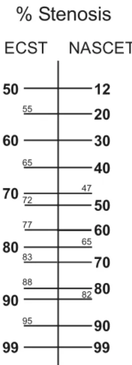
‘NASCET’ or ‘N’ method.3 The second method expresses the residual lumen as a percentage of the diameter of the carotid bulb and has been used in the European Carotid Surgery Trial (ECST)4 to become known as the European or ‘ECST’ or ‘E’ method.5The relationship between both methods is shown inFig. 1.
Several natural history studies6–15indicate that the risk of stroke in asymptomatic patients is low (0.1–
1.6% per year) for NASCET stenosis less than 75–80%
and higher (2.0–3.3% per year) with greater degrees of stenosis. Different cut-off points, ranges and methods of grading stenosis have been used in these natural history studies6–15 and randomized controlled trials.1,2,4,16 Universal agreement as to the best method for grading ICA stenosis and optimum cut-off points in relation to risk have not yet been established.
The aims of this paper based on data from the
*Corresponding author. Prof A.N. Nicolaides, MS, FRCS, The Cyprus Institute of Neurology and Genetics, P.O. Box 23462, 1683 Nicosia, Cyprus.
E-mail address:anicolai@cing.ac.cy
asymptomatic carotid stenosis and risk of stroke (ACSRS) natural history study17are to determine:
(a) the risk of ipsilateral ischaemic hemispheric neurological events associated with different degrees of ICA stenosis,
(b) which method of expressing percent stenosis provides a better expression of risk,
(c) which are the most appropriate ranges of stenosis for grouping patients at different risk, and (d) whether any independent clinical or biochemical
risk factors can be used in conjunction with the degree of stenosis to improve risk prediction.
Material and Methods
Aims of ACSRS study
The asymptomatic carotid stenosis and risk of stroke (ACSRS) is an ongoing international multicentre study under the auspices of the International Union of Angiology. Patients with asymptomatic 50–99% ste- nosis in relation to the carotid bulb diameter (ECST) are followed for at least 5 years to identify subgroups at high and low risk for future neurological events.
Approval has been obtained from the Multicenter Research Ethics Committee (North Thames, London, UK) and local ethics committees. Patients were admitted to the study after informed consent. The methodology used in the ACSRS study, eligibility of participating centres and quality control have been published in detail previously.17Only an outline of the methodology and results related to the degree of ICA stenosis are presented in this paper.
Admission to the study
Patients with ICA stenosis greater than 50% (ECST) on duplex scanning who never had any ipsilateral hemispheric or retinal symptoms and did not have any neurological abnormality on examination were eligible for admission to the study. Patients who had contralateral hemispheric symptoms were also included provided they had been asymptomatic for at least 6 months at the time of recruitment. The ratio of patients with stenosis 50–69 and 70–99% recruited into the study from any centre had to be 1:2 in order to avoid any selection bias from centres that had a policy to operate on patients with severe asymptomatic stenosis. The side with the more severe stenosis was considered to be the ipsilateral side for any patient with bilateral stenosis.
Risk factors and non-invasive investigations The following clinical risk factors and their duration and severity were recorded for each patient: age, gender, height and weight, hypertension, cardiac status (history of myocardial infarction or angina), diabetes, smoking, fibrinogen, fasting blood choles- terol, HDL, LDL, triglycerides, creatinine and haematocrit.
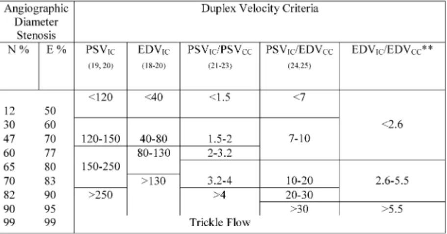
Duplex scanning was performed on admission to the study and subsequently every 6 months to grade ICA stenosis. The team that included a neurologist reviewed each patient at all visits to provide clinical information to document the rate of progression of ICA stenosis. Velocity criteria for grading the degree of stenosis are summarized inTable 1.3Because absolute velocity measurements could underestimate stenosis (e.g. in the presence of cardiac arrhythmia) or over- estimate stenosis (e.g. in the presence of severe contralateral disease), ultrasonographers at each centre were trained to use a combination of absolute velocity measurements and velocity ratios.3,18–25 Characteristics of vertebral artery flow whether cephalad, reversed or absent were also recorded. The entire duplex examination was recorded on videotape.
Fig. 1. The relationship between ECST and NASCET % stenosis. The conversion scale is based on the following equations: NZ(ECST stenosisK43)!(100/57) and EZ (NASCET % stenosis!(57/100)C43) (note: a 43% stenosis of the bulb reduces the lumen to the diameter of the lumen of the normal distal internal carotid artery).
Endpoints
Primary endpoints were ipsilateral hemispheric ischaemic stroke (including fatal stroke) defined as a hemispheric neurological deficit lasting for more than 24 h, any stroke, ipsilateral hemispheric TIAs, amauro- sis fugax and death from cardiovascular causes other than stroke.17For reported strokes a special form was filled locally and two members of the coordinating team, which included a neurologist made the final classification. The local team made the diagnosis of TIAs or amaurosis fugax.
Exit points
Exit points are any of the above endpoints, death from any cause, ipsilateral carotid endarterectomy or stent- ing. Patients who had carotid endarterectomy or stenting were excluded from subsequent analysis, as they would no longer be contributing to the natural history of asymptomatic carotid stenosis.
Quality control
The ultrasound methodology in the ACSRS study includes a set of procedures designed to control quality and monitor key components of measure- ments. These include instrumentation settings, method of recording data, standardization of the method of scanning and personnel training at the coordinating centre. Throughout the study’s perform- ance is compared to the set standards. Results of the
quality control, already published,17indicate that the goal of prospectively controlling quality in the ACSRS Study has been achieved.
Statistical analysis
The SPSS for Windows (release 10.01 Chicago, Ill, USA) statistical package was used throughout. The chi-square test was used to test the significance of the incidence of event rates in relation to grades of stenosis (Tables 2–4) and in univariate analyses between clinical risk factors and neurological events. Kaplan–
Meier curves were used for event-free survival rates and log rank tests for significance of difference between curves. Cox regression analysis was used to determine which clinical and biochemical risk factors as well as stenosis were independent predictors of risk. The level of significance was considered as p%0.05.
Role of the funding source
The sponsors of the study had no role in the study design, data collection, data analysis, data interpret- ation, writing of the report or decision to submit the report for publication.
Results
The results presented in this paper are based on the first 1115 patients recruited with a follow-up of 6–84
Table 1. Duplex velocity criteria selected for highest accuracy* (from Nicolaideset al., 1996)3
months (mean 37.1). A total of 108 ipsilateral hemi- spheric events have occurred: 18 were amaurosis fugax (AF), 44 TIAs and 48 strokes of which 46 were ischaemic (eight fatal) and two were haemorrhagic (both fatal). There have been a total of 163 deaths of which 105 were due to cardiovascular causes. Ipsilat- eral carotid endarterectomy has been performed in 116 patients with severe stenosis (median: 85% in relation to the bulb; interquartile range: 75–90) because the doctor or the patient requested it despite absence of neurological events. This was particularly so after publication of the ACST results. Eighteen patients have been lost to follow-up: six at 6 months, two at 12 months, one at 18 months, one at 24 months, three at 30 months, four at 36 months and one at 72 months. Of the 18 patients contact was lost with 12, five declined to re-attend and one emigrated. The number of patients followed up is 1115 for 6 months, 964 for 12 months, 851 for 18 months, 745 for 24 months, 640 for 30 months, 543 for 36 months, 459 for 42 months, 376 for 48 months, 323 for 54 months, 246 for 60 months, 147 for 66 months, 90 for 72 months, 43 for 78 months and 27 for 84 months.
Relation to ICA stenosis
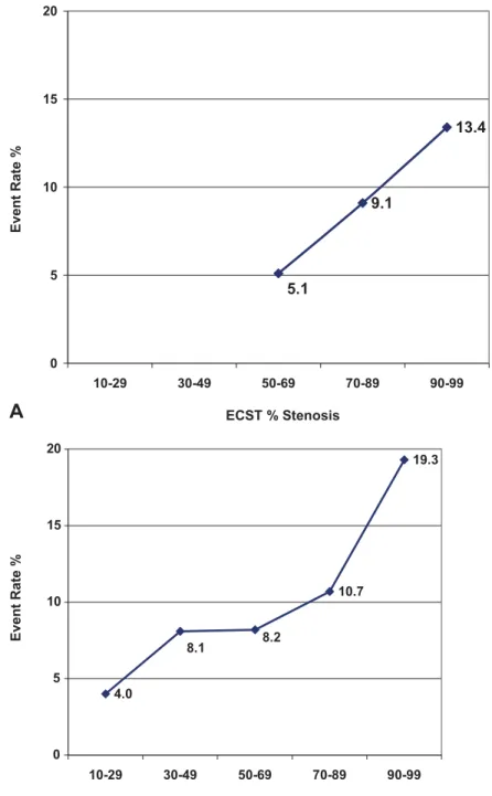
The numbers of different ipsilateral ischaemic hemi- spheric neurological events in relation to stenosis expressed by the ECST and NASCET methods are listed inTables 2 and 3, respectively. The incidence of ipsilateral hemispheric events appears to have a linear relationship with ECST stenosis (Fig. 2(A)), but not with NASCET stenosis (Fig. 2(B)). The cumulative event free-survival rate for all ipsilateral events in relation to the ECST and NASCET methods for stenosis is shown in Fig. 3. Fig. 3(B) demonstrates
that there is little difference in the event-free survival rate between the groups with NASCET percent stenosis 30–49, 50–69 and 70–89%.
Table 4 lists the number of different ipsilateral ischaemic hemispheric neurological events and Fig.
3(c) the cumulative event-free survival rate in relation to less than 60 and 60–99% NASCET stenosis, respectively. The difference is significant (pZ0.022, log rank test) but it is apparent that the 60% selection criterion for carotid endarterectomy recommended from the ACAS trial is not a good discriminator of risk since 37 (34%) of all 108 events have occurred in the group of patients with stenosis less than 60% by the NASCET method (Table 4).
Fig. 3(A) shows the cumulative event-free survival rate in relation to three groups: (a) 50–69% ECST equivalent to 12–46% NASCET, (b) 70–89% ECST equivalent to 47–81% NASCET and (c) 90–99%
ECST equivalent to 82–99% NASCET. Classification into these three groups identifies a low risk group (a) with a 7-year cumulative event rate of 8%, a moderate risk group (b) with a 7-year cumulative event rate of 18% and a high-risk group (c) with a 7-year cumulative event rate of 35%.
Other potential risk factors
Age, gender, hypertension, cardiac symptoms, atrial fibrillation, diabetes, smoking, pack-years, fibrinogen, fasting total cholesterol, HDL, LDL, triglycerides, BMI and contralateral stenosis of any degree or occlusion were not associated with an increased incidence of ipsilateral ischaemic hemispheric neurological events in a univariate analysis using chi-square and log rank tests; a history of contralateral hemispheric TIAs at least 6 months prior to admission to the study and
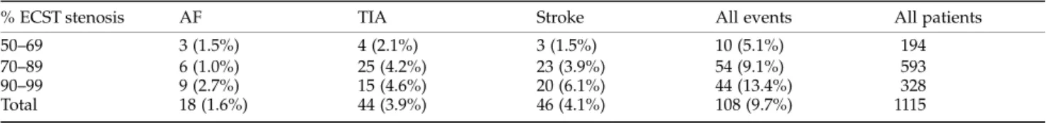
Table 2. The number of different ipsilateral ischaemic hemispheric neurological events in relation to the ECST method of grading internal carotid artery stenosis (chi-square 13.4;pZ0.037)
% ECST stenosis AF TIA Stroke All events All patients
50–69 3 (1.5%) 4 (2.1%) 3 (1.5%) 10 (5.1%) 194
70–89 6 (1.0%) 25 (4.2%) 23 (3.9%) 54 (9.1%) 593
90–99 9 (2.7%) 15 (4.6%) 20 (6.1%) 44 (13.4%) 328
Total 18 (1.6%) 44 (3.9%) 46 (4.1%) 108 (9.7%) 1115
Table 3. The number of different ipsilateral ischaemic hemispheric neurological events in relation to the NASCET method of grading internal carotid artery stenosis (chi-square 27.9;pZ0.007)
% NASCET stenosis AF TIA Stroke All events All patients
!30 1 (1.0%) 1 (1.0%) 2 (2.0%) 4 (4.0%) 101
30–49 3 (1.4%) 6 (2.9%) 8 (3.8%) 17 (8.1%) 209
50–69 3 (0.9%) 14 (4.0%) 12 (3.4%) 29 (8.2%) 352
70–89 8 (2.3%) 18 (5.2%) 11 (3.2%) 37 (10.7%) 344
90–99 3 (2.8%) 5 (4.6%) 13 (11.9) 21 (19.3%) 109
Total 18 (1.6%) 44 (3.9%) 46 (4.1%) 108 (9.7%) 1115
Table 4. The number of different ipsilateral ischaemic hemispheric neurological events in relation to less than 60 and 60–99% NASCET internal carotid artery stenosis (chi-square 4.6;pZ0.21)
% NASCET stenosis AF TIA Stroke All events All patients
!60 6 (1.2%) 15 (3.0%) 16 (3.2%) 37 (7.4%) 499
60–99 12 (1.9%) 29 (4.6%) 30 (4.7%) 71 (11.2%) 636
Total 18 (1.6%) 44 (3.9%) 46 (4.1%) 108 (9.7%) 1115
Fig. 2.The incidence of ipsilateral ischaemic hemispheric events (A) in relation to the ECST % stenosis of the internal carotid artery and (B) in relation to the NASCET % stenosis of the internal carotid artery.
elevated creatinine levels were (Fig. 4). The best cut off level for creatinine as determined by an ROC curve was 85mmol/L.
In a Cox regression analysis all three factors:
severity of ipsilateral stenosis (ECST), history of contralateral hemispheric TIAs and creatinine were independent predictors of risk (Table 5). Thus, after adjusting for confounding variables stenosis remained significant. Based on this Cox regression model the linear risk predictor score (x*Beta) (SPSS version 10.01) was calculated for every patient. This score is the sum of the product of mean-centered covariance values and their corresponding parameter estimates for each patient. The upper quartile of this score consisted of a high-risk group with a 44% cumulative event rate at 6 years and the remaining lower three quartiles consisted of a low-risk group with a 14% cumulative event rate at 6 years (Fig. 5). Fifty events including 25 strokes were in the high-risk group. Fifty-eight of the events including 21 strokes were in the low-risk group.
Discussion
The results of the ACSRS study demonstrate that the risk of ipsilateral ischaemic hemispheric events rises with increasing severity of ICA stenosis (Tables 2–3 and Fig. 2) and confirm the findings of previous natural history studies6–13irrespective of different cut- off points used: 50,6,8,10–137510,11and 80%9in relation to the distal internal carotid or 5012 and 80%12 in relation to the bulb.
The NASCET randomised controlled study used angiography and a cut-off point of 70% stenosis in relation to the distal internal carotid which is equivalent to 83% stenosis in relation to the bulb.
The ECST randomised controlled study used angio- graphy also but a cut-off point of 70% stenosis in relation to the bulb which is equivalent to 47% stenosis in relation to the distal internal carotid artery. Many vascular surgeons are under the impression that these cut-off points are similar! The only similarity is the value of 70%. In reality the difference in terms of plaque size or residual lumen is considerable. How- ever, with increasing degrees of stenosis the values of Fig. 3.(A) The ipsilateral hemispheric event-free cumulative
survival rate in relation to the ECST % stenosis of the internal carotid artery (50–69% group:nZ101; 70–89% group:nZ593;
90–99% group:nZ328). Overall log rank: 11.7pZ0.0026; 50–
69 vs79–89%pZ0.045; 79–89vs90–99%pZ0.020; 50–69vs 90–99%pZ0.0014; (B) the ipsilateral hemispheric event free cumulative survival rate in relation to the NASCET % stenosis of the internal carotid artery (12–30% group:nZ101;
30–49% group: nZ209; 50–69% group: nZ352; 70–89%
group: nZ354; 90–99% group: nZ109). Overall log rank:
11.7pZ0.0026; statistical significance between the four lower grades of stenosispO0.1; 90–99%vsany other groupp!0.01;
(C) the ipsilateral hemispheric event free cumulative survival rate in relation to the NASCET % stenosis of the internal carotid artery (!60% group:nZ497; 60–99% group:
nZ636). Log rank: 5.2pZ0.022. In view of the small number of patients that have been followed for 7 years (nZ27) the abrupt drop in the survival curve should be ignored.
the two methods converge and the discrepancy decreases (Fig. 1).
The method of expressing stenosis as a percentage of the distal internal carotid dates back to the 1960s when angiography was the only imaging method available.26,27 It was a standard method in North America until duplex ultrasound became available.
The duplex velocity criteria for grading internal carotid artery stenosis developed by Strandness and his team in the 1980s were in relation to the percent stenosis of the bulb.18 Their gold standard was angiography and microcalcification in the arterial wall was used to define the outline of the bulb. Several
studies have since redefined the velocity criteria in terms of percent stenosis of the distal internal carotid (Table 1). It can be seen in Table 1 that the velocity criteria for 70% stenosis ‘NASCET’ are very different from 70% stenosis ‘ECST’.
Radiology departments and vascular laboratories often express the results of angiography or duplex ultrasound as ‘percent stenosis’ without any indi- cation whether the ‘percent stenosis’ is expressed in terms of the bulb or the distal internal carotid. The ACST randomised controlled study used duplex to grade the degree of stenosis with 70% as the cut-off point, but neither the velocity criteria nor what 70%
stenosis refers to (bulb or distal internal carotid) has been stated.16 Without such a statement it is not possible to know whether some teams were expressing their results as percent stenosis of the bulb and others of the internal carotid. Much needs to be done in terms of education on this issue and for standardisation of methods published in vascular journals.
The results of the present study suggest that contralateral symptoms imply an inherent predisposi- tion with increased risk for the ipsilateral asympto- matic side irrespective of the degree of stenosis. This is supported by the finding that the annual stroke rate on the asymptomatic side of the ECST14 and NASCET15 studies in which patients presented initially with unilateral hemispheric symptoms was 1.5–2.0 times higher than the risk in studies of bilaterally asympto- matic patients.10–13 It is further supported by the Fig. 4.(A) The ipsilateral hemispheric event-free cumulative
survival rate in relation to the presence or absence of history of contralateral TIAs 6 months or more before admission to the study (history absent:nZ1014; history present:nZ101).
Log rank: 26.7 pZ0.00001; (B) The ipsilateral hemispheric event free cumulative survival rate in relation to the creatinine plasma levels (!85mmol/L group: nZ283;
O85mmol/L group: nZ835). Log rank: 7.8 pZ0.0051. In view of the small number of patients that have been followed for 7 years (nZ27) the abrupt drop in the survival curve should be ignored.
Fig. 5. The ipsilateral hemispheric event free cumulative survival rate in relation to the risk (Cox x*Beta) calculated for each patient according to the Cox regression model shown in Table 5. Upper quartilevslower three quartiles of risk: log rank: 27.5p!0.0001 (50 events including 25 strokes are in the upper quartile and 58 events including 21 strokes are in the rest of the population). In view of the small number of patients that have been followed for 7 years (nZ27) the abrupt drop in the survival curve should be ignored.
finding that in the control group of the ACST16 the ipsilateral stroke rate was double in those with a history of contralateral symptoms than those without.
The relationship between stenosis and event rate is linear if expressed as ECST per cent stenosis (Fig. 2(A)), but it is S-shaped if expressed as NASCET per cent stenosis (Fig. 2(B)). Linearity between ECST per cent stenosis and risk makes this method for grading stenosis more amenable to risk prediction without any transformation not only in clinical practice but also when linear logistic or Cox regression analysis is to be used.
The results of the ACSRS study, like previous natural history studies which included patients with lesions up to 99% stenosis,6,8–12 demonstrate that a considerable number of events occur at low grades of stenosis. In fact 37 (34%) of 108 ipsilateral ischaemic hemispheric events including 16 (35%) of the 46 strokes (Table 4) occurred in patients with stenosis less than 60% NASCET (!77% ECST), the selection criterion for carotid endarterectomy in asymptomatic patients as indicated from the findings of the ACAS trial. Only 10 (9%) of the events including 3 (3%) strokes occurred in patients with stenosis less than 70% ECST equivalent to approximately 50% NASCET (Table 2). This is because the 60% NASCET cut-off point is in the middle of the moderate risk-group (Fig. 3(B)). In contrast, the ECST 70% cut-off point is in the lower end of the moderate-risk group (Fig. 3(A)).
Of the clinical and biochemical risk factors studied only two were associated with an increased overall event rate in a univariate analysis: history of contral- ateral TIAs (Fig. 4(A)) and creatinine levels (Fig. 4(B)).
The fact that most of the established clinical and biochemical risk factors for atherosclerosis were not associated with an increased event rate is not surpris- ing because these risk factors are present in the majority of patients in the study who are all arteriopaths. All three risk factors, stenosis (ECST), history of contralateral TIAs and creatinine remained significant independent predictors in a Cox regression analysis (Table 5). Table 5 indicates that the relative risk between ECST 50–69 and 70–89% or between 70–
89 and 90–99% stenosis is 1.6; also, the risk increases three times if there is a history of contralateral TIAs and 2.1 times if creatinine is higher than 85mmol/L.
Epidemiology studies have demonstrated that mild to moderately raised serum creatinine is an indepen- dent predictor of cardiovascular risk and particularly ischaemic stroke.28,29A more recent study has demon- strated that elevated serum creatinine levels even within the normal range are associated with an increased risk of stroke in both normotensive and hypertensive men.30
The combination of all three significant risk factors (stenosis, history of ipsilateral TIAs and creatinine) allows one to identify a high-risk group with a 44%
cumulative event rate at 7 years (7.3% annual event rate) and a low-risk group with a 14% cumulative event rate at 7 years (2.3% annual event rate). The cumulative stroke rate at 7 years is 26% (4.3% annual stroke rate) in the high-risk group and 4.2% (0.7%
annual stroke rate) in the low risk group. However, this means that only 46% of all events including 54% of the strokes were in the high-risk group, the rest occurring in the low risk group. The contribution of plaque characterisation using image normalisation and texture feature extraction techniques in improving these findings so that a high-risk group can be identified that contains the majority of the strokes31 will be the subject of another paper.
The potential practical implications of the above findings are as follows. In patients with 50–69% ECST stenosis even in the presence of a history of contral- ateral TIAs or creatinine in excess of 85mmol/L or in the presence of both, the annual stroke rate is not higher than 1%. In patients with 70–89% ECST stenosis, in the presence of a history of contralateral TIAs and creatinine in excess of 85mmol/L the annual stroke rate is 3.2%. In the rest of the patients with 70–
89% ECST stenosis even when one of the other two risk factors is present the annual stroke rate is not higher than 1.5%. In patients with 90–99% ECST stenosis, in the absence of a history of contralateral TIAs and with creatinine less than 85mmol/L the annual stroke rate is 1%; in the presence of creatinine in excess of 85mmol/L it is 2.2% and in the presence of a history of contralateral TIAs it is 2.6%. In the presence of both, history of contralateral TIAs and creatinine in excess of 85mmol/L the annual stroke rate is 6.3%.
Our findings on risk assessment from the ACSRS study need to be validated by other natural history
Table 5. Results of Cox regression analysis with ipsilateral ischaemic hemispheric neurological events as the dependent variable; ECST stenosis (three groups: 50–69, 70–89, 90–99%), creatinine (two groups:!85mmol/L,O85mmol/L) and history of contralateral TIAs 6 months or more prior to admission to the study were the independent variables
Independent variables B SE p Exp(B) 95% CI for exp(B)
ECST stenosis 0.480 0.15 0.001 1.615 1.21–2.15
History of contralateral TIAs 1.099 0.23 0.0001 3.002 1.90–4.73
Creatinine 0.749 0.27 0.007 2.116 1.23–3.65
studies, the control group of the ACST study and the asymptomatic sides of the ECST and NASCET trials. If confirmed, they will provide a new approach to risk stratification and may help refine the indications for carotid endarterectomy.
Acknowledgements
This study was supported by a grant from the European Commission (Biomed II) Program (PL 650629) for the first 3 years and subsequently by a grant from the CDER Trust (UK).Conflict of interest. Prof A. Nicolaides is Chairman of the CDER Trust (UK Registered Charity), which was one of the sponsors of the study.
The ACSRS Study Group
R. Adovasio & B. Ziani (Trieste, Italy)
L. Middleton, M. Pantziaris &
T. Tyllis (Nicosia, Cyprus) F.P. Alo` & C.G. Cicilioni
(Ancona, Italy)
E. Minar & A. Willfort (Vienna, Austria)
G. Ambrosio (Venezia, Italy) L. Moggi & P. DeRango (Per- ugia, Italy)
A. Andreev (Stara Zagora, Bulgaria)
G. Nenci & S. Radicchia (Per- ugia, Italy)
G.M. Andreozzi, F. Verlato & G.
Camporese (Padova, Italy)
A. Nicolaides, S. Kakkos & D Thomas (London, UK) E. Arosio (Verona, Italy) L. Norgren & E. Ribbe (Lund,
Sweden) E. Barkauskas (Vilnius,
Lithuania)
S. Novo & R. Tantillo (Palermo, Italy)
A.A.B. Barros D’Sa & P. Bran- nigan (Belfast, UK)
D. Olinic (Cluj-Napoca, Romania)
V. Batchvarova & A. Dramov (Sofia, Bulgaria)
W. Paaske (Aarhus, Denmark) P. Belardi, GP Novelli & G.
Simoni (Genoa, Italy)
A. Pagnan (Castelfranco, Italy) P. Bell (Leicester, UK) P. Pauletto & V. Pagliara
(Padova, Italy) G.M. Biasi & P. Mingazzini
(Milan, Italy)
G. Pettina (Pistoia, Italy) N.M. Bornstein (Tel-Aviv,
Israel)
C. Pratesi & S. Matticari (Firenze, Italy)
D. Bouchier-Hayes & P. Fitz- gerald (Dublin, Ireland)
J. Polivka & P. Sevcik (Plzen, Czech Republic)
M.A. Cairols (Barcelona, Spain) P. Poredos, A. Blinc &V. Videc- nik (Ljubjana, Slovenia) P.G. Cao & P. DeRango (Per-
ugia, Italy)
A. Pujia (Cantanzaro, Italy) G.P. Carboni & C. Geoffredo
(Rome, Italy)
A. Raso, P. Rispoli & M. Con- forti (Torino, Italy)
M. Catalano (Milan, Italy) T. Robinson & M.S.J. Dennis (Leicester, UK)
B. Chambers, M. Goetzmann &
A. Dickinson (Victoria, Australia)
S. Rosfors (Stockholm, Sweden)
D. Clement & M. Bobelyn (Ghent, Belgium)
G. Rudofsky (Essen, Germany) S. Coccheri & E. Conti
(Bologna, Italy)
T. Schroeder & ML. Gronholdt (Copenhagen, Denmark) E. Diamantopoulos, E.A.
Andreadis (Athens, Greece)
G. Simoni, C.Finocchi, & G.
Rodriguez (Genoa, Italy) (continued)
P.B. Dimakakos & T. Kotsis (Athens, Greece)
C. Spartera, M Ventura & P.
Scarpelli (L’Aquila, Italy) B. Eikelboom (Utrecht, The
Netherlands)
M. Sprynger, B. Sadzot, C Hottermans and Moonen (Chenee, Belgium) L. Entz (Budapest, Hungary) P.R. Taylor (London, UK) Ferrari-Bardile, T. Aloi & M.
Salerno (Montescano, Italy)
A. Tovar-Pardo & J. Negreira (Madrid, Spain)
J. Fernandes e Fernandes & L.
Pedro (Lisbon, Portugal)
M. Vayssairat & JM Faintuch (Paris, France)
D.E. Fitzgerald & Anne O’Shaunnersy (Dublin, Ireland)
J. Valaikiene´ (Vilnius, Lithuania)
J. Fletcher (Westmead, Australia)
M.G. Walker (Manchester, UK) S. Forconi, R. Cappeli, A.R Wilkinson (Hull, UK) M. Bicchi, S. Arrigucci (Siena,
Italy)
V. Gallai & G. Cardaiolli (Per- ugia, Italy)
G. Geroulakos & S. Kakkos (London, UK)
L.F. Gomez-Isaza (Medellin, Colombia)
G. Gorgoyannis & N. Liasis (Athens, Greece)
M. Graf (Vienna, Austria) P. Guarini (Napoli, Italy) S. Hardy (Blackburn, UK) P. Harris & S. Aston (Liverpool, UK)
G. Iosa (Cesena, Italy) A. Katsamouris & A. Giannou- kas (Crete, Greece)
M. Krzanowski (Krakow, Poland)
G. Ladurner (Salzburg, Austria) J. Leal-Monedero (Madrid, Spain)
B.B. Lee (Seoul, Korea) C. Liapis & P. Galanis (Athens, Greece)
W. Liboni & E. Pavanelli (Torino, Italy)
E. Mannarino & G. Vaudo (Perugia, Italy)
P. McCollum & R. Levison (Dundee, UK)
G. Micieli & D. Bosone (Pavia, Italy)
International Advisory Committee
HJM Barnett (Canada) The late EF Bernstein (USA) D Clement (Belgium) Anne M Jones (USA) W Moore (USA) K Myers (Australia)
The late DE Strandness (USA) J Toole (USA)
M Tsapogas (USA/Greece) J van Gijn (The Netherlands)
References
1 North American Symptomatic Carotid Endarterectomy Trial Collaborators. Beneficial effect of carotid endarterectomy in symptomatic patients with high-grade carotid stenosis.N Engl J Med1991;325:445–453.
2 Executive Committee for the Asymptomatic Carotid Athero- sclerosis Study. Endarterectomy for asymptomatic carotid artery stenosis.J Am Med Assoc1995;273:1421–1428.
3 Nicolaides AN, Shifrin EG, Bradbury A, Dhanjil S, GriffinM,BelcaroGet al. Angiographic and duplex grading of internal carotid stenosis: can we overcome the confusion?
J Endovasc Surg1996;3:158–165.
4 European Carotid Surgery Trialists Collaborative Group. MRC European carotid surgery trial: interim results for symptomatic patients with severe (70–99%) or with mild (0–29%) carotid stenosis.Lancet1991;337:1235–1243.
5 De BrayJM,GlattB. Quantification of atheromatous stenosis in the extracranial internal carotid artery. Cerebrovasc Dis 1995;
5:414–426.
6 Johnson JM, Kennelly MM, Decesare D, Morgan S, Sparrow A. Natural history of asymptomatic carotid plaque.
Arch Surg1985;120:1010–1012.
7 BockRW,Gray-WealeAC,MockPA,StatsMA,RobinsonDA, IrwigLet al. The natural history of asymptomatic carotid artery disease.J Vasc Surg1993;17:160–169.
8 ChambersBR,NorrisJW. Outcome in patients with asympto- matic neck bruits.N Engl J Med1986;315:860–865.
9 Hennerici M, Hulsbomer HB, Hefter H, Lammerts D, Rautenberg W. Natural history of asymptomatic extracranial arterial disease. Results of a long-term prospective study.Brain 1987;110:777–791.
10 NorrisJW,ZhuCZ,BornsteinNM,ChambersBR. Vascular risks of asymptomatic carotid stenosis.Stroke1991;22:1485–1490.
11 Zhu CZ, Norris JW. A therapeutic window for carotid endarterectomy in patients with asymptomatic carotid stenosis.
Can J Surg1991;34:437–440.
12 Mackey AE, Abrahamowicz M, Langlois Y, Battista R, SimardD,BourqueFet al. Outcome of asymptomatic patients with carotid disease.Neurology1997;48:896–903.
13 NadareishviliZG,RothwellPM,BeletskyV,PagnielloA, NorrisJW. Long-term risk of stroke and other vascular events in patients with asymptomatic carotid artery stenosis.Arch Neurol 2002;59:1162–1166.
14 The European Carotid Surgery Trialists Collaborative Group.
Risk of stroke in the distribution of an asymptomatic carotid artery.Lancet1995;345:209–212.
15 Inzitari D, Eliasziw M, Gates P, Sharpe BL, Chan RKT, MeldrumHEet al. The causes and risk of stroke in patients with asymptomatic internal-carotid-artery stenosis.N Engl J Med2000;
342:1693–1700.
16 MRC Asymptomatic Carotid Surgery Trial (ACST) Collaborative Group. Prevention of disabling and fatal strokes by successful carotid endarterectomy in patients without recent neurological symptoms: randomized controlled trial. Lancet 2004;363:1491–
1502.
17 Nicolaides A, Sabetai M, Kakkos S, Dhanjil S, Tegos T, StevensJMet al. The asymptomatic carotid stenosis and risk of stroke (ACSRS) study.Int Angiol2003;22:263–272.
18 TaylorDC,StrandnessDE. Carotid artery duplex scanning.
J Clin Ultrasound1987;15:635–644.
19 Mitti RL, Broderick M, Carpentier JP, Goldberg HI, ListerudJ,Mishkin MMet al. Blinded-reader comparison of magnetic resonance angiography and duplex ultrasonography for carotid artery bifurcation stenosis.Stroke1994;25:4–10.
20 Zweibel WJ. Spectrum analysis in carotid sonography. Ultra- sound Med Biol1987;13:625–636.
21 Robinson ML, Sacks D, Perimutter GS, Marinelli DL.
Diagnostic criteria for carotid duplex sonography.Am J Radiol 1988;151:1045–1049.
22 Moneta GL, Edwards JM, Chitwood RW, Taylor Jr LM, LeeRW, CummingsCAet al. Correlation of North American Symptomatic Carotid Endarterectomy Trial (NASCET) angio- graphic definition of 70% to 99% internal carotid artery stenosis with duplex scanning.J Vasc Surg1993;17:152–159.
23 Moneta GL, Edwards JM, Papanicolaou G, Hatsukami T, TaylorJr LM,StrandnessJr DEet al. Screening for asympto- matic internal carotid artery stenosis: duplex criteria for discriminating 60% to 90% stenosis.J Vasc Surg1995;21:989–994.
24 BelcaroG,RuloAHF,VasdekisS,BonomoL,NicolaidesAN.
Correlation of duplex scanning with two plane angiography in patients with symptomatic carotid stenosis.Acta Chir Belg1988;
88:159–161.
25 Knox RA, Greene FM, Beach K, Phillips DJ, Chikos PM, Strandness Jr DE. Computer based classification of carotid arterial disease: a prospective assessment.Stroke1982;13:589–594.
26 Blaisdell WF, Clauss RH, Galbraith JG, Imparato AM, Wylie EJ. Joint study of extracranial arterial occlusion. IV. A review of surgical consideration.JAMA1969;209:1889–1895.
27 Toole JF, Castaldo JE. Accurate measurement of carotid stenosis: chaos in methodology.J Neuroimaging1994;4:222–230.
28 PocockSJ,McCormackV,GueyffierF,BoutitieF,FagardRH, BoisselJP. A score for predicting risk of death from cardiovas- cular disease in adults with raised blood pressure, based on individual patient data from randomized controlled trials.BMJ 2001;323:75–81.
29 MannJF,GersteinHC,Dulau-FloreaI,JohnE. Cardiovas- cular risk in patients with mild renal insufficiency.Kidney Int 2003;84(Suppl):S192–S196.
30 Wannamethee SG, Shaper AG, Perry IJ. Serum creatinine concentration and risk of cardiovascular disease: a possible marker for increased risk of stroke.Stroke1997;28:557–563.
31 NicolaidesA,KakkosS,SabetaiM,GriffinM,IoannidouE, FrancisSet al. Asymptomatic carotid stenosis and risk of stroke:
natural history study.Stroke2005;36:424 [Abstract].
Accepted 26 April 2005 Available online 13 June 2005