IRON DEFICIENCY ANEMIA-RELATED GUT MICROBIOTA DYSBIOSIS IN INFANTS AND
YOUNG CHILDREN: A PILOT STUDY
AUDRONE MULEVICIENE1*, FEDERICAD’AMICO2, SILVIATURRONI2, MARCOCANDELA2and AUGUSTINAJANKAUSKIENE1
1Clinic of Children’s Diseases, Institute of Clinical Medicine, Faculty of Medicine, Vilnius University, Vilnius, Lithuania
2Unit of Microbial Ecology of Health, Department of Pharmacy and Biotechnology, University of Bologna, Bologna, Italy
(Received: 31 July 2018; accepted: 4 September 2018)
Nutritional iron deficiency (ID) causes not only anemia but also malfunction of the entire human organism. Recently, a role of the gut microbiota has been hypothesized, but limited data are available especially in infants. Here, we performed a pilot study to explore the gut microbiota in 10 patients with iron deficiency anemia (IDA) and 10 healthy controls aged 6–34 months. Fresh stool samples were collected from diapers, and the fecal microbiota was profiled by next-generation sequencing of the V3–V4 hypervariable region of the 16S rRNA gene. Except for diet diversity, the breastfeeding status at the enrollment, the exclusive breastfeeding duration, and the introduction of complementary foods did not differ between groups. Distinct microbial signatures were found in IDA patients, with increased relative abundance of Enterobacteriaceae(mean relative abundance, patients vs. controls, 4.4% vs. 3.0%) andVeillonellaceae(13.7% vs. 3.6%), and reduced abundance ofCoriobacteriaceae (3.5% vs. 8.8%) compared to healthy controls. A decreased Bifidobacteriaceae/
Enterobacteriaceae ratio was observed in IDA patients. Notwithstanding the low sample size, our data highlight microbiota dysbalance in IDA worth for further investigations, aimed at unraveling the ID impact on the microbiome trajectory in early life, and the possible long-term consequences.
Keywords:iron deficiency anemia, gut microbiota, dysbiosis, infants, children Introduction
Anemia is a common disease found all over the world [1]. Half of the anemia cases are caused by iron deficiency (ID), and children are most
*Corresponding author; E-mail:audrone.muleviciene@santa.lt
First published online November 12, 2018
vulnerable to this disease due to high iron needs [1,2]. According to the World Health Organization, anemia is a moderate public health problem in Lithuania.
The disease prevalence was 26% in children aged 6–59 months in 2011 [1].
Moreover, previous study suggests that almost 50% of children in the first 2 years of life may experience ID [3]. Thesefigures are of particular concern, as ID significantly disturbs functions of the brain, heart, and skeletal muscles even in non-anemic stages [4]. The long-lasting health consequences are associated with delayed cognitive and social–emotional development [5–7].
Based on recent evidence [8–13], ID and ID anemia (IDA) may also be featured by unfavourable changes of the gut microbiota. Two studies addressing the effect of iron fortification on the intestinal microbiota in African children showed higher levels of potentially pathogenic enterobacteria than bifidobacteria and lactobacilli at the baseline [14,15]. Thesefindings are particularly relevant, as the gut microbiota is a key regulator of the host metabolic homeostasis [16], an integral component of the immune system [17–19], and essential for central nervous system development [20, 21]. Unbalanced microbial configurations have been found to result in lifelong consequences, including increased risk of metabolic and immunological diseases [16,17,20,22].
We performed a pilot study in a homogenous cohort of 10 infants and young children with nutritional IDA compared to 10 healthy controls (HCs) to provide insights into the early-life alterations in the intestinal microbial ecosystem.
Methods Subjects enrollment and sample collection
Ten infants and young children were recruited at a tertiary university hospital (Vilnius, Lithuania) with diagnosis of nutritional IDA (serum hemoglobin
<110 g/L, ferritin<12μg/L, and/or reticulocyte-hemoglobin equivalent<28 pg) meeting the following criteria: (1) anemia diagnosed for the first time in life, (2) otherwise healthy and not taking medicines for at least 4 weeks, (3) singleton pregnancy, and (4) vaginally delivered. Ten healthy children (HC) meeting the same (2–4) criteria served as controls. The study was approved by the Vilnius Regional Committee for Biomedical Research Ethics. Informed consent was obtained from the parents of all individual participants included in the study.
The parents of the subjects filled a questionnaire survey and provided information on their child’s nutrition. Fresh stool samples were collected from diapers and immediately frozen at −20 °C. Samples were delivered to the laboratory (Bologna, Italy), where they were stored at−80 °C until processing.
Microbial DNA extraction
Microbial DNA was extracted from feces using the repeated bead-beating plus column method, as previously described [23]. Briefly, 250 mg of sample were suspended in 1 ml of lysis buffer (500 mM NaCl, 50 mM Tris-HCl, pH 8, 50 mM EDTA, and 4% sodium dodecyl sulfate) and bead-beaten thrice in a FastPrep instrument (MP Biomedicals, Irvine, CA) at 5.5 movements/s for 1 min, in the presence of four 3-mm glass beads and 0.5 g of 0.1-mm zirconia beads (BioSpec Products, Bartlesville, OK). Samples were heated at 95 °C for 15 min and then centrifuged at 13,000 rpm for 5 min. Two hundred and sixty microliters of 10 M ammonium acetate were added to the supernatant, followed by 5-min incubation in ice and 10-min centrifugation at 13,000 rpm. One volume of isopropanol was added to each sample and incubated in ice for 30 min. Precipi- tated nucleic acids were washed with 70% ethanol, resuspended in 100μl of TE buffer, and treated with 2μl of 10 mg/ml DNase-free RNase at 37 °C for 15 min.
Proteinase K treatment and DNA purification were performed using the QIAamp DNA Stool Mini Kit (QIAGEN, Hilden, Germany). DNA concentration and quality were evaluated using NanoDrop ND-1000 spectrophotometer (NanoDrop Technologies, Wilmington, DE).
16S rRNA gene sequencing
The V3–V4 hypervariable region of the 16S rRNA gene was amplified using the 341F and 805R primers with added Illumina adapter overhang sequences, as previously described [24]. Polymerase chain reaction (PCR) products were cleaned up with Agencourt AMPure XP magnetic beads (Beckman Coulter, Brea, CA). Indexed libraries were prepared by limited-cycle PCR using Nextera technology, further cleaned up as described above, and pooled at equimolar concentrations. The sample pool was denatured with 0.2 N NaOH and diluted to 6 pM with 20% PhiX control. Sequencing was performed on Illumina MiSeq (Illumina, San Diego, CA) platform using a 2×250 bp paired-end protocol, according to the manufacturer’s instructions.
Bioinformatics and statistics
Raw sequences were processed using a pipeline combining PANDAseq [25]
and QIIME [26]. High-quality reads were clustered into operational taxonomic units (OTUs) at 97% similarity using UCLUST [27]. Taxonomy was assigned using the Ribosomal Database Project classifier against Greengenes database
(released on May 2013). Singleton OTUs were discarded to exclude chimera sequences. Alpha rarefaction was performed using the Faith’s phylogenetic diversity, observed OTUs, and Shannon index metrics. Beta diversity was estimated by computing weighted and unweighted UniFrac distances.
All statistical analysis was performed using R software version 3.3.2.
UniFrac distances were plotted using the vegan package, and data separation in the principal coordinates analysis (PCoA) was tested using a permutation test with pseudo-F ratios (function adonis). Significant differences in alpha or beta diversity as well as in taxon-relative abundances were assessed by Wilcoxon rank-sum test.
Continuous clinical variables are provided as mean and standard deviation (SD).
They were compared using Student’s t-test or Wilcoxon rank-sum test based on normality. Categorical variables are provided as absolute numbers and compared using the Pearson’s χ2 test. A p value <0.05 was considered as statistically significant.
Results
Ten infants and young children suffering from nutritional IDA (7 females, aged 6–32 months) and 10 HCs (2 females, aged 7–34 months) were recruited.
No significant differences in baseline characteristics were found between the two study groups, except for hemoglobin levels and birth weight, which are both higher in the control group (Student’st-test, p<0.05). With regard to diet, less diversity is observed for IDA patients compared to HC, with a significantly lower proportion of subjects consumingfish (5 IDA infants vs. 10 HC; Pearson’sχ2test, p=0.0098) and a tendency toward a lower number of individuals fed with meat, cereals, and eggs (TableI).
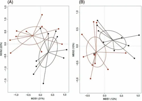
For the gut microbiota analysis, fecal samples were analyzed by next-generation sequencing of the 16S rDNA V3–V4 hypervariable region. The sequencing generated 1,234,956 high-quality reads (mean=61,748; SD=9,362) that were clustered into 4,610 OTUs at 97% identity. According to common alpha diversity metrics (i.e., the phylogenetic diversity–PD_whole_tree, the Shannon index for biodiversity and observed OTUs), no difference was found between IDA and HC infants (Supplementary Figure 1). On the other hand, the PCoA of both weighted and unweighted UniFrac distances reveals a significant segregation between the two sample groups (permutational multivariate ANOVA, p=0.02) (Figure1).
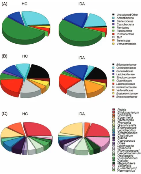
The most abundant phyla are Firmicutes (mean relative abundance, 54.0%), Actinobacteria (23.4%), and Bacteroidetes (14.2%). Even if statistical significance is not achieved (Wilcoxon rank-sum test, p>0.05), it is worth
noting that, compared to HC, IDA patients show an enrichment of Bacteroidetes (IDA vs. HC, 18.8% vs. 9.4%) and Proteobacteria (5.2% vs.
3.9%), with a corresponding reduction of Actinobacteria (19.3% vs. 27.5%) and Verrucomicrobia (0.7% vs. 3.0%) (Figure2A). At family level,Lachnos- piraceae (17.4%), Bifidobacteriaceae (17.2%), Ruminococcaceae (13.3%), and Bacteroidaceae (11.9%) are the most represented taxa in the gut micro- biota of the entire cohort. The gut microbial community of IDA patients appears significantly depleted in Coriobacteriaceae (3.5% vs. 8.8%;
p=0.004) and enriched in Veillonellaceae (13.7% vs. 3.6%; p=0.009) and Enterobacteriaceae (4.4% vs. 3.0%; p=0.04) compared to HC (Figure 2B).
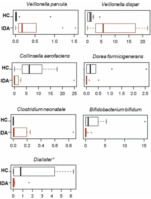
Accordingly, we found a significantly decreased Bifidobacteriaceae to Enterobacteriaceae ratio, which is a recently proposed index of gut health [15] in IDA patients (19.5 vs. HC, 93.3; p=0.02). At the genus level, the differences in the microbiota profiles are mainly attributable to an over- representation of Veillonella (mostly the Veillonella dispar and Veillonella parvula species) and diminished proportions of Collinsella, Bifidobacterium (B. bifidum),Dialister, andDoreain IDA patients compared to HC (Figures2C and 3). Although largely subdominant, the species Clostridium neonatale was identified only in IDA patients (Figure 3).
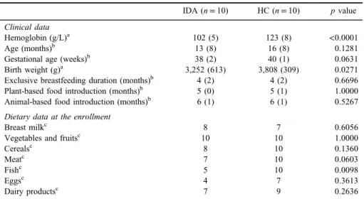
Table I.Baseline characteristics of study participants
IDA (n=10) HC (n=10) pvalue Clinical data
Hemoglobin (g/L)a 102 (5) 123 (8) <0.0001
Age (months)b 13 (8) 16 (8) 0.1281
Gestational age (weeks)b 38 (2) 40 (1) 0.0631
Birth weight (g)a 3,252 (613) 3,808 (309) 0.0271
Exclusive breastfeeding duration (months)b 4 (2) 4 (2) 0.6696
Plant-based food introduction (months)b 5 (0) 5 (1) 1.0000
Animal-based food introduction (months)b 6 (1) 6 (1) 0.5267
Dietary data at the enrollment
Breast milkc 8 7 0.6056
Vegetables and fruitsc 10 10 1.0000
Cerealsc 8 10 0.1360
Meatc 7 10 0.0603
Fishc 5 10 0.0098
Eggsc 4 7 0.3613
Dairy productsc 7 9 0.2636
Note:Baseline characteristics of iron deficiency anemia (IDA) group and healthy controls (HCs). Data are expressed as mean (standard deviation) or numbers.
aNormal distribution, Student’s t-test applied.
bNon-normal distribution, Wilcoxon rank-sum test applied.
cPearson’sχ2test applied.
Discussion
To date, only a few studies have investigated the impact of ID and IDA on the intestinal microbiota, in animal experiments [9,10],in vitrocolonic fermen- tation models [11,12], Indian women [13], and African infants, and school-aged children [14, 15]. Unlike these, this study included infants and young children living in a European town and aged 6–34 months, thus representing the most vulnerable age group, i.e., when a child exhausts endogenous iron stores and becomes dependent on iron-containing foods [28]. It is well established that this is a crucial time window for the child’s long-term health, characterized by a peculiar developmental trajectory of the gut microbiota, which undergoes distinctive compositional shifts, as solid foods are progressively introduced in the diet [29]. Microbiota alterations during this critical developmental window may impair the programing of the infant’s physiological systems, with long-term host metabolic and immunological effects [16,17,20, 22].
Figure 1.Gut microbiota community structure in infants suffering from iron deficiency anemia (IDA) and healthy controls (HCs). PCoA of Unweighted (A) and weighted (B) UniFrac distances
shows significant segregation between infants with IDA (red) and HC (black). Permutational multivariate ANOVA based on distance matrices (adonis),p=0.02
In line with the literature, the gut microbiota of the infants in this study cohort (all receiving complementary foods but the majority of them still breastfed) mainly consists of Firmicutes (with Ruminococcaceae, Lachnospiraceae, and Streptococcaceae as the most abundant families),Actinobacteria (dominated by
Figure 2.Taxonomic composition of intestinal bacterial communities in iron deficiency anemia (IDA) infants and healthy controls (HCs). Relative abundance of the most abundant phyla (A), families (B), and genera (C) in the gut microbiota of IDA infants and HCs. Only taxa with relative
abundance>0.02% in at least 15 samples were included
Figure 3.Discriminant species-level taxa between iron deficiency anemia (IDA) infants and healthy controls (HCs). Box plots showing the distribution of relative abundance values of discriminant
species between IDA infants and HCs. Only taxa with relative abundance>0.02% in at least 15 samples were included. Wilcoxon rank-sum test,p<0.05
Bifidobacteriaceae), and Bacteroidetes (represented by Bacteroidaceae). Interest- ingly, distinctive microbial signatures were identified in IDA patients compared to HC, especially an increase inEnterobacteriaceaeandVeillonella, and a reduction in Collinsella, Dialister, Bifidobacterium, andDorea. Such compositional traits could be partly accounted for by different iron needs for bacterial growth and metabolism.
Iron-low conditions are recognized to cause major shifts in micro- bial composition, leading to increased growth of bacteria, which are good iron scavengers (e.g., the health-promoting bifidobacteria and mainly Enterobacteriaceae, a family that includes known enteropathogens) or have no need for iron at all (e.g.,Lactobacillaceae) [30]. Recently, these data have been confirmed in in vitro colonic fermentation models inoculated with immobilized fecal microbiota from children aged 2.5 [11] and 6–10 years [12]. Specifically, ID conditions were found to result in decreased relative abundance of short-chain fatty acid producers (Lachnospiraceae, Ruminococcaceae, andBacteroidaceae), and increased proportions of Bifidobacteriaceae, Enterobacteriaceae, and Lactobacillaceae [11,12].
On the contrary, experiments in animal models showed that the gut microbiota composition depends not only on the dietary iron but also on the host iron homeostasis. In particular, a work with iron-replete rats receiving an iron- deficient diet revealed only minimal changes in the microbiota (i.e., decreased relative abundance ofBilophilaspp.,Eubacterium hallii, andCoprococcusspp.) [10]. The authors speculated that the fecal iron content was also determined by iron degradation from sloughed enterocytes, thus explaining the stable microbiota composition observed in low-dietary iron conditions. These results are in agree- ment withfindings that genetic modifications of iron metabolism in mice affect the gut microbial community [31]. Researchers concluded that higher iron avail- ability in the intestinal lumen increased the relative abundance ofLactobacillus murinus and Lactobacillus intestinalis. Nevertheless, they could not exclude positive effects on Lactobacillaceae expansion of other metals and minerals (e.g., manganese) that were increased in feces along with iron. In light of this, it is doubtful whether the decreased relative abundance of Lactobacillus acidophilus in IDA women in South India is actually related to host ID [13].
Similar results were obtained in African populations with high prevalence of anemia (non-IDA in most cases) and systemic inflammation. In fact, school-aged Ivorian children had higher relative abundance of enterobacteria (including Shigellaspp., enteroinvasiveE. coli, and/orSalmonellaspp.) than bifidobacteria and lactobacilli [14]. Six-month-old Kenyan infants also had high prevalence of enteropathogens, and the anemic group showed decreased abundance of
Actinomycetales and Streptococcus [15]. These findings could be associated with poor sanitation and hygiene. Conversely, our study included iron-deficient anemic, but otherwise healthy children from middle-class families; nevertheless, it showed a similar dysbiotic profile, with increased relative abundance of Enterobacteriaceae and altered Bifidobacteriaceae/Enterobacteriaceae ratio compared to HC. Such alterations (i.e., the expansion of enterobacteria) could reflect an altered intestinal environment created by host inflammatory responses [32]. Indeed, ID negatively affects cell-mediated and humoral immunity in children [33,34]. An impaired functioning of the host immune system may result in dysbiosis of the gut microbiota, as that observed in our IDA infant cohort, which, in turn, may further impair immunological responses, nurturing the inflammatory process and contributing to disease development [35].
An expansion of Enterobacteriaceae is typically found in inflammatory bowel disease (IBD), likely representing a hallmark of inflammation in the global population [36, 37]. Interestingly, IBD patients also usually have increased levels of Veillonellaceae [37] and lower relative abundances of Coriobacteriaceae [38], as we observed in IDA infants. Veillonella spp. are lactate-utilizing bacteria normally present in human microbiomes, especially in those of breast-fed infants [39], which may aid the immune system development in thefirst few months of life [40]. However, the abundances found in our work (mainly ofV. dispar, mean relative abundance in IDA is 9.0%) are far greater than those recently reported in a Swedish cohort of about 100 infants (1.5% in 1-year-old vaginally delivered infants) [39], suggesting an unusual succession of gut microbial communities in IDA. Further supporting these observations, other typical signatures of the infant microbiota, such as Bifidobacterium (mainly B. bifidum) andCollinsella, were found to be differentially represented between IDA infants and HC. IDA infants were also highly depleted in the short-chain fatty acid producerDorea, as well as in Dialister. The latter was found to be negatively correlated with infant morbidity in thefirst weeks after birth [41], and completely absent in the infant gut microbiota prior to the onset of type-I diabetes [42]. C. neonatale, a recently described lactose-utilizing lactate- producing species, isolated from the microbiota of neonates [43], was only detected in IDA infants.
Although the analysis was conducted at a single time point and on a small sample size, our data highlight a significant dysbalance of the gut microbiota in IDA infants. These findings pave the way for further, possibly longitudinal investigations, aimed at unravelling how the ID status may affect the microbiome developmental trajectory in early life, and the possible long-term consequences on the child’s health.
Conflict of Interest The authors declare no conflict of interest.
References
1. World Health Organization: The Global Prevalence of Anaemia in 2011. World Health Organization, Geneva, Switzerland, 2015. Available at http://www.who.int/nutrition/
publications/micronutrients/global_prevalence_anaemia_2011/en/
2. World Health Organization: Nutritional Anaemias: Tools for Effective Prevention and Control. World Health Organization, Geneva, Switzerland, 2017. Available athttp://www.
who.int/nutrition/publications/micronutrients/anaemias-tools-prevention-control/en/
3. Kiudeliene, R.: Diagnostic peculiarities of iron deficiency in early childhood. Dissertation, Kaunas Medicine University, Kaunas, Lithuania, 2009.
4. Georgieff, M. K.: Iron assessment to protect the developing brain. Am J Clin Nutr 106, 1588S–1593S (2017).
5. Lozoff, B., Jimenez, E., Smith, J. B.: Double burden of iron deficiency in infancy and low socioeconomic status: A longitudinal analysis of cognitive test scores to age 19 years. Arch Pediatr Adolesc Med160, 1108–1113 (2006).
6. Corapci, F., Radan, A. E., Lozoff, B.: Iron deficiency in infancy and mother-child interaction at 5 years. J Dev Behav Pediatr27, 371–378 (2006).
7. Armony-Sivan, R., Zhu, B., Clark, K. M., Richards, B., Ji, C., Kaciroti, N., Shao, J., Lozoff, B.: Iron deficiency (ID) at both birth and 9 months predicts right frontal EEG asymmetry in infancy. Dev Psychobiol58, 462–470 (2016).
8. Paganini, D., Zimmermann, M. B.: The effects of iron fortification and supplementation on the gut microbiome and diarrhea in infants and children: A review. Am J Clin Nutr 106, 1688S–1693S (2017).
9. Dostal, A., Chassard, C., Hilty, F. M., Zimmermann, M. B., Jaeggi, T., Rossi, S., Lacroix, C.:
Iron depletion and repletion with ferrous sulfate or electrolytic iron modifies the composition and metabolic activity of the gut microbiota in rats. J Nutr142, 271–277 (2012).
10. Dostal, A., Lacroix, C., Pham, V. T., Zimmermann, M. B., Del’homme, C., Bernalier- Donadille, A., Chassard, C.: Iron supplementation promotes gut microbiota metabolic activity but not colitis markers in human gut microbiota-associated rats. Br J Nutr 111, 2135–2145 (2014).
11. Dostal, A., Fehlbaum, S., Chassard, C., Zimmermann, M. B., Lacroix, C.: Low iron availability in continuousin vitrocolonic fermentations induces strong dysbiosis of the child gut microbial consortium and a decrease in main metabolites. FEMS Microbiol Ecol 83, 161–175 (2013).
12. Dostal, A., Lacroix, C., Bircher, L., Pham, V. T., Follador, R., Zimmermann, M. B., Chassard, C.: Iron modulates butyrate production by a child gut microbiotain vitro. MBio 6, e01453–01415 (2015).
13. Balamurugan, R., Mary, R. R., Chittaranjan, S., Jancy, H., Shobana Devi, R., Ramakrishna, B. S.: Low levels of faecal lactobacilli in women with iron-deficiency anaemia in South India. Br J Nutr104, 931–934 (2010).
14. Zimmermann, M. B., Chassard, C., Rohner, F., N’Goran, E. K., Nindjin, C., Dostal, A., Utzinger, J., Ghattas, H., Lacroix, C., Hurrell, R. F.: The effects of iron fortification on the gut microbiota in African children: A randomized controlled trial in Cote d’Ivoire. Am J Clin Nutr92, 1406–1415 (2010).
15. Jaeggi, T., Kortman, G. A., Moretti, D., Chassard, C., Holding, P., Dostal, A., Boekhorst, J., Timmerman, H. M., Swinkels, D. W., Tjalsma, H., Njenga, J., Mwangi, A., Kvalsvig, J., Lacroix, C., Zimmermann, M. B.: Iron fortification adversely affects the gut microbiome, increases pathogen abundance and induces intestinal inflammation in Kenyan infants. Gut 64, 731–742 (2015).
16. den Besten, G., van Eunen, K., Groen, A. K., Venema, K., Reijngoud, D. J., Bakker, B. M.:
The role of short-chain fatty acids in the interplay between diet, gut microbiota, and host energy metabolism. J Lipid Res54, 2325–2340 (2013).
17. Wu, H. J., Wu, E.: The role of gut microbiota in immune homeostasis and autoimmunity.
Gut Microbes3, 4–14 (2012).
18. Honda, K., Littman, D. R.: The microbiota in adaptive immune homeostasis and disease.
Nature535, 75–84 (2016).
19. Thaiss, C. A., Zmora, N., Levy, M., Elinav, E.: The microbiome and innate immunity.
Nature535, 65–74 (2016).
20. Fung, T. C., Olson, C. A., Hsiao, E. Y.: Interactions between the microbiota, immune and nervous systems in health and disease. Nat Neurosci 20, 145 (2017).
21. Sharon, G., Sampson, T. R., Geschwind, D. H., Mazmanian, S. K.: The central nervous system and the gut microbiome. Cell167, 915–932 (2016).
22. Rowland, I., Gibson, G., Heinken, A., Scott, K., Swann, J., Thiele, I., Tuohy, K.:
Gut microbiota functions: Metabolism of nutrients and other food components. Eur J Nutr 57, 1–24 (2018).
23. Biagi, E., Franceschi, C., Rampelli, S., Severgnini, M., Ostan, R., Turroni, S., Consolandi, C., Quercia, S., Scurti, M., Monti, D., Capri, M., Brigidi, P., Candela, M.: Gut microbiota and extreme longevity. Curr Biol26, 1480–1485 (2016).
24. Turroni, S., Rampelli, S., Biagi, E., Consolandi, C., Severgnini, M., Peano, C., Quercia, S., Soverini, M., Carbonero, F. G., Bianconi, G., Rettberg, P., Canganella, F., Brigidi, P., Candela, M.: Temporal dynamics of the gut microbiota in people sharing a confined environment, a 520-day ground-based space simulation, MARS500. Microbiome 5, 39 (2017).
25. Masella, A. P., Bartram, A. K., Truszkowski, J. M., Brown, D. G., Neufeld, J. D.:
PANDAseq: Paired-end assembler for illumina sequences. BMC Bioinformatics 13, 31 (2012).
26. Caporaso, J. G., Kuczynski, J., Stombaugh, J., Bittinger, K., Bushman, F. D., Costello, E. K., Fierer, N., Pena, A. G., Goodrich, J. K., Gordon, J. I., Huttley, G. A., Kelley, S. T., Knights, D., Koenig, J. E., Ley, R. E., Lozupone, C. A., McDonald, D., Muegge, B. D., Pirrung, M., Reeder, J., Sevinsky, J. R., Turnbaugh, P. J., Walters, W. A., Widmann, J., Yatsunenko, T., Zaneveld, J., Knight, R.: QIIME allows analysis of high-throughput community sequencing data. Nat Methods7, 335–336 (2010).
27. Edgar, R. C.: Search and clustering orders of magnitude faster than BLAST. Bioinformatics 26, 2460–2461 (2010).
28. Lonnerdal, B.: Development of iron homeostasis in infants and young children. Am J Clin Nutr 106, 1575S–1580S (2017).
29. Voreades, N., Kozil, A., Weir, T. L.: Diet and the development of the human intestinal microbiome. Front Microbiol5, 494 (2014).
30. Kortman, G. A., Raffatellu, M., Swinkels, D. W., Tjalsma, H.: Nutritional iron turned inside out: Intestinal stress from a gut microbial perspective. FEMS Microbiol Rev38, 1202–1234 (2014).
31. Buhnik-Rosenblau, K., Moshe-Belizowski, S., Danin-Poleg, Y., Meyron-Holtz, E. G.:
Genetic modification of iron metabolism in mice affects the gut microbiota. Biometals25, 883–892 (2012).
32. Baumler, A. J., Sperandio, V.: Interactions between the microbiota and pathogenic bacteria in the gut. Nature535, 85–93 (2016).
33. Hassan, T. H., Badr, M. A., Karam, N. A., Zkaria, M., El Saadany, H. F., Abdel Rahman, D. M., Shahbah, D. A., Al Morshedy, S. M., Fathy, M., Esh, A. M., Selim, A. M.: Impact of iron deficiency anemia on the function of the immune system in children. Medicine (Baltimore)95, e5395 (2016).
34. Aly, S. S., Fayed, H. M., Ismail, A. M., Abdel Hakeem, G. L.: Assessment of peripheral blood lymphocyte subsets in children with iron deficiency anemia. BMC Pediatr 18, 49 (2018).
35. Kato, L. M., Kawamoto, S., Maruya, M., Fagarasan, S.: The role of the adaptive immune system in regulation of gut microbiota. Immunol Rev260, 67–75 (2014).
36. Papa, E., Docktor, M., Smillie, C., Weber, S., Preheim, S. P., Gevers, D., Giannoukos, G., Ciulla, D., Tabbaa, D., Ingram, J., Schauer, D. B., Ward, D. V., Korzenik, J. R., Xavier, R. J., Bousvaros, A., Alm, E. J.: Non-invasive mapping of the gastrointestinal microbiota identifies children with inflammatory bowel disease. PLoS One7, e39242 (2012).
37. Gevers, D., Kugathasan, S., Denson, L. A., Vazquez-Baeza, Y., Van Treuren, W., Ren, B., Schwager, E., Knights, D., Song, S. J., Yassour, M., Morgan, X. C., Kostic, A. D., Luo, C., Gonzalez, A., McDonald, D., Haberman, Y., Walters, T., Baker, S., Rosh, J., Stephens, M., Heyman, M., Markowitz, J., Baldassano, R., Griffiths, A., Sylvester, F., Mack, D., Kim, S., Crandall, W., Hyams, J., Huttenhower, C., Knight, R., Xavier, R. J.: The treatment-naive microbiome in new-onset Crohn’s disease. Cell Host Microbe15, 382–392 (2014).
38. Maukonen, J., Kolho, K. L., Paasela, M., Honkanen, J., Klemetti, P., Vaarala, O., Saarela, M.:
Altered fecal microbiota in paediatric inflammatory bowel disease. J Crohns Colitis 9, 1088–1095 (2015).
39. Backhed, F., Roswall, J., Peng, Y., Feng, Q., Jia, H., Kovatcheva-Datchary, P., Li, Y., Xia, Y., Xie, H., Zhong, H., Khan, M. T., Zhang, J., Li, J., Xiao, L., Al-Aama, J., Zhang, D., Lee, Y. S., Kotowska, D., Colding, C., Tremaroli, V., Yin, Y., Bergman, S., Xu, X., Madsen, L., Kristiansen, K., Dahlgren, J., Wang, J.: Dynamics and stabilization of the human gut microbiome during thefirst year of life. Cell Host Microbe17, 852 (2015).
40. Arrieta, M. C., Stiemsma, L. T., Dimitriu, P. A., Thorson, L., Russell, S., Yurist-Doutsch, S., Kuzeljevic, B., Gold, M. J., Britton, H. M., Lefebvre, D. L., Subbarao, P., Mandhane, P., Becker, A., McNagny, K. M., Sears, M. R., Kollmann, T., Investigators, C. S., Mohn, W. W., Turvey, S. E., Finlay, B. B.: Early infancy microbial and metabolic alterations affect risk of childhood asthma. Sci Transl Med7, 307ra152 (2015).
41. Davis, J. C., Lewis, Z. T., Krishnan, S., Bernstein, R. M., Moore, S. E., Prentice, A. M., Mills, D. A., Lebrilla, C. B., Zivkovic, A. M.: Growth and morbidity of Gambian infants are influenced by maternal milk oligosaccharides and infant gut microbiota. Sci Rep7, 40466 (2017).
42. Kostic, A. D., Gevers, D., Siljander, H., Vatanen, T., Hyotylainen, T., Hamalainen, A. M., Peet, A., Tillmann, V., Poho, P., Mattila, I., Lahdesmaki, H., Franzosa, E. A., Vaarala, O., de Goffau, M., Harmsen, H., Ilonen, J., Virtanen, S. M., Clish, C. B., Oresic, M., Huttenhower, C., Knip, M., Group, D. S., Xavier, R. J.: The dynamics of the human infant gut microbiome in development and in progression toward type 1 diabetes. Cell Host Microbe17, 260–273 (2015).
43. Schonherr-Hellec, S., Klein, G., Delannoy, J., Ferraris, L., Friedel, I., Roze, J. C., Butel, M. J., Aires, J.: Comparative phenotypic analysis of“Clostridium neonatale”and Clostridium butyricumisolates from neonates. Anaerobe48, 76–82 (2017).