PhD DISSERTATION
DR. KÚTVÖLGYI GABRIELLA
KAPOSVÁR UNIVERSITY
FACULTY OF ANIMAL SCIENCE
2012
KAPOSVÁR UNIVERSITY
FACULTY OF ANIMAL SCIENCE
Department of Breeding and Production of Ruminants and Horse
Head of Doctoral School:
Dr. PÉTER HORN
Member of the Hungarian Academy of Sciences Supervisor:
DR. JÓZSEF STEFLER Candidate of Agricultural Sciences
Co-Supervisor:
DR. ANDRÁS KOVÁCS
Doctor of the Hungarian Academy of Sciences
DEVELOPMENT OF QUALIFICATION OF FRESH AND FROZEN STALLION SEMEN, INVESTIGATION OF FACTORS
AFFECTING SPERM QUALITY USING A NEW EVALUATION METHOD
Author:
DR. GABRIELLA KÚTVÖLGYI
KAPOSVÁR 2012
Szüleim és nıvérem emlékére
Dedicated to the memory of my parents and sister
TABLE OF CONTENTS
1. GENERAL INTRODUCTION...1
2. LITERATURE OVERVIEW ...6
2.1 Breeding soundness examination of the stallion ...6
2.2 Stallion semen ...7
2.3 Spermatozoa production, spermatogenesis, sperm maturation ...9
2.4 Structure of equine spermatozoa...11
2.5 Morphology of stallion spermatozoa ...15
2.5.1 Classification systems ...16
2.5.2 Preparation for morphologic evaluation ...20
2.5.3 Abnormal morphology of spermatozoa...23
2.6 Evaluation of the spermatozoa...33
2.7 Cryopreservation of equine spermatozoa ...41
2.7.1 History and present of sperm cryopreservation and its utilization ...41
2.7.2 Cryobiology of equine spermatozoa: principles, factors affecting on semen quality and freezability, species differences and individual variations ...43
2.7.3 Cryodamages and their evaluations...47
2.8 Sperm separation for in vitro embryo production...54
2.8.1 Comparison of swim up and density gradient centrifugation ...58
2.8.2 Comparison of different sperm preparation techniques...59
2.8.3 Preparation of low quality, small volume and low concentration sperm for ICSI...59
2.8.4 Improving of efficiency of sperm separations using treatment of spermatozoa ...61
2.8.5 Effect of acrosome on the success of ICSI ...63
2.9 Fertility of the stallion...64
2.9.1 Fertility parameters, subfertility ...64
2.9.2 Relationship between sperm quality and fertility ...68
2.9.3 Reproductive disorders of stallions; diagnosis, prognosis and possible treatment ...75
3. OBJECTIVES OF THE DISSERTATION ...79
4. MATERIALS AND METHODS ...81
4.1 Semen samples, main procedures and experimental design ...81
4.2 Sperm evaluation method...87
4.3 Data analyses, statistical methods ...94
5. RESULTS ...98
5.1 Experiment 1. Improvement of assessment of stallion sperm quality by Chicago sky blue and Giemsa viability and acrosome staining method ...98
5.2 Experiment 2. Analysis of the injuries of stallion spermatozoa during the whole freezing procedure ...105
5.3 Experiment 3. Use of pentoxifylline and hyaluronic acid for stallion sperm separation ...117
5.4 Experiment 4. Viability, acrosome integrity and morphology evaluation of sperm
samples from subfertile stallions (Case reports and their interpretation)... 124
6. DISCUSSION AND CONCLUSIONS... 150
6.1 Experiment 1. Improvement of assessment of stallion sperm quality by Chicago sky blue and Giemsa viability and acrosome staining method ... 150
6.2 Experiment 2. Analysis of the injuries of stallion spermatozoa during the whole freezing procedure... 152
6.3 Experiment 3. Use of pentoxifylline and hyaluronic acid for stallion sperm separation... 158
6.4 Experiment 4. Viability, acrosome integrity and morphology evaluation of sperm samples from subfertile stallions ... 163
7. NEW SCIENTIFIC RESULTS ... 169
8. SUMMARY ... 170
9. ÖSSZEFOGLALÁS ... 173
10. REFERENCES ... 176
11. PUBLICATIONS IN THE FIELD OF THE THESIS ... 201
12. PUBLICATIONS OUT OF THE FIELD OF THE THESIS... 202
13. CURRICULUM VITAE ... 204
14. KÖSZÖNETNYILVÁNÍTÁS... 205
15. APPENDIX
List of abbreviations Laboratory Stock solutions Microscopic pictures
“Life shield” from Tayos gold library (Ecuador) /collection of Padre Carlos Crespi,
presumed age of the artifact is 8000-10000 years/
1. GENERAL INTRODUCTION
The horse has been in close contact with humans for thousands of years. This species - presented in the largest populations of mammals in Eurasia 25 thousand years ago-, was an important part of human life in prehistoric times, which is attested by the 20- 30-thousand-year-old Palaeolithic art cave paintings from Lascaux Caves, Pech-Merle Cave, etc. Horses had also survived the glacial period. The date and place of horse domestication has long been subject to research. However, several evidence supports the hypothesis that horses were domesticated in the Eurasian Steppes (Dereivka centered in Ukraine) approximately 4000-3500 BCE, recent discoveries on Botai culture in northern Kazakhstan suggest that location of the earliest domestication of the horse was in this region during the Copper Age, dating to about 3500 BCE.
Pathological characteristics indicate that some horses were bridled, perhaps ridden and Botai culture used horses as a source of meat and milk (Outram et al. 2009). DNA studies seem to indicate that domestication probably occurred in multiple locations simultaneously (Jansen et al. 2002). Horse domestication was a great breakthrough, bringing horsepower to communications, transportation, farming and warfare. In the Scythian Empire, the horse was an important animal, enabling the Eurasian steppe population to cover long distances. It was also an essential companion in the afterlife as the archaeological findings from the Unesco World Heritage Frozen Scythian Tombs of the Altai Mountains shows. Phylogenetical analysis showed that these
„A csodák nem a természeti törvényekkel állnak ellentmondásban, csupán azzal, amit a természetrıl tudunk” - Szent Ágoston-
"Miracles are not contrary to nature, but only contrary to what we know about nature."
- Saint Augustine -
ancient horse’s mitochondrial DNA sequences were distributed throughout the tree defined by modern horses’ sequences and are closely related to them (Keyser-Tracqui et al. 2005). The horse later, for centuries had represented a decisive role in human cultures. The selection of excellent sires and dams had strategic importance to the expansion, conquest, and in battles.
According to the legend the horse was the first animal upon which artificial insemination (AI) was practised successfully. In 1322 an Arab chieftain sent a spy to the neighbouring rival’s camp. This man then scooped some of the semen from a recently mated mare’s vagina, diluted it with camel milk and rushed with this „diluted semen” back to base. The legend does not relate how the diluted semen was inseminated into the boss’s mare but it does avow that a healthy and exceptionally beautiful foal was born the following year (Bowen 1969, Pickett et al. 2000, Allen 2005). The first successful real insemination was performed by Spallanzani (1784) in a dog, which gave birth to three puppies 62 days later (Foote 2002). In 1897, Heape and other scientists in several countries reported successful use of AI in dogs, rabbits and horses. Pioneering efforts to establish AI as a practical procedure were begun in Russia in 1899 by Ivanov who studied AI in many domestic farm animals, dogs, foxes, rabbits, and poultry. Some of this research, especially in horses, is included in his paper from 1922. The Japanese scientist Ishikawa studied with Ivanov and after he had returned to Japan began a similar program in horses in 1912 (Nishikawa 1962). In Sweden, Lagerlöf became known for his research on infertility problems in bulls.
Other Scandinavians, such as Blom (1950), followed, publishing a steady stream of excellent papers on abnormal sperm morphology (Foote 2002). Intensive growth of AI occurred in the 1940s in the United States with close collaboration between dairy cow farmers and researchers. In Hungary Kaldrovics around 1890 then Threisz from 1902 successfully utilized equine AI in the Kisbér and Mezıhegyes Studs (Bölcsházy and Mészáros 1962). Dezsı Hámori after the World War II. introduced generally artificial insemination in order to stop the spread of infectious Trypanosomiasis. One year after the other artificial insemination stations had been established throughout the country.
In 1950 seventy four Artificial Insemination Stations were working and altogether 42.000 mares were inseminated with sperm of 240 stallions. In average 175 mares were booked for one stallion. Several stallions had more than 300 mares for breeding (Mészáros 1951). In 1953 the number of AI stations and mares inseminated was culminated (63.000 horses in 120 breeding stations) and one-third of the broodmares were bred with AI. The reorganization of agriculture from the mid-1950s caused that importance of horse breeding gradually declined, artificial insemination was also getting underplayed. In 1960 only 9000 mares were inseminated, to the year of 1965
the application of AI had been almost completely gone. In the years of 1970s, Magdolna Alhegyi started cryopreservation of stallion semen in the National Artificial Insemination Station because of a dramatically decline in the number of horses, only this way was feasible to ensure preservation of genetic diversity of bloodlines of rare breeds.
In the last decades the use of artificial insemination (AI) in equine reproduction has been increasing again worldwide in the horse industry, offering many advantages over natural service. Some of the reasons for this include the choice of a great number of stallions, safety for both the mare and the stallion, reduced risk of infectious disease transmission, and transport inconvenience. Although pregnancy rates have been shown to be equal or even higher after AI with fresh or chilled semen compared to natural service, lower fertility occurs when mares are inseminated with frozen semen (Jasko et al. 1992, Samper 2001, Janett et al. 2003). Some experimental and field studies however indicate that acceptable fertility with frozen semen is possible with good quality semen, selection of mares and stallions and good mare management, and may not differ significantly from the fertility of commercial cooled semen (Metcalf 1995, Barbacini et al. 1999, Loomis 2001). Based on my clinical experiences I agree with these findings.
Smith and Polge (1950) first froze equine spermatozoa in 1950 and the first equine pregnancy using frozen semen was reported in Barker and Gandier (1957). Recently the total AI with cryopreserved or cooled equine semen is 0.5 million AI/year all over the countries. From the total number in 0.1 million AI/year has been used frozen semen. Around 350.000 foals are born each year after successful artificial insemination (Central European Management Intelligence /CEMI/, frozen + fresh data from 2006). Frozen semen offers breeders additional benefits not available with cooled semen: Stallions need not be available for every second day collections, allowing for involvement in performance events during the breeding season; Illness, injury or death of a stallion does not prevent insemination of mares with his semen;
Frozen semen can be shipped in advance and maintained on the farm until the optimum time for insemination; It allows for international distribution; Less semen is wasted while all the semen collected for freezing is processed and stored resulting in an average of 10–12 insemination doses per ejaculate However some disadvantages of utilization of frozen semen limiting their general usage: Frozen semen results in lower fertility than with cooled semen for many stallions; More technical expertise is required for processing frozen semen than cooled semen; Increased costs associated with management of mares due to more frequent examinations (Loomis 2001).
The equine artificial insemination was getting a new impetus from the end of 90’s also in Hungary. In 2003 forty two equine AI stations were working in the country with 314 stallions and around 5000 mares were inseminated which was half of the total number of broodmares involved in the breeding. After the governmental financial support system was stopped many of them have closed. In 2010 twenty three AI breeding stations were working with 96 stallions (data of Central Agricultural Office /Nemzeti Élelmiszerlánc-biztonsági Hivatal/ and National Horse Information System /OLIR/, personal communication of Dr. Ferenc Flink and Csilla Végi). The ratio of AI in all breeding services decreased however the utilization of frozen semen and imported cooled semen increased in the recent years. The "conservation of genetic resources" programme and supports for in vitro and/or ex situ preservation of protected native and endangered farm animal breeds starting in 2012 hopefully will bring again upsurge in Hungarian horse breeding.
In the recent years modern breeding technologies like hysteroscopic low-dose insemination, fluorescence-activated sex sorting of stallion spermatozoa, embryo transfer, embryo freezing and bisection, transvaginal ultrasound-guided oocyte collection (OPU), intracytoplasmic sperm injection for fertilization (ICSI), gamete intrafallopian transfer (GIFT) and nuclear transfer (cloning), have all been applied to equids with encouraging success (Allen 2005).
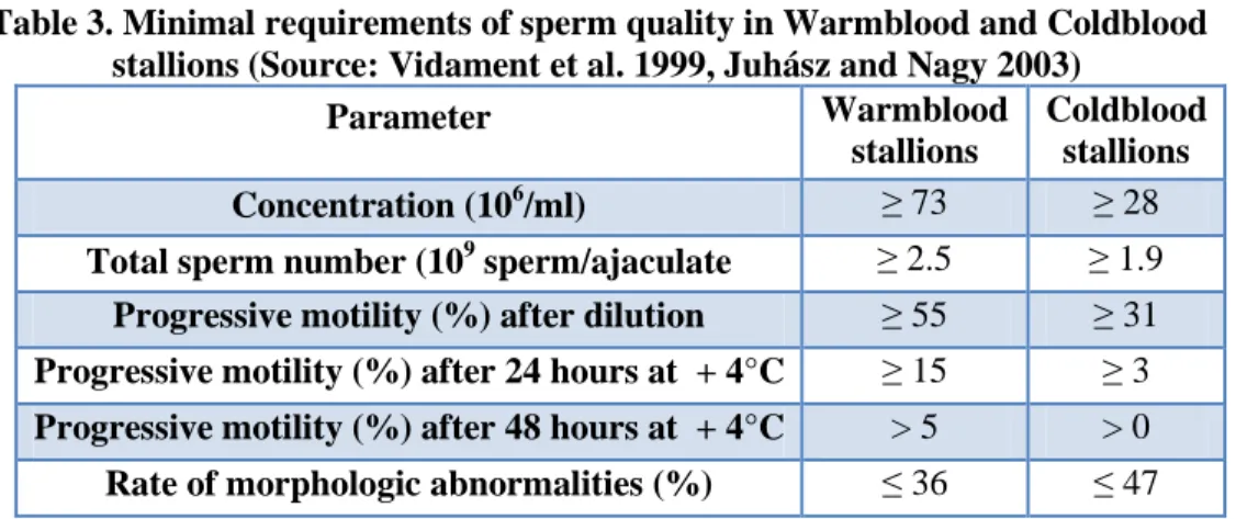
In the horse breeding selection of sire is based on performance, genetic potential and exterior. However excellent sport or race performance and noble appearance are not related to great fertility of the stallion. Unlike bulls, stallions have been not selected by the artificial insemination (AI) industry for many years and generations based on semen production, sperm quality and freezability. This explains that there is a wide variation in semen characteristics among individuals and in remarkable rate the semen quality is not sufficient. In some breeds (Kavak et al. 2004) or rather in some genetic lines increased proportion of morphologically abnormal spermatozoa was observed which is considered inheritable due to inbreeding, e.g. in the case of Kuhaylan Zaid Arabian line (Mészáros et al. 1951, Bölcsházy and Mészáros 1962). In more countries like in France or Netherlands semen evaluation is performed in the prospecting breeding stallions. In the Netherlands it has been recommended that minimum values of semen quality of 3-year-old stallions for registration in the studbook is a mean total number of motile, morphologically normal spermatozoa of 2 x 10⁹ and a mean of 50%
for motility and 50% of morphologically normal spermatozoa (Parlevliet et al. 1994) No such official recommendation exists in Hungary for the young stallions, although there is a Hungarian Standard for breeding stallion semen (7034/1999) which proposes at least
40 x 106 spermatozoa in 1 ml gel-free ejaculate, 20 ml gel-free volume, 40 % motility and ≤ 30% sperm with any morphologic aberrations, if less than half of these abnormal cells have primary defect. Moreover one insemination dose of diluted extended stallion semen is recommended to have at least 3-5 x 10 8 total number of live spermatozoa and to have ≥ 40 % progressive motile sperm at the time of insemination.
Both of healthy reproductive status of the broodmare and good quality semen of the breeding stallion are playing key-role in the successful fertilization and producing offspring. AI, cooled- and frozen storage of the semen give opportunity to allocate more doses from one ejaculate of the breeding stallion and to inseminate several mares even in more-hundred kilometres distance from the stud farm where the sperm was collected.
This is unrealizable if the stallion is used for naturel service. Considering this tendency in the world it is need to lay greater emphasis on examination of stallion sperm quality even before receiving the breeding permit. Conventional semen evaluation is very subjective mainly based on sperm concentration and movement of the spermatozoa. In spite of its limited applicability, motility and progressive motility are the most commonly used parameters in the evaluation of stallion semen, in both laboratories and studfarms, because it is easily accessible and quick to perform (Katila et al. 2001a). Various techniques and protocols are available for evaluation of the spermatozoon. However besides routine examination to combine several tests or use combined analyses of more features are needed for quality and fertility evaluation of equine semen (Katila 2001a, Colenbrander et al. 2003). It should be taken into consideration that if parameters of the fresh semen are behind the desired level, after chilled-transportation or cryopreservation these would be declined. Using multi-parametric semen analysis methods subfertile and infertile stallions would be identified and reason for decreased pregnancy results may be revealed (Colenbrander et al. 2003).
It is important that a reliable sperm quality control will be available in both of introduction of a new technology or annual or daily routine examination of the stallions in the practice. Kovács-Foote staining procedure (Kovács and Foote 1992) has been tested in many species since its first debut and may be suitable for complex quality control of the semen. In the equine species for the correct evaluation it was necessary to improve and modify the assessment technology, to achieve the ideal settings in every steps of the staining. Comparing the treatments in two sperm manipulation process and investigation of subfertile and fertile stallions, I defined new classification which could make the comparisons more sophisticated and which is capable of more complex evaluation.
2. LITERATURE OVERVIEW
2.1 Breeding soundness examination of the stallion
Annual assessments for actively breeding stallions prior to the breeding season can help with management decisions and provide a baseline for comparison if a problem should arise. Complex breeding soundness examination (BSE) of the stallion is an attempt to provide an estimate of a stallion's potential fertility and is composed of the following: History (past breeding records, illness, medications, pedigree); General physical examination (body condition, inheritable conditions, external genitalia, testicular evaluations, and measurements); Evaluation of libido and mating behavior;
Quantity and quality evaluation of semen; Ancillary procedures (bacterial cultures, cytology, special stains, hormone analysis) (Juhász et al. 2000, Steiner 2002). The Society for Theriogenology has developed Guidelines for Classification of Breeding Stallions (Kenney et al. 1983) based on breeding 40 mares by natural service or 120 mares by artificial insemination with a result of at least 75 % of the mares become pregnant. Stallions are classified as satisfactory, questionable, or unsatisfactory breeding prospects. To be classified as satisfactory, the following criteria must be met:
stallion demonstrates good libido and mating ability, normal penis without inflammatory lesions, free of venereal or transmissible diseases or potentially heritable defects; negative EIA (Coggins) test; two scrotal testes and epididymes which have normal size, consistency, and shape; total scrotal width greater than 8.0 centimeters; a stallion produces a minimum of one billion morphologically normal, progressively motile spermatozoa (corrected for season, e.g. 2 billions in May) in each of 2 ejaculates collected 1 hour apart. Under these criteria the stallion is classified as a questionable or unsatisfactory. Several stallions are not used for 40/120 mares and may be able to settle a smaller book of mares with adequate reproductive management. A recent report indicates that the mare book of Thoroughbred stallions in North America has increased considerably in the last 20 years, and that in 2005, 11% of stallions had a book greater than the traditional book of 40 mares. More importantly, over 50% of Thoroughbred mares were bred by stallions with books greater than 40 mares, and 35% of the mares were bred by stallions with books greater than 80 mares. Interestingly, foaling rates increased as the book increased, indicating that selected fertile stallions are able to couple with the larger books (Turner and McDonnell 2007, Brito 2007).
2.2 Stallion semen
In the process of BSE two semen samples are collected from the stallion one hour apart. The two ejaculates are considered representative if volume of the ejaculates are similar and the second contains approximately one-half the number of spermatozoa as the first with comparable or increased sperm motility. Immediately after collection, the sample is evaluated for colour, clarity, and volume. The gel portion of the ejaculate should be removed by filtration. Next, the motility of the sperm should be assessed and sperm concentration determined using a densometer, hemocytometer, or spectrophotometer. Sperm concentration multiplied by gel-free semen volume will give the total number of sperm. Live/dead ratio of spermatozoa and sperm morphology is also addressed as well as longevity of spermatozoa (Steiner 2002).
Composition of semen shows species specific and individual differences within species moreover changes occasionally in different ejaculates. Individual differences are related to spermatogenesis and secretion of accessory sex glands. Sperm production of the stallion depends on age, testicular volume, sperm reserve capacity of the epididymis and is also influenced by extrinsic factors as nutrition, temperature, breeding season, frequency of ejaculation, medication (Haraszti and Zöldág 1993, Pickett 1993a). Volume of equine semen is in avarege 70 ml (30-300 ml), sperm concentration is 30-800 x 106 sperm/ml. The light-absorbance of semen is primarily influenced by the spermatozoa, therefore opacity of the ejaculate is proportional to sperm concentration. The pH of the semen is determined by accessory sexual glands which is pH. 6.6-7.6 (Pécsi 2007). The semen is composed by two main parts: cellular elements (5-10% dispersed portion) and seminal plasma (90-95%). Spermatozoa compose the main part of cellular elements and other cells like nucleated epithelial cells of reproductive tracts, immature spermatozoa, lymphocytes, leucocytes are also found in this fraction. In addition protein, lipid, pigment particles and prostatic amyloids are also analysed from the semen. Seminal plasma is the product of accessory sex glands. Different types of bacteria may be also presented in the sperm related to bacterial flora of the praeputium (Haraszti and Zöldág 1993). Values for the most frequently used parameters of fresh equine semen are presented in Table 1.
Evaluation of daily sperm output (DSO) is a criterion used to assess the breeding potential of a stallion. In order to accurately predict the number of mares a stallion can breed or the number of AI doses a stallion can yield during a given time period it is also necessary to know the number of morphologically normal spermatozoa in ejaculates. (Stich et al. 2002, Einarsson et al. 2009). Collecting and evaluating total
spermatozoa number (TSN) per ejaculate found that DSO determinations can begin as early as on the 5th day for stallions with smaller testes, and on the 6–7th day for stallions with larger testes (Kavak et al. 2003b).
.
Table 1. Quantitative and qualitative parameters of fresh equine semen (Juhász et al. 2000, Juhász and Nagy 2003)
Parameter Mean ± SD Number of
Stallions Reference
Gel free volume (ml)
65 ± 26 45 ± 30 33.7 ± 2.13 51.6 ± 31.5 45.3 ± 30.9
63.1
398 417 165 8 47 245*
Parlevliet et al. 1994 Pickett 1993b Dowsett and Knott 1996
Long et al. 1993 Dowsett and Pattie 1982
Juhász and Nagy 2003
Concentration (106/ml)
206.1 ± 168.5 335 ± 232 164.13 ± 39.35
223 ± 148 178 ± 168 192.5 ± 169.6
398 417 165 8 47 245*
Parlevliet et al. 1994 Pickett 1993b Dowsett and Knott 1996
Long et al. 1993 Dowsett and Pattie 1982
Juhász 2003
Total sperm number (109)
11.29 ± 7.13 11.9 ± 9
6.34 ± 1.93 9.1 ± 4.7 7.21 ± 6.87 8.44 ± 4.61
398 417 165 8 47 245*
Parlevliet et al. 1994 Pickett 1993b Dowsett and Knott 1996
Long et al. 1993 Dowsett and Pattie 1982
Juhász and Nagy 2003
Total motility (%)
53 ± 15 76.43 72.1 ± 16 70.3 ± 17.4 70.3 ± 17.4
417 165 47 64 245*
Pickett 1993b Dowsett and Knott 1996 Dowsett and Pattie 1982
Jakso et al. 1991 Juhász and Nagy 2003 Progressive
motility (%)
68 ± 9 53.1 ± 16.2 52.7 ± 23.8 66.9 ± 10.4
398 8 64 245*
Parlevliet et al. 1994 Long et al. 1993 Jakso et al. 1991 Juhász and Nagy 2003 Live
spermatozoa (%)
65 ± 16 82.56
78.8
398 165 47
Parlevliet et al. 1994 Dowsett and Knott 1996 Dowsett and Pattie 1982
*data based on 245 ejaculates of 8 different stallions
Collecting one ejaculate per day for 10 consecutive days found that TSN decreased successively from day 1 to 8 in full-size horses, and from day 1 to 4 in small-size
horses, thereafter remaining approximately at the same number. The DSO, calculated as the mean of TSN of days 8–10 was 6.4 x 10⁹ (Kavak et al. 2003b, Einarsson et al.
2009).
2.3 Spermatozoa production, spermatogenesis, sperm maturation
Spermatogenesis is a process of division and differentiation of cells from diploid germinal cells termed spermatogonia, to haploid, mature spermatozoa in seminiferous tubules of the testis. Spermatozoa are released every 12.2 days in horses, however, the duration of spermatogenesis is around 57 days (Johnson et al. 1997). The 57-day- period may be divided into three phases, including: spermatocytogenesis, where stem cell spermatogonia divide by mitosis to produce other stem cells in order to continue the lineage throughout the adult life of the male, they also divide cyclically to produce committed spermatogonia and primary spermatocytes (19.4 days); meiosis, the period of replication (primary spermatocytes) and then reduction of genetic material into haploid spermatids (19.4 days); and spermiogenesis or spermiomorphogenezis which is a Sertoli cell aided differentiation of spermatids (18.6 days). Spermiation is the release of spermatids as spermatozoa into the lumen of seminiferous tubule. Transit of sperm through the efferent ductules and the epididymis is associated with significant maturation changes such as gaining the capacity for progressive motility; final condensation of the nucleus and further modification of the form of the acrosome;
alteration of plasma membrane surface (release, modification and adsorption of proteins and lipids); migration of cytoplasmic droplet from a proximal to distal midpiece position. The most important functional changes in sperm occur in the efferent ductules, caput and corpus of epididymis. Sperm taken from the distal part of corpus and cauda epididymis already have the potential to fertilize (Bart and Oko 1989, Gadella et al. 2001). Approximately 9 days are required for transportation of spermatozoa through the excurrent duct system. Therefore, a new population of spermatozoa is ejaculated every 64–66 days (Amann 1993, Card 2005). The cauda epididymis has properties that allow the sperm to be stored for several weeks. Seminal fluids secreted by accessory sex glands (ampulla, seminal vesicles, prostate, bulbourethral glands) added during ejaculation serve as a vehicle, stimulate the metabolism of sperm and provide the energy requirements for passage through the uterus (Bart and Oko 1989, Gadella et al. 2001). In contact of female reproductive tract, mainly in the isthmus of oviduct the spermatozoa undergo special changes termed ‘‘capacitation’’ which include reorganization of membrane proteins, metabolism of membrane phospholipids, reduction in membrane cholesterol levels.
Capacitation takes 6-8 hours in stallion spermatozoa (Yanagimachi 1994).
The process of spermatogenesis, steroidogenesis and testicular function are regulated by a complex interplay of endocrine, paracrine and autocrine signals. It is well established in many mammalian species, including the horse, that normal testicular function is dependent upon a functional pineal gland and hypothalamic – pituitary - testicular (HPT) axis. The pineal gland secretes melatonin that acts on the hypothalamus to regulate gonadotropin-releasing hormone (GnRH) output. GnRH stimulates secretion of the pituitary hormones, luteinizing hormone (LH) and follicle stimulating hormone (FSH), which in turn acts at the level of the testis. LH binds to receptors on testicular Leydig cells to stimulate production of testosterone and estrogens while FSH binds to the Sertoli cell to stimulate production of estrogens, inhibin (INH) and activin. These testicular steroid and protein hormones are components of the classic feedback mechanisms involved in the regulation of hypothalamic GnRH and pituitary LH and FSH (Amann 1993).
Recent studies in the horse have demonstrated the potential involvement of other hormones such as opioids, prolactin, growth hormone (GH), thyroid hormone and activin (Gerlach and Aurich 2000, Arai et al. 2006, Roser 2007). Moreover, evidence indicates that local paracrine - autocrine factors within the testes such as growth factors like IGF-1, oxytocin, vasopressin, pro-enkephalins, enkephalins, propiomelanocortin, β-endorphins, cytokines, transferring, modulate endocrine control of testicular function, steroidogenesis and spermatogenesis (Roser 2008). Systemic hormones and local testicular factors work together to initiate and maintain testicular function. Testosterone from the Leydig cells diffuses into the seminiferous tubules and in conjunction with FSH signals the Sertoli cells to produce local factors that initiate and maintain development of the germ cells. FSH is of critical importance in the initiation and expansion of spermatogenesis in mammals during puberty, however the role of FSH in the regulation of spermatogenesis in the adult mammal is still controversial. (Zirkin et al. 1994). Many of the local interactions in the equine testis are unknown. Inhibine and activin have endocrine, paracrine and autocrine functions.
Inhibin feeds back on the pituitary to inhibit the release of FSH. Activin has been shown to positively modulate the release of FSH at the level of the pituitary in other species, but its action in the stallion is unknown (Roser 2007). IGF-1 may be involved in testicular development (Hess and Roser 2001, Roser 2008).
Billions of spermatozoa are produced each day. Many of the cells produced are defective and some are eliminated through apoptosis and phagocytosis by the Sertoli cells, whereas others are passed into the ejaculate (Heninger et al. 2004, Card 2005).
Sertoli cells, the somatic component of the seminiferous epithelium, are critical for germ cell development during spermatogenesis. High environmental temperature,
other conditions such as fever, orchitis, periorchitis, hydrocele, scrotal hemorrhage, scrotal edema, scrotal dermatitis and improper scrotal descent can interfere with the thermoregulatory mechanism necessary to cool the testis and allow normal spermatogenesis (Varner et al. 1991, Johnson et al. 1997).
2.4 Structure of equine spermatozoa
The spermatozoon consists of the head, neck, and tail and is entirely covered by the plasma membrane. The tail is the longest part of the spermatozoon and consists of midpiece, principal piece, and end piece (Figs 1/A, 1/B, Amann and Graham 1993, Brito 2007). The length of equine spermatozoon is approximately 60 µm (Pesch and Bergman 2006, Brito 2007). Every part of the spermatozoa plays important role in the fertilization process. The plasma membrane surrounds the spermatozoon in total and is characterised by a regional specific glycoprotein and lipid, mainly phospholipids and cholesterol constitution. These so-called surface domains are important for the function of the membrane areas. For example, the part of the membrane at the equatorial segment is responsible for the contact to the oocyte membrane in fertilization (Busch and Holzmann, 2001, Pesch and Bergman 2006). Lipids are arranged as a bilayer. Proteins are intermingled with the lipids as integral or peripheral proteins. The ratio of cholesterol to phospolipids and the nature of phospholipids determine the flexibility of the membrane, which is “fluid” at body temperature (Hammerstedt et al. 1990, Juhász et al. 2000). Plasma membrane of stallion sperm contains approximately 57% phospholipids, 37% cholesterol and 6% glycolipids, such that the stallion sperm differs primarily with regard to its relatively high cholesterol content. Phospholipids are the major lipid components and they are largely composed of polyunsaturated fatty acids (PUFA) (Scott 1973) in particular, docosahexaenoic acid (DHA; 22:6 n-3, an omega-3 fatty acid) and docosapentaenoic acid (DPA; 22:5 n-6, an omega-6 fatty acid). Stallion sperm differ from those of the other mammalian species by having a surprisingly very high content of 22:5 fatty acids in their phosphocholineglycerides and phosphoethanolamineglycerides fractions of phospholipids and relatively few 22:6 fatty acids (Yanagimachi 1994, cited: Gadella et al. 2001).
The spermatozoon head is formed by the acrosome, the postacrosomal lamina, and the nucleus (Fig. 1/A). The anterior two-thirds of the nucleus is overlaid by the acrosome, which is a specialized vesicle formed from a double-layered membrane which contains hydrolytic enzymes essential for spermatozoon penetration of the zona pellucida of the oocyte. The acrosome is covered by the inner and outer acrosomal
membrane and can be divided into apical, principal and equatorial segments. The equatorial segment does not contain enzymes and is not involved in the acrosomal reaction, but the plasma membrane in this area fuses with the plasma membrane of the oocyte. The postacrosomal lamina may have a role in attachment of the spermatozoa to the oocyte. The nucleus compromises most of the spermatozoon head and contains the genetic material in the form of highly condensed DNA. The nucleus is contained by a double-layered nuclear envelope. The base of the nucleus terminates with the implantation fossa, where the outer layer of the double-layered nuclear envelope thickens to form the basal plate, which provides the attachment of the head to the capitulum of the neck. Position of the implantation fossa is often abaxial in the stallion, therefore abaxial position of the tail is considered normal in equine spermatozoa. This results in curvilinear movement of the sperm. The border between the head and neck is clearly defined by a posterior ring (Amann and Graham 1993, Juhász et al. 2000, Brito 2007).
The head of the equine spermatozoon is elliptical shape, slightly thicker at the posterior part. Reported means for dimensions of the stallion spermatozoon head include: 5.33 µm to 6.62 µm for length, 2.79 µm to 3.26 µm for maximum width, 0.43 to 0.52 for length/width ratio, 13.76 µm to 15.64 µm for perimeter and 11.43 µm2 to 16.28 µm2 for area. All reports indicate a significant stallion effect (Dott 1975, Bielanski and Kaczmarski 1979, Ball and Mohammed 1995, Gravance et al. 1996, 1997, Casey et al. 1997, Pesch and Bergman 2006, Brito 2007). The variation in the shape of normal stallion sperm heads is considerable, ranging from somewhat thinner and elongated to shorter and broader forms. The correct classification of sperm with extreme head shape morphology may be difficult, and the distinction of tapered and microcephalic sperm heads requires comparison among several sperm to establish what the “normal” sperm head shape for an individual stallion is (Brito 2007).
Substantial differences in sperm head shape and size were found between breeds and within breeds in stallions. However preparation of sperm for morphometry analyses was also important, sperm head size as determined from Feulgen-stained spermatozoa was smaller than that determined from live, unfixed spermatozoa (Ball and Mohammed 1995). Differences in sperm head size within breed have been reported in both Warmblood (Ball and Mohammed 1995) and Spanish Thoroughbred stallions (Hidalgo et al. 2008). Similarly, differences between breeds have been observed in Arabian, Warm-blood, Thoroughbred and Morgan stallions (Ball and Mohammed 1995). The results of study Phetudomsinsuk et al. (2008) confirmed the previous observations; morphometric characters of normal sperm heads were significantly different among individual Thai native croosbred (T) or control warmblood (purebred)
stallions, and between T and Purebred stallions. The heads of stallion spermatozoa were analysed by computer-assisted sperm head morphometry (ASMA) in several studies. The mean values for length, width, area and perimeter in the major cluster of sperm head dimensions of fertile stallions (>60% per cycle conception rate) were significantly different from those of the subfertile stallions (<40% per cycle conception rate). The range of values of the major cluster of fertile stallions was length: 4.9-5.7 µm, width: 2.5-3.0 µm, width/length ratio: 0.45-0.59, area: 10.3-12.1 µm2, and perimeter: 12.9-14.2 µm. On the basis of these values, a significantly higher percentage of normal sperm heads were found in the fertile group than in the subfertile group of stallions (52% versus 19%). The results suggest that a value of < 30% of spermatozoa with normal head morphometry may indicate impaired fertility in stallions, while a value > 40% would be indicative of a fertile stallion. The mean measurements for length, area and perimeter were significantly larger in the subfertile than the fertile group (5.77 vs 5.33 µm, 12.66 vs 11.37 µm2 and 14.59 vs 13.64 µm respectively). Sperm in subfertile stallions also tended to be more tapered than in fertile stallions (Gravance et al. 1996, Casey et al. 1997). The larger sperm heads found in subfertile stallions may reflect disturbances in spermatogenesis, particularly involving altered chromatin structure. However, it is important to note that subfertile stallions also had lower total sperm number and percentages of motile and normal sperm in the ejaculate than fertile stallions, which likely also influenced fertility (Brito 2007). Révay et al. (2004) measured the head area of bull spermatozoa after viability and acrosome staining using trypan blue and Giemsa stains, followed by X- and Y- chromosome-specific fluorescence in situ hybridisation (FISH). In all bulls, morphologically normal, viable cells with intact acrosomes were significantly smaller than dead cells with damaged acrosomes. No significant difference in the head area between X- and Y-chromosome-bearing viable, acrosome-intact spermatozoa was found in individual bulls. It seems that live/dead status of the sperm also influences head-morphometry and partly can be the reason for higher values of spermatozoa of subfertile males. Arruda et al. (2002) studied the effects of extenders and cryoprotectants on stallion sperm head mophometry using ASMA. The morphometric parameters such as length, perimeter and area were significantly smaller in cryopreserved sperm than in fresh-extended sperm. Changes in dimensions might be due to acrosomal damage or alteration in chromatin condensation associated with cryopreservation, or extender’s osmolarity.
The spermatozoon neck is a short linking segment between the head and the tail contains the connecting piece, the proximal centriole, which are the base of the dense fibers and the axoneme (Fig. 1/B). This region is the site, where beat of the tail is
initiated. The tail of the spermatozoon includes the middle piece, the principal piece and the end piece. The length of the tail is in average 54 µm (midpiece: 10 µm, principal piece: 40 µm, end piece: 4 µm). Diameter of the midpiece: 0.9 µm and of the principal piece: ≤ 0,6 µm. The midpiece is the widest part of the tail formed by the axoneme surrounded by the nine outer dense fibers and the mitochondrial sheath, extends from the caudal end of the neck to the annulus (or Jensen’s ring).
Mitochondrial sheath is the helically arranged mitochondria, which contains enzymes and cofactors for production of ATP. The axoneme and dense fibers of the midpiece continue through the principal piece, but the dense fibers become narrower and terminate at different levels in the distal principal piece. The principal piece is the longest part of the spermatozoon. It contains the axoneme, the dense fibers and a fibrous sheath, which is characteristic of this part. The axoneme consists of a central pair of microtubules surrounded by nine microtubular doublets which are the elements that contract to produce sperm tail movement. Axoneme microtubules extend from the neck region through the midpiece and principal piece into the end piece, where they terminate at slightly different sites. The dense fibers and fibrous sheath do not contract; however, they provide rigidity and flexibility at the same time for the flagellar movement. The end piece is the short terminal segment of the tail containing only the axoneme or single microtubules which are surrounded by the plasma membrane (Bielanski and Kaczmarski 1979, Amann and Graham 1993, Juhász et al.
2000, Brito 2007).
Figure 1/A. Structure of stallion spermatozoon (Source: Amann and Graham 1993, Brito 2007)
Figure 1/B. Structure of stallion spermatozoon (Source: Amann and Graham 1993, Brito 2007)
2.5 Morphology of stallion spermatozoa
The morphologic assessment of spermatozoa has a long history that begins with the scientist Van Leeuwenhoek in 1678. He noted ‘‘animalicula’’ in seminal fluid, and was the first to record heterogeneity in spermatozoal head shapes in men and other species. Sperm vary considerably in their morphology between and within species related to sperm competition and cooperation. High inter-male variability and high levels of sperm pleiomorphy within ejaculates are observed in species with low levels of sperm competition (e.g. some monogamous passerine birds in the nature or in domestic animals which are not selected for sperm quality: dog, horse) possibly because of relaxed selection pressure on sperm quality control (Birkhead and Immler 2006). It has been shown that specific morphologic abnormalities of spermatozoa are related to male sub- or infertility. Systematic studies of spermatogenesis and spermatozoal morphology resulted in the development of a number of classification systems (Card 2005).
2.5.1 Classification systems
During morphology evaluation of spermatozoa structure of the sperm is analysed, alterations compared to normal cells and the proportion of abnormal sperm are determined (Pécsi 2007). Abnormalities in spermatozoal morphology traditionally have been classified as primary, secondary, or tertiary according to their origin.
Primary defects are representing a failure of spermatogenesis caused by pathological processes in the seminiferous epithelium. Primary defect is therefore testicular in origin and includes such defects as nuclear vacuoles, pyriform heads, microcephalic sperm, Dag defect, and mitochondrial sheath defect. Secondary abnormalities are created in the excurrent duct system representing a failure of maturation and abnormal epididymal function. An example of a Secondary defect is a distal midpiece reflex (DMR). Tertiary abnormalities develop in vitro as a result of improper semen collection or handling procedures (Blom 1977, Barth and Oko 1989). Using the traditional classification system the assortment and its interpretation can be incorrect because the origin of some spermatozoal morphologic abnormalities is unknown and some defects can be either primary or secondary. Proximal droplets may be either the result of a disturbance of spermatogenesis (primary) or a disturbance of epididymal function (secondary). Detached heads may be due to a defect of the basal plate which connects the sperm head to the midpiece (primary), or it may be due to abnormal epididymal function (secondary) (Barth 1994) or it can be artifact produced by smearing the semen (tertiary) (Varner 2008). High proportion of cells with distal droplet can be the result of epididymal malfunction but may be caused by a lack of a haemolytic factor in seminal fluid which is one of the product of seminal vesicle that enhances cytoplasmic droplet release (Barth and Oko 1989). It is also important to note that definition for primary and secondary sperm defects denotes the origin and not the severity of a defect. Primary defects are not necessarily more deleterious to fertility than secondary defects as a common misinterpretation of this system shows.
Furthermore since adverse conditions that cause both types of abnormalities can affect epididymal function and spermatogenesis simultaneously, primary and secondary defects are equally important as indicators of disturbance of testicular function (Barth 1994).
Another classification system reported by Blom labels defects as Major or Minor according to their importance to fertility. Major defects include abnormal heads, midpieces, proximal droplets, double forms, which are thought to have a greater impact on fertility. The major defects are mostly those that have been associated with a presumed disturbance of spermatogenesis. Minor defects such as distal droplets or simple bent tail have an unknown role or no consequence for fertility. (Blom 1950,
1973, 1977, Barth and Oko 1989, Barth 1994, Card 2005). Although minor defects are of less importance with regard to fertility, they may cause a serious reduction in fertility when present in very high numbers (Blom 1977, Barth and Oko 1989).
Another concept regarding spermatozoal defects involves determining if the defect is compensable or non-compensable. A compensable defect (e.g. knobbed acrosome or bent tails) is one where the defective spermatozoa either do not reach the site of fertilization (uterotubal junction filters a proportion of abnormal sperm), or if the defective spermatozoon reaches the oocyte but not capable of penetrating the zona pellucida and the cortical reaction is not induced. These defects may be compensated by increasing sperm dosage. Defective spermatozoa which are not filtered and which are capable of penetrating the zona pellucida causing cortical reaction but are not supporting further embryonic development and cannot be compensated by higher number of sperm in the insemination dose. These non-compensable spermatozoal defects interfere with fertility and compete with normal spermatozoa for fertilization.
An example of a non-compensable defect is a defect in chromatin condensation or diadem defect (Saacke et al 2000, Barth 1994, Card 2005). Consequently it is important to realize that breeding doses with different sperm defects might also have different effects on fertility even when the percentages of normal sperm are the same (Brito 2007). Barth and Oko (1989) proposed limits of different group of sperm categories considering the fertilizing ability of the bull ejaculate: 1. Abnormalities of the nucleus that would allow oocyte penetration, zona reaction, or syngamy (but not fertilization or embryonic development) cannot be tolerated at levels than 15-20% of spermatozoa. 2. Abnormalities of the acrosome or sperm tail do not interfere with the ability of other normal cells to fertilize ova and thus can probably be tolerated at levels up to 25% of spermatozoa. 3. At least 70% of spermatozoa should be normal.
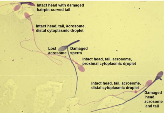
The current trend is to record the numbers of specific morphologic defects, such as knobbed acrosomes, proximal cytoplasmic droplets, swollen midpieces and coiled tails. This method of classification is considered superior to the traditional system because it reveals more specific information regarding a population of sperm, while avoiding false assumptions about the origin of these defects (Varner 2008). Any clustering method is used, normalities and abnormalities should always be consistently identified and categorized (Juhász and Nagy 2003). Simple, practical method which can also be used in routine examination is the classification that defines main morphologic categories according to subdomains of the sperm then divides them further into subcategories (Fig. 2).
Figure 2. Normal and abnormal sperm morphologic features (Source: Varner 2008) a) Normal spermatozoa (A)
b) Abnormal head morphology (B) macrocephalic (B1)
microcephalic (B2)
nuclear vacuoles or crater defects (B3) tapered head (B4)
pyriform head (B5) hour-glass head (B6) degenerate head (B7)
c) Acrosomal defects /knobbed acrosome/ (C)
d) Proximal cytoplasmic droplets (D1), distal cytoplasmic droplets (D2, D3) e) Midpiece abnormalities (E)
segmental aplasia of the mitochondrial sheath (E1)
roughed midpiece from uneven distribution of mitochondria /corkscrew defect/ (E2) enlarged mitochondrial sheath (E3)
bent midpiece (E4, E5, E6) double midpiece/double head (E7) f) Bent tail or hairpin tail (F)
bent principal piece (F1–F4)
single bend involving the midpiece-principal piece junction /DMR/ (F5) double bend involving the midpiece-principal piece junction (F6) g) Coiled tail (G)
h) Fragmented sperm /detached heads or tailless heads/ (H) i) Premature germ cells, round spermatids (I)
In the situation where a spermatozoon has more than one defect, the Society for Theriogenology guidelines suggests the most proximal defect is identified on each spermatozoa (Kenney et al. 1990). In this morphology evaluation system defects are prioritized based on the assumption that certain defects are more important or more deleterious to fertility than others (Brito 2007). The differential spermiogram system is similar to what occurs when we analyze the white cells in a leukogram. All of the abnormalites are counted regardless of one spermatozoon shows two or three defects.
Therefore, if a spermatozoon has a macrocephalic head and a midpiece defect, both are enumerated. The type of head defects and midpiece defects is recorded, too. To enumerate multiple defects simultaneously, a special cell counter is used. When abnormalities are counted on each spermatozoon, only one number is added to the total count when more than one defect are identified by pressing keys representing multiple categories of defects. The percentages of sperm defects when added to the percentage of normal sperm will not tally to 100% (Card 2005, Brito 2007). In the Primary and Secondary defect system, if a spermatozoon has three defects such as a knobbed acrosome, a swollen midpiece and a proximal droplet, one primary defect would be enumerated even though there is another primary (swollen midpiece) and a
secondary (proximal droplet) defect present. Using the Major–Minor system where a spermatozoon has a pyriform head, segmental aplasia of the mitochondrial sheath and a distal droplet, only one major head defect would be enumerated. If a system is used where the defects are prioritized so that only one is chosen per spermatozoon, the examiner will achieve a percentage distribution that will total to 100%. They do not have a record of the real distribution of all the spermatozoal defects in the ejaculate. It is not possible to track changes in individual defects. Primary–Secondary, Major–
Minor or differential spermiogram systems will identify the percentage of normal spermatozoa in the sample, which is a constant in all systems. Enumerating all defects for each spermatozoon allows the examiner to determine the potential of the defect to interfere with fertility, and analyse the changes in the patterns or cellular associations of the defects over time. Different defects found together represent more severe disturbances in spermatogenesis and could influence the prognosis (Veeramachaneni et al. 2006). The present Society for Theriogenology forms for stallion breeding soundness evaluation have the following categories listed in the differential spermiogram: normal sperm, abnormal acrosomal regions/heads, detached head, proximal droplets, distal droplets, abnormal midpieces, and bent/coiled tails. The presence of other cells (round germ cells, WBC, RBC, etc.) should also be indicated (Card 2005).
2.5.2 Preparation for morphologic evaluation
The standard evaluation of sperm morphology is performed with phase and/or light microscopy (Kenney et al. 1983) and computer-assisted methods have also been used (Ball and Mohammed 1995, Casey et al. 1997). However, available computer-assisted methods can only evaluate the sperm head, not count morphological abnormalities of mid-pieces, tails and acrosomes (Kavak et al. 2004). The morphology or structure of spermatozoa is typically examined with a light microscope (LM) at 1000 x magnification. Standard bright-field microscope optics can be used to examine air- dried semen smears prepared by specific stains. Giemsa, hematoxylin/eosin, eosin/nigrosin, Papanicolaou are the most widely used stains. Visualization of the structural detail of spermatozoa can be greatly enhanced by fixing the cells in buffered formol saline or a similar fixative, then viewing the unstained cells as a wet-mount with either phasecontrast or preferably, differential-interference contrast (DIC) microscopy (Kenney et al. 1983, Varner 2008). More detailed analysis can be done using transmission and scanning electron microscopy (Dott 1975, Dowsett et al. 1984, Barth and Oko, 1989).
Brito et al. (2011) demonstrated that sperm morphology evaluation results varied, depending on the evaluation method and clinician by analysing a diverse population
represented a wide range of fertility, from normal and fertile to severely sub-fertile stallions. There were significant differences among methods for all sperm morphology categories. Wet-mount preparation with phase-contrast microscopy appeared to be more sensitive for identification of abnormal stallion sperm when compared to eosin/nigrosin- and Papanicolaou-stained smears. The use of wetmount preparations seemed to reduce the introduction of some artifacts (detached sperm heads), but increased others (bent/coiled midpieces). The use of wet-mount preparations and phase-contrast facilitated the observation of acrosome defects, nuclear vacuoles, and cytoplasmic droplets, as demonstrated by the increased proportions of these defects when compared to stained smears. However one potential disadvantage of wet preparations is the observation of improperly oriented sperm, i.e., sperm that do not lie flat on the slide and therefore cannot be properly classified. A common concern with eosin/nigrosin is the hypotonicity of the stain and the possibility of introduction of artifactual tail defects, e.g., bending and coiling. Although the use of Papanicolaou- stained smears is the method recommended by the WHO (2010) for evaluation of human sperm, the results obtained with stallion sperm were extremely poor.
Abnormalities of sperm head and size were readily identified, but more subtle defects, including acrosome defects and nuclear vacuoles were difficult to observe with this stain. Moreover, staining of the tail was generally light and contributed to the difficulty of observing rough/swollen midpieces, principal piece defects, and cytoplasmic droplets.
Although reference materials with detailed descriptions and comprehensive documentation of sperm morphology are available for some species (WHO 2010, Barth and Oko 1989), similar materials have not been available for stallions however recent publication of Brito (2007) has attempted to document stallion sperm morphology in samples stained with eosin-nigrosin (Brito et al. 2011).
Percentages for the most frequently used morphologic categories of spermatozoa of stallions with normal fertility according to different publications are presented in Table 2.
Table 2. Means of spermatozoal morpologic characteristics
Morphologic categories
Mean ± SD (%)
Number of Stallions
and (samples)
Breed Reference
Normal morphology
66 ± 15*
61 ± 13 67.8 59.8 ± 9.4
57.5 ± 4 74.4 ± 4 49.7 ± 1.3
48.1 ±2.8 72.7 ± 1.5
398 5 (10) 168 (531)
8 (245) 6 (12) 7 (14) 5 (30) 4 (24) 8 (23)
Dutch warmblood■ different breeds 9 different breeds
different breeds Tori breed Estonian breed Thai native crossbred
Warmblood Swedish Warmblood
Parlevliet et al. 1994 Ball and Mohammed 1995
Dowsett and Knott 1996 Juhász and Nagy 2003
Kavak et al. 2004 ► Kavak et al. 2004 Phetudomsinsuk et al. 2008 Phetudomsinsuk et al. 2008
Einarsson et al. 2009
Head defect
2.8 ± 2.4 5.4 ± 2 3.9 ± 2.1 #
3 – 5 12.6 ± 1.7 13.9 ± 1.5 10.2 ± 0.5 13.4 ± 1.1 10.2 ± 0.7
398 168 (531)
16 (299) 8 (245)
6 (12) 7 (14) 5 (30) 4 (24) 8 (23)
Dutch warmblood■ 9 different breeds Thoroughbred different breeds
Tori breed Estonian breed Thai native crossbred
Warmblood Swedish Warmblood
Parlevliet et al. 1994 Dowsett and Knott 1996
Koyago et al. 2008 Juhász and Nagy 2003
Kavak et al. 2004 Kavak et al. 2004 Phetudomsinsuk et al. 2008 Phetudomsinsuk et al. 2008
Einarsson et al. 2009
Abnormal midpiece
5.1 ± 4.1 0.35 ± 2.3 4.5 – 6 5.1 ± 1.2 2.4 ± 1.1 16.5 ± 0.8 23.9 ± 2.1 5 ± 0.9
398 168 (531)
8 (245) 6 (12) 7 (14) 5 (30) 4 (24) 8 (23)
Dutch warmblood■ 9 different breeds 8 different breeds
Tori breed Estonian breed Thai native crossbred
Warmblood Swedish Warmblood
Parlevliet et al. 1994 Dowsett and Knott 1996
Juhász and Nagy 2003 Kavak et al. 2004 Kavak et al. 2004 Phetudomsinsuk et al. 2008 Phetudomsinsuk et al. 2008
Einarsson et al. 2009
Tail defect
2.1 ± 2.1 7.5 ± 2.2 11.5 ± 5.9 #
10 – 15 6.6 ± 1.4 6.4 ± 1.3 1.6 ±0.1 3.3 ± 0.6 3.7 ± 0.7
398 168 (531)
16 (299) 8 (245)
6 (12) 7 (14) 5 (30) 4 (24) 8 (23)
Dutch warmblood■ 9 different breeds Thoroughbred different breeds
Tori breed Estonian breed Thai native crossbred
Warmblood Swedish Warmblood
Parlevliet et al. 1994 Dowsett and Knott 1996
Koyago et al. 2008 Juhász and Nagy 2003
Kavak et al. 2004 Kavak et al. 2004 Phetudomsinsuk et al. 2008 Phetudomsinsuk et al. 2008
Einarsson et al. 2009
Detached head
1.5 ± 3 2.3 ±0.9 2.3 ±0.9 1.6 ± 0.3
168 (531) 6 (12) 7 (14) 8 (23)
9 different breeds Tori breed Estonian breed Swedish Warmblood
Dowsett and Knott 1996 Kavak et al. 2004 Kavak et al. 2004 Einarsson et al. 2009 Proximal
droplets
5.8 ±4.6*
6.7 ± 2.6
398 168 (531)
Dutch warmblood■ 9 different breeds
Parlevliet et al. 1994 Dowsett and Knott 1996
2.4 ± 2.6**# 2.5 – 4 17.3 ± 2.7
2.9 ±2.5 11.4 ± 1 5.5 ± 0.7 6.4 ± 1
16 (299) 8 (245)
6 (12) 7 (14) 5 (30) 4 (24) 8 (23)
Thoroughbred different breeds
Tori breed Estonian breed Thai native crossbred
Warmblood Swedish Warmblood
Koyago et al. 2008 Juhász and Nagy 2003
Kavak et al. 2004 Kavak et al. 2004 Phetudomsinsuk et al. 2008 Phetudomsinsuk et al. 2008
Einarsson et al. 2009
Distal droplets
4.3 ± 4.3*
4.8 ± 2.7 2 – 5 10.5 ± 0.8
5.8 ± 0.9 3.6 ± 1
398 168 (531)
8 (245) 5 (30) 4 (24) 8 (23)
Dutch warmblood■ 9 different breeds different breeds Thai native crossbred
Warmblood Swedish Warmblood
Parlevliet et al., 1994 Dowsett and Knott, 1996
Juhász and Nagy 2003 Phetudomsinsuk et al. 2008 Phetudomsinsuk et al. 2008
Einarsson et al. 2009
* percentage of live (unstained) spermatozoa (anilin blue-eosin staining)
■ Three-year-old young stallions
** The rate of spermatozoa with cytoplasmic droplets (proximal + distal droplets)
# Measurements from dismount semen (hematoxylin – eosin staining)
► Morphological evaluation was performed in wet preparations made from the formol-saline fixed samples
(Hancock 1957) and carbolfuchsin-eosin staining for assesment of head defect
2.5.3 Abnormal morphology of spermatozoa
The pathogenesis and effects on fertility of specific sperm defects have been more extensively studied in bulls than in stallions although several observations related to morphology of sperm from fertile and subfertile stallions have been published. The incidence of sperm head defects is relatively high and these are usually either the most or second most prevalent defects in the stallion ejaculate (Dowsett and Knott 1996, Dowsett et al. 1984, Love et al. 2000, Jasko et al. 1990). The pyriform and tapered head defect is the most common sperm nuclear abnormality. Small numbers of these defects are found in the semen of most bulls, even in bulls of good fertility. Pyriform head defects usually occur as a result of abnormal testicular functions through disturbances in intratesticular heat regulation or endocrine balance (Barth and Oko 1989). The swim-up technique, which is used to separate highly motile spermatozoa from the rest of the sperm population, did not significantly decrease the proportion of pyriform spermatozoa in the insemination droplets (Kawarsky et al. 1995). This finding is in agreement with previous reports that spermatozoa with pyriform heads have motility similar to that of spermatozoa with normal heads (Barth and Oko 1989).
These cells have generally normal intact acrosomes. In a study of Kawarsky et al (1995) visual assessment of the oocyte-spermatozoon interaction revealed pyriform spermatozoa binding to the zona pellucida (ZP), penetrating the ZP, and entering the perivitelline space. The rate of sperm penetration and the rates of cleavage and