THESES OF PhD DISSERTATION
KAPOSVÁR UNIVERSITY
FACULTY OF ANIMAL SCIENCE
Department of Breeding and Production of Ruminants and Horse
Head of Doctoral School:
Dr. PÉTER HORN
Member of the Hungarian Academy of Sciences Supervisor:
DR. JÓZSEF STEFLER Candidate of Agricultural Sciences
Co-Supervisor:
DR. ANDRÁS KOVÁCS
Doctor of the Hungarian Academy of Sciences
DEVELOPMENT OF QUALIFICATION OF FRESH AND FROZEN STALLION SEMEN, INVESTIGATION OF FACTORS
AFFECTING SPERM QUALITY USING A NEW EVALUATION METHOD
Author:
DR. GABRIELLA KÚTVÖLGYI
KAPOSVÁR 2012
1. Introduction
The horse has been in close contact with humans for thousands of years. In the last decades the use of artificial insemination (AI) in equine reproduction has been increasing worldwide in the horse industry, offering many advantages over natural service. Recently the total AI with cryopreserved or cooled equine semen is 0.5 million AI/year all over the countries. From the total number in 0.1 million AI/year has been used frozen semen. Around 350.000 foals are born each year after successful artificial insemination (Central European Management Intelligence /CEMI/, frozen + fresh data from 2006). Frozen semen offers breeders additional benefits not available with cooled semen. However stallion semen cryopreservation, despite its impact on the horse industry, is not an established technology. During the last years, a number of modifications have been proposed to the freezing process, however a large population of stallions still have poor semen quality and fertility after frozen-thawed. Only 30–
40% of stallions produce semen that is constantly suitable for cryopreservation with acceptable pregnancy results after AI, and a consistent variation on sperm freezability has been also observed among breeds. More than 50% of all spermatozoa are damaged by freezing process. Stallions show different susceptibilities to stress of dilution, freezing and thawing of their sperm, independently from the initial quality. Changes in plasma membrane structure and integrity appear to be an important component associated with reduced fertility of frozen–thawed spermatozoa.
Many valuable stallions produce poor quality semen, including bad sperm freezability, low number of sperm and low percentage of viable spermatozoa. Availability of semen of some very valuable stallions is reduced, because they are dead or it is not possible to collect semen from them anymore. In these cases sperm can be used for in vitro embryo production by intracytoplasmic sperm injection (ICSI). In vitro reproduction methods require the isolation of motile, morphologically normal, mature, viable, functionally intact spermatozoa. Standard sperm separation methods are not always effective with low numbers of total and viable sperm. Commonly there are two different main approaches to increase the effectiveness of sperm separation. One is modifying and developing separation methods and the other is adding chemical stimulators to the media to improve functional capacity of spermatozoa for successful fertilization. Hyaluronic acid (HA) was used successfully in combination with swim- up for separating bull spermatozoa. In human medicine, Pentoxifyllin (PX) is widely used to initiate motility in the case of immotile testicular or epididymal spermatozoa or for astheno-zoospermic men for ICSI. PX treatment after thawing also increased motility and progressive motility of equine spermatozoa.
A variety of techniques and protocols are available for evaluation of the spermatozoon. Over the past decades, a number of laboratory tests have been developed to determine properties of sperm function. These include quantitative sperm motion parameters, capacitation, basal and induced acrosome reactions, nuclear and mitochondrial sperm DNA but few have been adopted into routine clinical use.
Conventional semen evaluation is very subjective mainly based on sperm concentration and movement of the spermatozoa. In spite of its limited applicability, motility and progressive motility are the most commonly used parameters in the evaluation of stallion semen, in both laboratories and stud farms, because it is easily accessible and quick to perform. Evaluation of only a single variable, did not adequately explain differences among stallions’ fertility in most experiments.
However, combining results of assays that measured multiple sperm attributes improved the ability to evaluate fertilizing potential of equine spermatozoa. Besides routine examination to combine several tests or use combined analyses of more features are needed for quality and fertility evaluation of equine sperm. Using multi- parametric semen analysis methods subfertile and infertile stallions would be identified and reason for decreased pregnancy results may be revealed. Comparing freezing or sperm separation protocols can be also assessed by complex evaluation methods.
The use of fluorescent dyes and flow cytometry has provided the researcher and clinician with powerful tools to evaluate several sperm attributes. These procedures have been utilized to evaluate sperm viability, acrosome status, mitochondrial status, DNA integrity and stages of capacitation. There are developed multi-fluorescence- staining techniques used for flow cytometric evaluation, however flow cytometry analyses or fluorescence microscopy is generally not accessible to practitioners in the field. There is a difficulty and disadvantage and a relevant shortcoming of these techniques: In most cases they claim sperm washing or separation procedures and further incubations with the stains which can alter the original sperm quality parameters especially in equine semen in which spermatozoa are very sensitive to time-consuming processes. The shortcoming of the methods is that sperm morphology could not be assessed with them. Unfortunately there is no reliable computer aided automatised method which is able to perform complete morphology evaluation neither of all spermatozoa nor separately morphology of membrane-intact, viable sperm.
The maintenance of normal function of the plasma membrane is a crucial prerequisite for sperm viability as well as for reactivity at the site of fertilisation. A trypan blue- neutral red-Giemsa staining method for simultaneous evaluation of acrosome
integrity, sperm membrane, and overall morphology has been described for bull, boar and rabbit spermatozoa by Kovács and Foote (1992) and it was reported later that stain-permeable ("dead") sperm tails also could be distinguished (Nagy et al. 1999).
Since its introduction, this technique has been applied successfully to many other mammals. Simultaneous evaluation of the viability and acrosome integrity of sperm permits differentiation of true acrosome reaction from degenerative acrosome loss after cell death. After freezing and thawing of stallion semen, the number of spermatozoa with intact, unstained head membranes, but damaged, stained tail membranes, is increased significantly. These cells are considered immotile (Nagy et al. 1999). Therefore, unambiguous differentiation of the intact/damaged sperm tail membrane is very important for evaluating semen quality. The staining method showed acceptable repeatability and good agreement with flow cytometric measurements using fluorescein isothiocyanate-conjugated peanut agglutinin/propidium iodide (FITC-PNA/PI) staining of bull spermatozoa (Nagy et al.
2003). The staining method in combination of motility analysis or HOST was considered to be predictive test could be used for the prognosis of the potential fertility of semen correlation with fertility results (Domes 2003, Tartaglione and Ritta 2004). Some special characteristics and problems have been observed in stallion semen staining. One problem with the method was the length of the procedure (overnight Giemsa staining). Another problem was the differentiation of intact vs.
damaged sperm tails mainly in the case of frozen/thawed samples.
Many other factors such as management of the stallion (nutrition, housing, semen collection, semen processing and storage) and his mares (optimal time of insemination, reproductive status and conditions of the mares) may have a large impact on the success of insemination and overall fertility of the stallion. Most published fertility studies reporting no significant difference due to treatments are suspect, because too few males and/or females were used. Precision of commercial fertility data is low and conduct of a carefully planned fertility trial is very expensive in horses, even if biologically and ethically possible. Due to these observations finding accurate and reliable correlation between sperm characteristics and the future fertility of the sperm, rather in the case of frozen/thawed stallion semen is very difficult. Horse breeding unfortunately has declined in the past decade in Hungary which is shown from the breeding, insemination and foaling data in the country. During the experimental period there was no possibility to establish in vivo study and involve adequate number of mares (at least 100 mares/treatment) to an experiment. However I tried to use the complex staining method to verify the relationship between sperm quality of subfertile stallions and their reduced fertility in the indirect way.
Breeding stallions are selected primarily based on their pedigree, athletic performance, or other phenotypic characteristics. Fertility or fertility potential are usually secondary considerations. This explains that there is a wide variation in semen characteristics among individuals and in remarkable rate the semen quality is not sufficient.
Infertility or subfertility is the most common reproductive complaint of horse owners.
Diagnosis of the reason of infertility causes a confusing problem for veterinarians.
Subfertile stallions may participate in breeding if they have extraordinary genetic value, outstanding sport results, or in a small population of rare, native breeds (e.g.
Gidrán, Hucul) for the purpose of gene conservation. In these special cases thorough examination of the horse and his semen and the use of complex sperm evaluation are needed to define the further breeding management. Using subfertile frozen semen, appropriate in vitro sperm separation methods then deep intrauterine insemination or in vitro fertilization (ICSI) may be another chance to result in pregnancy.
2. Objectives of the dissertation
The objectives of the dissertation were: (1) to improve, validate and adapt the complex staining technique which evaluates sperm head and tail membrane integrity, acrosomal status and morphology for stallion spermatozoa, (2) to use this valuable method to evaluate sperm quality during and after two prominent sperm manipulation procedures (cryopreservation and sperm separation) and (3) to apply the technique to define detectable anomalies of semen from stallions with reduced fertility;
consequently draw an attention to the complexity and wide range of use of the staining technique in laboratory experiments as well as in quality control or determination of possible fertility potential of the fresh or processed equine sperm.
3. Materials and Methods
- The studies of Experiment 1, 2, 4 were performed between 2001-2008 in Hungary in co-operation with some Equine Artificial Insemination Stations in the country and in the spermatologic laboratory of Research Institute of Animal Breeding and Nutrition, Herceghalom. The Experiment 3 was investigated in 2004 at the Colorado State University, Department of Biomedical Sciences, Animal Reproduction and Biotechnology Laboratory, Fort Collins, Co. USA.
3.1 Semen samples
- Altogether thirty semen samples: fresh, diluted, centrifuged or frozen and thawed from 10 stallions were used for repeatability and methodology comparisons in Experiment 1. Smears were made from each of the 30 samples to compare the live/dead ratio for smears stained by 0.16% Chicago sky blue (CSB) and 0.27% trypan blue (TB) for evaluating the toxicity of CSB. Twenty semen samples from 15 stallions were used for densitometry.
- In the second experiment 10 fertile stallions were involved. All of the stallions were used as breeding stallion for artificial insemination. Three-four ejaculates were frozen from the 10 stallions (n=33), the collection dates performed randomly throughout between the years 2001-2004. Semen of Stallion 9 was used for more additional freezing (altogether 17 collecting days /Sept. 2003 – Jan. 2004/). Viability, acrosome status and morphology of the spermatozoa were evaluated after the technological steps of cryopreservation: fresh, centrifuged, and frozen-thawed semen was analysed.
- In the study of Experiment 3, poor to medium quality frozen semen (cryopreserved in 0.5-ml straws in 200 x 106/ml sperm concentration, using EZ-Freezin-LE extender, progressive motility after thawing was ≤ 30%) from 3 stallions was used, 3 replicates each. Two straws were thawed at 38 °C for 30 minutes and mixed. From this sperm suspension one hundred µl semen was allocated to each of 7 separation treatments.
- In the study of Experiment 4, semen samples of 14 fertile and 10 subfertile stallions were analysed. Stained smears were prepared from fresh ejaculates of 10 fertile and 10 subfertile stallions and from extended/chilled semen of 5 fertile and 4 subfertile stallions after 1 day storage at 4°C. To evaluate the semen samples, morphologic analysis alone and also viability and acrosome integrity examination combined with morphology were performed. Ages and breeds of the stallions were different. All of the stallions were used as a breeding stallion at different Breeding Stations in Hungary.
Stallions were categorized as “fertile” or “subfertile” by veterinarians of Breeding Stations based on the rates of pregnant or non-pregnant mares inseminated with sperm of the given stallion during the breeding season.
3.2 Freezing procedure
In Experiment 2 the freezing process followed the advised protocol of Vidament et al.
(2000) using modified INRA 82 extenders. After collection, the gel-free fraction of the ejaculate was diluted in centrifugation extender (E1): (INRA82 + centrifuged egg
yolk, 2%, v/v) in v:v 1:2 or 1:3 rates at 37 °C. After cooling to room temperature diluted semen was centrifuged for 10 min at 600xg in a 50 ml conical centrifugation tube. After centrifugation sperm pellets were resuspended in INRA 82 extender containing 2% egg yolk and 2,5 % glycerol (E2) to obtain 100×106 spermatozoa/ml at room temperature (22 °C). Semen was equilibrated for 60 min from 22°C to 4°C with -0.3°C/min cooling rate, then an additional 60 min at 4°C before freezing. Semen was packaged in 0.5 ml straws and freezing was performed by keeping 0.5 ml straws at 4 cm above liquid nitrogen for 10 min then plunging the straws in liquid nitrogen.
Thawing of 0.5 straws was done at 37 °C for 30 sec using a waterbath.
3.3 Sperm separation in Experiment 3
Mini-Percoll: Three aliquots (100 µl ) were incubated at 38°C in an atmosphere of 5% CO2 for 20 min in 0.25 ml Hepes-buffered chemically defined handling medium (HCDM) (P-NT: non-treated; P-PX: 3.5 mM PX (P 1784 Sigma, St Louis, MO); or P- HA: 1 mg/ml HA final concentration) before Percoll®-centrifugation and one aliquot was centrifuged through Percoll® without incubation (P-CON).
Our mini-Percoll discontinuous density gradients were prepared by carefully layering 0.4 ml of 90%, and 0.5 ml of 45% Percoll® solutions in a 1.5-ml microcentrifuge tube starting with the highest-density solution at the bottom. Incubated or not-incubated (P- CON) sperm was layered on top of the Percoll® gradients and centrifuged at 600 x g for 5 min. Then a 30-µl pellet was aspirated from the bottom and washed in 1 ml HCDM at 300 x g for 5 min.
Swim-up: At the same time 3 aliquots of 100 µl sperm were placed in 1 ml HCDM - without or with supplementation - for swim-up (SU-NT: non-treated; SU-PX: 3.5 mM PX; or SU-HA: 1 mg/ml HA final concentration) at 38°C in an atmosphere of 5% CO2
for 30 min in a 5 ml round-bottomed tube. After incubation 0.65 ml supernatant was collected and centrifuged in 1 ml HCDM at 300 x g for 5 min.
In every treatment, the final 30-µl pellet after washing aspirated from the bottom was analysed. For evaluating sperm concentrations 5 µl samples were taken from each final 30-µl sperm suspension then diluted in 95 µl distilled water. Concentration was determined using a hemacytometer. Recovery rate was calculated as a percentage of original concentration (200 million/ml) of the frozen semen. 10 µl of the sperm pellet was used making smear for Sperm head, tail and acrosome membrane integrity and morphology evaluation with CSB-Giemsa staining.
3.4 Sperm evaluation method
Viability staining, fixing and acrosome staining
Stallion spermatozoa are sensitive to pH, osmolality and temperature changes.
Therefore, phosphate-buffered saline (PBS) containing 0.06% K2HPO4 and 0.825%
NaCl was used for semen dilution at the same temperature as the sperm specimen was.
Fresh sperm and cooled semen - diluted in 1:1 or 1:2 rate with NFDSM-Glucose or egg-yolk-skim-milk-based (EY-SM) extender- , was diluted 1:4 with PBS before viability staining. Both the centrifuged and frozen/thawed semen processed at a final concentration of 100-200 million cells/ml in freezing extender containing egg-yolk and glycerol, diluted 1:9 with PBS just before making smear.
The viability test stain contained 0.16% Chicago sky blue 6B (Sigma-Aldrich St.
Louis, MO, USA, C-8679). The working solution was made from a 2.6% stock solution in distilled water diluted 1:15 with PBS. The 0.27% trypan blue working solution was prepared from 0.4% stock solution (Sigma T-8154) diluted 2:1 with PBS.
Both staining solutions are isotonic, have neutral pH, and are stable for a year in eye- drop bottles at room temperature. The fixative was composed of 86 ml 1 N HCl plus 14 ml of 37% formaldehyde solution and 0.2 g neutral red (Sigma N2880); it is stable for a year at room temperature and may be used repeatedly. The acrosome stain was 7.5% Giemsa stock solution (Sigma GS-500) in distilled water prepared freshly before use.
Staining procedure
Equal drops (20 µl) of viability stain and diluted semen were mixed gently on a slide flatly with the corner of another slide without scratching and touching the glass surface. Liquid layer is formed between the two slides and the droplets get mixed up with a slightly movement of the slides. The slides were attached parallel to each other and pulled to make two smears. The smears were air dried nearly vertically at room temperature. After drying, slides were fixed for 4 min. Both sides of the slides were rinsed with tap water and distilled water, then stained in Giemsa solution in an uncovered staining jar (not more than 14 slides per jar with 16 spaces) at 25-40° C for 2-4 h. Slides were rinsed with tap water, then differentiated in distilled water for 2 min, air dried in a nearly vertical position, and cover slipped with Entellan (Merck 1.07960, Darmstadt, Germany). Slides were evaluated at 1000 X magnification using oil-immersion objective and a yellow filter for better live/dead differentiation.
Viability evaluation
- Three hundred cells were counted on each slide and classified into five categories in Experiment 1 and Experiment 3: intact head, intact tail and acrosome membrane (Intact); intact head and tail, damaged acrosome (IHITDA); intact head, damaged tail (IHDT); damaged head, intact tail (DHIT); and damaged head-, tail-, acrosome membrane (DHDTDA).
- Eight sperm categories were classified based on membrane integrity combined morphology for the light microscopy examination in Experiment 2 and Experiment 4: intact head, intact tail and acrosome membrane, normal morphology (IHITIA);
intact head, tail and acrosome, proximal cytoplasmic droplet (IPD); intact head, tail and acrosome, distal cytolasmic droplet (IDD); intact head, tail and acrosome; bent, curved, broken midpiece or tail (IBT); intact head, tail, damaged acrosome (IHITDA);
intact head, damaged tail (IHDT); damaged head, intact tail (DHIT); and damaged head, damaged tail, damaged acrosome (DHDTDA). Two-three hundred cells were evaluated in each sample.
- In the statistical analysis of Experiment 2 additional and combined categories were also evaluated:
1. Damaged spermatozoa with CD [DCD]
2. Damaged) sperm with bent, curved, broken midpiece or tail [DBT]
3. All cells with intact membranes [IHITA + IPD + IDD + IBT] Intact
4. Intact sperm with CD + Intact sperm with bent tail [IPD + IDD + IBT] ICDBT 5. All (intact and damaged) spermatozoa with CD [IPD + IDD + DCD] IDCD
6. All (intact and damaged) sperm with bent, curved, broken midpiece or tail [IBT + DBT] IDBT
7. All (intact and damaged) spermatozoa with CD or BT [IDCD + IDBT] IDCDBT
Morphological evaluation
- Three hundred cells were classified in 5 simple morphological categories in Experiment 3:
1. normal
2. proximal cytoplasmic droplets 3. distal cytoplasmic droplets
4. midpiece and tail defect (midp+tail) 5. abnormal head (head)
- Cells were classified in 9 morphologic categories in Experiment 4:
1. Normal morphology (normal)
2. Head abnormalities (head) (microcephal, macrocephal, tapered, pyriform, nuclear vacuoles, acrosome defects)
3. Midpiece defect (midp) (swollen, bent, DMR, mitochondrial sheath defect, corkscrew, bowed)
4. Tail abnormalities (tail) (broken, bent, hairpin-curved, distal coiled tail) 5. Coiled tail defect (coiled) (tightly coiled tail, dag-like defect)
6. Detached head (detached)
7. Proximal cytoplasmic droplet (PD) 8. Distal cytoplasmic droplet (DD)
9. Multiple forms (multiple) (e.g. double midpiece, head, tail)
Scanning and transmission electron-microscopic investigations were carried out as well from sperm of Stallion 9 in Experiment 2.
3.5 Data analyses, statistical methods
In Experiment 1 the Bland-Altman statistical method (Bland and Altman 1986, Nagy et al. 2003) was used to assess repeatability and agreement between the CSB and TB stains. The analysis was calculated with the “intact” and the categories of the IHDT and DHDTDA merged, counting all damaged tails (DT).
Figure 1. A Spermatozoon with intact head and damaged tail membrane.
CSB/Giemsa staining. The stained tail is indicated with dotted lines.
The histogram shows the RGB values of the selected area. Bar = 2 µm.
For densitometry analysis, individualdigital images were taken from smears stained by CSB/Giemsa and by TB/Giemsa. The Magic Wand Mask tool of the Corel Photo- Paint 8 software was used to select the tail or head areas. For analysis of the individual digital images, red-green-blue (RGB) histograms were drawn for each different area of spermatozoa (Fig. 1). The histogram plotted the brightness value of every pixel in the selected area of the image. Values ranged from zero to 255 (from darkest to brightest), and the histogram indicated how many pixels were at each brightness level.
Means of the composite RGB values of the selected area on each of the intact or damaged tails (midpiece and principal piece, at least 4000 pixels per selected area measurement) and the stained and unstained heads (without acrosome, at least 1000 pixels/measurement) were registered from each picture. Differences between means of RGB values of live vs. dead tails and separate live vs. dead heads from each photo were used to compare the two stains. Altogether, 120 photos were measured and 480 histograms of the total RGB value were made from the different areas. After evaluating the data for normality, the paired two-tailed T-test (SPSS 11.0. statistical analysis program, SPSS Inc. Chicago, IL) was used to compare the RGB differences between the stained and unstained tails or heads for CSB and TB staining.
In the Experiment 2 paired T test statistical analysis was performed to compare the mean values of the percentages of different sperm categories in fresh, centrifuged and frozen samples using „R” software.
In Experiment 3 data (recovery rates and percentages of different cell types in the selected sperm after the 7 treatments) were arcsin transformed to achieve normality on the data and evaluated by GLM analysis of variance of SAS (SAS Inst. Inc., Cary, NC, USA). Differences among means were tested using Tukey's honestly significant difference (h. s.d.) procedure. In all cases, significance was set at p < 0.05 level. Data are presented as Least squares means and standard errors (LS means ± SE).
In Experiment 4 the evaluation of 10 subfertile stallion samples and the discussion with incorporation of previous data and observations from the stallions were interpreted in case reports. Mean values calculated from the results of the viability and morphology evaluations of stallions with good fertility (Fertile stallions), the average values, minimum requirements and the acceptable limits of the different sperm morphologic categories in fertile stallions according to the literature and to the guidelines of Hungarian standard for breeding stallion semen were considered as bases of comparison.
4. Results
4.1 Experiment 1. Improvement of assessment of stallion sperm quality by Chicago sky blue and Giemsa viability and acrosome staining method
CSB/Giemsa staining showed good repeatability and agreement with standard TB/Giemsa measurements (Fig. 2).
-10 -9 -8 -7 -6 -5 -4 -3 -2 -1 0 1 2 3 4 5 6 7 8 9 10
0 10 20 30 40 50 60 70 80 90 100
Average of paired measurements (%) Differences of paired measurements (%)
d d + 2 corr SD = 6.19%
d - 2 corr SD = -6.13%
d = 0.03%
2 corr SD = 6.16%
Figure 2. Agreement between the TB/Giemsa and CSB/Giemsa staining methods for counting intact cells on smears. The differences between the paired measurements are plotted against their average. The mean of the differences (d) and the limits of agreement (d ± 2corrSD) are presented (n =30).
The 95% limits of agreement (d ± 2 corrSD) were -6.13 and 6.19% (Fig. 2). This interval was small and close to the d ± 2 SD of the repeated measurements of both methods (TB: -5.19, 5.75%; CSB: -6.44, 4.90%).
CSB resulted in similar live/dead sperm head differentiation, but a better tail differentiation than TB. This was verified by densitometry analysis. (Fig. 3). The 80%
greater differences in the brightness levels between the live and dead tails after CSB compared to TB staining allows easier differentiation.
The background after CSB staining is more uniform compared to TB. TB viability stain can be replaced by CSB for staining stallion sperm, thereby providing more reliable evaluation. Fixation for 4 min resulted in darker “dead” staining with acceptable background. Giemsa exposure for 2-4 h was sufficient for acrosome staining.
60 70 80 90 100 110 120 130 140 150 160 170 180 190
1 2 3 4 5 6 7 8 9 10 11 12 13 14 15 16 17 18 19 20
Individual samples (n =20)
RGB values intact tail CSB
damaged tail CSB intact tail TB damaged tail TB
Figure 3. Means of RGB values of stained (damaged) and unstained (intact) spermatozoon tails in the individual samples.
4.2 Experiment 2. Analysis of the injuries of stallion spermatozoa during the whole freezing procedure
During the cryopreservation procedure, neither all membrane-intact (Intact) nor IHITIA cells proportion was changed after centrifugation but both were decreased significantly in the frozen/thawed semen (78±9; 78±8; 38±11% and 58±16; 58±15; 26±9%, in fresh, centrifuged and frozen sperm, respectively). After freezing/thawing not only the proportion of DHDTDA sperm was higher but IHDT also increased considerably compared to fresh and centrifuged semen /19±7% vs. 4±3; 4±3; p<0.01/ (Fig. 4).
Damages and depletion of acrosome of viable cells were uncharacteristic after freezing/thawing since the proportion of IHITDA was less than 1 %. Fresh ejaculates and frozen/thawed semen samples of stallions show individual characteristics in point of viability and morphology. I found individual susceptibility to centrifugation and differences among stallions in the proportion of sperm types. Centrifugation may cause similar morphologic alterations (bent-looped, coiled tail of sperm) as induced by cold-, warm and hypoosmotic shock and this occurred intensively in some stallions. Increasing IBT cells were observed after centrifugation related to 3 stallions /Group II/ (19±4%, p<0,01), which had fresh semen also containing high percentage of this cell type /14±5%/ (Fig. 5).
58,5 6,5 5,7 7,50,2 3,9 0,8
17,0
58,0 6,2 4,7 9,4 4,00,60,1
17,2
25,6 3,4 2,1
6,7 0,7 19,1
4,1 38,5
0%
10%
20%
30%
40%
50%
60%
70%
80%
90%
100%
fresh centrifuged frozen
DHDTDA DHIT IHDT IHITDA IBT IDD IPD IHITIA
a a
a a b
b b
b a a
a a
Figure 4. Distribution of different sperm categories during the freezing procedure (10 stallions, 33 freezing). a, b means in the same cell category differ, p < 0.01
66,7 3,9
44,1 13,7
65,9 3,9
44,1 18,9
27,1 3,0
23,0 13,2
0%
10%
20%
30%
40%
50%
60%
70%
80%
90%
100%
%
Group I
Group II
Group I
Group II
Group I
Group II
fresh centrifuged frozen
DHDTDA DHIT IHDT IHITDA IBT IDD IPD IHITIA
Figure 5. Distribution of different sperm categories during the freezing procedure in the two groups of stallions. (mean values of replicates, in Group I: 7 stallions, n = 21 and in Group II: 3 stallions, n = 12)
Proportion of IBT was also high in the frozen/thawed semen (13±5%) in these 3 stallions, besides that rate of IHITIA sperm decreased considerable from 44% to 23%.
All the sperm with CD-s (IDCD) were slightly decreased during the process (15±9;
13±8; 12±8 %) and sperm with midpiece- and tail defect (IDBT) were mildly increased after centrifugation considering all the examined stallions (10±7; 12±10; 12±10 %).
Proportion of IDCDBT didn’t change during the freezing procedure (25±15; 26±15;
24±15% fresh, centrifuged and frozen respectively). The relative ratios of IDBT, IDCD and IDCDBT during the process might be explained in some cases with the effect of centrifugation which results in curve of the midpiece containing CD which is very often entrapped in the bend.
4.3 Experiment 3. Use of pentoxifylline and hyaluronic acid for stallion sperm separation
Percoll method was successfully modified by reducing the volume of separating media (Mini-Percoll 0.4 ml 90% and 0.5 ml 45% Percoll in a 1.5 ml microcentrifuge tube), the time of centrifugation and use higher g-force (600 x g for 5 min) to increase the yield of viable sperm separation for ICSI when low volume and few numbers of equine sperm are available. The purpose of this study was to compare the effectiveness of mini-Percoll (P) and swim-up (SU) method for low numbers of sperm treated or non-treated with hyaluronic acid (HA) or pentoxifylline (PX). Mini-Percoll separation of sperm suspension without incubation with additional chemicals (P-CON) or after incubation with 3.5 mM pentoxifylline (P-PX) resulted in the most morphologically normal, intact sperm according to high proportion of normal cells (92 and 91%), the most intact sperm (54 and 57 %) and the best recovery rate (13 and 13 %, respectively). All Mini-Percoll separations resulted in more „normal”, and less sperm with droplets (proximal + distal droplets) and midpiece + tail defect compared to all swim-ups (91-92% vs.71-78%; 1%
vs. 4-7%; 6-7% vs.16-19% respectively, p<0.01).
After separations fairly high percentage of the spermatozoa had damaged head but intact tail (DHIT). This ratio was highest in P-CON, P-NT (Mini-Percoll separation after incubation of sperm suspension without additional chemicals) and SU-PX (3.5 mM pentoxifylline supplementation in the swim-up medium) treatments (14%, 13%
and 13%, respectively). P-PX resulted in less DHIT cells compared to P-CON (8 vs.
14 %) but it was not significant (P>0.05). Rate of membrane-intact sperm with damaged acrosome (IHITDA) was the highest after P-PX separation (17±1.6 %).
These spermatozoa are loosing or already have lost acrosomal material what can be advantage in ICSI fertilization of oocyte. PX seems to promote acrosome reaction,
maybe mainly on sperm with destabilized membranes. There were individual differences among stallions in the reactivity of acrosome-membranes. 1 mg/ml hyaluronic acid (HA) supplementation increased the recovery rate during swim-up, but not viability and proportion of normal cells in any of the treatments.
48 1,5 17 33
0%
10%
20%
30%
40%
50%
60%
70%
80%
90%
100%
thawed P-CON P-PX P-HA P-NT SU-NT SU-HA SU-PX
%
DHDTDA IHDT DHIT IHITDA Intact
a a
b b b
a a
a a
b b b
a,b in the same sperm category indicate significant differences between values (p<0.01)
Treatment
Figure 6. Distribution of viability sperm categories after different treatments
4.4 Experiment 4. Viability, acrosome integrity and morphology evaluation of sperm samples from subfertile stallions
At subfertile stallions in every case some alterations were detected and quality of the sperm in respect of membrane-integrity or morphology was lagged behind those in fertile stallions were observed. In several cases serious morphologic abnormalities or dramatic reduction in the proportion of intact, viable, morphological normal sperm with an increase rate of different membrane-damaged sperm categories were found in the ejaculate. In Stallion “H” remarkable differences were found in the results of viability evaluation between sperm samples diluted with egg-yolk-skim-milk-based (EY-SM) and non-fat dry skim milk (NFDSM)-Glucose extender. Spermatozoa showed poor sperm survival in NFDSM diluted-cooled semen. Every semen samples of subfertile stallions were unsatisfactory regarding to the strict guidelines of the Hungarian Standard for breeding stallion semen (7034/1999) which allows ≤ 30%
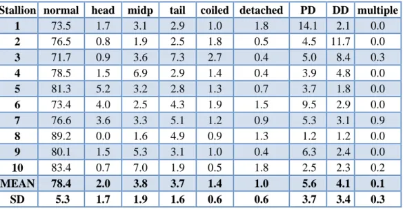
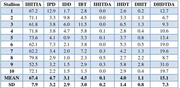
sperm with any morphologic aberrations, if less than half of these abnormal cells have primary defect. Results of morphology and viability evaluation of fresh semen of fertile and subfertile stallions are shown in Table 1-4.
Table 1. Percentages of sperm in different morphologic categories (fresh semen of fertile stallions semen)
Stallion normal head midp tail coiled detached PD DD multiple
1 73.5 1.7 3.1 2.9 1.0 1.8 14.1 2.1 0.0
2 76.5 0.8 1.9 2.5 1.8 0.5 4.5 11.7 0.0
3 71.7 0.9 3.6 7.3 2.7 0.4 5.0 8.4 0.3
4 78.5 1.5 6.9 2.9 1.4 0.4 3.9 4.8 0.0
5 81.3 5.2 3.2 2.8 1.3 0.7 3.7 1.8 0.0
6 73.4 4.0 2.5 4.3 1.9 1.5 9.5 2.9 0.0
7 76.6 3.6 3.3 5.1 1.2 0.9 5.3 3.1 0.9
8 89.2 0.0 1.6 4.9 0.9 1.3 1.2 1.2 0.0
9 80.1 1.5 5.3 3.1 1.0 0.4 6.3 2.4 0.0
10 83.4 0.7 7.0 1.9 0.5 1.8 2.5 2.3 0.2
MEAN 78.4 2.0 3.8 3.7 1.4 1.0 5.6 4.1 0.1 SD 5.3 1.7 1.9 1.6 0.6 0.6 3.7 3.4 0.3
Table 2. Percentages of sperm in different morphologic categories (fresh semen of subfertile stallions)
Stallion normal head midp tail coiled detached PD DD multiple
A 24.7 41.0 2.0 6.0 4.7 1.7 15.0 4.0 1.0
B 28.0 41.0 4.7 4.0 5.7 3.7 10.0 3.0 0.0
C * 32.5 16.0 9.5 1.0 2.5 4.0 31.0 3.5 0.0 C ** 30.0 19.3 11.3 2.3 5.5 4.0 24.3 3.3 0.0
D 31.4 9.5 1.9 0.9 1.2 6.9 47.0 1.1 0.2
E 40.5 6.7 11.3 13 2.5 1.3 22.7 2 0
F 52.5 21.6 9.4 3.8 3.8 1.3 6.6 1.3 0.0
G 50.0 1.0 13.2 3.6 2.9 2.1 14.7 11.5 1.0
I ■ 52.7 2.3 22.7 3.7 2.3 7.0 4.7 4.7 0.0
I ■ ■ 42.0 2.0 21.3 10.8 5.0 7.5 2.1 9.3 0.0
J 50.5 3.3 8.8 2.0 1.5 1.7 5.0 26.8 0.3
* Result of morphology evaluation of semen collected in June
** Result of morphology evaluation of semen collected in August
■ Result of morphology evaluation of semen collected in June
■ ■ Result of morphology evaluation of semen collected in July
Table 3. Percentages of sperm in different viability categories (fresh semen of fertile stallions)
Stallion IHITIA IPD IDD IBT IHITDA IHDT DHIT DHDTDA
1 67.2 12.9 1.7 2.8 0.0 2.6 0.2 12.7
2 71.1 3.3 9.8 4.5 0.0 3.3 1.3 6.7
3 61.8 3.8 6.0 11.5 0.0 6.5 1.3 9.3
4 71.8 3.8 4.7 5.8 0.1 2.8 0.4 10.6
5 73.6 4.1 0.9 3.3 0.1 3.7 0.8 13.4
6 62.1 7.3 2.1 3.8 0.0 5.3 0.5 19.0
7 62.2 3.4 2.0 7.2 0.3 4.2 1.3 19.6
8 79.8 2.9 1.0 2.3 0.5 2.7 2.2 8.7
9 52.5 3.2 1.5 2.9 0.3 5.8 2.8 31.0
10 72.1 2.2 1.5 1.3 0.0 2.9 0.4 19.7
MEAN 67.4 4.7 3.1 4.5 0.1 4.0 1.1 15.1
SD 7.9 3.2 2.9 3.0 0.2 1.4 0.8 7.3
Table 4. Percentages of sperm in different viability categories (fresh semen of subfertile stallions)
Stallion IHITIA IPD IDD IBT IHITDA IHDT DHIT DHDTDA
A 9.5 3.0 1.0 6.5 1.0 8.0 0.0 71.0
B 14.0 6.0 0.0 5.0 0.0 4.0 5.0 66.0
C * 12.6 14.7 1.8 4.9 0.0 12.3 5.8 48.0
C ** 10.0 10.7 3.3 3.3 0.0 2.0 0.7 70.0
D 12.0 41.5 0.5 1.0 1.5 11.0 9.0 23.5
E 1.5 4.5 0.5 5.0 1.5 28.0 0.0 59.0
F 19.0 3.0 0.7 5.0 0.7 9.0 6.0 56.7
G 36.3 10.0 8.0 10.0 0.3 12.7 0.9 21.8
H # 35.8 8.3 0.8 11.3 1.0 3.5 1.3 38.3
H # # 12.0 2.5 1.5 18.0 0.0 5.0 0.0 61.0
I ■ 37.7 2.3 4.3 13.3 0.0 2.7 4.0 35.7
I ■ ■ 30.0 0.0 11.0 17.5 0.5 5.0 2.5 33.5
J 47.0 3.7 21.3 11.3 0.2 1.1 2.2 13.5
* Result of viability evaluation of semen collected in June
** Result of viability evaluation of semen collected in August
# Fresh semen diluted with egg-yolk-skim-milk-based (EY-SM) extender
# # Fresh semen diluted with non-fat dry skim milk (NFDSM) extender
■ Result of viability evaluation of semen collected in June
■ ■ Result of viability evaluation of semen collected in July
5. Discussion, conclusions and recommendations
5.1 Using the TB/Giemsa method for staining stallion sperm, differentiation of intact or damaged sperm tails was problematic, mainly with frozen and thawed samples.
Stallion spermatozoa are small and the larger number of seminal plasma and extender proteins binding to TB make colour differentiation of live and dead tails less clear.
The aim of the Experiment 1 was to improve Kovács-Foote staining method using another viability stain and optimizing each steps of the staining procedure to distinguish more accurately the different cell types, especially in stallion sperm.
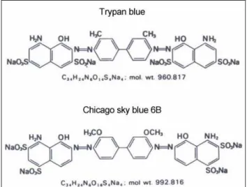
We searched for other supravital stains in the “acid disazo dye” group with the aim of finding a dye with more affinity to the proteins of the tail of membrane-permeable stallion spermatozoa. We selected Chicago sky blue 6B to compare to trypan blue. TB and CSB dyes have the capacity to bind directly, presumably by hydrogen binding, to different proteins including those with linear structures (Lillie 1977). CSB has a stronger affinity for the proteins of the sperm tail than does TB. This is probably due to the structure of the molecules, because CSB has two more groups that are capable of hydrogen binding than TB (Fig. 7).
Figure 7. Chemical structures of TB and CSB (Lillie 1977)
In our preliminary study, fixation for more than one day after viability staining caused pale discoloration of the head. Therefore, fixing smears as soon as possible after the viability staining, is advised. Fixation for 4 minutes resulted in darker dead staining with acceptable background. Acrosomes of equine spermatozoa are stained more rapidly than those of other domestic mammals. Giemsa staining below 20° C does not work; it is more effective at 25-40° C. Contact with air is also important, especially
with short dye exposure. Background caused by seminal plasma and extender proteins was greatly reduced on slides stained for only 2-4 h with Giemsa. In conclusion after staining with 0.16% CSB and 4 min fixation, 2-4 h Giemsa staining at 25-40° C is recommended for stallion semen. This improved method for equine spermatozoa can be used in routine practice and research.
For evaluating membrane integrity based on staining characteristics of the sperm cell subdomains, we generally classified the cells into five practical categories: intact head, tail and acrosome membrane; intact head, tail and damaged or lost acrosome;
intact head with damaged tail; damaged head with intact tail; damaged head, tail and acrosome. This viability evaluation also can be performed in combination with morphological assessment. Consequently, more informative classifications of sperm among the live, intact cells can be made. Intact sperm with no morphological abnormalities and those with different morphologic aberrations (categorizing the most common defects such as proximal cytoplasmic droplets; distal cytoplasmic droplets;
midpiece or tail defects) can be identified and together with the four cell types with damaged membranes in any part of the sperm, a useful complex classification system with 8 combined categories would be applied. Sperm based on morphology are also can be classified into five simple or nine more differentiated categories (details are found in the Materials and Methods).
5.2 The results of Experiment 2 clearly demonstrate that the most sensitive subdomain for the freezing/thawing stress is the flagellum. 57.6% of the spermatozoa had damaged tail membrane after cryopreservation. Although it seems the sperm head area and shape influence sperm freezability, the damage of the midpiece and tail membrane is also of great significance during the freezing process. The rate of IHITDA cell type after freezing/thawing was lower (1.8 % of the Intact cells) than in other studies detected by combined fluorescence staining methods. Differences can be explained by the discrepant staining methodologies because fluorescens stainings normally included 1-2 washing steps and a short period incubation while using TB/CSB-Giemsa staining the smears are made after a quick dilution of frozen/thawed sperm. Another reason may be the different classification of the cell types. Fresh ejaculates and frozen/thawed semen samples of stallions show individual characteristics in point of viability and morphology. I found individual susceptibility also to centrifugation.
Spermatozoa response to sublethal effects characteristically. Cold-, warm and hypoosmotic shock induce bent-looped, coiled tail of sperm due to changes in water- permeability of cell membrane. We found that centrifugation also may cause similar morphologic alterations and this occurred intensively in some stallions. Percentage of
the spermatozoa with normal morphology among viable cells is very important. The importance of midpiece and tail defect can be higher in the frozen semen, because it could show equal or higher proportion among the viable sperm than the cells with normal morphology. In this approach it is also important to examine the incidence of the abnormal sperm among the „live cells”. In the effected group of stallions the proportion of IBT among viable sperm with intact membranes was 30.3 % in the frozen/thawed semen while in the non-effected group this ration was 8.6%. These sperm are either selectively filtered throughout the female genital tract or unable to penetrate the zona pellucida at the fertilization place. In this aspect fertility of the sperm can be improved with higher number of spermatozoa in the insemination dose.
Regarding to the literature spermatozoa with retained cytoplasmic droplets are fairly common in the equine semen. Proximal droplets (PDs) found on ejaculated spermatozoa are generally considered indicative of a defect of testicular origin and have been implicated in the depressed fertility of bulls and boars. The effect of a retained DD on fertility is lesswell defined, although today, retained distal droplets are concerned to be more detrimental to fertility than previously suspected. According to the theories that sperm with CD-s are partly filtered out during the sperm transport in the female genital tract but one portion of these cells reach the oocyte however supposed to fail to bind zona pellucida, otherwise enzymes of the droplets affect on the normal spermatozoa without defect; CD-s are considered to be a semi-conpensable defect. High proportion of sperm with CD-s among intact spermatozoa may have a negative effect on the fertility of frozen semen.
The ratio of intact, viable spermatozoa is the most important parameter of the quality of frozen semen. However, for the further development of cryopreservation technologies or determination of freezability of individual stallion sperm and usability of frozen semen, it is also important to define accurately the localization of cell injury during the cryopreservation process for which each of the part of the sperm need to be assessed. Our staining method is well-applicable for subdomain-specific examination of spermatozoa. Conservation of semen of stallions representing valuable, promising or rare breeds and bloodlines for future use, or storage is important way to preserve genetic diversity. The complex staining method may assist in selecting the optimal sperm cryopreservation technique individually in the case of high priority stallions to improve the frozen sperm quality and its fertility potency. It would be a very useful innovation to work out computer aided automatised technique for evaluation the stained smears.
5.3. Standard sperm separation methods are not always effective with low numbers of total and viable sperm. In addition, stallion spermatozoa are very sensitive to protracted procedures. Numerous studies have previously been carried out to compare swim-up and Percoll® separation of spermatozoa with very varied results. I found Percoll separation superior compared to swim-up. P-CON and P-PX were the most effective separation procedures when beginning with low numbers of sperm. Twenty five to 35% of the sperm with intact midpiece and tail membrane - which are considered motile (Nagy et al. 1999) -, have damaged head or acrosome membranes after separations. This could affect the success and results of ICSI procedures, in which final selection of sperm is based on motility. The results point out a weakness of this method since there is a quite high proportion of the sperm having intact tail but damaged head and acrosome consequently these cells could be also functionally damaged. The reason for good sperm concentration (recovery rate) but poor viability (survival rate) and morphology of the SU-HA selected sperm may be attributed to the detrimental effect of the final washing procedure after SU. PX is beneficial if Percoll® separation is delayed, but there is a need to clarify its effect on acrosome exocytosis and the influence of absence of acrosome for further development of equine embryos produced by ICSI.
5.4 Our results of Experiment 4 have pointed out to the importance of defining of the ratio of membrane-intact and morphologically normal spermatozoa. Taking this into account in determining the sperm number in the insemination dose is recommended. It is also important to define the type of abnormalities of spermatozoa, because the decision of further sperm manipulation methods depends on these results. If the abnormality is compensable (e.g. microcephal head defect, DMR, bent, coiled tail), the sperm concentration is satisfactory and a 20-30% of normal, viable sperm is also present in the ejaculate, the increasing of insemination dose could be solution for the problem. In the case of semi-compensable or non-compensable defects, for example the presence of high proportion of cytoplasmic droplets which alter biological properties of normal cells and affect negatively on development processes after fertilization of oocyte, sperm separation may help to isolate normal viable spermatozoa from the defected sperm cells and from the affected seminal plasma. This portion after dilution with semen extender can be used more effectively for AI immediately after preparation or after cooled-transportation. Besides of standard parameters of routine sperm evaluations (volume, sperm concentration, total sperm number, motility and progressive motility) using the complex staining method for analysis of fresh ejaculate and of 24-hours-chilled-stored semen (longevity test), subfertile and infertile stallions would be identified and reason for decreased
pregnancy results may be revealed. For this intention the method would be installed into the annual control examination of the stallions’ semen. Subfertile stallions may participate in breeding if they have extraordinary genetic value, outstanding sport results, or in a small population of rare, native breeds for the purpose of gene conservation. In these special cases with thorough examination of the horse and his semen and to use of complex evaluation system, changes in semen quality can be monitored and the breeding management would be adjusted to these alterations.
Enhanced breeding management of subfertile stallions (e.g. changes in semen handling procedures: centrifugation, change of extender and dilution rate, recalculating of insemination dose, sperm separation; decrease in number of mares mated/inseminated; increased mare management: determination of the optimal time of natural service or AI, and/or ovulation induction) can provide better pregnancy results.
6. New scientific results
1. I improved the Kovács-Foote staining to distinguish different cell types more accurately: Chicago sky blue (CSB) resulted in similar sperm head, but better tail live/dead differentiation compared to trypan blue (TB). After staining with 0.16%
CSB and 4 minutes fixation, 2-4 hours Giemsa staining at 25-40°C is recommended for stallion semen. I validated the improved technique: CSB/Giemsa staining showed good repeatability and high agreement with the standard TB/Giemsa method.
2. I developed an evaluation system combining the viability and acrosome integrity examination with morphology analysis in order to define the proportion of intact sperm with no morphological abnormalities and those with the most common morphologic aberrations (proximal-, distal cytoplasmic droplets and midpiece or tail defects). Altogether with different membrane-damaged spermatozoa, cells were classified in eight categories. The new evaluation system was used for monitoring changes during cryopreservation process, and to define detectable anomalies as causes of subfertility of different stallions. In all studied subfertile stallions relationship was found between qualitative sperm parameters and the degree of reduced fertility. I have verified that high proportion of sperm with cytoplasmic droplets among intact spermatozoa has a negative effect on the fertility of equine semen. Using this multi- parametric semen analysis method, subfertile and infertile stallions can be identified and reason for decreased pregnancy results may be revealed.
3. During the cryopreservation procedure, the proportion of all membrane-intact cells and the ratio of intact, morphologically normal sperm was not changed after centrifugation but was decreased significantly after freezing/thawing. Damages and depletion of acrosome of viable cells were uncharacteristic after freezing/thawing since the rate of IHITDA was lower than 1%. I found individual susceptibility to centrifugation which caused similar morphologic alterations (bent, coiled sperm tail) as induced by cold-, warm- and hypoosmotic shocks.
4. Percoll method was successfully modified by reducing the volume of separating media (Mini-Percoll: 0.4 ml 90% and 0.5 ml 45% Percoll in a 1.5 ml microcentrifuge tube), the time of centrifugation and use higher g-force (600 x g for 5 min) to increase the yield of viable sperm separation for ICSI when low volume and few numbers of equine sperm are available. Mini-Percoll separation without incubation and additional chemical supplementation (P-CON) or after incubation with 3.5 mM pentoxifylline (P-PX) resulted in the most morphologically normal, intact sperm according to high proportion of normal cells, the most intact sperm and the best recovery rate compared to mini- percoll after incubation of spermatozoa with 1 mg/ml hyaluronic acid (P-HA) and all swim-up treatments. Twenty five to 35% of the sperm with intact midpiece and tail membrane, - which are considered motile -, have damaged head or acrosome membranes after separations. This could affect on the success of ICSI procedures, in which selection of sperm is based on motility. Rate of viable sperm with damaged acrosome (IHITDA) was the highest after P-PX separation. These spermatozoa are loosing or already have lost acrosomal material what can be advantage in ICSI fertilization of oocyte.
7. Publications in the field of the thesis
Scientific papers published in reviewed journals
1. Kútvölgyi G., Nagy Sz., Czimber Gy., Balogh A., Stefler J., Kovács A. (2003) Ménspermiumok élı/elhalt és akroszóma festése (Viability and acrosome staining of stallion spermatozoa); Állatenyésztés és Takarmányozás (Hungarian Journal of Animal Production) 52. 2. 137-143. (in Hungarian, with English summary).
2. Kútvölgyi G., Stefler J., Kovács A. (2006) Viability and acrosome staining of stallion spermatozoa by Chicago sky blue and Giemsa. Biotech. Histochem. Vol.
81. (4-6) p.109 – 117. Erratum in: Biotech. Histochem. 2007. 82: 45.
3. Morrell JM., Mari G., Kútvölgyi G., Meurling S., Mislei B., Iacono E., Rodriguez-Martinez H. (2011). Pregnancies following artificial insemination with spermatozoa from problem stallion ejaculates processed by Single Layer Centrifugation with Androcoll-E; Reproduction in Domestic Animals. 46 (4): 642- 645.
Poster and oral presentations in Conferences
1. Kútvölgyi G., Balogh A., Nagy Sz., Czimber Gy., Stefler J., Kovács A. (2003) Shorter (2 hours) live/dead and acrosome staining of stallion spermatozoa;
Reproduction in Domestic Animals 38: p340. Abstract P24. (ESDAR Congress, September 4-6, 2003; Dublin).
2. Kútvölgyi G., Czimber Gy., Nagy Sz., Stefler J., Kovács A. (2004) An unusual response of spermatozoa to centrifugation in case of an Arabian stallion; 15th International Congress on Animal Reproduction (ICAR), 2004 August, Porto Seguro, Brazil, Abstracts. Vol 2. p.499.
3. Kútvölgyi G., Suh T., Carnevale E., Seidel G. Jr. (2005) Use of pentoxifylline and hyaluronic acid for stallion sperm separation; The 31st Annual Conference of the International Embryo Transfer Society, Copenhagen, Denmark, 8-12 January 2005; Abstr. in: Reproduction, Fertility and Development. 17 (1,2) p.310.
4. Kútvölgyi G., Suh T., Carnevale E., Seidel G. Jr. (2005) Morphologic evaluation after using pentoxifylline and hyaluronic acid for stallion sperm separation; The 9th Annual Conference of the European Society for Domestic Animal Reproduction (ESDAR), Murcia, Spain, 1-3 September, 2005; Abstract in:
Reproduction in Domestic Animals. Vol. 40: p407. (Abstract P271).
5. Kútvölgyi G., Reiczigel J., Stefler J., Kovács A. (2006) Effect of Morinda citrifolia on the membrane integrity of stallion spermatozoa; 10th International
Symposium on Spermatology Madrid, 17-22 September 2006, Abstract in the proceedings: P3-28, p.110.
6. Kútvölgyi G., Czimber Gy., Nagy Sz., Jancsik V., Kovács A., Stefler J. (2006) Mén ondósejtek károsodásainak elemzése a mélyhőtési folyamat során (Analysis of the injuries of stallion spermatozoa during the whole freezing procedure);
Állatbiotechnológiai kutatások Magyarországon konferencia (Animal- Biotechnology Research in Hungary, Hungarian Academy of Sciences Conference), September 29. 2006., Budapest, oral presentation, abstract in the proceedings: p.23 (in Hungarian).
7. Horváth A., Kútvölgyi G., Molnár M., Pribenszky Cs., Harnos A., Szenci O.
(2007) A magas hidrosztatikai nyomás alkalmazása a ménondó mélyfagyasztási protokolljában; 14. Szaporodásbiológiai Találkozó. 2007. október 5. Keszthely, Hungary. (Oral presentation by Horváth A. in Hungarian)
8. Mari G., Iacono E., Kútvölgyi G., Mislei B., Rodriguez-Martinez H., Morrell JM.
(2010) Stallion spermatozoa prepared by single layer centrifugation with androcollTM-E are capable of fertilization in vivo; (Poster presentation at ESDAR 2010, Eger, Hungary) Abstract in: Reproduction in Domestic Animals 45.
supplement 3., p.97.
8. References
1. Bland JM, Altman DG. 1986. Statistical methods for assessing agreement between two methods of clinical measurement. Lancet i 8476: 307–310.
2. Domes U. 2003. Untersuchungen über die Bestimmung der Spermaqualität - insbesondere Motilität und Membranintegrität – und Zusammenhänge zur Fertilität von Besamungshengsten. Inaugural Dissertation zur Erlangung der tiermedizinischen Doktorwürde der Tierärztlichen Fakultät der Ludwig-Maximilians-Universität München.
3. Kovács A, Foote RH. 1992. Viability and acrosome staining of bull, boar and rabbit spermatozoa. Biotech. Histochem. 67. 119-124.
4. Lillie RD. 1977. H. J. Conn’s Biological Stains, 9th ed., Williams & Wilkins Co., Baltimore. pp. 158-163.
5. Nagy Sz, Házas G, Bali Papp Á, Iváncsics J, Szász F, Szász F Jr, Kovács A, Foote RH.
1999. Evaluation of sperm tail membrane integrity by light microscopy. Theriogenology 52: 1153-1159.
6. Nagy Sz, Jansen J, Topper EK. 2003. Validation of a light microscopic analysis of bull sperm quality by flow cytometry. Reprod. Dom. Anim. 38. No 4. p334. Abstract P3.
7. Tartaglione CM, Ritta MN. 2004. Prognostic value of spermatological parameters as predictors of in vitro fertility of frozen-thawed bull semen. Theriogenology 62: 1245- 1252.
8. Vidament M, Ecot P, Noue P, Bourgeois C, Magistrini M, Palmer E. 2000. Centrifugation and addition of glycerol at 22° C instead of 4° C improve post-thaw motility and fertility of stallion spermatozoa. Theriogenology 54: 907-919.


