Gray matter differences in the anterior cingulate and orbitofrontal cortex of young adults with Internet gaming disorder: Surface-based morphometry
DEOKJONG LEE1,2, JINSICK PARK3, KEE NAMKOONG1,2, IN YOUNG KIM3* and YOUNG-CHUL JUNG1,2*
1Department of Psychiatry, Yonsei University College of Medicine, Seoul, Republic of Korea
2Institute of Behavioral Science in Medicine, Yonsei University College of Medicine, Seoul, Republic of Korea
3Department of Biomedical Engineering, Hanyang University, Seoul, Republic of Korea
(Received: October 24, 2017; revised manuscript received: December 28, 2017; second revised manuscript received: January 31, 2018;
accepted: February 18, 2018)
Background and aims: Altered risk/reward decision-making is suggested to predispose individuals with Internet gaming disorder (IGD) to pursue short-term pleasure, despite long-term negative consequences. The anterior cingulate cortex (ACC) and the orbitofrontal cortex (OFC) play important roles in risk/reward decision-making.
This study investigated gray matter differences in the ACC and OFC of young adults with and without IGD using surface-based morphometry (SBM).Methods:We examined 45 young male adults with IGD and 35 age-matched male controls. We performed region of interest (ROI)-based analyses for cortical thickness and gray matter volume (GMV) in the ACC and OFC. We also conducted whole-brain vertex-wise analysis of cortical thickness to complement the ROI-based analysis. Results:IGD subjects had thinner cortices in the right rostral ACC, right lateral OFC, and left pars orbitalis than controls. We also found smaller GMV in the right caudal ACC and left pars orbitalis in IGD subjects. Thinner cortex of the right lateral OFC in IGD subjects correlated with higher cognitive impulsivity. Whole-brain analysis in IGD subjects revealed thinner cortex in the right supplementary motor area, left frontal eyefield, superior parietal lobule, and posterior cingulate cortex.Conclusions:Individuals with IGD had a thinner cortex and a smaller GMV in the ACC and OFC, which are critical areas for evaluating reward values, error processing, and adjusting behavior. In addition, in behavioral control-related brain regions, including frontoparietal areas, they also had thinner cortices. These gray matter differences may contribute to IGD pathophysiology through altered risk/reward decision-making and diminished behavioral control.
Keywords:cortical thickness, gray matter volume, Internet gaming disorder, risk/reward decision-making, surface-based morphometry
INTRODUCTION
Since Young (1998b) presented the concept approximately two decades ago, behavioral addictions to Internet-related activities have emerged as an important mental health issue in young people (Kuss, Griffiths, Karila, & Billieux, 2014).
Of these behavioral disorders, Internet gaming disorder (IGD) has been widely investigated as a subject of great interest (Kuss, 2013). Enhanced reward sensitivity and decreased loss sensitivity are indicated in IGD cases (Dong, DeVito, Huang, & Du, 2012; Dong, Hu, & Lin, 2013). Problems with error monitoring (Dong, Shen, Huang, & Du, 2013) and difficulty in appropriately con- trolling behavior (Ko et al., 2014) are also reported in IGD.
Consequently, an imbalance between enhanced reward- seeking and diminished behavioral control in IGD pro- motes impaired risk/reward decision-making (Dong &
Potenza, 2014). In IGD, altered risk/reward decision- making, which is characterized by decision-making defi- cits under risky conditions and preference for immediate reward, is closely related to pursuing short-term pleasure
from Internet games, despite long-term negative conse- quences (Pawlikowski & Brand, 2011; Yao et al., 2015).
A meta-analysis of decision-making revealed that that the orbitofrontal cortex (OFC) and anterior cingulate cortex (ACC) brain regions were most consistently involved in risk/reward-related decisions (Krain, Wilson, Arbuckle, Castellanos, & Milham, 2006). Specifically, the OFC is thought to assign reward values to behavioral choices, based on the perceived or expected outcomes of the behavior (Wallis, 2007). The ACC is suggested to encode a reward prediction error (the difference between a predicted reward and an actual outcome) (Hayden, Heilbronner, Pearson, &
* Corresponding authors: In Young Kim, MD, PhD; Department of Biomedical Engineering, Hanyang University, 04763 Wangsimni-ro, Seongdong-gu, Seoul 133 791, Republic of Korea; Phone: +82 2 2291 1713; Fax: +82 2 2220 4949; E-mail:iykim@hanyang.ac.kr;
Young‑Chul Jung, MD, PhD; Department of Psychiatry, Yonsei University College of Medicine, 03722 Yonsei‑ro, Seodaemun‑gu, Seoul 120 752, Republic of Korea; Phone: +82 2 2228 1620; Fax:
+82 2 313 0891; E‑mail:eugenejung@yuhs.ac
This is an open-access article distributed under the terms of theCreative Commons Attribution-NonCommercial 4.0 International License, which permits unrestricted use, distribution, and reproduction in any medium for non-commercial purposes, provided the original author and source are credited, a link to the CC License is provided, and changes–if any–are indicated.
DOI: 10.1556/2006.7.2018.20 First published online March 12, 2018
Platt, 2011) and play a crucial role in error monitoring and adjusting behaviors (Amiez, Joseph, & Procyk, 2005).
Individuals with IGD have reported altered functional ac- tivity of the ACC and the OFC in response to several mental tasks, which could affect their ability to make risk/reward- related decisions. In a previous functional imaging study using the Probabilistic Guessing Task, individuals with IGD showed increased activation in the OFC during gain con- ditions and decreased activation in the ACC during loss conditions (Dong, Huang, & Du, 2011). Individuals with IGD also demonstrated altered activation in the ACC and the OFC in response to the STROOP Task, indicating a diminished capacity to perform error monitoring and exert cognitive control over their behavior (Dong, DeVito, Du, &
Cui, 2012;Dong, Shen, et al., 2013). Notably, thesefindings are consistent with reported structural changes in the OFC and the ACC associated with IGD (Lin, Dong, Wang, & Du, 2015;Yuan et al., 2011). A recent study, which combined a cross-sectional and longitudinal design, indicated that def- icits in orbitofrontal gray matter are a marker of IGD (Zhou et al., 2017). A relationship between altered gray matter in the ACC and dysfunctional cognitive control is reported in IGD (Lee, Namkoong, Lee, & Jung, 2017; Wang et al., 2015). Given the influence of altered gray matter on func- tional neural activity (Honey, Kötter, Breakspear, & Sporns, 2007), we hypothesize that altered gray matter in the OFC and the ACC contributes to maladaptive risk/reward decision-making in IGD.
Several neuroanatomical techniques are used to investi- gate gray matter, including surface-based morphometric (SBM) analysis, which provides a sensitive method for measuring morphological properties of the brain using geometric models of the cortical surface (Fischl et al., 2004). SBM analysis has numerous potential advantages for investigations of cortical morphology: it can be utilized to measure cortical folding patterns (Fischl et al., 2007) and to mask out subcortical tissues (Kim et al., 2005). In addition, SBM analysis provides meaningful information on cortical thickness, whereas comparable techniques, such as voxel-based morphometry (VBM), are limited to asses- sing cortical shape (Hutton, Draganski, Ashburner, &
Weiskopf, 2009). Although VBM studies have found re- gional gray matter volume (GMV) alterations in individuals with IGD (Yao et al., 2017), there has not been sufficient SBM analysis, including assessment of cortical thickness, for IGD. Some SBM studies found a thinner OFC in adolescents with IGD than in controls (Hong et al., 2013;
Yuan et al., 2013). However, SBM analysis of young adults with IGD has not been performed. Furthermore, although adolescents and young adults with IGD are reported to have smaller GMV of the ACC (Lee et al., 2017; Wang et al., 2015), there has been no study of cortical thickness of the ACC. Because GMV and cortical thickness provide differ- ent kinds of information about neuropsychiatric disorders (Lemaitre et al., 2012;Winkler et al., 2010), we speculate that the combined measures of GMV and cortical thickness can offer a more complete picture of altered gray matter in IGD.
The purpose of this study was to compare ACC and OFC gray matter in young adults with and without IGD. Using SBM analysis, we analyzed GMV and cortical thickness in
Internet game addicts. We hypothesized that young adults with IGD would have a smaller GMV and a thinner cortex in the ACC and the OFC. We anticipate that these gray matter alterations correlate with an increased tendency to make decisions founded in short-term gratification, such as the pleasure of gaming, rather than assessment of long-term risks, such as negative psychosocial consequences. To test our hypothesis, we conducted a region of interest (ROI)- based analysis, focused on the ACC and the OFC, to investigate GMV and cortical thickness in young adults with IGD. We then used correlation analyses to investigate the relationship between altered gray matter and the clinical features of IGD. For a secondary analysis, we performed a whole-brain vertex-wise analysis of cortical thickness to examine cortical thickness alterations outside of the ACC and OFC, as a complement to the ROI-based analysis.
MATERIALS AND METHODS
Participants
Participants for this study were recruited through online advertisements, flyers, and word of mouth. Only males were included in the study. The participants were evaluated for their Internet use patterns and screened for IGD using a previously established Internet Addiction Test (IAT;
Young, 1998a). The participants who scored 50 points or above on the IAT and reported that their main use of the Internet was playing games were then classified as candi- dates, with a diagnosis of IGD. These candidates then underwent a clinician-administered interview to assess the core components of their addiction, including tolerance, withdrawal, adverse consequences, and excessive use with a loss of sense of time (Block, 2008). As such, a total of 80 subjects took part in the study; these included 45 male adults with IGD and 35 healthy male controls, who were all right-handed and aged between 21 and 26 years (mean:
23.6±1.6).
All subjects received the structured clinical interview for DSM-IV Axis I disorders (First, Spitzer, & Williams, 1997) to evaluate the presence of major psychiatric disorders and the Korean version of the Wechsler Adult Intelligence Scale (Wechsler, 2014) to assess Intelligence Quotient (IQ).
Considering that IGD often has psychiatric comorbidities (Kim et al., 2016), we performed the Beck Depression Inventory (BDI; Beck, Steer, & Brown, 1996) for depres- sion, the Beck Anxiety Inventory (BAI; Beck, Epstein, Brown, & Steer, 1988) for anxiety, and the Wender Utah Rating Scale (WURS;Ward, 1993) for childhood symptoms of attention-deficit hyperactivity disorder (ADHD). Finally, because IGD is closely associated with high impulsivity (Choi et al., 2014), we used the Barratt Impulsiveness Scale –version 11 (BIS-11; Patton & Stanford, 1995) to test impulsivity. The BIS-11 consists of three subscales:
cognitive impulsivity, motor impulsivity, and non-planning impulsivity. All subjects were medication-naive during assessment. Exclusion criteria for all subjects were major psychiatric disorders other than IGD, low intelligence that impeded ability to complete self-reports, neurological, or medical illness, and contraindications on the MRI scan.
Data acquisition and image processing
Brain MRI data were collected using a 3T Siemens Magne- tom MRI scanner equipped with an eight-channel head coil.
A high-resolution structural MRI was acquired in the sagit- tal plane by means of a T1-weighted spoiled 3D gradient echo sequence (echo time=2.19 ms, repetition time= 1,780 ms,flip angle=9°,field of view=256 mm, matrix= 256×256, transversal slice thickness=1 mm). All MRI data were visually inspected for the presence of artifacts.
FreeSurfer 5.3.0 (http://surfer.nmr.mgh.harvard.edu/) was employed for SBM analyses of cortical thickness and GMV.
The processing stream included the disposal of non-brain tissue using a hybrid approach (Ségonne et al., 2004), correc- tion of intensity non-uniformity (Sled, Zijdenbos, & Evans, 1998), segmentation of gray–white matter tissue (Dale, Fischl,
& Sereno, 1999), tessellation of gray–white matter boundary and topologically correction (Ségonne, Pacheco, & Fischl, 2007), surface inflation andflattening (Fischl, Sereno, & Dale, 1999), transformation into a spherical space atlas (Fischl, Sereno, Tootell, & Dale, 1999), and automatic parcellation of human cerebral cortex (Fischl et al., 2004). Cortical thickness was determined by estimating the distance between the gray–white matter boundary (inner surface) and the pial surface (outer surface). The data were smoothed using a 10-mm full-width at half maximum Gaussian kernel.
Imaging data analysis
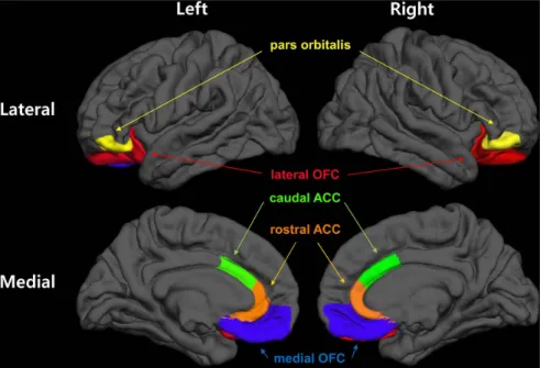
ROI-based analyses were performed to compare GMV and cortical thickness between individuals with IGD and con- trols. ROIs were defined using the Desikan–Killiany cortical atlas (Desikan et al., 2006). ROIs included both sides of the ACC (caudal/rostral ACC) and the OFC (lateral/medial OFC, pars orbitalis) (Figure1). To assess group differences
(individuals with IGD vs. controls) in GMV and cortical thickness, mean values of GMV, and cortical thickness within each ROI were extracted using FreeSurfer. For each ROI, we conducted an analysis of covariance with SPSS 24.0 (SPSS Inc., Chicago, IL, USA) for a significance level of p=.05. Age, IQ, and the intracranial volume (ICV) of each subject were entered as covariates in analysis for GMV. Age and IQ were entered as covariates in analysis for cortical thickness, but ICV was not included as a covariate, as previous studies have suggested that cortical thickness is not affected by ICV (Buckner et al., 2004). To assess the brain-behavior relationships, we performed a correlation analysis for gray matter alterations (GMV and cortical thickness in the OFC and the ACC) and the self- reporting scales (IAT and BIS).
To complement ROI analysis, the surface-based whole- brain analyses for cortical thickness were also performed using general linear models in FreeSurfer’s Query, Design, Estimate, Contrast module after controlling for age and IQ of each subject. As an exploratory investigation for whole-brain, a cluster-forming threshold of uncorrected p<.005 was employed for a vertex-wise comparison. We exclusively reported clusters with a significant number of vertices greater than 200 to reduce the possibility of generating false positives (Fung et al., 2015; Wang et al., 2014).
Ethics
This study was carried out under the guidelines for the use of human participants established by the Institutional Review Board at Yonsei University. The Institutional Review Board of the Yonsei University approved the study. Following a complete description of the scope of the study to all participants, written informed consent was obtained.
Figure 1. Regions of interest (ROIs). ROIs were defined according to the Desikan–Killiany cortical atlas. ROIs for the anterior cingulate cortex (ACC) included both sides of the caudal ACC (green) and the rostral ACC (orange). ROIs for the orbitofrontal cortex (OFC) included
both sides of the lateral OFC (red), medial OFC (blue), and the pars orbitalis (yellow)
RESULTS
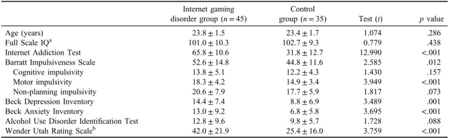
Demographic and clinical characteristics of subjects The participants in the control and IGD groups were matched by age and full-scale IQ (Table1). Subjects with IGD scored significantly higher on tests of Internet addiction (IA) and impulsivity compared with controls (IAT: p<.001; BIS: p=.012). In addition, members of the IGD group scored significantly higher on tests of depression, anxiety, and childhood ADHD symptoms com- pared with healthy controls (BDI: p=.001; BAI: p<.001;
WURS: p<.001). Total ICV was not significantly different between controls and subjects with IGD (1,600.39± 149.09 cm3for IA group; 1,624.02±138.96 cm3for controls;
p=.467).
ROI-based analyses
ROI-based analyses of cortical thickness found that sub- jects with IGD had a thinner cortex in the right rostral ACC, the right lateral OFC, and the left pars orbitalis than the cortex in controls (rostral ACC:p=.011; lateral OFC:
p=.021; pars orbitalis:p=.003; Table2). Thesefindings remained significant after including comorbid conditions
(BDI, BAI, and WURS) as covariates (rostral ACC:
p=.008; lateral OFC: p=.044; pars orbitalis: p=.014).
ROI-based analyses for GMV showed that subjects with IGD had smaller GMV in the right caudal ACC and the left pars orbitalis, compared with controls (caudal ACC:
p=.042; pars orbitalis:p=.021). Thesefindings remained significant in the caudal ACC (p=.013) after including comorbid conditions (BDI, BAI, and WURS) as covariates but not in the pars orbitalis (p=.098). Relative to controls, subjects with IGD did not have a larger GMV or thicker cortex in ROIs.
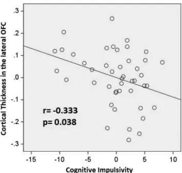
In IGD subjects, a thinner cortex in the right lateral OFC significantly correlated with higher cognitive impulsivity scores, after comorbid conditions (BDI, BAI, and WURS) were included as covariates (r=−.333,p=.038; Figure2).
We found no statistical correlation between gray matter alterations, specifically a smaller GMV and a thinner cortex, and IAT scores.
Whole-brain vertex-wise analysis
A whole-brain vertex-wise analysis of cortical thickness showed that subjects with IGD had a thinner cortex in the right supplementary motor area (SMA; peak Talairach coordinate:X=7,Y=21,Z=53; Figure3A). In addition,
Table 1. Demographics and clinical variables of participants Internet gaming
disorder group (n=45)
Control
group (n=35) Test (t) pvalue
Age (years) 23.8±1.5 23.4±1.7 1.074 .286
Full Scale IQa 101.0±10.3 102.7±9.3 0.779 .438
Internet Addiction Test 65.8±10.6 31.8±12.7 12.990 <.001
Barratt Impulsiveness Scale 52.6±14.8 44.8±11.6 2.585 .012
Cognitive impulsivity 13.8±5.1 12.2±4.3 1.430 .157
Motor impulsivity 18.3±4.2 14.9±3.4 3.949 <.001
Non-planning impulsivity 20.6±7.9 17.7±5.9 1.817 .073
Beck Depression Inventory 14.4±7.4 8.8±6.9 3.489 .001
Beck Anxiety Inventory 13.0±9.2 6.8±5.8 3.695 <.001
Alcohol Use Disorder Identification Test 12.8±9.6 9.8±5.7 1.728 .088
Wender Utah Rating Scaleb 42.0±21.9 25.4±16.0 3.759 <.001
Note. Values are expressed as means±SD.
aIntelligence Quotient (IQ) was assessed using the Wechsler Adult Intelligence Scale.
bWender Utah Rating Scale was performed to assess childhood ADHD symptoms.
Table 2. Region of interest-based comparison of cortical thickness and gray matter volume between young males with Internet gaming disorder (IGD) and controls (IGD group<control group)
Side
Internet gaming disorder group (n=45)
Control
group (n=35) Test (F) pvalue Cortical thickness (mm)
Rostral anterior cingulate cortex Right 2.86±0.20 2.98±0.19 6.747 .011
Lateral orbitofrontal cortex Right 2.71±0.14 2.79±0.14 5.540 .021
Pars orbitalis Left 2.71±0.20 2.86±0.21 9.453 .003
Gray matter volume (mm3)
Caudal anterior cingulate cortex Right 2,353.24±556.33 2,606.89±540.76 4.285 .042
Pars orbitalis Left 2,298.00±323.25 2,457.83±298.86 5.523 .021
Note. Values are expressed as means±SD.
subjects with IGD had a thinner cortex in the left frontal eye field (FEF; peak Talairach coordinate: X=−10, Y=17, Z=45; Figure 3B), the left posterior cingulate cortex (PCC; peak Talairach coordinate:X=−9,Y=−30, Z=40; Figure 3B), and the left superior parietal lobule (SPL; peak Talairach coordinate: X=−15, Y=−62, Z=61; Figure 3C) than controls. Members of the IGD group did not have any areas of the brain with a thicker cortex compared with controls.
DISCUSSION
Using SBM analysis, we compared the gray matter of the ACC and OFC in young adults with IGD with that of matched healthy controls. Ourfindings support the hypoth- esis that young adults with IGD have thinner cortices and smaller GMVs in the ACC and the OFC than controls. We performed an ROI-based analysis and found that subjects with IGD have a thinner cortex in the right rostral ACC, right lateral OFC, and left pars orbitalis than controls.
Previous studies have reported a thinner cortex in the lateral OFC and pars orbitalis of adolescents with IGD (Hong et al., 2013; Yuan et al., 2013). This study focused on young adults and found similar results with respect to cortical thickness in the OFC and in the rostral ACC. In subjects with IGD, a thinner right lateral OFC cortex correlated with higher cognitive impulsivity, reflecting a tendency to make decisions based on short-term gratification. In addition, we found that subjects with IGD had a smaller GMV in the right caudal ACC and the left pars orbitalis. This finding is consistent with previous VBM studies, which reported that subjects with IGD have smaller GMVs in the ACC and the OFC (Yuan et al., 2011;Zhou et al., 2011). As in previous studies (Hutton et al., 2009;Tomoda, Polcari, Anderson, &
Teicher, 2012), our results of GMV and cortical thickness coincided partially, but we also found differences. Our findings suggest that cortical thickness does not coincide completely with GMV, indicating that GMV and cortical thickness should be considered together for a more accurate picture of gray matter alterations.
An important finding of this study is that young adults with IGD have gray matter alterations in the ACC; specifi- cally, these individuals have a thinner right rostral ACC cortex, as well as a smaller GMV in the right caudal ACC, compared with controls. The rostral part of the ACC is implicated in error-related responses, including affective processing, and the caudal part of the ACC is associated with detection of conflict to recruit cognitive control (Van Veen & Carter, 2002). Because regional cortical thickness is associated with behavior (Bledsoe, Semrud-Clikeman, &
Pliszka, 2013; Ducharme et al., 2012), the thinner rostral Figure 2. Correlation analysis for brain-behavior relationships.
Partial correlation between cortical thickness in the right lateral orbitofrontal cortex (OFC) and cognitive impulsivity score of the Barratt Impulsiveness Scale (BIS) after controlling for covariates (age, IQ, BDI, BAI, and WURS). To depict partial correlation, variables were regressed onto covariates using a linear regression.
Scatter plots were generated using calculated non-standardized residuals. The cortical thickness of the right lateral OFC significantly correlated with cognitive impulsivity in IGD subjects
(r=−.333,p=.038)
Figure 3. Whole-brain vertex-wise analysis of cortical thickness. A statistical threshold ofp<.005 (uncorrected) was employed for a vertex- wise comparison. Compared with controls, subjects with IGD had a thinner cortex in the (A) right supplementary motor area (SMA; peak Talairach coordinate:X=7,Y=21,Z=53; number of vertices: 271), (B) left frontal eyefield (FEF; peak Talairach coordinate:X=−10, Y=17,Z=45; number of vertices: 224) and the left posterior cingulate cortex (PCC; peak Talairach coordinate:X=−9,Y=−30,Z=40;
number of vertices: 215), and (C) left superior parietal lobule (SPL; peak MNI coordinate:X=−15,Y=−62,Z=61; number of vertices: 216)
ACC cortex in IGD may contribute to the failure to respond to the negative consequences of excessive gaming using impaired error processing. Also, the smaller GMV of the caudal ACC in Internet game addicts may contribute to the loss of cognitive control over excessive gaming. In addition, ourfindings of gray matter differences in the right side of the ACC are consistent with previous evidence that monitoring and related behavioral control is lateralized to the right hemisphere (Stuss, 2011).
Here, we found that young adult males with IGD had a thinner cortex in the right lateral OFC compared with controls. In general, the OFC contributes to the monitoring of reward values assigned to different decisions; in particu- lar, the right lateral part of the OFC has been implicated in the inhibitory processes that suppress previously rewarded choices (Elliott & Deakin, 2005; Elliott, Dolan, & Frith, 2000) and promote the selection of delayed monetary rewards over immediate rewards (McClure, Laibson, Loewenstein, & Cohen, 2004). Moreover, recently, the role of the right lateral OFC was proposed to include integration of prior outcome-based information with current perceptual information to make anticipatory signals about upcoming choices (Nogueira et al., 2017). On the whole, this evidence suggests that the right lateral OFC regulates decision- making using internal and external information in aflexible and adaptive manner. Lesions to the lateral OFC impair decision-making related to a delayed reward, leading to short-term and impulsive decisions (Mar, Walker, Theobald, Eagle, & Robbins, 2011). Here, the cortical thickness of the right lateral OFC in IGD subjects significantly correlated with cognitive impulsivity, which is defined as “making quick decisions”(Stanford et al., 2009). Recently, cognitive impulsivity was closely related to reward-based learning and decision-making (Cáceres & San Martín, 2017). Therefore, based on the combination of our findings and the existing literature, we speculate that a thinner right lateral OFC cortex prevents individuals with IGD from effectively inte- grating information to estimate reward values, thereby contributing to preference for short-term pleasure and impulsive decision-making.
Another important finding was that subjects with IGD demonstrated smaller GMV and a thinner cortex in the left pars orbitalis compared with controls. The pars orbitalis is located at the anterior portion of the inferior frontal gyrus, and the inferior frontal gyrus tends to coactivate with the lateral OFC (Zald et al., 2012). Moreover, the pars orbitalis, along with other orbitofrontal regions, has been associated with reward-related information processing and decision-making (Dixon & Christoff, 2014). In particular, the left side of the pars orbitalis has been shown to be closely connected with the middle temporal gyrus and is implicated in cognitively controlled memory retrieval (Badre, Poldrack, Paré-Blagoev, Insler, & Wagner, 2005). Given that adaptive response selec- tion involves strategic control of the memory system (Poldrack
& Packard, 2003), gray matter alterations within the left pars orbitalis may make it difficult to guide behavior based on prior information (Badre & Wagner, 2007). Therefore, in view of the literature, our findings suggest that smaller GMV and thinner cortex in the left pars orbitalis of IGD subjects may contribute to their uncontrolled Internet use by impairing their ability to adjust their behavior based on prior information.
In the whole-brain vertex-wise analysis, we found that subjects with IGD had a thinner cortex in the right SMA, the left FEF, the left SPL, and the left PCC compared with controls. The right SMA plays a role in connecting cogni- tion and behavior (Nachev, Kennard, & Husain, 2008) and is an important area for response inhibition (Picton et al., 2007). Neuronal activity in the PCC is modulated by external environmental changes, and this modulation may be associated with a cognitive set shift for behavioral adaptation (Pearson, Heilbronner, Barack, Hayden, & Platt, 2011). The FEF and the SPL are also crucial brain regions that are involved in top-down attention control (Corbetta &
Shulman, 2002). Proper coordination of the frontal and parietal regions is suggested to be essential for adaptive action planning (Andersen & Cui, 2009). Although neither the FEF nor SPL regions were ROIs in this study, we suggest that a thinner cortex in these areas of the brain, particularly in frontoparietal areas, plays important roles in diminished behavioral control in individuals with IGD.
This diminished behavioral control may alter risk/reward decision-making, resulting in difficulty in suppressing urges and the pursuit of short-term gratification.
This study has limitations that should be considered. First, thefinding of a thinner cortex in the ACC and the OFC by ROI-based analysis was not confirmed in the whole-brain analysis. We speculate that this discrepancy was primarily driven by differences in methodology. For example, the ROI-based analysis was performed by calculating the mean cortical thickness within the manually delineated area and group differences were investigated by subsequent statistical analysis; in contrast, the whole-brain analysis employed a generalized linear model to estimate vertex-wise group differences in cortical thickness. Because the ROI-based and whole-brain approaches offer different kinds of information, these two methods are suggested to be complementary (Giuliani, Calhoun, Pearlson, Francis, & Buchanan, 2005).
Our currentfindings would be clarified by further research to reduce errors in the ROI-based and whole-brain vertex-wise analyses, particularly, errors derived from spatial normaliza- tion processes. Second, although this study defined ROIs on the assumption that structural alterations in the OFC and the ACC underlie the impaired risk/reward decision-making in IGD, there was no direct measurement of decision-making capacity by neuropsychological tests. Thus, careful consid- eration should be made when linking our imagingfindings to dysfunctional risk/reward decision-making in IGD. Third, although IGD diagnosis in this study was made using the IAT scale and clinical interviews, the DSM-5 diagnostic criteria for IGD were not applied. The DSM-5 IGD diagnos- tic criteria are widely used, since DSM-5 identified IGD as one of the conditions requiring further study (Petry &
O’Brien, 2013). To accumulate reliable evidences for IGD, it is necessary to apply a consistent diagnostic tool. Thus, future IGD studies should apply the DSM-5 diagnostic criteria. Fourth, although we limited this study to subjects with IGD who reported that online gaming was their primary use of the Internet, most subjects also participated in other online activities, including social networking. Thus, a future- combined structural and functional study design that mea- sure neural activities in response to gaming-specific stimuli would enhance ourfindings. Fifth, we used a cross-sectional
design in this study. Future research that utilized longitudinal study designs to measure cortical thickness changes during adolescence and early adulthood would investigate whether there is a causal relationship between our imaging results and excessive Internet gaming. Sixth, our sample for this study was small and only included male subjects. Gender differ- ences are reported with respect to the clinical features of IGD (Ko, Yen, Chen, Chen, & Yen, 2005). Larger studies that include both men and women will be necessary to expand our understanding of IGD.
CONCLUSIONS
We performed an SBM analysis of young adult males with IGD to investigate gray matter alterations in the ACC and the OFC, which were related to risk/reward decision-making.
The ROI-based comparison with controls demonstrated that IGD subjects had a thinner cortex in the right rostral ACC, the right lateral OFC, and the left pars orbitalis, and a smaller GMV in the right caudal ACC and the left pars orbitalis. A thinner cortex in the right lateral OFC correlated with higher cognitive impulsivity in IGD subjects, providing possible insight to decision-making based on short-term gratification in IGD. The whole-brain analysis of IGD subjects found they had a thinner cortex in behavioral control-related brain regions, including frontoparietal areas. Ourfindings suggest that gray matter alterations may provide information about IGD pathophysiology, by reflecting altered risk/reward decision-making and diminished behavioral control.
Funding sources: This research was supported by the Original Technology Research Program for Brain Science through the National Research Foundation of Korea funded by the Ministry of Science, ICT, and Future Planning (NRF-2015M3C7A1064906).
Authors’ contribution: DL and Y-CJ conceived and designed the study. DL recruited participants and drafted the manuscript. JP analyzed and interpreted the data. IYK and KN provided critical revision of the manuscript and important intellectual content. All authors had full access to all data in the study and take responsibility for the integrity of the data and the accuracy of the data analysis. All authors critically reviewed and approved the final version of this manuscript for publication. IYK and Y-CJ were contributed equally to this study as co-corresponding authors.
Conflict of interest: The authors declare no conflict of interest.
REFERENCES
Amiez, C., Joseph, J. P., & Procyk, E. (2005). Anterior cingulate error-related activity is modulated by predicted reward.
European Journal of Neuroscience, 21(12), 3447–3452.
doi:10.1111/j.1460-9568.2005.04170.x
Andersen, R. A., & Cui, H. (2009). Intention, action planning, and decision making in parietal-frontal circuits. Neuron, 63(5), 568–583. doi:10.1016/j.neuron.2009.08.028
Badre, D., Poldrack, R. A., Paré-Blagoev, E. J., Insler, R. Z., &
Wagner, A. D. (2005). Dissociable controlled retrieval and generalized selection mechanisms in ventrolateral prefrontal cor- tex.Neuron, 47(6), 907–918. doi:10.1016/j.neuron.2005.07.023 Badre, D., & Wagner, A. D. (2007). Left ventrolateral prefrontal cortex and the cognitive control of memory. Neuropsycho- logia, 45(13), 2883–2901. doi:10.1016/j.neuropsychologia.
2007.06.015
Beck, A. T., Epstein, N., Brown, G., & Steer, R. A. (1988). An inventory for measuring clinical anxiety: Psychometric prop- erties.Journal of Consulting and Clinical Psychology, 56(6), 893–897. doi:10.1037/0022-006X.56.6.893
Beck, A. T., Steer, R. A., & Brown, G. K. (1996). Beck Depres- sion Inventory-II.San Antonio, 78(2), 490–498. doi:10.1037/
t00742-000
Bledsoe, J. C., Semrud-Clikeman, M., & Pliszka, S. R. (2013).
Anterior cingulate cortex and symptom severity in attention- deficit/hyperactivity disorder.Journal of Abnormal Psycholo- gy, 122(2), 558–565. doi:10.1037/a0032390
Block, J. J. (2008). Issues for DSM-V: Internet addiction. The American Journal of Psychiatric, 165(3), 306–307.
doi:10.1176/appi.ajp.2007.07101556
Buckner, R. L., Head, D., Parker, J., Fotenos, A. F., Marcus, D., Morris, J. C., & Snyder, A. Z. (2004). A unified approach for morphometric and functional data analysis in young, old, and demented adults using automated atlas-based head size normalization: Reliability and validation against manual mea- surement of total intracranial volume. Neuroimage, 23(2), 724–738. doi:10.1016/j.neuroimage.2004.06.018
Cáceres, P., & San Martín, R. (2017). Low cognitive impulsivity is associated with better gain and loss learning in a probabilistic decision-making task. Frontiers in Psychology, 8, 204.
doi:10.3389/fpsyg.2017.00204
Choi, S.-W., Kim, H., Kim, G.-Y., Jeon, Y., Park, S., Lee, J.-Y., Jung, H. Y., Sohn, B. K., Choi, J. S., & Kim, D. J. (2014).
Similarities and differences among Internet gaming disorder, gambling disorder and alcohol use disorder: A focus on impulsivity and compulsivity. Journal of Behavioral Addic- tions, 3(4), 246–253. doi:10.1556/JBA.3.2014.4.6
Corbetta, M., & Shulman, G. L. (2002). Control of goal-directed and stimulus-driven attention in the brain. Nature Reviews.
Neuroscience, 3(3), 201–215. doi:10.1038/nrn755
Dale, A. M., Fischl, B., & Sereno, M. I. (1999). Cortical surface- based analysis: I. Segmentation and surface reconstruction.
Neuroimage, 9(2), 179–194. doi:10.1006/nimg.1998.0395 Desikan, R. S., Ségonne, F., Fischl, B., Quinn, B. T., Dickerson,
B. C., Blacker, D., Buckner, R. L., Dale, A. M., Maguire, R. P., Hyman, B. T., Albert, M. S., & Killiany, R. J. (2006). An automated labeling system for subdividing the human cerebral cortex on MRI scans into gyral based regions of interest.Neuro- image, 31(3), 968–980. doi:10.1016/j.neuroimage.2006.01.021 Dixon, M. L., & Christoff, K. (2014). The lateral prefrontal cortex
and complex value-based learning and decision making.Neu- roscience and Biobehavioral Reviews, 45,9–18. doi:10.1016/j.
neubiorev.2014.04.011
Dong, G., DeVito, E., Huang, J., & Du, X. (2012). Diffusion tensor imaging reveals thalamus and posterior cingulate cortex ab- normalities in Internet gaming addicts.Journal of Psychiatric
Research, 46(9), 1212–1216. doi:10.1016/j.jpsychires.2012.
05.015
Dong, G., DeVito, E. E., Du, X., & Cui, Z. (2012). Impaired inhibitory control in‘Internet addiction disorder’: A functional magnetic resonance imaging study.Psychiatry Research: Neu- roimaging, 203(2), 153–158. doi:10.1016/j.pscychresns.2012.
02.001
Dong, G., Hu, Y., & Lin, X. (2013). Reward/punishment sensitiv- ities among Internet addicts: Implications for their addictive behaviors. Progress in Neuro-Psychopharmacology and Biological Psychiatry, 46, 139–145. doi:10.1016/j.pnpbp.
2013.07.007
Dong, G., Huang, J., & Du, X. (2011). Enhanced reward sensitivity and decreased loss sensitivity in Internet addicts: An fMRI study during a guessing task.Journal of Psychiatric Research, 45(11), 1525–1529. doi:10.1016/j.jpsychires.2011.06.017 Dong, G., & Potenza, M. N. (2014). A cognitive-behavioral model
of Internet gaming disorder: Theoretical underpinnings and clinical implications.Journal of Psychiatric Research, 58,7– 11. doi:10.1016/j.jpsychires.2014.07.005
Dong, G., Shen, Y., Huang, J., & Du, X. (2013). Impaired error- monitoring function in people with Internet addiction disorder:
An event-related fMRI study.European Addiction Research, 19(5), 269–275. doi:10.1159/000346783
Ducharme, S., Hudziak, J. J., Botteron, K. N., Albaugh, M. D., Nguyen, T.-V., Karama, S., Evans, A. C., & Brain Develop- ment Cooperative Group. (2012). Decreased regional cortical thickness and thinning rate are associated with inattention symptoms in healthy children.Journal of the American Acad- emy of Child and Adolescent Psychiatry, 51(1), 18–27.e2. e12.
doi:10.1016/j.jaac.2011.09.022
Elliott, R., & Deakin, B. (2005). Role of the orbitofrontal cortex in reinforcement processing and inhibitory control: Evidence from functional magnetic resonance imaging studies in healthy human subjects. International Review of Neurobiology, 65, 89–116. doi:10.1016/S0074-7742(04)65004-5
Elliott, R., Dolan, R. J., & Frith, C. D. (2000). Dissociable functions in the medial and lateral orbitofrontal cortex: Evi- dence from human neuroimaging studies. Cerebral Cortex (New York, NY), 10(3), 308–317. doi:10.1093/cercor/10.3.308 First, M., Spitzer, R., & Williams, J. (1997). Structured clinical interview for the diagnostic and statistical manual.
Washington, DC: American Psychiatric Press.
Fischl, B., Rajendran, N., Busa, E., Augustinack, J., Hinds, O., Yeo, B. T., Mohlberg, H., Amunts, K., & Zilles, K. (2007).
Cortical folding patterns and predicting cytoarchitecture.Ce- rebral Cortex (New York, NY), 18(8), 1973–1980. doi:10.1093/
cercor/bhm225
Fischl, B., Sereno, M. I., & Dale, A. M. (1999). Cortical surface- based analysis: II: Inflation, flattening, and a surface-based coordinate system.Neuroimage, 9(2), 195–207. doi:10.1006/
nimg.1998.0396
Fischl, B., Sereno, M. I., Tootell, R. B., & Dale, A. M. (1999).
High-resolution intersubject averaging and a coordinate system for the cortical surface.Human Brain Mapping, 8(4), 272–284.
doi:10.1002/(SICI)1097-0193(1999)8:4<272::AID-HBM10>
3.0.CO;2-4
Fischl, B., Van Der Kouwe, A., Destrieux, C., Halgren, E., Ségonne, F., Salat, D. H., Busa, E., Seidman, L. J., Goldstein, J., Kennedy, D., Caviness, V., Makris, N., Rosen, B., & Dale, A. M. (2004). Automatically parcellating the human cerebral
cortex. Cerebral Cortex (New York, NY), 14(1), 11–22.
doi:10.1093/cercor/bhg087
Fung, G., Deng, Y., Zhao, Q., Li, Z., Qu, M., Li, K., Zeng, Y. W., Jin, Z., Ma, Y. T., Yu, X., Wang, Z. R., Shum, D. H., &
Chan, R. C. (2015). Distinguishing bipolar and major de- pressive disorders by brain structural morphometry: A pilot study.
BMC Psychiatry, 15(1), 298. doi:10.1186/s12888-015-0685-5 Giuliani, N. R., Calhoun, V. D., Pearlson, G. D., Francis, A., &
Buchanan, R. W. (2005). Voxel-based morphometry versus region of interest: A comparison of two methods for analyzing gray matter differences in schizophrenia. Schizophrenia Re- search, 74(2), 135–147. doi:10.1016/j.schres.2004.08.019 Hayden, B. Y., Heilbronner, S. R., Pearson, J. M., & Platt, M. L.
(2011). Surprise signals in anterior cingulate cortex: Neuronal encoding of unsigned reward prediction errors driving adjust- ment in behavior.The Journal of Neuroscience, 31(11), 4178– 4187. doi:10.1523/JNEUROSCI.4652-10.2011
Honey, C. J., Kötter, R., Breakspear, M., & Sporns, O. (2007).
Network structure of cerebral cortex shapes functional con- nectivity on multiple time scales.Proceedings of the National Academy of Sciences of the United States of America, 104(24), 10240–10245. doi:10.1073/pnas.0701519104
Hong, S.-B., Kim, J.-W., Choi, E.-J., Kim, H.-H., Suh, J.-E., Kim, C.-D., Klauser, P., Whittle, S., Yűcel, M., Pantelis, C., & Yi, S. H. (2013). Reduced orbitofrontal cortical thickness in male adolescents with Internet addiction. Behavioral and Brain Functions: BBF, 9(1), 11. doi:10.1186/1744-9081-9-11 Hutton, C., Draganski, B., Ashburner, J., & Weiskopf, N. (2009).
A comparison between voxel-based cortical thickness and voxel-based morphometry in normal aging. Neuroimage, 48(2), 371–380. doi:10.1016/j.neuroimage.2009.06.043 Kim, J. S., Singh, V., Lee, J. K., Lerch, J., Ad-Dab’bagh, Y.,
MacDonald, D., Lee, J. M., Kim, S. I., & Evans, A. C. (2005).
Automated 3-D extraction and evaluation of the inner and outer cortical surfaces using a Laplacian map and partial volume effect classification. Neuroimage, 27(1), 210–221.
doi:10.1016/j.neuroimage.2005.03.036
Kim, N. R., Hwang, S. S.-H., Choi, J.-S., Kim, D.-J., Demetrovics, Z., Király, O., Nagygyörgy, K., Griffiths, M. D., Hyun, S. Y., Youn, H. C., & Choi, S. W. (2016). Characteristics and psychiatric symptoms of Internet gaming disorder among adults using self-reported DSM-5 criteria.Psychiatry Investi- gation, 13(1), 58–66. doi:10.4306/pi.2016.13.1.58
Ko, C.-H., Hsieh, T.-J., Chen, C.-Y., Yen, C.-F., Chen, C.-S., Yen, J.-Y., Wang, P. W., & Liu, G. C. (2014). Altered brain activation during response inhibition and error processing in subjects with Internet gaming disorder: A functional magnetic imaging study.
European Archives of Psychiatry and Clinical Neuroscience, 264(8), 661–672. doi:10.1007/s00406-013-0483-3
Ko, C.-H., Yen, J.-Y., Chen, C.-C., Chen, S.-H., & Yen, C.-F.
(2005). Gender differences and related factors affecting online gaming addiction among Taiwanese adolescents. Journal of Nervous and Mental Disease, 193(4), 273–277. doi:10.1097/
01.nmd.0000158373.85150.57
Krain, A. L., Wilson, A. M., Arbuckle, R., Castellanos, F. X., &
Milham, M. P. (2006). Distinct neural mechanisms of risk and ambiguity: A meta-analysis of decision-making.Neuroimage, 32(1), 477–484. doi:10.1016/j.neuroimage.2006.02.047 Kuss, D. J. (2013). Internet gaming addiction: Current perspec-
tives. Psychology Research and Behavior Management, 6, 125–137. doi:10.2147/PRBM.S39476
Kuss, D. J., Griffiths, M. D., Karila, L., & Billieux, J. (2014).
Internet addiction: A systematic review of epidemiological research for the last decade.Current Pharmaceutical Design, 20(25), 4026–4052. doi:10.2174/13816128113199990617 Lee, D., Namkoong, K., Lee, J., & Jung, Y. C. (2017). Abnormal
gray matter volume and impulsivity in young adults with Internet gaming disorder.Addiction Biology. Advance online publication. doi:10.1111/adb.12552
Lemaitre, H., Goldman, A. L., Sambataro, F., Verchinski, B. A., Meyer-Lindenberg, A., Weinberger, D. R., & Mattay, V. S.
(2012). Normal age-related brain morphometric changes: Non- uniformity across cortical thickness, surface area and gray matter volume? Neurobiology of Aging, 33(3), 617.e1–617.e9.
doi:10.1016/j.neurobiolaging.2010.07.013
Lin, X., Dong, G., Wang, Q., & Du, X. (2015). Abnormal gray matter and white matter volume in‘Internet gaming addicts’. Addictive Behaviors, 40,137–143. doi:10.1016/j.addbeh.2014.
09.010
Mar, A. C., Walker, A. L., Theobald, D. E., Eagle, D. M., &
Robbins, T. W. (2011). Dissociable effects of lesions to orbitofrontal cortex subregions on impulsive choice in the rat. The Journal of Neuroscience, 31(17), 6398–6404.
doi:10.1523/JNEUROSCI.6620-10.2011
McClure, S. M., Laibson, D. I., Loewenstein, G., & Cohen, J. D.
(2004). Separate neural systems value immediate and delayed monetary rewards.Science (New York, NY), 306(5695), 503– 507. doi:10.1126/science.1100907
Nachev, P., Kennard, C., & Husain, M. (2008). Functional role of the supplementary and pre-supplementary motor areas.Nature Reviews. Neuroscience, 9(11), 856–869. doi:10.1038/nrn2478 Nogueira, R., Abolafia, J. M., Drugowitsch, J., Balaguer-Ballester, E., Sanchez-Vives, M. V., & Moreno-Bote, R. (2017). Lateral orbitofrontal cortex anticipates choices and integrates prior with current information.Nature Communications, 8,14823.
doi:10.1038/ncomms14823
Patton, J. H., & Stanford, M. S. (1995). Factor structure of the Barratt Impulsiveness Scale.Journal of Clinical Psychology, 51(6), 768–774. doi:10.1002/1097-4679(199511)51:6<768::
AID-JCLP2270510607>3.0.CO;2-1
Pawlikowski, M., & Brand, M. (2011). Excessive Internet gaming and decision making: Do excessive World of Warcraft players have problems in decision making under risky conditions?
Psychiatry Research, 188(3), 428–433. doi:10.1016/j.
psychres.2011.05.017
Pearson, J. M., Heilbronner, S. R., Barack, D. L., Hayden, B. Y., &
Platt, M. L. (2011). Posterior cingulate cortex: Adapting behavior to a changing world.Trends in Cognitive Sciences, 15(4), 143–151. doi:10.1016/j.tics.2011.02.002
Petry, N. M., & O’Brien, C. P. (2013). Internet gaming disorder and the DSM-5. Addiction (Abingdon, England), 108(7), 1186–1187. doi:10.1111/add.12162
Picton, T. W., Stuss, D. T., Alexander, M. P., Shallice, T., Binns, M. A., & Gillingham, S. (2007). Effects of focal frontal lesions on response inhibition.Cerebral Cortex (New York, NY), 17(4), 826–838. doi:10.1093/cercor/bhk031
Poldrack, R. A., & Packard, M. G. (2003). Competition among multiple memory systems: Converging evidence from animal and human brain studies.Neuropsychologia, 41(3), 245–251.
doi:10.1016/S0028-3932(02)00157-4
Ségonne, F., Dale, A. M., Busa, E., Glessner, M., Salat, D., Hahn, H. K., & Fischl, B. (2004). A hybrid approach to the skull
stripping problem in MRI. Neuroimage, 22(3), 1060–1075.
doi:10.1016/j.neuroimage.2004.03.032
Ségonne, F., Pacheco, J., & Fischl, B. (2007). Geometrically accurate topology-correction of cortical surfaces using non- separating loops. IEEE Transactions on Medical Imaging, 26(4), 518–529. doi:10.1109/TMI.2006.887364
Sled, J. G., Zijdenbos, A. P., & Evans, A. C. (1998). A nonpara- metric method for automatic correction of intensity nonunifor- mity in MRI data. IEEE Transactions on Medical Imaging, 17(1), 87–97. doi:10.1109/42.668698
Stanford, M. S., Mathias, C. W., Dougherty, D. M., Lake, S. L., Anderson, N. E., & Patton, J. H. (2009). Fifty years of the Barratt Impulsiveness Scale: An update and review.Personal- ity and Individual Differences, 47(5), 385–395. doi:10.1016/j.
paid.2009.04.008
Stuss, D. T. (2011). Functions of the frontal lobes: Relation to executive functions. Journal of the International Neuropsy- chological Society: JINS, 17(5), 759–765. doi:10.1017/
S1355617711000695
Tomoda, A., Polcari, A., Anderson, C. M., & Teicher, M. H.
(2012). Reduced visual cortex gray matter volume and thick- ness in young adults who witnessed domestic violence during childhood. PLoS One, 7(12), e52528. doi:10.1371/journal.
pone.0052528
Van Veen, V., & Carter, C. S. (2002). The timing of action- monitoring processes in the anterior cingulate cortex.Journal of Cognitive Neuroscience, 14(4), 593–602. doi:10.1162/
08989290260045837
Wallis, J. D. (2007). Orbitofrontal cortex and its contribution to decision-making.Annual Review of Neuroscience, 30,31–56.
doi:10.1146/annurev.neuro.30.051606.094334
Wang, H., Jin, C., Yuan, K., Shakir, T. M., Mao, C., Niu, X., Niu, X., Niu, C., Guo, L., & Zhang, M. (2015). The alteration of gray matter volume and cognitive control in adolescents with Internet gaming disorder. Frontiers in Behavioral Neurosci- ence, 9,64. doi:10.3389/fnbeh.2015.00064
Wang, Y., Deng, Y., Fung, G., Liu, W.-H., Wei, X.-H., Jiang, X.-Q., Lui, S. S., Cheung, E. F., & Chan, R. C. (2014). Distinct structural neural patterns of trait physical and social anhedonia:
Evidence from cortical thickness, subcortical volumes and inter-regional correlations.Psychiatry Research: Neuroimag- ing, 224(3), 184–191. doi:10.1016/j.pscychresns.2014.09.005 Ward, M. F. (1993). The Wender Utah Rating Scale: An aid in the retrospective.The American Journal of Psychiatry, 1(50), 885.
doi:10.1176/ajp.150.6.885
Wechsler, D. (2014). Wechsler Adult Intelligence Scale–Fourth Edition (WAIS–IV). San Antonio, Texas: Psychological Corporation.
Winkler, A. M., Kochunov, P., Blangero, J., Almasy, L., Zilles, K., Fox, P. T., Duggirala, R., & Glahn, D. C. (2010). Cortical thickness or grey matter volume? The importance of selecting the phenotype for imaging genetics studies. Neuroimage, 53(3), 1135–1146. doi:10.1016/j.neuroimage.2009.12.028 Yao, Y. W., Liu, L., Ma, S. S., Shi, X. H., Zhou, N., Zhang, J. T.,
et al. (2017). Functional and structural neural alterations in Internet gaming disorder: A systematic review and meta- analysis.Neuroscience and Biobehavioral Reviews, 83,313– 324. doi:10.1016/j.neubiorev.2017.10.029
Yao, Y.-W., Wang, L.-J., Yip, S. W., Chen, P.-R., Li, S., Xu, J., Zhang, J. T., Deng, L. Y., Liu, Q. X., & Fang, X. Y. (2015).
Impaired decision-making under risk is associated with
gaming-specific inhibition deficits among college students with Internet gaming disorder.Psychiatry Research, 229(1), 302– 309. doi:10.1016/j.psychres.2015.07.004
Young, K. S. (1998a). Caught in the net: How to recognize the signs of Internet addiction – And a winning strategy for recovery. New York, NY: Wiley.
Young, K. S. (1998b). Internet addiction: The emergence of a new clinical disorder. CyberPsychology & Behavior, 1(3), 237– 244. doi:10.1089/cpb.1998.1.237
Yuan, K., Cheng, P., Dong, T., Bi, Y., Xing, L., Yu, D., Zhao, L., Dong, M., von Deneen, K. M., Liu, Y., Qin, W., & Tian, J.
(2013). Cortical thickness abnormalities in late adolescence with online gaming addiction. PLoS One, 8(1), e53055.
doi:10.1371/journal.pone.0053055
Yuan, K., Qin, W., Wang, G., Zeng, F., Zhao, L., Yang, X., Liu, P., Liu, J., Sun, J., von Deneen, K. M., Gong, Q., Liu, Y., &
Tian, J. (2011). Microstructure abnormalities in adolescents with Internet addiction disorder. PLoS One, 6(6), e20708.
doi:10.1371/journal.pone.0020708
Zald, D. H., McHugo, M., Ray, K. L., Glahn, D. C., Eickhoff, S. B., & Laird, A. R. (2012). Meta-analytic connecti- vity modeling reveals differential functional connectivity of the medial and lateral orbitofrontal cortex. Cerebral Cortex (New York, NY), 24(1), 232–248. doi:10.1093/
cercor/bhs308
Zhou, F., Montag, C., Sariyska, R., Lachmann, B., Reuter, M., Weber, B., Trautner, P., Kendrick, K. M., Markett, S., &
Becker, B. (2017). Orbitofrontal gray matter deficits as marker of Internet gaming disorder: Converging evidence from a cross-sectional and prospective longitudinal design.
Addiction Biology. Advance online publication. doi:10.1111/
adb.12570
Zhou, Y., Lin, F.-C., Du, Y.-S., Zhao, Z.-M., Xu, J.-R., & Lei, H.
(2011). Gray matter abnormalities in Internet addiction: A voxel-based morphometry study. European Journal of Radi- ology, 79(1), 92–95. doi:10.1016/j.ejrad.2009.10.025