Endocannabinoid Tone Regulates Human Sebocyte Biology
No´ra Za´ka´ny1,8, Attila Ola´h1,8, Arnold Markovics1, Erika Taka´cs1, Andrea Aranya´sz1, Simon Nicolussi2, Fabiana Piscitelli3, Marco Allara`3, A´gnes Po´r4, Ilona Kova´cs4, Christos C. Zouboulis5, Ju¨rg Gertsch2, Vincenzo Di Marzo3, Tama´s Bı´ro´6,9and Tama´s Szabo´7,9
We have previously shown that endocannabinoids (eCBs) (e.g., anandamide) are involved in the maintenance of homeostatic sebaceous lipid production in human sebaceous glands and that eCB treatment dramatically increases sebaceous lipid production. Here, we aimed to investigate the expression of the major eCB synthesizing and degrading enzymes and to study the effects of eCB uptake inhibitors on human SZ95 sebocytes, thus exploring the role of the putative eCB membrane transporter, which has been hypothesized to facilitate the cellular uptake and subsequent degradation of eCBs. We found that the major eCB synthesizing (N-acyl phosphatidylethanolamine- specific phospholipase D, and diacylglycerol lipase-a and -b) and degrading (fatty acid amide hydrolase, monoacylglycerol lipase) enzymes are expressed in SZ95 sebocytes and also in sebaceous glands (except for diacylglycerol lipase-a, the staining of which was dubious in histological preparations). eCB uptake-inhibition with VDM11 induced a moderate increase in sebaceous lipid production and also elevated the levels of various eCBs and related acylethanolamides. Finally, we found that VDM11 was able to interfere with the proinflammatory action of the TLR4 activator lipopolysaccharide. Collectively, our data suggest that inhibition of eCB uptake exerts anti-inflammatory actions and elevates both sebaceous lipid production and eCB levels; thus, these inhibitors might be beneficial in cutaneous inflammatory conditions accompanied by dry skin.
Journal of Investigative Dermatology(2018)138,1699e1706;doi:10.1016/j.jid.2018.02.022
INTRODUCTION
Sebaceous glands (SGs) are important players and regulators of human skin homeostasis. In addition to their obvious function, that is, production of lipid-rich sebum, by which they contribute to the cutaneous lipid barrier and thermoregulation, they also play a role in the endocrine and immune systems of the skin and serve as stem cell reservoirs (Dajnoki et al., 2017; Lupi, 2008;
Porter, 2001; To´th et al., 2011; Zouboulis et al., 2008, 2014).
Their clinical significance is also remarkably high, because overproduction and pathologically altered composition of sebum in seborrhea is a key step in the pathogenesis of acne, one of the most prevalent human skin diseases (Kurokawa et al., 2009; To´th et al., 2011; Zouboulis et al., 2008, 2014). On the other hand, lack of sufficient sebum production in adulthood may contribute to dry skin syndrome, xerosis, or even skin aging and atopic dermatitis (AD) (Kim et al., 2014; Mischo et al., 2014;
Shi et al., 2015; Zampeli et al., 2012; Zouboulis and Boschnakow 2001). Moreover, the unique composition of sebum is thought to play an important role in regulating the growth of the cutaneous microbiota by restricting unwanted microbes and promoting preferred ones, thus making homeostatic SG functions important orchestrators of skin- microbiota crosstalk (Pappas, 2009). Hence, via the subsequent pathological alterations in the cutaneous microbiota, disorders of sebaceous lipid production (SLP) may contribute to the patho- genesis of several diseases, including AD (Shi et al., 2015).
Therefore, a better understanding of the (dys)regulation of SG biology and identification of additional regulators of homeostatic SLP are clinically relevant topics of investigative dermatology.
Unfortunately, human SGs are challenging to study, because primary sebocytes cannot be kept in culture for more than a few passages, and there are no adequate animal model systems for assessing the whole complexity of human SG biology (To´th et al., 2011; Zouboulis et al., 2008, 2014). Therefore, in this study, we used the human immortalized SZ95 sebocyte cell line (Zouboulis et al. 1999), which is a widely accepted model system for studying human SG functions in vitro (To´th et al., 2011;
Zouboulis et al., 2008, 2014).
A growing body of evidence suggests that SGs are not only
“innocent” targets of the complex cutaneous neuroendocrine
1Department of Physiology, Faculty of Medicine, University of Debrecen, Debrecen, Hungary;2Institute of Biochemistry and Molecular Medicine, National Centre of Competence in Research TransCure, University of Bern, Bern, Switzerland;3Institute of Biomolecular Chemistry, Consiglio Nazionale delle Ricerche, Pozzuoli, Italy;4Department of Pathology, Gyula Kene´zy University Hospital, University of Debrecen, Debrecen, Hungary;
5Departments of Dermatology, Venereology, Allergology and Immunology, Dessau Medical Center, Brandenburg Medical School Theodore Fontane, Dessau, Germany;6Department of Immunology, Faculty of Medicine, University of Debrecen, Debrecen, Hungary; and7Department of Pediatrics, Faculty of Medicine, University of Debrecen, Debrecen, Hungary
8These authors contributed equally to this work.
9These authors contributed equally to this work.
Correspondence: Tama´s Bı´ro´, University of Debrecen, Egyetem te´r 1, Debrecen, H-4032, Hungary. E-mail:biro.tamas@med.unideb.hu Abbreviations: 2-AG, 2-arachidonoylglycerol; AD, atopic dermatitis; AEA, arachidonoylethanolamide; DAGL, diacylglycerol lipase; eCB, endocanna- binoid; ECS, endocannabinoid system; EMT, endocannabinoid membrane transporter; FAAH, fatty acid amide hydrolase; LPS, lipopolysaccharide;
MAGL, monoacylglycerol lipase; NAPE-PLD, N-acyl
phosphatidylethanolamine-specific phospholipase D; OEA, oleoylethanola- mide; PEA, palmitoylethanolamide; SG, sebaceous gland; SLP, sebaceous lipid production
Received 11 September 2017; revised 19 February 2018; accepted 19 February 2018; accepted manuscript published online 6 March 2018;
corrected proof published online 2 May 2018
ORIGINAL ARTICLE
ª2018 The Authors. Published by Elsevier, Inc. on behalf of the Society for Investigative Dermatology. www.jidonline.org 1699
system (Slominski, 2005; Slominski and Wortsman, 2000), but also act as its master regulators by producing several hormones and paracrine signal molecules (e.g., androgen hormones, corticotropin-releasing hormone, several adipo- kines, etc.) (Kova´cs et al., 2016; To´th et al., 2011; Zouboulis et al., 2008, 2014). Furthermore, we have previously shown that, besides the aforementioned regulators, human SGs are also important players in the endocannabinoid system (ECS) of the skin (Dobrosi et al., 2008; Ola´h and Bı´ro´, 2017).
The ECS is a complex signaling system, comprising endogenous ligands (i.e., the endocannabinoids [eCBs]
such as arachidonoylethanolamide [AEA], also known as anandamide; 2-arachidonoylglycerol [2-AG]); and receptors (e.g., CB1and CB2, etc.) and multiple enzymes involved in the synthesis (e.g., N-acyl phosphatidylethanolamine- specific phospholipase D [NAPE-PLD], diacylglycerol lipase [DAGL]-a and -b, etc.) and degradation (e.g., fatty acid amide hydrolase [FAAH], monoacylglycerol lipase [MAGL]) of the eCBs and related non-eCB mediators (such as monoacylglycerols and acylethanolamides, such as pal- mitoylethanolamide [PEA], oleoylethanolamide [OEA], etc.). Moreover, a putative eCB membrane transport mech- anism (usually referred to as putative eCB membrane transporter [EMT]), postulated to facilitate cellular uptake and release of AEA and 2-AG, also belongs to this system (Chicca et al., 2012; Ligresti et al., 2016; Maccarrone, 2017; Maccarrone et al., 2015; Nicolussi and Gertsch, 2015; Solymosi and Ko¨falvi, 2016). We have previously shown that human SGs are capable of producing the major eCBs (namely AEA and 2-AG), and we could also show, in line with the early data of Sta¨nder et al. (2005) that SGs express CB1 (mostly in differentiated cells) and CB2 re- ceptors (predominantly in proliferating, basal layer sebo- cytes) (Dobrosi et al., 2008; Sta¨nder et al., 2005). Moreover, we found that locally produced eCBs, acting through a CB2- coupled signaling pathway, are key players in the mainte- nance of the homeostatic SLP, because selective gene silencing of CB2 significantly reduced basal SLP of SZ95 sebocytes, whereas eCB treatment of the cells led to greatly increased lipogenesis (Dobrosi et al., 2008). In this way, we provided evidence, to our knowledge previously unre- ported, that human SGs have a functionally active ECS, and that treatment of human sebocytes with exogenously administered AEA or 2-AG dramatically increases SLP.
However, we did not have any data about either the expression of the enzyme apparatus involved in the syn- thesis and degradation of the eCBs or the role of the local eCB tone created by these enzymes in human sebocytes.
Thus, within the confines of this highly focused study, we aimed to explore the expression of the major members of the ECS in vitro in human sebocytes and in situ in human skin, and we also wanted to investigate whether pharma- cological modulation of eCB homeostasis was indeed able to regulate SLP.
RESULTS
Major enzymes of eCB metabolism are expressed in cultured human sebocytes and in situ in human SGs
First, by using human immortalized SZ95 sebocytes, we investigated expression of the major enzymes involved in the
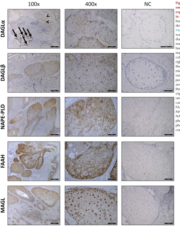
synthesis and degradation of AEA (NAPE-PLD and FAAH, respectively) and 2-AG (DAGLa and -band MAGL, respec- tively). We found that, irrespective of the confluence level of the cells, all the mentioned enzymes were expressed in hu- man sebocytes both at the mRNA (quantitative PCR) and protein (Western blot) levels (see Supplementary Figure S1aed online). To further confirm these results, we also investigated their expression in human skin samples (appropriately labeled positive controls are shown in Supplementary Figure S2online). With the sole exception of DAGLa, which exhibited questionable expression compared with the endogenous positive control sweat glands (Czifra et al., 2012), our findings nicely confirmed our in vitro data about the expression of these enzymes in human SGs in situ (Figure 1).
Sebocytes exhibit a pharmacologically inhibitable eCB uptake process
Next, we assessed whether pharmacologically inhibitable AEA uptake by the putative EMT (Chicca et al., 2012;
Nicolussi and Gertsch, 2015) was observable in these cells.
By monitoring the uptake of radiolabeled [3H]AEA by the cells, we found that acute inhibition (15 minutes, 10mmol/L) of AEA cellular uptake using the reference eCB transport in- hibitor UCM707 (Lo´pez-Rodrı´guez et al., 2003; Rau et al., 2016) was able to significantly alter the eCB uptake pro- cess. UCM707 reduced the intracellular [3H]AEA signal, which was accompanied by a significant increase in extra- cellular [3H]AEA levels compared with vehicle control. As a consequence of [3H]AEA uptake inhibition, the overall hy- drolysis of [3H]AEA by FAAH to arachidonic acid and [3H]
ethanolamine was reduced (Figure 2a). Theoretically, a decrease of [3H]AEA uptake and [3H]ethanolamine levels could be explained not only by the inhibition of the eCB transport process, but also by nonspecific inhibition of FAAH resulting in a reduced driving force for [3H]AEA uptake (Chicca et al., 2012; Nicolussi et al., 2014a). To exclude this possibility, we assessed how two different EMT inhibitors (UCM707 and VDM11, both widely used to abrogate cellular uptake of eCBs) (De Petrocellis et al., 2000; Lo´pez-Rodrı´guez et al., 2003) influence FAAH activity in SZ95 sebocytes compared with the reference FAAH inhibitor URB597 (Mor et al., 2004). We found that SZ95 sebocytes exhibit very low constitutive FAAH activity (2.320.25 pmol/minute/mg protein, n¼10), and that, unlike URB597, neither UCM707 nor VDM11 exerted substantial FAAH inhibition (half maximal inhibitory concentration values were above 25 mmol/L for both EMT inhibitors, whereas the half maximal inhibitory concentration of URB597 was below 100 nmol/L) (see Supplementary Table S1online). Taken together, these findings suggest that in human sebocytes, eCB uptake can be inhibited by VDM11, most likely in a FAAH-independent manner.
The EMT plays a role in the degradation of eCBs in human sebocytes
Next, we investigated the eCB transport process using one of the aforementioned EMT inhibitors, VDM11 (De Petrocellis et al., 2000). Cells were treated with this com- pound or vehicle for 24 hours, and eCB content of the samples was analyzed by liquid chromatography-mass N Za´ka´nyet al.
Sebocyte Endocannabinoid System
Journal of Investigative Dermatology (2018), Volume 138 1700
spectrometry. We found that VDM11 treatment significantly increased AEA content of the samples (Figure 2b), whereas elevation of 2-AG concentration did not reach statistical significance (Figure 2c). Besides the two major eCBs, we also investigated the presence and potential alterations of two ECS-related acylethanolamides, PEA and OEA. We found that treatment by VDM11 tended to increase PEA and significantly elevated OEA concentrations (Figure 2d and e).
Collectively, these data provided evidence that administra- tion of VDM11 can increase the levels of certain eCBs and eCB-like mediators in human sebocytes. These results suggest that VDM11 might therefore promote ho- meostatic eCB and eCB-like mediator signaling in these cells.
Administration of eCB membrane transport inhibitors mimics lipogenic actions of direct eCB treatment, whereas selective FAAH inhibition does not influence SLP
It is well described that direct eCB treatment of sebocytes results in dramatically increased SLP (Dobrosi et al., 2008;
Ola´h et al., 2014, 2016b). Thus, next we wanted to assess how treatment with eCB uptake inhibitors influences the functions of SGs. Using noncytotoxic concentrations (deter- mined by MTT assay; seeSupplementary Figure S3online) of the aforementioned VDM11, we explored its effect on SLP.
By using fluorescent Nile Red staining, we found that its low micromolar concentrations induced a moderate but significant increase in SLP after 48-hour treatments (Figure 3a). Repetition of the experiment with AM404
Figure 1. Major enzymes of the endocannabinoid metabolism are expressed in human sebaceous glands in situ.Immunohistochemistry of human skin sections was performed as described in theSupplementary Materials. Specific immunopositivity was visualized by 3,30-
diaminobenzidine (brown color), and nuclei were counterstained with hematoxylin (blue color). Original magnifications¼ 100 in the left column and400 in the middle and right columns. Scale bars¼200mm in the left column and 50mm in the middle and right columns. Arrows indicate sweat glands (endogenous positive control for DAGLa), and arrowheads mark sebaceous glands on the same image. Negative controls (right column) were obtained by omitting the primary antibody in all cases. DAGL, diacylglycerol lipase;
FAAH, fatty acid amide hydrolase;
MAGL, monoacylglycerol lipase;
NAPE-PLD,N-acyl
phosphatidylethanolamine-specific phospholipase D, NC, negative control.
N Za´ka´nyet al.
Sebocyte Endocannabinoid System
www.jidonline.org 1701
(another well-known AEA uptake inhibitor) (Beltramo et al., 1997; Nicolussi and Gertsch, 2015) yielded very similar re- sults (seeSupplementary Figure S4a and b online). Although both VDM11 and AM404 mimicked the lipogenic actions of the prototypic eCB AEA (Dobrosi et al., 2008), their effi- ciencies at elevating SLP were far exceeded by direct AEA treatment (Figure 3a).
As discussed, several lines of evidence show that eCB uptake inhibitors may concentration-dependently inhibit not only the putative EMT but FAAH as well (Beltramo et al., 1997; Chicca et al., 2017; Nicolussi and Gertsch, 2015).
Although our current measurements of the FAAH activity of sebocytes already provided strong evidence that (i) SZ95 sebocytes exhibit very low constitutive FAAH activity and (ii) in SZ95 sebocytes, both UCM707 and VDM11 can be used at 10mmol/L without the risk of having substantial impact on FAAH activity (seeSupplementary Table S1), we also inten- ded to investigate biological effects of the selective reference FAAH inhibitor URB597 (Mor et al., 2004). We found that
Figure 2. Effects of the inhibitors of the putative endocannabinoid membrane transporter in human sebocytes.(a) AEA transport measurement.
Cells were treated with the reference AEA uptake inhibitor UCM707 or vehicle for 15 minutes, and intra- and extracellular amounts of radiolabeled AEA or EtNH2were detected as described in theSupplementary Materials.
Results are expressed as the percentage of vehicle control (100%, solid line) as meanstandard error of the mean of three independent experiments, each run in triplicate. (bee) AEA, 2-AG, PEA, and OEA determinations of the samples (i.e., cells and their supernatants together) were performed as described in theSupplementary Materials. Results are expressed as mean standard error of the mean of 3 or 4 independent cultures. *P<0.05, **P<
0.01 and ***P<0.001 mark significant differences compared with the vehicle control. 2-AG, 2-arachidonoylglycerol; AEA, N-arachidonoylethanolamine (i.e., anandamide); EtNH2, ethanolamine; EMT, (putative) endocannabinoid membrane transporter; M, mol/L; n.s., not significant; OEA,
oleoylethanolamide; PEA, palmitoylethanolamide.
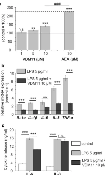
Figure 3. Noncytotoxic concentrations of VDM11 moderately but significantly increase sebaceous lipid synthesis and exert remarkable anti- inflammatory effects.(a) Sebaceous lipid production of SZ95 sebocytes was assessed by Nile Red staining after 48-hour treatments. Results are expressed as the percentage of the vehicle control (100%, solid line) as meanstandard error of the mean of four independent determinations. One additional experiment yielded similar results. **P<0.01 and ***P<0.01 mark significant differences compared with the vehicle control.###P<0.001.
(b) Quantitative PCR.IL-1a, IL-1b, IL-6, IL-8andTNF-amRNA expressions were determined after 3-hour LPS treatment with or without VDM11. Data are presented by using theDDCT method regarding18S RNA-normalized mRNA expressions of the vehicle control as 1 (solid line). Data are expressed as meanstandard deviation of three determinations. One additional experiment yielded similar results. **P<0.01 and ***P<0.001, as indicated.
(c) ELISA. IL-6 and IL-8 content of the sebocyte supernatants was determined after 24-hour LPS treatment with or without VDM11. Data are expressed as meanstandard deviation of three determinations. Two additional experiments yielded similar results. ***P<0.001, as indicated. AEA, N-arachidonoylethanolamine (i.e., anandamide); LPS, lipopolysaccharide;
M, mol/L; n.s., not significant compared with the vehicle control.
N Za´ka´nyet al.
Sebocyte Endocannabinoid System
Journal of Investigative Dermatology (2018), Volume 138 1702
administration of URB597 led to a different cell physiology outcome than application of VDM11 and AM404. Indeed, our data showed that noncytotoxic concentrations (MTT assay, see Supplementary Figure S5a online) of URB597 did not influence SLP (48-hour treatments, see Supplementary Figure S5b) compared with the vehicle control group, indi- cating that abrogation of eCB uptake, but not inhibition of their FAAH-mediated intracellular degradation, leads to the elevation of the SLP. Such lack of effect by URB597 can most likely be ascribed to the aforementioned very low levels of FAAH activity in SZ95 sebocytes.
Lipogenic action of direct AEA treatment is not further increased by co-administration of VDM11
Next, we assessed how co-administration of AEA and VDM11 affect SLP. We found that VDM11 was unable to further promote the AEA-induced, already elevated SLP (see Supplementary Figure S6 online) of human sebocytes, sug- gesting that the pro-lipogenic eCB signaling activated by 30 mmol/L AEA is exhaustive and has no further “reserve capacity.”
Up to 10mmol/L, VDM11 does not induce apoptosis of human sebocytes
Elevation of SLP is the hallmark of sebocyte differentiation, which is usually followed by programmed cell death (Dobrosi et al., 2008; Fischer et al., 2017; To´th et al., 2011;
Zouboulis et al., 2008, 2014). Because VDM11 was shown to moderately promote SLP (Figure 3a, and see Supplementary Figure S4b), we also wanted to know if it induced early apoptotic processes. We found that, although the most effective lipogenic concentration of VDM11 tended to decrease mitochondrial membrane potential in course of 48-hour treatments (seeSupplementary Figure S7online), this did not reach the level of significance, suggesting that within the studied timeframe, it may indeed be devoid of obvious pro-apoptotic effects.
VDM11 suppresses lipopolysaccharide (LPS)-induced proinflammatory cytokine expression of human sebocytes From a clinical point of view, induction of a moderate (not seborrheic or acnegenic) increase in the homeostatic SLP would be highly desirable in the treatment of skin dryness (Kim et al., 2014; Mischo et al., 2014; Shi et al., 2015;
Zampeli et al., 2012; Zouboulis and Boschnakow, 2001).
Thus, our results suggest that administration of VDM11 (and probably other EMT inhibitors too) may have beneficial ef- fects in such conditions. Considering that skin dryness is frequently accompanied by cutaneous inflammation (e.g., in the case of AD) (Peng and Novak, 2015; Sugiura et al., 2014), we finally investigated how VDM11 affects immune proper- ties of human sebocytes. To this end, we applied LPS (5mg/
ml, 3-hour treatments) (Ola´h et al., 2016b) to induce a proinflammatory response. Co-administration of VDM11 (10 mmol/L) efficiently suppressed LPS-induced expression of IL-1a, IL-1b, IL-6, IL-8, and TNF-a by human sebocytes (quantitative PCR) (Figure 3b). Moreover, as shown by sub- sequent ELISA analyses, VDM11 treatment significantly suppressed LPS-induced release of IL-6 and led to only a minor, nonsignificant decrease in IL-8 secretion (24-hour treatments) (Figure 3c). Concentrations of the other three
cytokines were below (TNF-a) or around (IL-1aand IL-1b) the detection limit of the respective assays (data not shown).
DISCUSSION
Skin dryness and the often accompanying overwhelming cutaneous inflammatory processes (e.g., in AD, etc.) can dramatically impair quality of life of many patients. On the other hand, appropriate moisturization and emollient treat- ment of the skin can alleviate symptoms, and in some cases, they are even able to prevent the onset of AD (Hoppe et al., 2015; Ola´h et al., 2017; Sawatzky et al., 2016; Sugiura et al., 2014). Although inappropriate epidermal ceramide produc- tion is thought to be the most important player in these processes, a growing body of evidence now supports the concept that dysregulation of SG functions and the subse- quent alterations in the SLP are also fundamental. Indeed, sebostasis, SG hypoplasia, and reduced SLP (with patholog- ically reduced squalene and wax ester content) were shown to occur in AD, and an inverse correlation between the prevalence of acne and AD (the former characterized by increased, and the latter by decreased, sebum production) was also observed (Shi et al., 2015). These findings, together with those showing that sebaceous lipids are important reg- ulators of the growth of cutaneous microbiota (Pappas, 2009), collectively suggest that controlled, moderate “sebostimula- tion” (ideally without altering the physiological composition of the sebum) may be beneficial in diseases characterized by skin dryness.
eCB signaling, a recently emerging regulator of cutaneous biology (Maccarrone et al., 2015), appears to be a very promising subject of study in this field. We have previously shown that (i) human sebocytes are able to produce the two major eCBs (AEA and 2-AG); (ii) CB2 receptor-coupled signaling contributes to the maintenance of homeostatic SLP; and (iii) direct AEA or 2-AG treatments dramatically increase lipogenesis (Dobrosi et al., 2008). On the other hand, (e)-cannabidiol and several further nonpsychotropic phytocannabinoids (e.g., (e)-D9-tetrahydrocannabivarin) were shown to normalize arachidonic acid- and other mediator-induced excessive lipid synthesis and exerted complex (combined lipostatic, anti-proliferative, and anti- inflammatory) anti-acne effects, whereas others (namely (e)-cannabigerol and (e)-cannabigerovarin) were able to slightly but significantly promote SLP (Ola´h et al., 2014, 2016b). Hence, in this study we aimed to unveil hidden as- pects of the ECS of human sebocytes.
Here, we provide evidence that major members of the ECS (i.e., NAPE-PLD, DAGLa and -b, MAGL, and FAAH) are expressed both in vitro in human sebocytes (see Supplementary Figure S1aed) and (with the sole exception of DAGLa, which exhibited a dubious immunostaining pattern), also in situ in SGs of the human skin (Figure 1), which nicely confirms the available murine data of MAGL (Ma et al., 2011) and FAAH (Wohlman et al., 2016) expression in SGs.
Moreover, besides the expression of the enzymes, we could also show that eCB transport is functionally active and pharmacologically inhibitable in human sebocytes (Figure 2a). This process is more likely to be involved in (re-) uptake/degradation rather than synthesis/release of the eCBs, because VDM11 (10 mmol/L for 24 hours) significantly N Za´ka´nyet al.
Sebocyte Endocannabinoid System
www.jidonline.org 1703
elevated AEA levels in the samples and tended to increase 2- AG concentration as well. Although this compound was proven to only negligibly alter FAAH activity in sebocytes (seeSupplementary Table S1), we also observed a significant elevation in OEA levels and a tendency toward increase in PEA concentrations (Figure 2bee).
Next, we tested the effects on the viability, lipid synthesis, and immune responses of human sebocytes. We found that noncytotoxic concentrations of VDM11 and AM404 (see Supplementary Figures S3,S4a, andS7) induced a moderate but significant increase in SLP (Figure 3a, and see Supplementary Figure S4b) and that VDM11 interfered with the proinflammatory effects of LPS (Figure 3b and c). Certain sebocyte-derived cytokines (e.g., IL-6) can induce differenti- ation of CD4þ/CD45RAþnaı¨ve T cells into T helper type 17 cells (Mattii et al., 2017), further supporting the concept that dysregulation of sebocyte biology can contribute not only to the development of acne, but also to other (partly) T helper type 17-driven inflammatory dermatoses, such as AD or psoriasis. Thus, the suppression of IL-6 expression and release that we have shown here (Figure 3b and c) is likely to be clinically relevant and promises to be beneficial in such conditions.
Collectively, these findings strongly suggest that abrogation of eCB degradation of sebocytes, and the subsequent eleva- tion of the “eCB-tone,” promotes SLP. However, quite unex- pectedly, by using the FAAH inhibitor URB597, we found that, although it was successful in suppressing the FAAH activity of the sebocytes (seeSupplementary Table S1), it had no effect on SLP (Supplementary Figure S5). An explanation of this unexpected finding remains to be uncovered in future targeted studies but may lie in the fact that relatively low levels of FAAH activity (measured as the capability of cell membranes to hydrolyze radiolabeled AEA) was detected in SZ95 sebocytes. Further possible interpretations about the possible underlying mechanisms are presented in the Supplementary Materialsonline.
As discussed, a moderate (i.e., not excessive, seborrheic/
acnegenic) elevation of physiological SLP would be highly desirable in the management of diseases accompanied by skin dryness (e.g., AD) (Pappas, 2009; Shi et al., 2015). The fact that the lipogenic effect of VDM11 was far weaker than those usually seen upon direct AEA (Figure 3a) or other lipogenic (e.g., 2-AG, arachidonic acid, and linoleic acidþ testosterone) treatments (Dobrosi et al., 2008; Ge´czy et al., 2012; Ola´h et al., 2014, 2016b), together with our reported anti-inflammatory effect (Figure 3b and c), indicates that VDM11 (and maybe other eCB transport inhibitors) may be beneficial in treating such diseases. Obviously, however, the exact impact of VDM11 treatment on the sebaceous lip- idome must be thoroughly investigated in future specific studies to exclude the possibility of its potential acnegenic transformation.
Anti-inflammatory effects of elevated eCB tone are not unprecedented in either murine or human skin. Since the groundbreaking work of Karsak et al. (2007), many other studies have shown that the homeostatic ECS is one of the master regulators of cutaneous immune responses, keeping under control local allergic and inflammatory processes (Ola´h et al., 2016a).
Moreover, the fact that VDM11 could significantly increase OEA and tended to elevate PEA levels (Figure 2d and e) highlights the possibility that SGs may contribute to the cutaneous PEA and OEA metabolism and supply of their local micromilieu with these pleiotropic anti-inflammatory mole- cules (Facci et al., 1995; Impellizzeri et al., 2015; Pontis et al., 2016; Yang et al., 2016). Based on these data, there is a possibility that SG hypoplasia and hypofunction observed in AD (Shi et al., 2015) may be accompanied not only by reduced sebum production, but also by impaired PEA (and/or OEA) supply, which might contribute to the development and worsening of atopic inflammation. Because a PEA-containing cream was recently shown to efficiently alleviate symptoms of AD patients (ATOPA study,Eberlein et al., 2008), clinical studies are urgently invited to explore the possible role of SG hypoplasia-derived putative PEA/OEA deficiency in the development of AD symptoms. Further speculations about the possible role and targets of PEA and OEA are mentioned in theSupplementary Materials.
Our findings show that (i) human sebocytes express the most important enzymes involved in eCB and eCB-like mediator metabolism; (ii) human sebocytes are involved in the cutaneous metabolism of PEA and OEA; (iii) VDM11 increases or tends to increase the levels of eCBs and related acylethanolamides; (iv) VDM11 induces a moderate increase in SLP; and (v) VDM11 induces remarkable anti- inflammatory actions in human sebocytes (see Supplementary Figure S8online). Thus, human SGs may be important in regulating the supply of key eCBs and related acylethanolamides to their tissue microenvironment, and targeting the ECS holds out promise to control cutaneous inflammation. All in all, our data should encourage the exploitation of selected, SG-targeting ECS modulators in appropriate clinical trials to alleviate symptoms of cutaneous diseases (e.g., AD) characterized by skin dryness and inflammation.
MATERIALS AND METHODS
Detailed descriptions of the applied materials and methods can be found in theSupplementary Materials. Briefly, lipid synthesis was investigated by fluorescent Nile Red staining, and viability and cell death were assessed by MTT assay and combined fluorescent DilC1(5)-SYTOX Green labeling, respectively. Gene expression was studied by quantitative PCR (mRNA level), ELISA, Western blot, and immunohistochemistry (protein level). The uptake of AEA into cells was determined by measuring the cellular uptake of radiolabeled AEA, as described before (Nicolussi et al., 2014a, 2014b), whereas levels of the eCBs were quantified by isotope dilution-liquid chro- matography coupled with single quadrupole mass spectrometric analysis (Marsicano et al., 2002), and FAAH activity was determined according to our previously established and optimized protocol (Ortar et al., 2003). Data were analyzed by Origin Pro Plus 6.0 software (Microcal, Northampton, MA) using the Student two-tailed, unpairedttest, andP-values less than 0.05 were regarded as sig- nificant differences. Graphs were plotted by using Origin Pro Plus 6.0 software. Primary human material was collected after obtaining written informed consent, adhering to the Helsinki Declaration, and after obtaining permission from the Institutional Research Ethics Committee and Government Office for Hajdu´-Bihar County N Za´ka´nyet al.
Sebocyte Endocannabinoid System
Journal of Investigative Dermatology (2018), Volume 138 1704
(document numbers: IX-R-052/01396-2/2012, IF-12817/2015, IF- 1647/2016, and IF-778-5/2017).
ORCIDs
Attila Ola´h:http://orcid.org/0000-0003-4122-5639 Simon Nicolussi:http://orcid.org/0000-0003-1260-3282 CONFLICT OF INTEREST
CCZ owns an international patent on the SZ95 sebaceous gland cell line (WO2000046353).
ACKNOWLEDGMENTS
This project was supported by Hungarian (Lendu¨let LP2011-003/2015, TA´MOP-4.2.4.A/2-11-1-2012-0001 National Excellence Program, National Research, Development and Innovation Office 120552, 121360, 125055, and GINOP-2.3.2-15-2016-00015 I-KOM Teaming) research grants. SN and JG were supported by National Centre of Competence in Research TransCure, Switzerland. AO’s work was supported by the Ja´nos Bolyai Research Schol- arship of the Hungarian Academy of Sciences. The authors are grateful to No´ra Czako´ for her expert contribution and to Rena´ta Uzonyi and Judit Szabo´-Papp for their technical support.
SUPPLEMENTARY MATERIAL
Supplementary material is linked to the online version of the paper atwww.
jidonline.org, and athttps://doi.org/10.1016/j.jid.2018.02.022.
REFERENCES
Beltramo M, Stella N, Calignano A, Lin SY, Makriyannis A, Piomelli D.
Functional role of high-affinity anandamide transport, as revealed by selective inhibition. Science 1997;277(5329):1094e7.
Chicca A, Marazzi J, Nicolussi S, Gertsch J. Evidence for bidirectional endocannabinoid transport across cell membranes. J Biol Chem 2012;287:
34660e82.
Chicca A, Nicolussi S, Bartholoma¨us R, Blunder M, Aparisi Rey A, Petrucci V, et al. Chemical probes to potently and selectively inhibit endocannabinoid cellular reuptake [e-pub ahead of print] Proc Natl Acad Sci USA 2017;114:
E5006e15.
Czifra G, Szo¨ll}osi AG, To´th BI, Demaude J, Bouez C, Breton L, et al. Endo- cannabinoids regulate growth and survival of human eccrine sweat gland- derived epithelial cells. J Invest Dermatol 2012;132:1967e76.
Dajnoki Z, Be´ke G, Kapita´ny A, Mo´csai G, Ga´spa´r K, Ru¨hl R, et al. Sebaceous gland-rich skin is characterized by TSLP expression and distinct immune sur- veillance which is disturbed in rosacea. J Invest Dermatol 2017;137:1114e25.
De Petrocellis L, Bisogno T, Davis JB, Pertwee RG, Di Marzo V. Overlap between the ligand recognition properties of the anandamide transporter and the VR1 vanilloid receptor: inhibitors of anandamide uptake with negligible capsaicin-like activity. FEBS Lett 2000;483:52e6.
Dobrosi N, To´th BI, Nagy G, Do´zsa A, Ge´czy T, Nagy L, et al. Endocanna- binoids enhance lipid synthesis and apoptosis of human sebocytes via cannabinoid receptor-2-mediated signaling. FASEB J 2008;22:3685e95.
Eberlein B, Eicke C, Reinhardt H-W, Ring J. Adjuvant treatment of atopic eczema: assessment of an emollient containing N-palmitoylethanolamine (ATOPA study). J Eur Acad Dermatol Venereol 2008;22:73e82.
Facci L, Dal Toso R, Romanello S, Buriani A, Skaper SD, Leon A. Mast cells express a peripheral cannabinoid receptor with differential sensitivity to anandamide and palmitoylethanolamide. Proc Natl Acad Sci USA 1995;92:3376e80.
Fischer H, Fumicz J, Rossiter H, Napirei M, Buchberger M, Tschachler E, et al.
Holocrine secretion of sebum is a unique DNase2-dependent mode of programmed cell death. J Invest Dermatol 2017;137:587e94.
Ge´czy T, Ola´h A, To´th BI, Czifra G, Szo¨ll}osi AG, Szabo´ T, et al. Protein kinase C isoforms have differential roles in the regulation of human sebocyte biology. J Invest Dermatol 2012;132:1988e97.
Hoppe T, Winge MCG, Bradley M, Nordenskjo¨ld M, Vahlquist A, To¨rma¨ H, et al. Moisturizing treatment of patients with atopic dermatitis and ich- thyosis vulgaris improves dry skin, but has a modest effect on gene expression regardless of FLG genotype. J Eur Acad Dermatol Venereol 2015;29:174e7.
Impellizzeri D, Ahmad A, Bruschetta G, Di Paola R, Crupi R, Paterniti I, et al.
The anti-inflammatory effects of palmitoylethanolamide (PEA) on endotoxin-induced uveitis in rats. Eur J Pharmacol 2015;761:28e35.
Karsak M, Gaffal E, Date R, Wang-Eckhardt L, Rehnelt J, Petrosino S, et al.
Attenuation of allergic contact dermatitis through the endocannabinoid system. Science 2007;316(5830):1494e7.
Kim J, Nakasaki M, Todorova D, Lake B, Yuan C-Y, Jamora C, et al. p53 In- duces skin aging by depleting Blimp1þsebaceous gland cells. Cell Death Dis 2014;5:e1141.
Kova´cs D, Lova´szi M, Po´liska S, Ola´h A, Bı´ro´ T, Veres I, et al. Sebocytes differentially express and secrete adipokines. Exp Dermatol 2016;25:
194e9.
Kurokawa I, Danby FW, Ju Q, Wang X, Xiang LF, Xia L, et al. New de- velopments in our understanding of acne pathogenesis and treatment. Exp Dermatol 2009;18:821e32.
Ligresti A, De Petrocellis L, Di Marzo V. From phytocannabinoids to canna- binoid receptors and endocannabinoids: pleiotropic physiological and pathological roles through complex pharmacology. Physiol Rev 2016;96:
1593e659.
Lo´pez-Rodrı´guez ML, Viso A, Ortega-Gutie´rrez S, Fowler CJ, Tiger G, de Lago E, et al. Design, synthesis, and biological evaluation of new inhibitors of the endocannabinoid uptake: comparison with effects on fatty acid amidohydrolase. J Med Chem 2003;46:1512e22.
Lupi O. Ancient adaptations of human skin: why do we retain sebaceous and apocrine glands? Int J Dermatol 2008;47:651e4.
Ma W-X, Yu T-S, Fan Y-Y, Zhang S-T, Ren P, Wang S-B, et al. Time- dependent expression and distribution of monoacylglycerol lipase during the skin-incised wound healing in mice. Int J Legal Med 2011;125:
549e58.
Maccarrone M. Metabolism of the endocannabinoid anandamide: open questions after 25 years. Front Mol Neurosci 2017;10:166.
Maccarrone M, Bab I, Bı´ro´ T, Cabral GA, Dey SK, Di Marzo V, et al. Endo- cannabinoid signaling at the periphery: 50 years after THC. Trends Phar- macol Sci 2015;36:277e96.
Marsicano G, Wotjak CT, Azad SC, Bisogno T, Rammes G, Cascio MG, et al.
The endogenous cannabinoid system controls extinction of aversive memories. Nature 2002;418(6897):530e4.
Mattii M, Lova´szi M, Garzorz N, Atenhan A, Quaranta M, Lauffer F, et al.
Sebocytes contribute to skin inflammation by promoting the differentiation of T helper 17 cells [e-pub ahead of print] Br J Dermatol 2017.https://doi.
org/10.1111/bjd.15879(accessed 20 February 2018).
Mischo M, von Kobyletzki LB, Bru¨ndermann E, Schmidt DA, Potthoff A, Brockmeyer NH, et al. Similar appearance, different mechanisms: xerosis in HIV, atopic dermatitis and ageing. Exp Dermatol 2014;23:446e8.
Mor M, Rivara S, Lodola A, Plazzi PV, Tarzia G, Duranti A, et al. Cyclo- hexylcarbamic acid 30- or 4’-substituted biphenyl-3-yl esters as fatty acid amide hydrolase inhibitors: synthesis, quantitative structure-activity relationships, and molecular modeling studies. J Med Chem 2004;47:
4998e5008.
Nicolussi S, Chicca A, Rau M, Rihs S, Soeberdt M, Abels C, et al. Correlating FAAH and anandamide cellular uptake inhibition using N-alkylcarbamate inhibitors: from ultrapotent to hyperpotent. Biochem Pharmacol 2014a;92:
669e89.
Nicolussi S, Gertsch J. Endocannabinoid transport revisited. Vitam Horm 2015;98:441e85.
Nicolussi S, Viveros-Paredes JM, Gachet MS, Rau M, Flores-Soto ME, Blunder M, et al. Guineensine is a novel inhibitor of endocannabinoid uptake showing cannabimimetic behavioral effects in BALB/c mice. Phar- macol Res 2014b;80:52e65.
Ola´h A, Ambrus L, Nicolussi S, Gertsch J, Tubak V, Keme´ny L, et al. Inhibition of fatty acid amide hydrolase exerts cutaneous anti-inflammatory effects both in vitro and in vivo. Exp Dermatol 2016a;25:328e30.
Ola´h A, Bı´ro´ T. Targeting cutaneous cannabinoid signaling in inflammation—
a “high”-way to heal? EBioMedicine 2017;16:3e5.
Ola´h A, Markovics A, Szabo´-Papp J, Szabo´ PT, Stott C, Zouboulis CC, et al. Differential effectiveness of selected non-psychotropic phyto- cannabinoids on human sebocyte functions implicates their introduc- tion in dry/seborrhoeic skin and acne treatment. Exp Dermatol 2016b;25:701e7.
N Za´ka´nyet al.
Sebocyte Endocannabinoid System
www.jidonline.org 1705
Ola´h A, Szabo´-Papp J, Soeberdt M, Knie U, Da¨hnhardt-Pfeiffer S, Abels C, et al. Echinacea purpurea-derived alkylamides exhibit potent anti- inflammatory effects and alleviate clinical symptoms of atopic eczema.
J Dermatol Sci 2017;88:67e7.
Ola´h A, To´th BI, Borbı´ro´ I, Sugawara K, Szo¨llo˜si AG, Czifra G, et al. Can- nabidiol exerts sebostatic and antiinflammatory effects on human sebo- cytes. J Clin Invest 2014;124:3713e24.
Ortar G, Ligresti A, De Petrocellis L, Morera E, Di Marzo V. Novel selective and metabolically stable inhibitors of anandamide cellular uptake. Bio- chem. Pharmacol 2003;65:1473e81.
Pappas A. Epidermal surface lipids. Dermatoendocrinol 2009;1:72e6.
Peng W, Novak N. Pathogenesis of atopic dermatitis. Clin Exp Allergy 2015;45:566e74.
Pontis S, Ribeiro A, Sasso O, Piomelli D. Macrophage-derived lipid agonists of PPAR-aas intrinsic controllers of inflammation. Crit Rev Biochem Mol Biol 2016;51:7e14.
Porter AM. Why do we have apocrine and sebaceous glands? J R Soc Med 2001;94:236e7.
Rau M, Nicolussi S, Chicca A, Gertsch J. Assay of endocannabinoid uptake.
Methods Mol Biol 2016;1412:191e203.
Sawatzky S, Schario M, Stroux A, Lu¨nnemann L, Zuberbier T, Blume- Peytavi U, et al. Children with dry skin and atopic predisposition: outcome measurement with validated scores for atopic dermatitis. Skin Pharmacol Physiol 2016;29:148e56.
Shi VY, Leo M, Hassoun L, Chahal DS, Maibach HI, Sivamani RK. Role of sebaceous glands in inflammatory dermatoses. J Am Acad Dermatol 2015;73:856e63.
Slominski A. Neuroendocrine system of the skin. Dermatology 2005;211:
199e208.
Slominski A, Wortsman J. Neuroendocrinology of the skin. Endocr Rev 2000;21:457e87.
Solymosi K, Ko¨falvi A. Cannabis: a treasure trove or Pandora’s box? Mini Rev Med Chem 2016;17:1223e91.
Sta¨nder S, Schmelz M, Metze D, Luger T, Rukwied R. Distribution of cannabinoid receptor 1 (CB1) and 2 (CB2) on sensory nerve fibers and adnexal structures in human skin. J Dermatol Sci 2005;38:
177e88.
Sugiura A, Nomura T, Mizuno A, Imokawa G. Reevaluation of the non- lesional dry skin in atopic dermatitis by acute barrier disruption: an abnormal permeability barrier homeostasis with defective processing to generate ceramide. Arch Dermatol Res 2014;306:
427e40.
To´th BI, Ola´h A, Szo¨llosi AG, Czifra G, Bı´ro´ T. “Sebocytes’ makeup”: novel mechanisms and concepts in the physiology of the human sebaceous glands. Pflu¨g Arch Eur J Physiol 2011;461:593e606.
Wohlman IM, Composto GM, Heck DE, Heindel ND, Lacey CJ, Guillon CD, et al. Mustard vesicants alter expression of the endo- cannabinoid system in mouse skin. Toxicol Appl Pharmacol 2016;303:
30e44.
Yang L, Guo H, Li Y, Meng X, Yan L, Zhang D, et al. Oleoylethanolamide exerts anti-inflammatory effects on LPS-induced THP-1 cells by enhancing PPARasignaling and inhibiting the NF-kB and ERK1/2/AP-1/STAT3 path- ways. Sci Rep 2016;6:34611.
Zampeli VA, Makrantonaki E, Tzellos T, Zouboulis CC. New pharmaceutical concepts for sebaceous gland diseases: implementing today’s pre-clinical data into tomorrow’s daily clinical practice. Curr Pharm Biotechnol 2012;13:1898e913.
Zouboulis CC, Baron JM, Bo¨hm M, Kippenberger S, Kurzen H, Reichrath J, et al. Frontiers in sebaceous gland biology and pathology. Exp Dermatol 2008;17:542e51.
Zouboulis CC, Boschnakow A. Chronological ageing and photoageing of the human sebaceous gland. Clin Exp Dermatol 2001;26:600e7.
Zouboulis CC, Katsambas AD, Kligman AM, editors. Pathogenesis and treat- ment of acne and rosacea. Berlin: Springer; 2014.
Zouboulis CC, Seltmann H, Neitzel H, Orfanos CE. Establishment and char- acterization of an immortalized human sebaceous gland cell line (SZ95).
J Invest Dermatol 1999;113:1011e20.
N Za´ka´nyet al.
Sebocyte Endocannabinoid System
Journal of Investigative Dermatology (2018), Volume 138 1706
1
SUPPLEMENTARY MATERIAL
Endocannabinoid tone regulates human sebocyte biology
Nóra Zákány1*, Attila Oláh1*, Arnold Markovics1, Erika Takács1, Andrea Aranyász1, Simon Nicolussi2, Fabiana Piscitelli3, Marco Allarà3, Ágnes Pór4, Ilona Kovács4, Christos C. Zouboulis5, Jürg Gertsch2, Vincenzo Di Marzo3, Tamás Bíró6#, Tamás
Szabó7#
1Departments of Physiology, 6Immunology and 7Pediatrics, Faculty of Medicine, University of Debrecen, Nagyerdei krt. 98. Debrecen, H-4032, Hungary; 2Institute of
Biochemistry and Molecular Medicine, NCCR TransCure, University of Bern, Bern, Switzerland; 3Institute of Biomolecular Chemistry, C.N.R., Pozzuoli, Italy; 4Department
of Pathology, Gyula Kenézy County Hospital, Debrecen, Hungary; 5Departments of Dermatology, Venereology, Allergology and Immunology, Dessau Medical Center,
Brandenburg Medical School Theodore Fontane, Dessau, Germany
*These authors contributed equally. #These authors contributed equally.
Correspondence should be addressed to Tamás Bíró (University of Debrecen, Egyetem tér 1. Debrecen, H-4032, Hungary; e-mail: biro.tamas@med.unideb.hu; phone/FAX:
+3652-417-159).
2 SUPPLEMENTARY METHODS
Materials
VDM11 ((5Z,8Z,11Z,14Z)-N-(4-Hydroxy-2-methylphenyl)-5,8,11,14- eicosatetraenamide) and AM404 (N-(4-Hydroxyphenyl)-5Z,8Z,11Z,14Z- eicosatetraenamide) together with AEA were purchased from Cayman Chemical Company (Ann Arbor, MI, USA). URB597 (Cyclohexylcarbamic acid 3'- (Aminocarbonyl)-[1,1'-biphenyl]-3-yl ester) and UCM707 ((5Z,8Z,11Z,14Z)-N-(3- Furanylmethyl)-5,8,11,14-eicosatetraenamide) were obtained from were obtained from Tocris Bioscience (Bristol, UK) or Cayman Chemical Company (Ann Arbor, MI, USA and Tallinn, Estonia; in case of AEA uptake assay); [ethanolamine-1-3H]-AEA (60 Ci/mmol) was purchased from American Radiolabeled Chemicals, Inc. (St. Louis, MO, USA); whereas manufacturer of γ-irradiated lipopolysaccharide from Escherichia coli 026:B6 (LPS) was Sigma-Aldrich (St. Louis, MO, USA), respectively. LPS was dissolved in filtered distilled water. The solvent of all other compounds was absolute ethanol (Sigma-Aldrich) except for the [3H]-AEA uptake assay where UCM707 was dissolved in DMSO. Control cultures were always treated with appropriate amount of vehicles.
Cell culturing
Human immortalized SZ95 sebocytes, originated from human facial sebaceous glands (Zouboulis et al., 1999; Tóth et al., 2011; Zouboulis et al., 2014), were cultured in Sebomed® Basal Medium (Biochrom, Berlin, Germany) supplemented with 10 (V/V)%
fetal bovine serum (Life Technologies Hungary Ltd., Budapest, Hungary), 1 mM CaCl2, 5 ng/ml human epidermal growth factor (Sigma-Aldrich), MycoZap™ Plus-CL (1:500;
3
Lonza, Budapest, Hungary). The medium was changed every other day, and cells were sub-cultured at 60-70% confluence.
Determination of intracellular lipids
For quantitative measurement of sebaceous (neutral) lipid content, cells (20,000 cells/well) were cultured in 96-well “black-well/clear-bottom” plates (Greiner Bio-One, Frickenhausen, Germany) in quadruplicates, and were treated with compounds as indicated. Subsequently, supernatants were discarded, cells were washed twice with phosphate-buffered saline (PBS; 115 mM NaCl, 20 mM Na2HPO4, pH 7.4; all from Sigma-Aldrich), and 100 μl of a 1 μg/ml Nile Red (Sigma-Aldrich) solution in PBS was added to each well. The plates were then incubated at 37°C for 20 min, and fluorescence was measured on FlexStation 3 multi-mode microplate reader (Molecular Devices, San Francisco, CA, USA). Results are expressed as percentage of the relative fluorescence units in comparison with the vehicle controls using 485 nm excitation and 565 nm emission wavelengths.
Determination of cellular viability
The viability of the cells was determined by measuring the conversion of the tetrazolium salt MTT (Sigma-Aldrich) to formazan by mitochondrial dehydrogenases.
Cells were plated in 96-well plates (20,000 cells/well) in quadruplicates, and were treated as indicated for 2 days. Cells were then incubated with 0.5 mg/ml MTT for 2 hrs, and concentration of formazan crystals (as an indicator of number of viable cells) was determined colorimetrically at 565 nm by using FlexStation 3 multi-mode
4
microplate reader (Molecular Devices). Results were expressed as percentage of vehicle controls regarded as 100%.
Determination of apoptosis
A decrease in the mitochondrial membrane potential is one of the earliest markers of apoptosis (Green and Reed, 1998; Susin et al., 1998). Therefore, to assess the process, mitochondrial membrane potential of SZ95 sebocytes was determined using 1,1′,3,3,3′,3′-hexamethylindodicarbo-cyanine iodide containing MitoProbe™ DilC1(5) Assay Kit (Life Technologies Hungary Ltd.). Cells (20,000 cells/well) were cultured in 96-well “black-well/clear-bottom” plates (Greiner Bio One) in quadruplicates and were treated as indicated for 48 hrs. After removal of supernatants, cells were incubated for 30 minutes with DilC1(5) working solution (50 μl/well), then washed with PBS, and the fluorescence of DilC1(5) was measured at 630 nm excitation and 670 nm emission wavelengths using FlexStation 3 multi-mode microplate reader (Molecular Devices).
Relative fluorescence values were expressed as percentage of vehicle controls regarded as 100%. As a positive control for apoptosis, we applied carbonyl cyanide m- chlorophenyl hydrazone (CCCP; Life Technologies Hungary Ltd.) dissolved in the DilC1(5) working solution (1:200 for 30 min).
Determination of necrosis
Necrotic processes were determined by SYTOX Green staining (Life Technologies Hungary Ltd.). The dye is able to penetrate (and then bind to the nucleic acids) only to necrotic cells with ruptured plasma membranes, whereas healthy cells with intact surface membranes show negligible SYTOX Green staining. Cells were cultured in 96-
5
well “black-well/clear-bottom” plates (Greiner Bio One), and treated as indicated for up to 48 hrs. Supernatants were then discarded, and the cells were incubated for 30 minutes with 1 μM SYTOX Green dye. Following incubation, cells were washed with PBS, the culture medium was replaced, and fluorescence of SYTOX Green was measured at 490 nm excitation and 520 nm emission wavelengths using FlexStation 3 multi-mode microplate reader (Molecular Devices). Relative fluorescence values were expressed as percentage of vehicle controls regarded as 100%. As a positive control for necrosis, lysis buffer (1:100 in the SYTOX Green working solution for 30 min; Life Technologies Hungary Ltd.) was applied.
Due to their spectral properties, DilC1(5) and SYTOX Green dyes were always administered together, enabling us to investigate necrotic and early apoptotic processes of the same cultures. Selective decrease of DilC1(5) intensity indicated mitochondrial depolarization (i.e. the onset of early apoptotic processes), whereas increase of SYTOX Green staining intensity revealed necrotic cell death.
RNA isolation, reverse transcription and quantitative “real-time” PCR (Q-PCR)
Q-PCR was performed on an ABI Prism 7000 sequence detection system (Applied Biosystems, Foster City, CA, USA) or Stratagene Mx3005P QPCR System (Agilent Technologies, Santa Clara, CA, USA) using the 5’ nuclease assay. Total RNA was isolated using TRIzol (Life Technologies Hungary Ltd.), DNase treatment was performed according to the manufacturer’s protocol, and then 1 μg of total RNA was reverse-transcribed into cDNA by using High Capacity cDNA Kit from Life Technologies Hungary Ltd. PCR amplification was performed by using the TaqMan primers and probes (assay IDs: Hs00174092_m1 for interleukin [IL]-1α,
6
Hs00174097_m1 for IL-1β, Hs00985639_m1 for IL-6, Hs00174103_m1 for IL-8, Hs00174128_m1 for tumor necrosis factor [TNF]-α; Hs00419593_m1 for NAPE-PLD;
Hs00391374_m1 for DAGLα; Hs00373700_m1 for DAGLβ, Hs00155015_m1 for FAAH and Hs00200752_m1 for MAGL) and the TaqMan universal PCR master mix protocol (Applied Biosystems). As internal control, transcripts of 18S RNA or peptidylprolyl isomerase A (PPIA) were determined (assay IDs: Hs03928905_g1 and Hs99999905_m1, respectively). The amount of the transcripts was normalized to those of the housekeeping gene using the ΔCT method. Finally, when indicated, the results were further normalized to the expression of the vehicle control (ΔΔCT method).
Determination of cytokine release (ELISA)
500,000 cells were seeded in Petri-dishes (d=15 mm) in 1.5 ml culture medium. On the next day, cells were treated as indicated. Supernatants were collected, and the released amounts of IL-1α, IL-1β, IL-6, IL-8, and TNFα were determined according to the manufacturers’ (IL-1β, IL-6, IL-8, and TNFα: BD Pharmingen, Franklin Lakes, NJ, USA; IL-1α: R&D Systems, Inc., Minneapolis, MN, USA) protocols. In case of IL-6 and IL-8, supernatants were diluted (1:50), whereas for the other three cytokines, non- diluted supernatants were used.
Western blotting
Cells were harvested in lysis buffer (20 mM Tris-HCl, pH 7.4, 5 mM EGTA, 1 mM 4- (2-aminoethyl) benzensulphonyl fluoride, protease inhibitor cocktail diluted 1:100, (all from Sigma-Aldrich) and the protein content was measured by a modified BCA protein assay (Pierce, Rockford, IL, USA). The samples were then subjected to sodium dodecyl
7
sulphate-polyacrylamide gel electrophoresis. 10% Mini Protean TGX gels (Bio-Rad, Hercules, CA, USA) were loaded with equal (40 μg) amount of protein per lane.
Samples were then transferred to nitrocellulose membranes, by using Trans-Blot® Turbo™ Nitrocellulose Transfer Packs and Trans-Blot Turbo™ System (both from Bio- Rad), and then probed with mouse-anti-human FAAH, rabbit-anti-human NAPE-PLD, MAGL and DAGLα (all from Abcam, Camebridge, UK), or goat-anti-human DAGLβ (Santa Cruz Inc., Heidelberg, Germany) specific primary antibodies (all overnight at 4°C in 1:200 in 5% milk containing PBS). As secondary antibody, horseradish peroxidase-conjugated rabbit IgG Fc segment-specific antibodies (developed in goat, 1:1000 in 5% milk containing PBS, Bio-Rad) were used, and the immunoreactive bands were visualized by a SuperSignal® West Pico Chemiluminescent Substrate enhanced chemiluminescence kit (Pierce) using a KODAK Gel Logic 1500 Imaging System (Eastman Kodak Company, Kodak, Tokyo, Japan).
Immunohistochemistry
The immunohistochemical investigation of NAPE-PLD, FAAH, MAGL (Novus Biologicals, Littleton, USA) and DAGLβ (Bioss Inc., Massachusetts, USA) was performed on 3 formalin fixed paraffin embedded, skin samples rich in sebaceous glands, all diagnosed as trichilemmal cyst. The expression pattern of DAGLα (Santa Cruz Biotechnology Inc., Santa Cruz, CA, USA) in sebaceous gland was examined on 3 frozen skin samples obtained from vertex.
Serial 4 μm thick sections were cut from paraffin blocks and frozen tissues as well.
Frozen sections were then fixed in pre-cooled acetone for 10 min, whereas heat-induced