III./7.3. Diagnosis
In this chapter, an introduction to the indications and
limitations of methods used in the diagnosis of nervous system tumors is provided.
Introduction
When studying this chapter, text and related figures should be studied together in order to become familiar with the characteristic neuroimaging signs of tumors.
Clinical symptoms and signs of nervous system tumors are usually not specific, therefore further examinations are needed to confirm the diagnosis. Although physical examination may correctly localize the lesion, naturally it doesn’t give information about the type of lesion.
Limitations and difficulties in the diagnostic process
The main problem in the diagnosis of the brain and spinal tumors is that the nervous system is covered with bony structures. However, this obstacle was overcome by the introduction of modern neuroimaging methods.
CT, MRI and nuclear medicinal methods provide images showing the location, size and relation to surrounding structures, and even metabolic features of tumors. Exact histological diagnosis is however not possible with these methods.
Neuroimaging methods
When a nervous system tumor is suspected, neuroimaging examination is indicated.
In Hungary, brain CT scan is used most often primarily because it is quick, and it has a relatively low expense and easy access. A CT scan is able to detect tumors because the X-ray absorption (density) of a tumor is different from that of normal brain tissue. The tumor itself is seen as a
“mass lesion” above a certain size, i.e. it causes displacement and compression of surrounding structures. If a tumor is suspected, an intravenous contrast agent should be injected. The enhancing effect of tumors is explained by the damage of the blood-brain barrier and the neovascularisation of the tumor. Extraaxial tumors (meningioma,
vestibular schwannoma) show intensive and homogeneous enhancement, because of the complete absence of blood-brain barrier. Certain tumors have a density identical to that of brain tissue, thus they are not or hardly seen in non-enhanced scans; using contrast agents, these tumors can be made visible.
Radiologic features of benign and malignant
Based on morphological characteristics, benign and malignant brain tumors can often be distinguished. Benign tumors are characterized by sharp borders, smooth surface, expansive growth, only a minimal reactive perifocal edema, homogenous structure and contrast
enhancement, and the absence of tumor necrosis or hemorrhage. On the other hand, malignant tumors are characterized by an inhomogeneous internal structure and contrast enhancement, irregular borders, marked
tumors perifocal oedema, and mass effect. It is important to emphasize that contrast enhancement alone does not differentiate between benign and malignant tumors, other morphological signs should be taken into account.
Based on CT images, it is not always possible to differentiate between primary and metastatic brain tumors, however metastasis is suspected if perifocal edema is relatively large compared to tumor size and a ring-like enhancement occurs. Multiple tumors are also more likely to be
metastases, however, 50% of metastases are solitary and primary tumors may also be multifocal.
The diagnostic value of CT is limited by several factors:
- compared to MRI, CT is less able to differentiate between tissues - small lesions are not seen because of limited spatial resolution - due to artifacts caused by the bony structures, posterior fossa lesions are difficult to examine
- the use of contrast agent is contraindicated in many patients - spinal cord cannot be tested properly
For these reasons, currently a CT scan is usually followed by MRI.
The advantages of MRI
MRI is the ideal choice in the diagnosis of tumors, because it is the most sensitive method in both brain and spinal lesions. It is often carried out after CT scans to clarify the nature of the lesion and to help surgical planning. Compared with CT scans, the ability of MRI to differentiate between tissues and its spatial resolution are significantly better. Bony structures do not cause disturbing artifacts in MRI, thus posterior fossa and spinal lesions can also be examined. However, the use of a contrast agent is also often important in MRI. The morphological characteristics of benign and malignant tumors are similar to those described with CT.
In case of metastases, it is a common situation that MRI shows multiple metastases where CT has only shown a solitary metastasis due to the significantly higher sensitivity and resolution of MRI.
Special MRI techniques In addition to routine MRI imaging, special techniques may provide important additional data about the location of the tumor and the tumor’s relationship to the surrounding structures. Multi-planar MRI imaging is the best way to visualize the relationship of the tumor to the surrounding structures, which is a valuable help to the neurosurgeon in planning the operation.
Effort has been made to develop special imaging techniques that may provide information about the histological type of the tumor.
MRI-spectroscopy (which is a complementary method to routine sequences) performs molecular analysis of a selected tissue volume.
Based on the presence or absence, or the ratio of some typical brain metabolites, MRI-spectroscopy may provide some information about the benign or malignant nature and the histological type of a tumor, but it does not replace histological examination.
The determination of hemispheric dominance and the exploration of eloquent areas are crucial in surgical planning, and special MRI methods provide non-invasive and valuable help for this purpose. Functional MRI (fMRI), using the BOLD (Blood Oxygenation Level Dependent)
technique, is able to display functioning brain areas, connecting function
The role of functional MRI in surgical planning
with morphology. This is also important because anatomical structures may be displaced due to the mass effect of tumors. During fMRI, the patient carries out a specific task, which causes local changes of blood circulation in brain areas functioning during the given task, thereby visualizing activated brain areas. With this method, the exact location of speech centers or the primary motor cortex can be determined.
Fig. 6: Identifying eloquent areas with fMRI (BOLD technique)
Preoperative and postoperative images of a right parietal oligodendroglioma, grade II, show activated brain areas during movement of the left hand.
(A) Based on the activity produced by movements of the right hand, the tumor has a precentral location. However, activity produced by movements of the left hand is along the
posterior border of the tumor. (B) After surgery, the previous tumor site is filled with cerebrospinal fluid. After the removal of the tumor, the paresis has resolved and left hand movements have normalized. Since a mass effect is no longer present, brain configuration has slightly changed, so the activities corresponding to the hand areas are located more
anterior in comparison to the preoperative image.
DTI
With the help of the Diffusion Tensor Imaging (DTI), central nervous system tracts can also be displayed (fiber tracking, MRI tractography).
DTI shows whether the tumor destroys, infiltrates, or merely dislocates an important tract, e.g. the corticospinal tract.
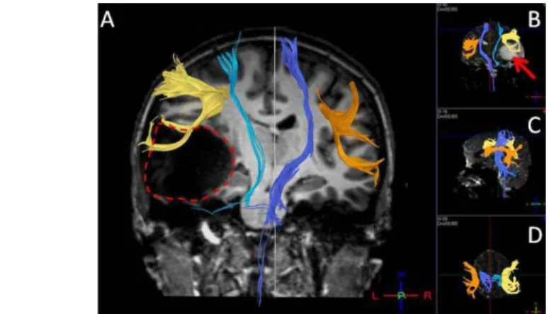
Fig. 7: Dislocation of tracts caused by a large left sided temporal ganglioglioma with a strong mass effect. (A) The tumor dislocates the left corticospinal tract (light blue) towards
the opposite side and the left arcuate fascicle (yellow) upwards. The tracts are shown on T1-weighted anatomical images, where the tumor (its borders shown with a red dashed line) appears as a low signal intensity mass. For help, the right corticospinal tract (dark blue) and the right arcuate fascicle (orange) are also plotted. (B, C, D) Images of tracts in
the three directions of space; the DTI derived measures were placed on T2-weighted images (b=0), in which the tumor appears as a high signal density area (red arrow in
figure B).
Differential diagnosis of tumors
Imaging also has an important role in the differential diagnosis of
tumors. Brain abscess, other infections (e.g. encephalitis, multiple sclerosis) or cerebrovascular diseases, various benign cystic lesions may appear as tumor-like, space-occupying lesions. In differentiating an abscess and a tumor, diffusion-weighted images (DWI) and
MRI-spectroscopy are helpful.
CT or MRI?
In most cases, MRI is the diagnostic technique of choice, but sometimes an additional CT examination is required if bone structure or
calcifications are areas of interest. Bone and calcifications are poorly seen with MRI.
The role of nuclear medicine
Nuclear medicine (SPECT, PET) also has a role in neuro-oncological diagnosis. It may provide functional information to complement
morphological CT and MR data. Nuclear medicine techniques may help in estimating the degree of tumor malignancy and in differential
diagnosis (e.g. tumor vs. inflammation or vascular lesion), and in post-operative follow-up (e.g. tumor relapse vs. scar tissue or radiation necrosis).
Current indications of angiography
Angiography (digital subtraction angiography, DSA) is not used today in the diagnosis of tumors. DSA may be indicated in special cases, if information about the tumor’s blood supply is important for treatment.
Furthermore, using endovascular methods, it is possible to occlude the major blood vessels supplying the tumor, which may lead to a reduction in size that facilitates surgical removal, or it may even result in complete necrosis that would make surgery unnecessary.
Indications for CSF examination
CSF examination is not part of the routine diagnosis of brain tumors, it is relatively infrequently performed. If the tumor reaches the surface of the brain or the ventricles, detached tumor cells may be detected in the CSF.
CFS examination is however necessary to confirm the diagnosis of meningeal carcinomatosis.
Electrophysiology
Electrophysiological studies are sometime needed during the process of patient evaluation to obtain functional information. For example, if the tumor is associated with epilepsy an EEG is indicated, or visual evoked potentials (VEP) show the degree of optic nerve lesion when compressed by a tumor. Likewise, brainstem acoustic evoked potential (BAEP) shows the degree of cochlear nerve dysfunction in case of a vestibular schwannoma, or somatosensory evoked potentials (SSEP) and motor evoked potentials (MEP) show spinal cord dysfunction in spinal tumors.
Endocrine tests
Hormone tests are necessary in pituitary gland and other sellar tumors.
Genetic testing
Genetic test can be requested if the nervous system tumor is part of an inherited disorder.
Brain biopsy
Imaging methods are able to detect and in most cases to differentiate intracranial lesions, however it is not equal to histological diagnosis.
Tissue samples for histological analysis may be obtained during surgery or via stereotactic biopsy if the indication of surgery is not clear and a tissue sample is needed to plan the treatment.
When is brain biopsy needed?
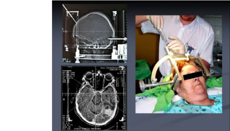
The procedure is carried out with a stereotactic targeting device fixed to the head. The device actually is a coordinate system, which allows the surgeon to perform the biopsy with high accuracy from areas selected with CT/MRI images.
The stereotactic “frame” is fixed to the cranial bones under local anesthesia, and then the imaging study is performed. On these images, both “frame” and the lesion are seen, and the exact coordinates of the sampling area is calculated. Sampling is carried out through a small drilled hole.
Fig. 8: Stereotactic biopsy. The coordinates are calculated by the CT images and the biopsy is being carried out.
How is it performed?
Throughout the intervention the patient is awake, using only local anesthesia. Complications (e.g. bleeding) are rare, they are under 1% rate in a large series. Before the intervention, antiplatelets and anticoagulants should be discontinued, coagulation parameters should be normal.
Twenty-four hours of observation is needed after the intervention.
If a brain abscess or CNS lymphoma is suspected based on neuroimaging studies, urgent biopsy is recommended.
Biopsy is also recommended in multiple brain tumors, where the primary tumor is unknown.
Among the tumors of the pineal region, germinoma is a common tumor type. It is treated with irradiation (not by surgical removal), which leads to complete remission. These patients often present with acute symptoms of elevated IC pressure and dilation of brain ventricles. With an
endoscopic method, both tumor sampling and – to treat the
hydrocephalus - fenestration of the third ventricle can be carried out.
Low malignant potential low-grade gliomas are also considered for biopsy, because the exact histological diagnosis may influence the planned therapy.
In summary, stereotactic biopsy is useful in obtaining histological diagnosis quickly and safely, which helps in planning the treatment.
For further reading
Illustrated, useful references for the imaging of tumors can be found at:
http://www.radiologyassistant.nl/en/47f86aa182b3a