Theoretical Design, Synthesis, and In Vitro Neurobiological Applications of a Highly E ffi cient Two-Photon Caged GABA Validated on an Epileptic Case
Balázs Chiovini,* Dénes Pálfi, Myrtill Majoros, Gábor Juhász, Gergely Szalay, Gergely Katona, Milán Szo ̋ri, Orsolya Frigyesi, Csilla Lukácsné Haveland, Gábor Szabó, Ferenc Erdélyi, Zoltán Máté, Zoltán Szadai, Miklós Madarász, Miklós Dékány, Imre G. Csizmadia, Ervin Kovács,* Balázs Rózsa,*
and Zoltán Mucsi*
Cite This:ACS Omega2021, 6, 15029−15045 Read Online
ACCESS
Metrics & More Article Recommendations*
sı Supporting InformationABSTRACT: In this paper, we present an additional, new cage-GABA compound, called 4-amino-1-(4′-dimethylaminoisopropoxy- 5′,7′-dinitro-2′,3′-dihydro-indol-1-yl)-1-oxobutane-γ-aminobutyric acid (iDMPO-DNI-GABA), and currently, this compound is the only photoreagent, which can be applied for GABA uncaging without experimental compromises. By a systematic theoretical design and successful synthesis of several compounds, the best reagent exhibits a high two-photon efficiency within the 700−760 nm range with excellent pharmacological behavior, which proved to be suitable for a complex epileptic study. Quantum chemical design showed that the optimal length of the cationic side chain enhances the two-photon absorption by 1 order of magnitude due to the cooperating internal hydrogen bonding to the extra nitro group on the core. This feature increased solubility while suppressing membrane permeability. The efficiency was demonstrated in a systematic, wide range of in vitro single-cell neurophysiological experiments by electrophysiological as well as calcium imaging techniques. Scalable inhibitory ion currents were elicited by iDMPO- DNI-GABA with appropriate spatial−temporal precision, blocking both spontaneous and evoked cell activity with excellent efficiency. Additionally, to demonstrate its applicability in a real neurobiological study, we could smoothly and selectively modulate neuronal activities during artificial epileptic rhythmsfirst time in a neural network of GCaMP6f transgenic mouse brain slices.
■
INTRODUCTIONCaged compounds are excellent tools to simulate and modulate neuronal activity patterns from the subcellular to network level.1 Although highly efficient excitatory caged molecules have already been synthesized,2−4 the development of inhibitory caged molecules for two-photon (2P) microscopy has proven to be a more difficult task.5,6 The previously reported photo-activable caged-GABA compounds are just at the limit of their usability, and they have exhibited several drawbacks in their stability, solubility, 2P uncaging efficiency, or antagonistic effect on GABAAreceptors.6−9During complex physiological brain activity patterns, excitatory and inhibitory processes appear in a delicate balance, which is upset during pathological progressions, such as epileptic activities. The 2P
uncaging technique provides an opportunity for rapid, light- initiated release of neurotransmitters.5,10−14
The precise, spatially and temporally controlled in situ release of neurotransmitters (mainly glutamate)15,16is a major benefit of the 2P uncaging technique, which enabled numerous studies of ion channel kinetics,17,18receptor distributions,19−21 synaptic transmission,21−24 synaptic integration,25−27 post-
Received: March 3, 2021 Accepted: May 20, 2021 Published: June 3, 2021
© 2021 The Authors. Published by
Downloaded via 193.224.44.80 on September 28, 2021 at 08:37:37 (UTC). See https://pubs.acs.org/sharingguidelines for options on how to legitimately share published articles.
synaptic mechanisms,28,29 and network circuitry.30−33 Caged glutamate is the most widely used caged neurotransmitter for possibly two main reasons. One is the obvious central role of glutamate in neural signaling, which renders it as the most needed tool. The second is the properties of the available caged glutamate compounds that permit precise research to be performed. These properties are the high uncaging efficiency, the low effect on target receptors in the absence of illumination, and a suitable pharmacological profile, all stemming from the structure of the caged molecule. So far, numerous cage scaffolds were developed simultaneously,3such as nitrobenzyl,34−36 coumarine,37−40 pyridine,5 β-carboline,41 and quinolone,7but nitroindoline-based (NI)9compounds are the most widely used. The nitroindilines mononitroindolyl glutamate (MNI-Glu)8,10,42,43and the improved dinitroindolyl glutamate (DNI-Glu)44,45show good pharmacological charac- ter, and they have been used successfully previ- ously.8,15,27,43,45−47
Compared to other photostimulation procedures (e.g., optogenetics), the advantage of 2P uncaging is the rapid, repeatable stimulation within a micron volume on a specific segment of the selected cell. Furthermore, it does not require altering the genetic code and protein composition of cells.
While cage molecules carrying the excitatory glutamate neurotransmitter have been developed for 2P experiments (e.g., DNI-Glu15,48 and MNI-Glu),8,10,42,43 relatively little attention and“success”have been achieved in the development of a cage compound carrying the inhibitory GABA molecule.
Although the focus has been on glutamate, GABA is also a prime candidate for a caged compound as it is fundamental in inhibitory−excitatory balance as the primary inhibitory neuro- transmitter. Several caged GABA compounds have been
reported previously in studies of GABA receptor kinetics and distribution in neural and glial compartments.9,34−36,38−40
However, these compounds exhibited a relatively low 2P uncaging efficiency and limited concentrations that can be employed without a substantial effect on GABAAreceptors in the absence of illumination and suboptimal pharmacological properties.6,8 Our ultimate goal is to design a photoreagent, which is able to modulate simulated and real activity patterns too. On one hand, it allows a better understanding of their structure−activity relationship. On the other hand, it opens new therapeutic possibilities for the elimination or correction of abnormal brain activity patterns such as epilepsy. Although optogenetic neuronal modulation of epileptic activity is increasingly used,49,50to understand how neurons are capable of classifying the excitatory and inhibitory inputs to their dendrites, both in health and disease, we need to use appropriate techniques. In the present work, we aimed to develop a novel caged GABA compound, with high uncaging efficacy and a reliable pharmacological profile besides the minimal off-target effect. For this reason, we have carried out a complex chemical and physiological validation process, including computational design, chemical synthesis, struc- ture−activity relationship, in vitro neurophysiological testing, and their functional application in an epileptic study. To be concise, we focused only on a few derivatives. For the physiological studies, we applied four modern methods: 2P imaging, GABA uncaging, electrophysiology, and the trans- genic mouse model, which was made in-house. As a result, we proved that 4-amino-1-(4′-dimethylaminoisopropoxy-5′,7′-di- nitro-2′,3′-dihydro-indol-1-yl)-1-oxobutane-γ-aminobutyric acid (iDMPO-DNI-GABA) is the most effective compound among the designed and prepared molecules. Finally, using Figure 1.Conceptual strategy of the present work. The present work is originated from DNI-Glu15,48retaining the effective dinitro substitution as DNI-GABA (1, in step I). Subsequently, we introduced the novel cationic side-chain conception as iDMPO-DNI-GABA (2a) and iDMBO-DNI- GABA (2b, step II) in contrast to the anionic ones.9,53Finally, we reverse the binding type of GABA as DNI-CO-GABA (3) in step (III).
iDMPO-DNI-GABA, we have shown the selective blockade of neuronal activity associated to epileptic rhythms.
■
RESULTS AND DISCUSSIONOur neurobiological goal was to selectively modulate the activity of neurons in a network during in vitro epileptic rhythms. Preliminary neurobiological studies and quantum chemical computations have shown only a moderate quantum yield on the previously reported caged GABA compounds (CDNI-GABA,40 DPNI GABA49), and they suffered by a number of side effects. Consequently, to realize our goal, we have carried out a structure−activity development on the caged scaffold by quantum mechanics (QM) methods in order to explore the best photoreagents, having high 2P absorption (TPA).
To meet the rigorous demands of 2P uncaging experiments, first we have designed several novel concept caged GABA compounds,51and their TPAs as well as photoreactivities were computed, with the aim to improve the uncaging efficacy.
Second, among many models, the four relevant derivatives were selected for synthesis and the subsequent multilevel neuropharmacological studies (1, 2a, 2b, 3). Our original starting point was the chemical structure of DNI-Glu, which was confirmed to be an outstanding caged glutamate compound reported in several publications by both exper- imental and QM methods.15,52Therefore, thefirst and directly derived GABA compound is DNI GABA (1,Figure 1, step I), which is used primarily as the reference point. According to our hypothesis, the TPA can be originated by the quadrupole moment of a molecule, which is in contrast with the one- photon transition, which is depending on the dipole moment.
To increase the TPA, we extended the size of the molecule in all dimensions in space and charge on the side chain (Figure 1, step II). An analogous strategy was attempted previously in the case of CDNI-GABA,8,47 extending the side chain of the indoline core; however, the incorporated negative side chain has yielded only little benefit. Contrary to the net neutral CDNI-GABA, we plan to include additional positively charged and branched side chains at position 4 (Figure 1, step II), with different lengths for compounds 2a and 2b. This resulted in the net double positive charge of these molecules, which effectively inhibits their penetration through the cell membrane and simultaneously increases its water solubility. For the fourth compound (3), we plan to block chemically the GABA amino group, which is assumed to be responsible for the undesired GABAA receptor activity. In this concept, we prepared a reversely bound nitroindolyl-caged-GABA with an introduced carbamide linker, DNI-CO-GABA (3,Figure 1, step III).
Quantum Chemical Modeling. Preparatory uncaging tests showed that GABA is successfully released from 1, 2a, and2bbut not from3, regardless of illumination strength. We hypothesized that the highly stable urea-type linker in 3 prevents the photochemical reaction, so this compound was excluded from further experiments.
In order to describe the remaining compounds1,2a, and2b, we carried out a detailed theoretical study involving 2P excitation (Dalton 2020.alpha)54 and mechanistic study (Gaussian 16)55 at the B3LYP/6-31G(d,p)//PCM(water) level of theory. Quantum chemical modeling has confirmed that the optimal length of the cationic side chain enhances TPA by 1 order of magnitude due to an internal hydrogen bonding (HB).
Conformational Study.Caged GABA derivatives2aand2b have numerous conformers at the GABA side chain; however, for the sake of simplicity, we selected only its linear GABA conformer, which represents the lowest energy arrangement.
Apart from this, the amino alkyl side chains can maintain two relevant conformers, a linear arrangement (I) and a ring arrangement (II), as shown in Scheme 1. The Gibbs-free-
energy difference of the two conformers was corrected by an explicit solvent model, resulting from the HB of water and the NO2 group. It is noteworthy that for 2a, the energetically preferred conformer is undoubtedly the cyclic 2a-II. For 2b, instead of the cyclic2b-II, the linear2b-Iconformer appears to be the more relevant conformer. This energy difference can be explained by the much stronger HBs in2a-II, in contrast to the solo HB in2b-II. In2a-II, the more optimal arrangement of the 9-member ring is beneficial, compared to the 10-member variant in2b. The weak HB for2b-IIis overcome by the HB effect of H2O to NO2, which was estimated as ca. 3 kJ mol−1by the same method (see theSupporting Information).
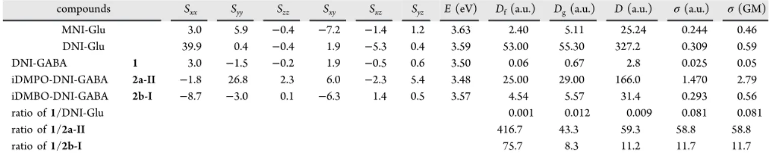
Quantum Chemical Modeling of TPA.We computed the TPA cross-section (σ) of DNI-GABA (1) and the most relevant conformers of iDMPO-DNI-GABA (2a-II) as well as iDMBO-DNI-GABA (2b-I) and compared to the recomputed values of MNI-Glu and DNI-Glu using the Dalton 2020.alpha program in the gas phase at the B3LYP/6-31G(d,p) level of theory.54 The 2P transition moment tensor components (S) were calculated and analyzed (Table 1).
The two diagonal tensor components SxxandSyy represent only high values for1, while other tensor components proved to be almost negligible, which corresponds to its rather flat chromophore. It appears that a charge transfer exists from OMe to NO2 along the direction x and it behaves as an antenna. For compounds 2a-II and 2b-I, more off-diagonal Scheme 1. Energetic Comparison of the Two Relevant Conformers of 2a and 2b; With Respect to the Linear Form (I), theΔG Values Are Corrected by the Explicit Effect of the Aqueous Solvation, Indicated by the Arrows (ΔG*)a
aHB = hydrogen bonding.
tensor components play important roles with higher values, referring to the more three-dimensional-shaped chromophores, compared to 1 and DNI-Glu, which contribute to the larger net transition probability values for these molecules.
The TPA cross-section values (σ) calculated from linear transition probability (D) and the excitation energy (E) (Table 2) are based on a method published earlier.56−59 Here, we
focused only on the dominant conformers of compounds2a-II and2b-I. The results showed about 58.8 times largerσvalue for iDMPO-DNI-GABA (2a-II) and 11.7 times largerσvalue for iDMBO-DNI-GABA (2b-I) than for DNI-GABA (1).
These results predict a 1 order of magnitude increase in TPA for theS0−S1 excitations of 2a and2b, which influences the overall photochemical process positively.
Quantum Chemical Modeling of the Photochemical Uncaging Process Versus the Ground-State Hydrolysis Mechanism. The reaction mechanism of the photochemical
release of GABA from1and the two forms of2aand2bwere modeled and compared by quantum chemical tools (Figure 2, Table 1) based on an earlier method.15 The explored photochemical mechanisms for these GABA derivatives were analogous to those of the previously published DNI-Glu. All the caged GABA derivatives (1,2a, and2b) followed the same pathways with close enthalpy levels according to the substituents and the conformers. The initial state A(S0) represents the ground state, and after the excitation, it follows the following sequence of states:A(S0)→B(S1)→C(S1)→ D(T1)→TS1→E(T1)→TS2→F(T1)→TS3→G(T1)
→ H(S0). This process involves the excitation [A(S0) → B(S1)], followed by the geometrical relaxation [B(S1) → C(S1)]. The high-energy excited singlet state C(S1)tends to transform to the energetically close triplet state D(T1) via intersystem crossing (ISC). In this triplet state, an acyl transfer reaction can occur preferably in two low-energy elementary steps [TS1→E(T1)→TS2→F(T1)], while the acyl group migrates from the indoline nitrogen to the nitro oxygen. The E(T1)minima were also confirmed in an earlier publication.52 The forming F(T1) triplet state is considerably stable, lying close to its ground-state analogue [I(S0)], which allows the spontaneous de-excitation via a second ISC. ThisF(T1)state also leads to the triplet product state [G(T1)] through TS3, resulting in the ground-state zwitterionic free GABA by the Table 1. Computed 2P Transition Tensor Components (S), 2P Transition Energy (E in eV), Probabilities (Df,Dgand Din a.u.) Cross-Section Values (σ in a.u.) for the S0−S1Transition of MNI-Glu, DNI-Glu, DNI-GABA (1), and the Relevant Conformers of iDMPO-DNI-GABA (2a-II) and iDMBO-DNI-GABA (2b-I) Computed at the B3LYP/6-31G(d,p) Level of Theory by Using Dalton 2020.alpha, Assuming a Monochromatic Light Source with Linear Polarization
compounds Sxx Syy Szz Sxy Sxz Syz E(eV) Df(a.u.) Dg(a.u.) D(a.u.) σ(a.u.) σ(GM)
MNI-Glu 3.0 5.9 −0.4 −7.2 −1.4 1.2 3.63 2.40 5.11 25.24 0.244 0.46
DNI-Glu 39.9 0.4 −0.4 1.9 −5.3 0.4 3.59 53.00 55.30 327.2 0.309 0.59
DNI-GABA 1 3.0 −1.5 −0.2 1.9 −0.5 0.6 3.50 0.06 0.67 2.8 0.025 0.05
iDMPO-DNI-GABA 2a-II −1.8 26.8 2.3 6.0 −2.3 5.4 3.48 25.00 29.00 166.0 1.470 2.79
iDMBO-DNI-GABA 2b-I −8.7 −3.0 0.1 −6.3 1.4 0.5 3.57 4.54 5.57 31.4 0.293 0.56
ratio of1/DNI-Glu 0.001 0.012 0.009 0.081 0.081
ratio of1/2a-II 416.7 43.3 59.3 58.8 58.8
ratio of1/2b-I 75.7 8.3 11.2 11.7 11.7
Table 2. Comparison of Biological Efficiency in IPSC Maximum Amplitude and Area (Relative to DNI-GABA, 1)
iDMPO-DNI-GABA,2a iDMBO-DNI-GABA,2b
IPSC max IPSC area IPSC max IPSC area
4.78 5.88 1.44 1.12
Figure 2. Left: Summary of the schematic potential enthalpy (ΔH) profile of the photochemical (read and blue) and ground-state (black) mechanism for compounds1,2a, and2bat the B3LYP/6-31G(d,p)//PCM(water) level of theory. Right: Computed enthalpy values (ΔH, in kJ mol−1) of different states (A−I,TS1−TS4) for compounds1,2a, and2b, relative to stateA(S0). ISC is estimated from the scanning of the reagent and reactant. (ainitial state;b,cestimated by scanning along the reaction coordinate).
N−O bond cleavage. G(T1) finally de-excites to the ground state [H(S0)]. The reactive species I(S0) can undergo a
ground-state rearrangement of theO-acyl form to the N-acyl, reaching back to the starting position A(S0) via TS4. The Scheme 2. Synthesis of the Three Cage-GABA Derivatives, iDMPO-GABA (2a), iDMBO-GABA (2b), and DNI-CO-GABA (3)
Figure 3.(A) Maximal intensityz-projection image of a cortical pyramidal neuronfilled with Alexa 594; the yellow dot indicates the location of the 2P GABA uncaging site in the presence of DNI-GABA (1) and iDMPO-DNI-GABA (2a); in 2.5 mM. (B) Representative example showing 2P uncaging (740 nm)-evoked somatic IPSCs of the same neuron in the presence of DNI-GABA (1) and iDMPO-DNI-GABA (2a) cage compounds, respectively. (C) Comparison of the uncaging-evoked IPSCs of iDMPO-DNI-GABA (2a) to that of DNI-GABA (1) and iDMBO-DNI-GABA (2b) compounds [n= 4, mean±standard error of the mean (SEM), respectively]. The green line represents the uncaging time. (D,E) Same data as in (C). Bar graphs of IPSC amplitude (left) and amplitude area (right), respectively. (F) Data of (D,E) are normalized to the IPSC amplitudes of iDMPO-DNI-GABA (2a). Normalized to iDMPO-DNI-GABA (2a): iDMBO-DNI-GABA (2b) and DNI-GABA (1) max: 0.31±0.11 and 0.22± 0.02 area: 0.19±0.09 and 0.16±0.02. Asterisks indicate significance (*p< 0.05,***p< 0.001).
process ofA→C→D→F→I→Acan be considered as an undesired“short circuit” of the uncaging process, which only emits and dissipates the laser energy to heat, recovering the starting state.
In conclusion of the photochemical pattern, no significant differences were found between the enthalpy profiles and their values of1,2a, and2b. However, the predicted TPA differed significantly; it proved to be 58.8 and 11.7 times larger values
for2aand2b, respectively, as compared to1. Based on these findings, three compounds were selected and subjected to biological testing, with predicted low (1), medium (2b), and high (2a) photoactivity.
Chemical Synthesis.The synthesis and the purification of DNI-GABA (1) were carried out according to a previously published pathway with a medium overall yield (35%, for details, see the Materials and Methods section).44 The Figure 4.(A) Image of the measured pyramidal neuronfilled with Fluo-4 and Alexa 594. The yellow dot indicates the location of the 2P GABA uncaging site in the presence iDMPO-DNI-GABA (2a) (2.5 mM). (B) Photolysis of iDMPO-DNI-GABA (2a) with 740 nm laser light (arrowhead) evokes IPSCs in the pyramidal neuron that is blocked by bath-applied gabazine (100μM); example traces (left), average responses from four cells (right) (n= 4, mean± SEM). Somatically evoked action potential (Ap) block: action potential amplitude before and after2a uncage: 76.50±4.88 and 31.24±0.63 mV, respectively (n= 3 cells),p= 0.0096. (C) Maximal intensityz-projection image of a cortical pyramidal neuronfilled with Fluo-4 and Alexa 594. The yellow dot represents the location of 2P uncaging, and the yellow line indicates the 2P imaging site along the dendrite. Somatically evoked Ap-associated backpropagated Ca2+transients before and after2auncage: 1.24±0.13 and 0.03±0.07 (in ΔF/F0), respectively (n= 3 cells),p= 0.00504. (D) Current injection induces somatically recorded single backpropagation action potential and triggers calcium transients at the proximal dendritic segment (left and right black traces, respectively). Uncaging of iDMPO-DNI GABA (2a) blocks somatically evoked backpropagation action potentials and the corresponding Ca2+signal (left and right red traces). (E) Somatically recorded spontaneous backpropagation action potentials can be blocked by single (left) or repetitive GABA uncaging (right).
synthesis of2aand 2bwas started on an analogous synthetic route (Scheme 2), building up the molecule by alkylation of the 4-hydroxy-indole (7) using 1-chloro-N,N-dimethylpropan- 2-amine or 3-chloro-N,N-2-trimethylpropan-1-amine in the presence of NaH. Then, indole derivatives were reacted with NaBH3CN, leading to the corresponding indoline derivatives 8a,b. The isolated8acontains minor (27%) regioisomer also, which is different on its side chain due to the formation of the asymmetric aziridine cation from 1-chloro-N,N-dimethylpro- pan-2-amine. In the subsequent step, they were acylated byN- Boc-GABA, resulting in the last intermediates (6a,b), respectively. Before the final dinitration step, the Boc protecting group was removed by trifluoro acetate (TFA), and then, the intermediates were nitrated by 3 equiv of NO2· BF4in dry dichloromethane (DCM) at a low temperature (0− 5 °C), yielding the desired products 2a and 2b. After purification by preparative high-performance liquid chroma- tography (HPLC) (eluent A: 0.1%TFA−water; eluent B:
MeCN), the overall yields were between 20 and 25% with an excellent chemical purity (>99%) as a TFA salt. The free GABA content was appropriately low (<1 ppm), confirmed by an HPLC method developed earlier.60 The minor and major isomers of2awere separated by preparative HPLC and tested in uncaging experiments. As the isomers yielded equivalent results,2awas subsequently used in biological experiments as an isomeric mixture. The synthesis of compound3followed a similar synthetic strategy, starting from the known 4-OMe- indoline 7 by reacting 1,1′-carbonyldiimidazole (CDI) and then GABA-OtBu. The resulting intermediate 13 was dinitrated by NO2·BF4in dry DCM, yielding thefinal product.
In all the cases, the last steps and the subsequent purification by preparative HPLC as well as lyophilization were carried out in darkness to avoid unwanted photochemical degradation that could lead to the appearance of free GABA. According to our accumulated experience,51 the TFA salt proved to be more stable and more resistant toward hygroscopic degradation in the solid form.
In VitroCharacterization of the Selected Caged GABA Compounds (1, 2a, and 2b). Neurophysiological experi- ments were carried out in three stages. In the primary assay, we compared the inhibitory postsynaptic currents (IPSCs) evoked with the three compounds (1, 2a, and 2b) under the same experimental conditions, which resulted in iDMPO-DNI- GABA (2a) as the most active candidate. In the secondary assay (the side-activity and applicability test), we examined the effect of 2a on intrinsic cellular properties and evaluated its ability to modulate neuronal input and output. Finally, we used iDMPO-DNI-GABA (2a) to modulate the participation of single neurons in a rhythmic, epileptic population activity in genetically modified, GCaMP6f mouse brain slices for thefirst time.
Primary Assay and the Activity Measurement: iDMPO- DNI-GABA (2a) Uncaging Evokes Significantly Larger IPSCs Compared to the Other Compounds. To select the most effective caged GABA compound for neurophysiological experiments, we compared IPSC amplitudes evoked with the three compounds on cortical pyramidal neurons (Figure 3) systematically under the standard conditions. Pairwise comparisons were made on the same neurons by wash-in and wash-out of the compounds (all at a 2.5 mM concentration) to get unquestionably comparable values. The spatial location and pattern of uncaging sites and the temporal order of their activation as well as all other parameters (e.g.,
laser intensity and pixel dwell time) were unchanged. Under these well-defined conditions, we found that uncaging of iDMPO-DNI-GABA (2a) evoked approximately 5-fold larger IPSCs on target neurons compared to the ones evoked by uncaging of DNI-GABA (1). This difference largely remained fixed when we compared iDMPO-DNI-GABA (2a) to iDMBO-DNI-GABA (2b), while uncaging of iDMBO-DNI- GABA (2b) evoked only marginally larger IPSCs compared to DNI-GABA (1, Figure 3D−F, in Table 2). Therefore, we selected iDMPO-DNI-GABA (2a) as the best candidate and limited further experiments on it exclusively. Moreover, the membrane permeability of the compound is very low.
Secondary Assay and the Compatibility and Applicability Test: iDMPO-DNI-GABA (2a) Effectively Modulates Neuronal Input and Output Signals without Significant Toxic Effects on the Basic Physiological Properties.In this stage, we tested extensively and thoroughly the effects and effectivity of iDMPO-DNI-GABA (2a) and characterized its ability to modulate neuronal input and output signals, the basic physiological properties, excitation wavelength dependence, laser power, reversal of the uncaging current, and the spatial precision on the soma and the dendrite. First, we tested how the bath application of2acould affect the basic physiological properties of pyramidal neurons (Figure S1). We patched and filled pyramidal cells with Fluo-4 Ca2+-sensitive and Alexa 594 anatomical dyes and measured thefluorescence and membrane potential changes to somatically injected current ramps (ramp test). We did notfind any significant difference in the resting membrane potential of patched neurons neither in the shape, frequency, half width, amplitude, rise time, and decay time of action potentials in the presence of 2a compared to control periods before wash-in (Figure S1B, Table S1). The Ca2+
fluorescence signals of evoked backpropagating action potentials also showed no significant differences in amplitude (52±0.4 vs 47±0.2%,n= 4 cells,p= 0.3) or area (111.66± 6.92 vs 102.96±2.89,n = 4 cells,p = 0.3) before and after application of iDMPO-DNI-GABA (2a;Figure S1C,D). Next, we validated that the effects of 2a uncaging were indeed mediated by released GABA. Irradiation at 740 nm for 1 ms near the cell body of a patched pyramidal neuron produced a robust outward current (Figure 4), which was abolished by bath application of 100 μM gabazine, a specific blocker of GABAAreceptors61(control: 48.72±16.977 pA vs gabazine:
3.77±1.92 pA, n= 4, p= 0.05) (Figure 4B).
Measuring the uncaging evoked IPSCs while holding the patched cells at different membrane potential values (10 mV intervals from 0 to−60 mV) showed that the reversal of the uncaging evoked currents are close to the reported chloride reversal potential62(−60.77±4.87 mV,n= 12 cells) (Figure S2). We also explored the wavelength dependence of photolysis-evoked IPSCs (Figure S3). Varying excitation wavelength between 700 and 820 nm showed that the amplitudes of IPSCs were the largest at 740 nm, revealing the 2P cross-section peak of iDMPO-DNI-GABA (2a) (Figure S3C). IPSC amplitudes also showed a linear relationship with applied laser power at a given wavelength (Figure S4). Next, we measured the spatial accuracy of the 2P uncaging of 2a (Figure S5). We denoted a series of points in a straight line at different distances from the soma (Figure S5C,D) or dendrite (Figure S6A,B) of pyramidal neurons and measured the IPSCs evoked by uncaging at these points one at a time. Uncaging closer to the soma evoked larger-amplitude IPSCs, and the IPSC amplitude approached 0 at a distance of 9.23±1.85μm
from the membrane of the soma (n = 6 cells). Apart from these, a further important question is whether iDMPO-DNI- GABA (2a) can block effectively neuronal output signals. To
test this, we somatically evoked single bAp-s and measured the associated calcium transients in the proximal dendritic segment (Figure 4D). Moreover, in another aspect, we raised the resting Figure 5.(A) Maximal intensityz-projection of the cortical region in Thy1-GCaMP6f transgenic mice for pyramidal cell population imaging in vitro. Yellow circles indicate measured pyramidal neurons. The red circle indicates the selectively modulated cell. (B) Representativefluorescence Ca2+responses recorded by multiple line scanning of pyramidal neurons during evoked epileptic events. The image shows the activity pattern of the simultaneously measured 18 pyramidal neurons in (A). Triangles indicate repetitive uncaging on the modulated cell (#3). (C) Ca2+transients from (B) with associated LFP recording (green). The red transient indicates the Ca2+signal of the cell modulated by iDMPO-DNI-GABA (2a), while the other neurons’activity remained intact (black). The colored bar represents the time interval of repetitive GABA uncaging. (D) Enlarged view from (C) shows the activities of cells#2, #3 (modulated), and #9. (E) Same maximal intensity z-projection image as in (A) shows another manipulated pyramidal cell,#5. Dashed circles indicate uncaging locations at the soma and at the edge of thefield of view as a reference [red (p1) and yellow (p2), respectively]. (F) Trace-by-trace modulation of the pyramidal cell activities by iDMPO-DNI-GABA (2a). The colored bar represents the time interval of uncaging. (G) Activities of neurons before, during, and after iDMPO-DNI-GABA uncaging (before: 0.46±0.04 Hz, during: 0.03±0.02 Hz, and after: 0.45±0.05 Hz,n= 12,p< 0.001). (H) Representative traces show cell#5 activities when the uncaging location was set at the cell [red; p1 on (E)] and at the reference uncaging location without a cell [yellow on (E); p2].
membrane potential of the pyramidal neurons, eliciting bursting, and then performed single or repetitive uncaging near the soma to prevent neuronal firing. We found that 2P uncaging of iDMPO-DNI-GABA (2a) is able to generate sufficient neuronal hyperpolarization to block single action potentials or even action potential burst (Figure 4D,E).
Furthermore, dendritic Ca2+ signals corresponding to blocked action potentials were also diminished (Figure 4D right).
According to the literature, we compared antagonist behavior of iDMPO-DNI-GABA (2a) and the well-known CDNI- GABA molecule.9,40 Both CDNI-GABA and iDMPO-DNI- GABA decreased the evoked IPSC amplitude and area significantly (Figure S8C−I). We found that the rate of changes in these self-controlled experiments was bigger in CDNI-GABA than in iDMPO-DNI-GABA. Besides this, we concluded that there is no significant (n.s.) difference between the two GABA cage compounds when compared to each other by their normalized data (Figure S8J). As we found that uncaging of iDMPO-DNI-GABA (2a) can evoke IPSCs repetitively with high spatial and temporal precision and could effectively block neuronal activity, which are necessary for selective and specific uncaging experiments, we moved on to test the compound in a functional experiment.
Tertiary Assay and the Direct Application: Rhythmic Epileptic Activity Can Be Selectively Modulated by iDMPO- DNI-GABA (2a). In the final stage, we have shown that even during large-scale pathological network activities, such as in vitro epileptic events, rapid and precise uncaging of GABA with iDMPO-DNI-GABA (2a) can modulate the participation of neurons in the ongoing network activity. Monitoring the activity of hundreds of neurons requires widespread labeling and fast sampling of individual neurons. Therefore, we used transgenic mice expressing GCaMP6f under the Thy1 promoter (Figure S7), which predominantly labels pyramidal cells,63 and multiple line scanning with 2P calcium imaging, combined with 2P uncaging. To generate population activity in brain slices, we washed in 4-aminopyridine (in 50μM), which triggered epileptic-like events involving most of the neuronal network (Figure 5). These rhythmic events were followed by LFP electrodes placed in close proximity to the imaging site.
After the pathological population activity was established, we selected a single, rhythmically active neuron and uncaged iDMPO-DNI-GABA (2a) near its soma locally, discretely, and repetitively. We found that uncaging suppressed the Ca2+
responses [before: 0.46 ± 0.04 Hz (mean ± SEM), during:
0.03 ± 0.02 Hz (mean ± SEM), and after: 0.45 ± 0.05 Hz (mean± SEM),n= 12 cells] of the targeted neuron reliably and its participation in the ongoing population activity was entirely blocked (Figure 5). This effect persisted through multiple successive uncaging periods, while previous activity resumed unchanged between suppressions. Other measured neurons were unaffected by this manipulation and maintained their activity during the uncaging periods. The selective modulation of cells was also sustainable trace by trace without any negative effect of their normal physiological conditions even during longer experiments (Figure 5E−H).
In summary, our novel caged GABA compound enables the reproduction of somatic or dendritic inhibitory inputs selectively and exclusively on individual cells due to the precise spatial and temporal control of 2P uncaging. As a result of thorough chemical development and biological testing, iDMPO-DNI-GABA (2a) is a well-tuned and potent GABA cage molecule.
■
CONCLUSIONSOur designed and synthetized cage GABA compound, iDMPO-DNI-GABA (2a), equipped with a novel cationic side chain, proved to be highly effective for the selective modulation of single or multiple neurons in physiological and epileptic networks. Quantum chemical study indicated that this compound could undergo the same photochemical mechanism upon excitation as DNI-Glu; however, its theoretical 2P cross- section increased by 1 order of magnitude for the optimal side- chain length. This benefit can be attributed to the cooperating internal HB.
An effective five-step synthesis was elaborated with a good overall yield, and the measured 2P efficiency of iDMPO-DNI- GABA (2a) proved to be very effective. The cationic side chain also increased its water solubility (>2.5 M in the buffer), while membrane permeability could be suppressing. The neuro- biological efficiency of the compound was demonstrated in a wide range of in vitro single-cell neurophysiological experi- ments by electrophysiological and calcium imaging. Scalable and stable inhibitory ion currents were elicited by iDMPO- DNI-GABA with appropriate spatial−temporal precision, blocking spontaneous and evoked cell activity with a high efficiency, with no significant side effects. With this structurally fine-tuned cage-GABA photoreagent, neuronal activities could be modulated selectively in GCaMP6f transgenic mouse brain slices during epileptic rhythms for thefirst time, allowing novel in vivo neuroscientific applications and therapeutic methods in the future.
■
MATERIALS AND METHODSChemical Synthesis and Analytical Methods. Amino acid derivatives were obtained from Bachem, and other chemicals were purchased from Sigma-Aldrich. Reagents were of the highest purity available. 1H, 13C, and NMR spectra were recorded in dimethyl sulfoxide-d6 (DMSO-d6) with a Bruker Avance III spectrometer operating at 500 and 125 MHz. High-resolution mass spectrometry (HRMS) spectra were recorded using an Agilent 6230 TOF LC/MS spectrometer. In some cases, preparative HPLC was applied, Agilent Prep HPLC, on a Gemini 250 ×50.00 mm; 10μm, C18, 110A column in 0.2% TFA or ammonium carbonate in water (eluent A) and the acetonitrile (eluent B) liquid phase using the gradient method.
Preparation of DNI-GABA (1).Synthesis of tert-Butyl (4- (4-Methoxyindolin-1-yl)-4-oxobutyl)carbamate (5). Synthe- sis of 4 has been previously reported.1 4-(tert- Butoxycarbonylamino)butyric acid, N-Boc-GABA (12.46 g, 61.3 mmol, 1.0 equiv), was dissolved in EtOAc (480 mL).
N,N-Diisopropylethylamine (11 mL, 63.1 mmol, 1.2 equiv) was added. The mixture was cooled to 0 °C; 1-ethyl-3-(3- dimethylaminopropyl)carbodiimide (EDC, 10.1 mL, 57.1 mmol, 0.95 equiv) was added, and the solution was stirred at room temperature for 10 min. 4-Methoxyindoline (4, 13.7 g, 92.0 mmol, 1.5 equiv) was added, and the mixture was stirred for 60 min. The mixture was cooled to 0°C; 1 M HCl was added (pH = 3); the phases were separated; and the organic phase was washed carefully with saturated aq NaHCO3 solution (200 mL), distilled water (2× 200 mL), and brine (200 mL). The organic phase was dried over anhydrous magnesium sulfate, and the solvent was evaporated under reduced pressure. This residue (12.10 g) was purified byflash chromatography (eluent: DCM and ethanol from 0 to 20%
using gradient elution) to give 11.75 g (57%) of6as a colorless oil.
1H NMR (500 MHz, DMSO-d6):δ(ppm) 7.70 (1H, d,J= 8.0 Hz, C[7]−H), 7.12 (1H, t, J = 8.1 Hz, C[6]−H), 6.83 (1H, br s, NH), 6.65 (1H, d,J= 8.2 Hz, C[5]−H), 4.05 (2H, t, J= 8.4 Hz, 2×C[1]−H), 3.78 (3H, s, 3×C[9]−H), 3.02− 2.96 (4H, m, 2×C[2]−H, 2×C[11]−H), 2.40 (2H, t,J= 7.0 Hz, 2×C[13]−H), 1.68 (2H, m, 2×C[12]−H), 1.37 (9H, s, 9× C[16]−H). 13C NMR (151 MHz, DMSO-d6): δ (ppm) 170.5 (C[10]), 155.6 (C[14]), 155.5 (C[4]), 144.3 (C[8]), 128.5 (C[6]), 118.0 (C[3]), 109.0 (C[7]), 105.9 (C[5]), 77.4 (C[15]), 55.1 (C[9]), 47.7 (C[1]), 39.9 (C[11]), 32.2 (C[2]), 28.2 (C[16]), 25.0 (C[12]), 24.6 (C[13]). HRMS (ES+): found 335.19627, C18H27O4N2 ([M + H]+) requires 335.19653.
Synthesis of 4-Amino-1-(4-methoxyindolin-1-yl)butan-1- one (6). tert-Butyl (4-(4-methoxyindolin-1-yl)-4-oxobutyl)- carbamate (5, 10.1 g, 30.0 mmol) was dissolved in TFA (30 mL), and the solution was stirred at room temperature for 60 min. Methanol (100 mL) was added, and the solvent was evaporated under reduced pressure. Methanol (100 mL) was added again, and the solvent was evaporated. Diethyl ether (50 mL) was added, and the solid precipitate wasfiltered, washed with diethyl ether, and dried. This residue (TFA salt of7, a colorless solid, 9.67 g, 92%) was used without further purification.
1H NMR (500 MHz, DMSO-d6):δ(ppm) 7.87 (3H, br s, NH3), 7.70 (1H, d,J= 8.2 Hz, C[7]−H), 7.13 (1H, t,J= 8.2 Hz, C[6]−H), 6.67 (1H, d,J= 8.2 Hz, C[5]−H), 4.06 (2H, t, J= 8.6 Hz, 2×C[1]−H), 3.78 (3H, s, 3 ×C[9]−H), 3.01 (2H, t,J= 8.6 Hz, 2×C[2]−H), 2.88 (2H, m, 2×C[11]− H), 2.55 (2H, t,J= 7.0 Hz, 2×C[13]−H), 1.85 (2H, m, 2× C[12]−H).13C NMR (151 MHz, DMSO-d6):δ(ppm) 169.9 (C[10]), 155.5 (C[4]), 144.1 (C[8]), 128.6 (C[6]), 118.1 (C[3]), 109.0 (C[7]), 106.1 (C[5]), 55.2 (C[9]), 47.6 (C[1]), 38.5 (C[11]), 31.6 (C[2]), 24.6 (C[13]), 22.0 (C[12]). HRMS (ES+): found 235.14392, C13H19O2N2([M + H]+) requires 235.14410.
Synthesis of 4-Amino-1-(4-methoxy-5,7-dinitroindolin-1- yl)butan-1-one, DNI-GABA (1).The solution of compound6 (0.5 g; 2.1 mmol) in acetonitrile (6 mL) was cooled to 10°C, and nitronium tetra-fluoroborate (1.11 g; 8.4 mmol, 4 equiv) was added portionwise. The reaction mixture was stirred at room temperature for 60 min. After the reaction was completed, NaHCO3 (1.50 g) was added and stirred for 30 min. The mixture was filtered and evaporated. After evaporation, the residue was purified by preparative HPLC (water−acetonitrile−0.1% TFA, using the gradient method).
After purification, the fractions were lyophilized. DNI-GABA· TFA1 was isolated as a yellow powder (0.14 g, yield 20%).
1H NMR (500 MHz, DMSO-d6): δ (ppm) 8.35 (1H, s, C[6]−H), 7.78 (3H, br s, 3×NH), 4.31 (2H, t,J= 8.2 Hz, 2
×C[1]−H), 4.02 (3H, s, 3×C[9]−H), 3.40 (2H, t,J= 8.2 Hz, 2×C[1]−H), 2.83 (2H, m, 2×C[11]−H), 2.69 (2H, t,J
= 7.0 Hz, 2×C[13]−H), 1.83 (2H, p,J= 7.5 Hz, 2×C[12]− H). 13C NMR (126 MHz, DMSO-d6): δ (ppm) 170.9 (C[10]), 151.9 (C[4]), 139.7 (C[7]), 137.5 (C5), 133.8 (C[8]), 129.4 (C[6]), 121.5 (C[3]), 61.0 (C[9]), 49.8 (C[1]), 38.2 (C[11]), 31.5 (C[12]), 26.8 (C[13]), 22.2 (C[2]). HRMS (ES+): found 325.11357, C13H17O6N4([M + H]+) requires 325.11426.
Synthesis of iDMPO-DNI-GABA (2a). Synthesis of 1- ((1H-Indol-4-yl)oxy)-N,N-dimethylpropan-2-amine (8a).
NaOMe (2.0 g, 41.25 mmol, 1.1 equiv) was dissolved in anhydrous MeOH (15 mL) and toluene (50 mL), and 4- hydroxyindole (7; 5.0 g, 3.75 mol, 1.0 equiv) in toluene (50 mL) was added. The mixture was stirred at reflux temperature for 60 min. 1-Chloro-N,N-dimethylpropan-2-amine (4.56 g, 3.75 mmol, 1.0 equiv) in toluene (3 mL) was added. The reaction mixture was stirred at reflux temperature for 2.5 h and cooled to room temperature. 100 mL of ethyl acetate was added, and the mixture wasfiltered through a perlite layer. The organic solution was washed with saturated aqueous sodium carbonate solution (50 mL) and water (3×50 mL) and dried over anhydrous magnesium sulfate, and the solvent was evaporated under reduced pressure. This residue (3.0 g, 37%) was used without further purification. Pure 8a was isolated by preparative HPLC (eluent water−acetonitrile− 0.1% TFA, gradient method).
1H NMR (500 MHz, DMSO-d6):δ(ppm) 11.18 (1H, br s, indole NH), 10.06 (1H, br s, aliphatic NH), 7.26−7.24 (1H, m, C[6]−H), 7.07 (1H, d,J= 8.2 Hz, C[7]−H), 7.03−7.00 (1H, m, C[1]−H), 6.56−6.55 (2H, m, C[2]−H, C[5]−H), 4.37 (1H, dd,J= 11.2, 3.9 Hz, C[9]−H), 4.29 (1H, dd,J= 11.2, 6.5 Hz, C[9]−H), 2.87 (6H, m, 6 ×C[12]−H), 2.84− 2.81 (1H, m, C[10]−H), 1.40 (3H, d,J= 6.9 Hz, 3×C[11]− H).13C NMR (126 MHz, DMSO-d6):δ(ppm) 150.9, 137.4, 123.8, 121.6, 118.3, 105.7, 100.3, 98.4, 66.3, 59.7, 40.1, 10.9.
HRMS (ES+): found 219.14882, C13H19ON2 ([M + H]+) requires 219.14919.
Synthesis of 1-(Indolin-4-yloxy)-N,N-dimethylpropan-2- amine (9a). 1-((1H-Indol-4-yl)oxy)-N,N-dimethylpropan-2- amine (8a, 3.00 g, 13.7 mmol, 1 equiv) was dissolved in acetic acid (15 mL), and the mixture was cooled down to 0°C.
Sodium cyanoborohydride (0.86 g, 13.7 mmol, 1 equiv) was added, and the mixture was stirred at room temperature for 60 min. The mixture was cooled to 0°C, and aqueous sodium hydroxide (20 m/m %, ca. 35 mL) was added (pH = 9). The organic compounds were extracted by DCM (4×40 mL), the combined organic layer was dried over anhydrous magnesium sulfate, and the solvent was evaporated under reduced pressure. This residue (9a, 3.0 g, 99%) was used without further purification. HRMS (ES+): found 221.16445, C13H21ON2([M + H]+) requires 221.16484.
Synthesis of tert-Butyl (4-(4-(2-(Dimethylamino)propoxy)- indolin-1-yl)-4-oxobutyl)carbamate (10a). 4-(tert- Butoxycarbonylamino)butyric acid, N-Boc-GABA (3.99 g, 19.6 mmol, 1.4 equiv), was dissolved in EtOAc (60 mL).
N,N-Diisopropylethylamine (5.7 mL, 32.7 mmol, 2.35 equiv) was added. The mixture was cooled to 0°C; EDC (5.7 mL, 32.4 mmol, 2.35 equiv) was added, and the solution was stirred at room temperature for 10 min.9aindoline (3.0 g, 13.6 mmol, 1.0 equiv) in EtOAc (80 mL) was added dropwise, and the mixture was stirred for 60 min. The mixture was cooled to 0
°C; 1 M HCl was added (pH = 3); the phases were separated;
and the organic phase was washed carefully with saturated aq NaHCO3solution (40 mL), distilled water (3×40 mL), and brine (50 mL). The organic phase was dried over anhydrous magnesium sulfate, and the solvent was evaporated under reduced pressure. This residue (4.87 g) was used without further purification. Pure 10a was isolated by preparative HPLC (eluent water−4 g NH4HCO3/10 L water−acetonitrile, gradient method).
1H NMR (300 MHz, DMSO-d6):δ(ppm) 7.69 (1H, d,J= 7.8 Hz, Ar-H), 7.10 (1H, t,J= 8.1 Hz, Ar-H), 6.84 (1H, br s, NH), 6.67 (1H, d, J = 8.2 Hz, Ar-H), 4.12−4.00 (3H, m,
C[1]−H, 2×C[9]−H), 3.86 (1H, dd,J= 9.7, 6.3 Hz, C[1]− H), 3.07−2.93 (4H, m, 2×C[14]−H, 2×C[16]−H), 2.90 (1H, q,J= 6.2 Hz, C[10]−H), 2.41 (2H, t,J= 6.9 Hz, 2 × C[2]−H), 2.25 (6H, s, 6×C[12]−H), 1.68 (2H, q,J= 6.9 Hz, 2×C[15]−H), 1.38 (9H, s, 9×C[19]−H), 1.05 (3H, d,J
= 6.8 Hz, 3×C[11]−H).13C NMR (75 MHz, DMSO-d6):δ (ppm) 170.4, 155.6, 154.8, 144.3, 128.5, 118.4, 109.0, 106.9, 77.4, 69.7, 57.5, 47.6, 41.1, 32.2, 28.2, 24.7, 24.4, 12.3. HRMS (ES+): found 406.26911, C22H36O4N3 ([M + H]+) requires 406.27003.
Synthesis of 4-Amino-1-(4-(2-(dimethylamino)propoxy)- indolin-1-yl)butan-1-one (11a).10a(4.87 g, 12.0 mmol) was dissolved carefully in TFA (15 mL) at 0°C, and the solution was stirred at room temperature for 60 min. Methanol (20 mL) was added, and the solvent was evaporated under reduced pressure. Methanol (20 mL) was added again, and the solvent was evaporated. The TFA salt of11a, a brownish oil (3.52 g, 55%), was used without further purification. Pure 11a was isolated by preparative HPLC (water−4 g NH4HCO3/10 L water−acetonitrile, gradient method).
1H NMR (300 MHz, DMSO-d6):δ(ppm) 10.26 (1H, br s, NH), 7.93 (2N, br s, NH2), 7.74 (1H, d,J = 7.8 Hz, Ar-H), 7.15 (1H, t,J= 8.1 Hz, Ar-H), 6.72 (1H, d,J= 8.2 Hz, Ar-H), 4.41−4.17 (10H, m, 2×C[9]−H), 4.08 (2H, t,J= 8.3 Hz, 2× C[1]−H), 3.80 (1H, q,J= 4.7 Hz, C[10]−H), 3.09 (2H, t,J= 8.3 Hz, 2×C[16]−H), 2.96−2.79 (8H, m, 2×C[14]−H, 6× C[12]−H), 2.56 (2H, t,J= 6.8 Hz, 2×C[2]−H), 1.86 (2H, q, J= 6.9 Hz, 2×C[15]−H), 1.36 (3H, d, J= 6.9 Hz, 3× C[11]−H).13C NMR (75 MHz, DMSO-d6):δ(ppm) 170.06, 153.87, 144.36, 128.67, 118.88, 109.80, 107.25, 66.35, 59.61, 47.64, 38.50, 31.75, 24.59, 22.04, 10.91. HRMS (ES+): found 306.21753, C17H28O2N3([M + H]+) requires 306.21760.
Synthesis of 4-Amino-1-(4-(2-(dimethylamino)propoxy)- 5,7-dinitroindolin-1-yl)butan-1-one (2a). The solution of compound 11a (8.0 g; 26.1 mmol) in acetonitrile (80 mL) was cooled to 10°C, and nitronium tetra-fluoroborate (6.90 g;
52.2 mmol, 2 equiv) was added portionwise. The reaction mixture was stirred at room temperature for 4 h. After the reaction was completed, THF (70 mL) and NaHCO3(17 g) were added and stirred for 30 min. The mixture wasfiltered, dried over MgSO4, and evaporated. The isomer mixture of2a and2a_2(3.50 g, 34%) was obtained as a brownish oil. Pure 2a and 2a_2 were separated by preparative HPLC (eluent water−4 g NH4HCO3/10 L water−acetonitrile, gradient method).
For 2a: 1H NMR (400 MHz, DMSO-d6): δ (ppm) 9.95 (1H, br s, NH), 8.41 (1H, s, C[6]−H), 7.81 (3H, br s, 3× NH3), 5.04−5.00 (1H, m, C[10]−H), 4.39−4.30 (2H, m, 2× C[9]−H), 3.45−3.39 (2H, m, 2×C[1]−H), 3.38−3.34 (2H, m, 2×C[16]−H), 2.90 (6H, s, 6×C[12]−H), 2.86 (2H, br s, 2×C[15]−H), 2.70 (2H, t,J= 7.0 Hz, 2×C[2]−H), 1.89− 1.77 (2H, m, 2× C[14]−H), 1.19 (3H, d, J= 6.4 Hz, 3 × C[11]−H). 13C NMR (101 MHz, DMSO-d6): δ (ppm) 171.07, 147.70, 139.67, 138.36, 134.32, 130.82, 121.97, 73.64, 61.05, 49.80, 39.4, 38.23, 31.64, 27.15, 22.18, 17.09. HRMS (ES+): found 396.18743, C17H26N5O4+ ([M + H]+) requires 396.18776.
For2a_2:1H NMR (400 MHz, DMSO-d6):δ(ppm) 10.01 (1H, br s, NH), 8.42 (1H, s, C[6]−H), 7.83 (3H, br s, 3× NH3), 4.50−4.42 (1H, m, C[9]−H), 4.34 (2H, t,J= 8.0 Hz, 2
×C[10]−H), 3.45−3.39 (2H, m, 2×C[1]−H), 3.43 (2H, t,J
= 8.0 Hz, 2×C[16]−H), 2.90−2.80 (8H, m, 6×C[12]−H, 2
×C[15]−H), 2.71 (2H, t,J= 7.8 Hz, 2×C[2]−H), 1.89−
1.77 (2H, m, 2× C[14]−H), 1.35 (3H, d, J= 6.8 Hz, 3 × C[11]−H). 13C NMR (101 MHz, DMSO-d6): δ (ppm) 171.07, 150.00, 140.95, 137.23, 134.26, 130.44, 122.13, 71.31, 60.01, 49.83, 39.40, 38.18, 31.58, 26.76, 22.13, 10.30. HRMS (ES+): found 396.18830, C17H26N5O4+ ([M + H]+) requires 396.18776.
Synthesis of iDMBO-DNI-GABA (2b). Synthesis of 3- ((1H-Indol-4-yl)oxy)-N,N,2-trimethylpropan-1-amine (8b).
NaOMe (8.0 g, 165 mmol, 1.1 equiv) was dissolved in anhydrous MeOH (80 mL) and toluene (80 mL), and 4- hydroxyindole (20.0 g, 150 mol, 1.0 equiv) in toluene (100 mL) and anhydrous MeOH (80 mL) were added. The mixture was stirred at reflux temperature for 60 min. 3-Chloro-N,N,2- trimethylpropan-1-amine (57.3 g, 37.5 mmol, 2.5 equiv) in xylene (50 mL) was added. The reaction mixture was stirred at reflux temperature for 6 h and cooled to room temperature.
100 mL of ethyl acetate was added, and the mixture was filtered through a perlite layer. The organic solution was washed with saturated aqueous sodium carbonate solution (200 mL) and water (3×100 mL) and dried over anhydrous magnesium sulfate, and the solvent was evaporated under reduced pressure. Hexane (70 mL) was added, and the solid precipitate was filtrated and dried. This residue (8b, 20.0 g, 57%) was used without further purification. Pure S5 was isolated by preparative HPLC (eluent water−acetonitrile− 0.1% TFA, using the gradient method).
1H NMR (600 MHz, DMSO-d6):δ(ppm) 11.15 (1H, br s, NH), 9.73 (1H, br s, NH), 7.23 (1H, t,J= 2.6 Hz, Ar-H), 7.03 (1H, d, J= 8.1 Hz, Ar-H), 6.99 (1H, t, J= 7.8 Hz, Ar-H), 6.50−6.47 (2H, m, 2×Ar-H), 4.03−3.98 (2H, m, 2×C[9]− H), 3.31−3.26 (1H, m, C[12]−H), 3.16−3.10 (1H, m, C[12]−H), 2.90 (3H, d, J = 4.2 Hz, 3 × C[13]−H), 2.84 (3H, d,J= 4.2 Hz, 3×C[13]−H), 2.48 (1H, m, C[10]−H), 1.13 (3H, d,J= 6.8 Hz, 3×C[11]−H).13C NMR (151 MHz, DMSO-d6): δ (ppm) 158.6 (q, J = 34.3 Hz), 151.7, 137.4, 123.6, 121.7, 118.4 (q,J= 294 Hz), 116.3, 105.2, 100.0, 98.4, 69.9, 60.5, 43.5, 42.7, 29.3, 15.0. HRMS (ES+): found 233.16449, C14H21ON2+ ([M + H]+) requires 233.16484.
Synthesis of 3-(Indolin-4-yloxy)-N,N,2-trimethylpropan-1- amine (9b).3-((1H-Indol-4-yl)oxy)-N,N,2-trimethylpropan-1- amine (8b, 19.61 g, 89 mmol, 1 equiv) was dissolved in acetic acid (62 mL), and the mixture was cooled down to 0 °C.
Sodium cyanoborohydride (5.58 g, 89 mmol, 1 equiv) was added, and the mixture was stirred at room temperature for 60 min. The mixture was cooled to 0°C, and aqueous sodium hydroxide (20 m/m %) was added (pH = 10). The organic compounds were extracted by ethyl acetate (3×100 mL); the combined organic layers were dried over anhydrous magnesium sulfate andfiltered, and the solvent was evaporated under reduced pressure. The crude product was recrystallized from ethyl acetate and yielded9b(19.5 g, 99%) as a yellowish oil.
1H NMR (600 MHz, DMSO-d6):δ (ppm) 9.65 (1H, br s, NH), 7.20 (1H, t,J= 8.0 Hz, Ar-H), 6.78 (1H, d,J= 7.8 Hz, Ar-H), 6.75 (1H, d,J= 8.2 Hz, Ar-H), 3.96 (2H, d,J= 5.7 Hz, 2 ×C[9]−H), 3.66 (2H, t,J = 8.0 Hz, 2×C[1]−H), 3.21 (1H, dd,J= 12.8, 6.6 Hz, C[12]−H), 3.11−3.01 (3H, m, 2× C[2]−H, C[12]−H), 2.87 (3H, s, 3×C[13]−H, NMe), 2.81 (3H, s, 3×C[13]−H, NMe), 2.43 (1H, dq,J= 12.7, 6.4 Hz, C[10]−H), 1.13 (3H, d,J= 6.8 Hz, 3×C[11]−H, C−Me).
13C NMR (151 MHz, DMSO-d6):δ(ppm) 158.5 (q,J= 33.7 Hz), 155.15, 143.32, 129.24, 120.63, 116.6 (q, J= 294 Hz), 108.61, 108.49, 70.28, 60.25, 45.50, 43.07, 42.95, 29.07, 26.33,
14.86. HRMS (ES+): found 235.18014, C14H23ON2+ ([M + H]+) requires 235.18049.
Synthesis of tert-Butyl (4-(4-(3-(Dimethylamino)-2- methylpropoxy)indolin-1-yl)-4-oxobutyl)carbamate (10b).
4-(tert-Butoxycarbonylamino)butyric acid, N-Boc-GABA (10.31 g, 34 mmol, 1.13 equiv), was dissolved in EtOAc (80 mL). N,N-Diisopropylethylamine (6.6 mL, 37.9 mmol, 1.25 equiv) was added. The mixture was cooled to 0 °C; EDC (5.62, 5.7 mL, 32 mmol, 1.06 equiv) was added, and the solution was stirred at room temperature for 10 min. 9b indoline (7.0 g, 30.2 mmol, 1.0 equiv) in EtOAc (80 mL) was added dropwise, and the mixture was stirred for 60 min. The mixture was cooled to 0°C; 1 M HCl was added (pH = 3); the phases were separated; and the organic phase was washed carefully with saturated aq Na2CO3 solution (100 mL), distilled water (3×50 mL), and brine (50 mL). The organic phase was dried over anhydrous magnesium sulfate, and the solvent was evaporated under reduced pressure. The 10b product (5.31 g, 41%) was used without further purification.
Pure10b was isolated by preparative HPLC (eluent water− acetonitrile−0.1% TFA, using the gradient method).
1H NMR (600 MHz, DMSO-d6):δ(ppm) 9.69 (1H, br s, NH), 7.71 (1H, d,J= 8.1 Hz, Ar-H), 7.12 (1H, t,J= 8.1 Hz, Ar-H), 6.84 (1H, br s, NH), 6.64 (1H, d,J= 8.2 Hz, Ar-H), 4.08 (2H, t,J= 8.5 Hz, 2×C[17]−H), 3.93 (2H, d,J= 5.7 Hz, 2×C[9]−H), 3.24−3.18 (1H, m, C[1]−H), 3.11−3.02 (3H, m, C[1]−H, 2×C[12]−H), 2.98 (2H, dd,J= 6.8, 6.0 Hz, 2×C[15]−H), 2.87 (3H, d,J= 4.0 Hz, 3×C[13]−H), 2.81 (3H, d,J= 4.0 Hz, 3×C[13]−H), 2.48−2.34 (3H, m, 2
×C[2]−H, C[10]−H), 1.68 (2H, q,J= 7.1 Hz, 2×C[16]− H), 1.37 (9H, s, 9×C[20]−H), 1.08 (3H, d,J= 6.8 Hz, 3× C[11]−H). 13C NMR (151 MHz, DMSO-d6): δ (ppm) 170.53, 158.3 (q,J= 33.7 Hz), 155.68, 154.49, 144.35, 128.50, 118.54, 116.6 (q, J= 294 Hz), 109.33, 106.87, 77.41, 70.08, 60.30, 47.67, 43.19, 42.87, 39.41, 32.24, 29.13, 28.25, 24.51, 24.47, 14.88. HRMS (ES+): found 420.28489, C23H38O4N3+ ([M + H]+) requires 420.28568.
Synthesis of 4-Amino-1-(4-(3-(dimethylamino)-2- methylpropoxy)indolin-1-yl)butan-1-one (11b). 10b(5.1 g, 12.2 mmol, 1 equiv) was dissolved in TFA (10 mL) at−10°C and then stirred at room temperature for 2 h. After completion (followed by HPLC), 15 mL of MeOH was added, the mixture was concentrated under reduced pressure, 15 mL of MeOH was added again, and the mixture was concentrated under reduced pressure to give the crude product (4.6 g), which was used without further purification. The pure product was isolated by preparative HPLC (0.1% TFA in water− acetonitrile, using the gradient method), and then, the collected fractions were lyophilized. 11b was obtained as a yellowish solid.
1H NMR (500 MHz, DMSO-d6):δ(ppm) 9.66 (1H, br s, NH), 7.88 (3H, br s, 3×NH), 7.71 (1H, d,J= 8.1 Hz, Ar-H), 7.13 (1H, t,J= 8.1 Hz, Ar-H), 6.66 (1H, d,J= 8.2 Hz, Ar-H), 4.08 (2H, t,J= 8.5 Hz, 2×C[17]−H), 3.94 (2H, d,J= 5.7 Hz, 2×C[9]−H), 3.25−3.18 (1H, m, C[1]−H), 3.11−3.02 (3H, m, C[1]−H, 2 ×C[12]−H), 2.92−2.84 (5H, m, 3 × C[13]−H, 2 × C[15]−H), 2.81 (3H, d, J = 4.0 Hz, 3 × C[13]−H), 2.56 (2H, t,J= 6.9 Hz, 2×C[2]−H), 2.42 (1H, td,J= 7.1, 6.5, 6.3 Hz, C[10]−H), 1.86 (2H, q,J= 7.1 Hz, 2× C[16]−H), 1.08 (3H, d,J= 6.8 Hz, 3×C[11]−H).13C NMR (126 MHz, DMSO-d6): δ (ppm) 169.95, 154.50, 144.17, 128.51, 118.55, 109.25, 107.03, 70.06, 60.25, 47.56, 43.19, 42.82, 38.45, 31.67, 29.11, 24.47, 21.98, 14.85. HRMS (ES+):
found 320.23316, C18H30O2N3+ ([M + H]+) requires 320.23325.
Synthesis of 4-Amino-1-(4-(3-(dimethylamino)-2-methyl- propoxy)-5,7-dinitroindolin-1-yl)butan-1-one (2b).11b(305 mg, 0.96 mmol, 1 equiv) was dissolved in DCM (8 mL) and cooled to−20°C. Nitronium tetrafluoroborate (381 mg, 2.86 mmol, 3.0 equiv) was added infive portions, and the reaction mixture was stirred at −20 °C, followed by HPLC. After completion (20−30 min), 2 g of solid NaHCO3 was added (pH > 7). The mixture wasfiltered, dried over MgSO4,filtered, and concentrated under reduced pressure to give the crude product, which was purified by preparative HPLC (0.1% TFA in water−acetonitrile, gradient method), and then, the collected fractions were lyophilized. 2b (98 mg, 25%) was obtained as a yellowish solid.
1H NMR (400 MHz, DMSO-d6):δ (ppm) 9.68 (1H, br s, NH), 8.38 (1H, s, C[6]−H), 7.87 (3H, br s, 3×NH3), 4.32 (2H, t,J= 8.1 Hz, 2×C[1]−H), 4.20 (2H, d,J= 5.3 Hz, 2× C[9]−H), 3.41 (2H, t,J= 8.1 Hz, 2 ×C[2]−H), 3.22 (1H, dd,J= 13.0, 6.0 Hz, C[12]−H), 3.08 (1H, dd,J= 13.0, 7.4 Hz, C[12]−H), 2.89−2.80 (8H, m, 6×C[13]−H, 2×C[17]−H), 2.42 (1H, td,J= 12.5, 6.8 Hz, C[10]−H), 2.70 (2H, t,J= 7.2 Hz, 2×C[16]−H), 1.84 (2H, q,J= 7.4 Hz, 2×C[15]−H), 1.08 (3H, d,J= 6.8 Hz, 3×C[11]−H).13C NMR (101 MHz, DMSO-d6):δ(ppm) 170.9, 150.8, 139.8, 137.1, 133.8, 129.4, 121.8, 75.3, 59.6, 49.8, 39.4, 38.2, 31.6, 29.7, 26.9, 22.1, 14.5.
HRMS (ES+): found 410.20303, C18H28O6N5+ ([M + H]+) requires 410.20341.
Synthesis of DNI-CO-GABA (3). Preparation of (1H- Imidazol-1-yl)(4-methoxyindolin-1-yl)methanone (12). 4- Methoxyindoline (5, 4.20 g, 28.15 mmol) was dissolved in DCM (200 mL). Triethylamine (5.89 mL, 42.23 mmol, 1.5 equiv) was added. CDI (6.85 g, 42.23 mmol, 1.5 equiv) was added in six portions, and the reaction mixture was stirred at room temperature for 30 min. The organic mixture was concentrated under reduced pressure to give the crude product. Diethyl ether (15 mL) was added, and white crystals were formed which werefiltered and died in a desiccator. 12 (6.84 g, 100%) was used without further purification.
1H NMR (500 MHz, DMSO-d6): δ (ppm) 8.22 (1H, s, C[11]−H), 7.66−7.64 (1H, m, C[12]−H), 7.30−7.15(2H, m, C[6]−H, C[13]−H), 7.10−7.06 (1H, m, C[7]−H), 6.77 (1H, m, C[5]−H), 3.85 (2H, t,J= 8.2 Hz, 2×C[1]−H), 3.81 (3H, s, 3×C[9]−H), 3.01 (2H, t,J= 8.2 Hz, 2×C[2]−H).13C NMR (126 MHz, DMSO-d6): δ (ppm) 155.7 (C[4]), 148.1 (C[10]), 142.9 (C[8]), 137.0 (C[11]), 128.9 (C[13]), 128.7 (C[6]), 119.5 (C[12]), 118.2 (C[3]), 109.1 (C[7]), 107.2 (C[5]), 55.4 (C[9]), 50.8 (C[1]), 25.1 (C[2]). HRMS (ES+):
found 244.10760, C13H14O2N3+ ([M + H]+) requires 244.10805.
Preparation of tert-Butyl 4-(4-Methoxy-2,3-dihydro-1H- indene-1-carboxamido)butanoate (13). 12 (2.0 g, 8.22 mmol) was suspended in DMSO (10 mL), and triethylamine (2.3 mL, 16.50 mmol, 2.0 equiv) and 1.76 g (9.00 mmol, 1.1 equiv) of tert-butyl 4-aminobutanoate hydrochloride were added. The reaction mixture was stirred for 8 h at 110 °C;
then, 200 mL of ethyl acetate was added, and the mixture was washed with distilled water (3×80 mL). The organic solution was concentrated under reduced pressure to give the crude product as a brownish oil, which was purified by recrystalliza- tion from diethyl ether. 13 (1.58 g, 57%) was obtained as a white solid.