Review on the neuroprotective effects of environmental enrichment in models of Parkinson’s disease
Review Article
Introduction
Parkinson’s disease (PD) is a progressive neurodegenerative disorder, affecting around 1% of the population over the age of 60.
With this ratio it is the second most common neurodegenerative disease after Alzheimer’s disease [1]. As the population is getting older, the number of PD patients is continuously increasing. PD is characterized by the loss of dopaminergic (DAergic) neurons of the substantia nigra pars compacta (SNpc), and the presence of Lewy bodies (intracytoplasmic α-synuclein inclusions) in the surviving neurons [2, 3]. The decreased dopamine (DA) level in the striatum leads to disrupted connections to the thalamus and motor cortex [4].
thus resulting in mainly motor symptoms. The most typical signs include tremor, rigidity and bradykinesia [5]. Although the exact cause of the disease is still unidentified, it is proven that several genes have a role in PD, such as the genes coding α- synuclein, Parkinson disease protein 7 (PARK7), parkin and phosphatase and tensin homologue-induced kinase 1 (PINK1) [6]. We must also take enviro- nmental factors into consideration, as enough evidence exists that age, gender, tobacco, caffeine consumption and pesticide exposure are associated with the development of the disease [7]. The therapy which would avoid the progression of the dopaminergic cell degeneration is still unknown, current therapeutical options of PD mainly focus on the substitution of DA, however, several side- effects have been described [8, 9]. As pharmacological therapy has its limitations numerous studies focusing on non-pharmacological methods have been emerging during the last few decades. It has been
ISSN:2572-7656
*Address for Correspondence: Jungling A, Department of Anatomy, MTA-PTE PACAP Research Group, University of Pecs Medical School, Pecs, Hungary, Tel: +36-72/536-392/31828; E-mail: junglingadel@gmail.com Received: February 17, 2018; Accepted: April 04, 2018; Published: April 05, 2018
Jungling A
*, Reglodi D, Tamas A
Department of Anatomy, MTA-PTE PACAP Research Group, Centre for Neuroscience, University of Pecs Medical School, Pecs 7624, Hungary.
Abstract
Parkinson’s disease is the second most common neurodegenerative disease after Alzheimer’s disease. Although its exact cause is still unidentified, it is proven that several genes and also environmental factors have a role in its development. As pharmacological therapy has its limitations numerous studies focusing on non-pharmacological methods have been emerging during the last few decades. It has been described that several environmental factors can change the prevalence and outcome of Parkinson’s disease. The positive environmental factors, stimulus-rich environment and exercise can be modeled by environmental enrichment. The aim of the current review is to summarize data on the role of environmental enrichment in different models of Parkinson’s disease. Based on the results it is assumable that enriched environment is able to alter both behavioral and cellular changes appearing in models of PD, hence it could provide a non-pharmaceutical, non- invasive method for possible prevention and treatment of Parkinson’s disease without the side- effects of pharmacotherapy.
Keywords: Enriched Environment; Parkinson’s Disease; MPTP; 6-OHD.
Positive environmental factors, stimulus-rich environment and exe- rcise can be modeled in laboratory by environmental enrichment (EE).
The beneficial effects of EE were first proposed by Donald Hebb, when he described that rats raised at home with his children along with other toys presented improved cognitive performance compared to their mates in the laboratory [14]. Since then thousands of studies have reinforced the positive effects of EE. It has an impact on both developing and intact or damaged mature brain [15, 16]. EE stimulates synapse formation, increases angiogenesis [17]. gliogenesis in cortical regions [18] and has an effect on cortical thickness [19]. Our research group has also described its protective effects in case of postnatal asphyxia [20] monosodium-glutamate-induced toxicity [21] as well as neonatal and adult retinal lesions [22, 23]. Importantly, EE is proven to be neuroprotective in numerous models of psychiatric and neurodegenerative diseases, such as drug addiction, depression, Huntington’s, Alzheimer’s and Parkinson’s disease [16, 24, 25].
The aim of the current review is to summarize data of studies on the role of environmental enrichment in different models of Parki- nson’s disease.
Animals and environments Animals
In this review we summarize results obtained from mouse and rat models of PD. The details and results of each study are summarized in Table 1. Mice were treated with MPTP injections, while rats received 6-OHDA treatment to induce PD. One of the most common models of PD is the administration of MPTP [26-28]. MPTP is converted to the toxic MPP+ by monoamine oxidase-B (MAO-B) of glial cells, which is taken up into neurons by the dopamine transporter (DAT);
MPP+ inhibits NAD(H)-linked mitochondrial oxidation at the level of
Complex I of the electron transport system [29] leading to the loss of
Duration of EE Cellular and biochemical effects Behavioral effects Author, date Rats treated with 6-0HDA
3 months postnatal 5 weeks decreased DAergic cell loss recovery in free rearing activity
higher distance covered Juigling, 2017 adult b efore 6-OHDA inj.
after 6-OHDA inj.
3 weeks 3 weeks
less TH-positive neuronal loss pre served nigrostriatal pathway prevented early neuronal degenerati on increased GFAP-p
reduced rotational behavior Anastasia 2009
8 weeks before 6-0HDA ital. 5 weeks enhanced neuronal migration towards striatum better performance an beam ‘,•alking Urakawa, 2007 adult after 6-OHDA inj. 4 or 7 weeks increased cell proliferation in s.n.
increased number o f necrvb am NG2-positive cells increased number af newb om GFAP-positive cells no effect on the number of
reduced rotational behavior (7 weeks) Steiner, 2006
adult before 6-OHDA inj. higher number o f residual TH-positive cells improved skilled movement accuracy
no rotational asymetry Jadavji, 2006
adult before 6-OHDA inj. 13 weeks increased survival of grafted striatal DAergic neurons reduction of drug-induced rotation better performance in spatial alternation test
Dobrossy, 2000 Mice treated with INIPTP
8-9 weeks before MPTP inj.
after MPTP inj. 40 days 20 days
restored DOPAC and HVA levels in striatum increased DA turnover in striatum increased D1R expression in the midbrain
inhibited NIPTP-induced hyperlocatnation similar muscle strength to saline-treated mice
Hilario, 2016
8-12 weeks 70 days increased generation of neural precursors (only
with levodopa Klaissle_ 2012
10 weeks 12
one week after first MPTP
inj. 3 weeks restoration of TH-positive neurons increased number of total years (in young) recovered free-standing rear behaviour improved motor control (in aged)
Goldberg. 2011
6 months 3 months 3 months less decreased expression of TH lower loss of TH-pasitive neurons reduction of DAT
less decreased spontaneous activity reduced anxiety
Yuan, 2009
5-7 months 2-3 months no TH-positive cell death
reduced DAT expression decreased VMAT2 expression increased GDNFexpression
Faherrv, 2005
3 months before 21PTP inj. higher number of surviving TH-positive cells decreased DAT expression in striatum increased BDNFf expression
Bezard 2003 months
Age at PD Age at EE
3 months
2 months after MPTP inj.
6 weeks
SNpc cells, thus recapitulating the pathology of PD [30, 31]. In cells MPP+ can be sequestered into intracellular vesicles by the vesicular monoamine transporter 2 (VMAT2), which can offer some degree of protection against MPTP [32, 33]. 6-OHDA injections result in oxida- tive stress-induced apoptosis of the DAergic cells, as the toxin itself is highly unstable and after being taken up by the cells it degrades causing formation of superoxide, hydrogen peroxide and hydroxyl radicals [34]. One experiment used both 6-OHDA and quinolinic acid injections into the striatum [35].
Most studies were performed in young or adult animals, between 8-week- to 7-month- old individuals. Only one experiment investiga- ted the effects of enriched housing conditions in aging, in 12-month- old mice [36] suggesting that this experimental setup gives a better model of the human disease. It is known that PD has increased prevalence in the older population, and its symptoms appear mostly after the age of 60 [37, 38]. It is also established that animals of older ages are more vulnerable to MPTP and 6-OHDA insult [10, 39]
therefore it is crucial to explore possible treatment options in models of PD in aged animals. Yuan and coworkers performed their examinations on SAMP8 mice, which are senescence- accelerated- prone animals, serving as useful models for age-dependent diseases, such as PD [40, 41].
Environmental enrichment
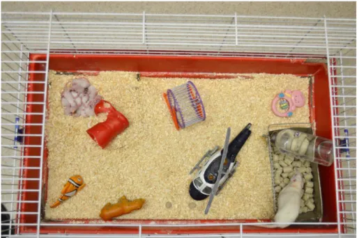
The term ‘environmental enrichment’ refers to altered housing conditions, where the animals are placed into larger cages, compared to standard cages (SE: standard environment). Regular cages typically only consist of nesting material, while enriched cages are suppleme- nted with different toys, running wheels, platforms, tunnels, rotating rods of different shape, size and material [Figure 1]. There is no standard protocol to create an enriched cage, but the main aim is to provide complex sensorimotor and cognitive stimuli, which is ensu- red by the continuous change of the supplies [16]. In the reviewed studies the frequency of changing the toys varied between daily to weekly changes. Several experiments included a social enrichment factor, as higher number of animals was kept in the enriched cages [36, 42, 43].
Overall, the time spent in enriched housing conditions changed between 3 weeks to 3 months. Mice were exposed to EE following MPTP injections for either 3 weeks [9, 36] or 70 days [44] or prior to MPTP injections for 40 days [9], 2 months [45] or 3 months [42, 43].
Rats were reared in enriched cages for 3 [46], 5 [35, 47], 6 [48] or
13 weeks [49] before and 3 [46], 4 or 7 weeks [50] after inducing PD
by 6-OHDA. The age when the animals were put into enriched cages
was also different in each study [Table 1].
Figure 1: Representative picture of an enriched cage in our laboratory (47).
Summary of results Behavioral signs
Behavioral signs and the possible improvement of motor symp- toms were assessed in the majority of studies. The capacity of EE to reverse motor deficits was proven by several types of tests in mice.
After MPTP-injections and subsequent 3 weeks of EE young mice showed increased number of total rears. The decreased ratio of foot faults to total activity in the parallel rod activity chamber test indi- cated improved motor control in aged mice, while a recovery of free- standing rears was visible both in young and aged groups [36].
In mice, placed in EE prior to MPTP lesion, numerous tests have proven the beneficial effects of EE. Evaluating the mean number of movements, enriched animals showed less decreased spontaneous activity [42]. Hilario et al observed hyperlocomotion in MPTP- treated animals, which was prevented by EE, based on the total distance traveled in open field test. Grip test demonstrated increased muscle strength after enrichment alone, while MPTP-treated enriched animals had similar performances to saline-treated groups [9]. Apart from motor symptoms PD patients may have emotional and cognitive problems which were also assessed by elevated plus maze instrument; the test showed reduced anxiety levels in enriched mice [42].
Most experiments conducted on rats involved behavioral examinations. Rats raised in EE for 6 weeks before unilateral 6- OHDA treatment (into the nigrostriatal bundle) had significantly higher reaching success in skilled reaching task. Furthermore EE animals had better error scores in skilled walking tests and made less severe placement errors compared to control animals [48]. The evaluation of apomorphine-induced rotations revealed an absent rotational asymmetry in enriched rats, as SE animals rotated significantly more towards the side contralateral to the lesion [48].
Our research group has also tested the effects of prophylactic enrichment; furthermore we were the first to provide evidence for the
protective effect of early, postnatal enrichment, when rats were kept in enriched cages for 5 weeks directly after birth and received 6- OHDA injections only several months later in adulthood [47]. In open field test we explored a reduced free rearing activity in both standard and enriched rats, however, enriched animals showed a better ability for compensation, as some degree of recovery was visible on the tenth postoperative day. When evaluating the distance covered by the animals enriched rats had better performance, they did not move significantly less in the postoperative period, similarly to their standard mates. In this study we did not observe asymme- trical behavioral signs [47]. Urakawa and co-workers assessed motor symptoms with beam walking test and observed better performance in enriched animals. In cylinder test they did not find differences in asymmetrical forelimb use between enriched and control animals [35]. In rats receiving intrastriatal dopaminergic grafts prior to the lesion the graft itself could not inhibit functional impairments caused by 6-OHDA [49] in contrast, grafted enriched animals showed reduced drug-induced rotational behavior and increased activity and better performance in spatial alternation test.
Rats kept in EE after 6-OHDA injections also displayed incre- ased physical activity on a rotarod [50]. In this experimental setup animals showed a trend towards improved drug- induced rotational behavior compared to control rats after 7 weeks of enrichment, and they exerted significantly reduced rotational behavior when compa- red to animals reared in EE for only 4 weeks [50]. In order to explore functional impact of EE before and after 6-OHDA- induced lesion amphetamine-induced rotational behavior was measured, which showed reduced number of rotations in enriched rats [46].
Cellular and biochemical effects
A frequent strategy to evaluate the severity of the lesion is to examine the nigral dopaminergic cell loss and the striatal changes.
In case of enrichment prior to the lesion the number of TH-
immunoreactive (-ir) neurons [42, 45] and TH mRNA expression were
higher [42] compared to mice kept in standard cages. Three months of preconditioning in EE completely protected the DAergic cells of the SNpc [43]. The expression of monoamine transporters were assessed in numerous studies, while searching for an explanation for the reduced toxicity. In enriched mice, a reduced number of DAT-ir cells [42] and lower expression of DAT mRNA was described in the SN [42, 43]. Bezard and co-workers described significantly decreased DAT binding and DAT mRNA levels in the striatum of mice raised in EE compared to SE animals [45]. Following MPTP exposure the decrease of VMAT2 can also contribute to the protective effect of EE.
It caused a significant decrease in VMAT2 mRNA expression of the SN in adult mice compared to standard animals [43]. The expression of different growth factors is often examined in PD models. Mice accommodated in EE in adulthood had higher GDNF mRNA expression in the SN, and even more raise could be detected after receiving MPTP [43]. The raise in BDNF levels in the striatum of EE mice may suggest that the protection of environmental effects could be mediated by the increase of expression of growth factors [45].
induced lesion a restoration in the number of TH-expressing neurons was visible, which was confirmed by retrograde Fluorogold labeling indicating preserved nigrostriatal pathway [46]. In enriched animals reduced cell degeneration in the anterior SNpc, and induced nigral astrocytic reaction (higher numbers of GFAP-labeled cells) were found early after the toxin exposure [46].
Discussion
By virtue of these above summarized studies, it is assumable that enriched environment is able to alter both neurochemical and behavioral changes in models of PD. There are various studies which describe effects of EE explaining its potential protective role in neurodegenerative diseases [24, 25]. Mice reared in EE from weaning to adulthood show alterations in the expression of striatal genes involved in cell proliferation, differentiation, intercellular signaling and cell metabolism. Among others an increased level of protein kinase C lambda, which is suggested to play an important role in neuroprotection and the plasticity of nervous system [51, 52]. In models of Huntington’s disease EE has been shown to delay motor symptoms and cognitive deficits. In Alzheimer’s disease an enhanced memory and learning and in case of epilepsy increased resistance to seizures have been described after EE. In stroke models enrichment resulted in an improved functional recovery of motor and cognitive deficits [24, 25].
The studies summarized in this review all point towards the protective effects of enriched circumstances in PD. Exercise and increased physical activity are important components of enrichment.
It has been shown that exercise alone could convey neuroprotective effects of MPTP-induced lesion, however, it was at a reduced degree compared to EE mice. This protection was supported by evaluating the death of TH- positive neurons. Also, an increased level of GDNF in SN and BDNF in striatum; and decreased DAT and VMAT2 transporter mRNA expressions were found in mice performing exercise [43]. Similarly to these growth factors, our research group described that 3-week-long exposure to EE also induces elevation in PACAP levels in most areas of the CNS. The peptide PACAP has strong neuroprotective effects, its increase could play a role in the beneficial effects of EE [53].
The fact that research groups found significant differences in the behavior of enriched animals compared to standard controls indicates that enriched environment potentially provides functional ameliora- tion and improvement of motor symptoms. The improvement was described in skilled and non-skilled motor tasks, exploration of rotati- onal movements, which are suitable to assess the severity of symptoms after unilateral lesions [48].
The fact, that some investigations found asymmetrical signs, while others did not might be due to the difference in the extent of injury.
As we know more than the half of dopaminergic neurons should be lost to lead to asymmetrical behavior [10, 54]. The functional outcome of enrichment was different when animals spent different amount of time in enlarged cages, only long-term stimulation could induce improvement in rotational behavior [46, 50]. Dobrossy et al. also described that EE on its own could not prevent drug-induced ipsila- teral rotation, however it could enhance the functional improvement of animals additionally receiving intrastriatal dopaminergic grafts [49]. Open field tests were used in both mouse and rat models and revealed better performances during the examination of free rearing activity, distance covered and spontaneous activity. Although hypokinesia is one of the main characteristics of PD one study described hyperlocomotion after MPTP injections, which had been Both young and aged mice showed a significant recovery in the
mean number of TH- positive neurons in the SNpc when kept in EE for 3 weeks after MPTP-lesioning [36]. Klaissle and coworkers descri- bed that enrichment could increase the number of newly formed neural precursors in SN if co-treated with levodopa. Also, the incre- ased formation of NG2+ (neuro-glial antigen-2) cells in enriched animals depended on levodopa treatment [44]. These cells are capable to differentiate into oligodendrocytes, and even to neurons in vitro, which offer a possible regenerative capacity in the midbrain [44].
Exposure to EE both before and after MPTP injection led to restoration of 3,4- dihydroxyphenylacetic acid (DOPAC) and homovanillic acid (HVA) levels in the striatum, but it was not able to prevent DA depletion. Animals, which received MPTP and were housed in EE had an increased DA turnover [9]. Hilario et al. also evaluated changes in the expression of DAergic-related genes in the midbrain (SN+VTA), among which only the expression of dopaminergic receptor D1 (D1R) was reduced after EE. The gene expression of the cholinergic system was also examined in this study [9].
In rats living in enriched cages prior to the lesion, examinations revealed higher number of residual TH-positive cells in the medial and ventral tegmental area, SNpc and lateral part of SN. In contrast, cortical thickness and brain weight measurements did not find significant differences between SE and EE rats [48]. In case of postnatal enrichment, in concordance with previous data we found that enriched circumstances could rescue the dopaminergic cells [47].
When animals were reared in EE for 5 weeks prior to lesion no significant differences were found between control and enriched animals regarding DAergic cell loss, however an increased number of migrating doublecortin-positive (DCX-positive) cells were found in the lesioned striatum of enriched animals [35]. These cells were suggested to be detached from neuroblasts in the subventricular zone (SVZ). Moreover, it has been proven that enriched circumstances can improve the survival of DAergic grafted cells in the striatum [49].
Exposure to enriched environment after 6-OHDA injection did not change the number of TH-positive cells compared to lesioned controls. However enrichment led to increased cell proliferation, additionally the number of newborn NG2- and GFAP-positive cells was significantly higher in enriched lesioned rats [50] similarly to the results of Klaissle et al. [44].
When enrichment was applied both before and after 6-OHDA-
reported earlier by several research groups [55, 56]. This symptom can be explained by pre-synaptic dopamine 1 receptor hypersensibili- zation due to the severe loss of nigral DA [57, 58]. EE- applied both before and after MPTP injections- prevented hyperlocomotion while decreasing the expression of D1R [9]. In this experiment EE did not have an effect on DA levels; it only restored grip strength, which was described in similar models [59, 60]. These observations were confirmed by several rat studies, when no significant difference was found it the percentage of surviving neurons of the SN, but enriched animals still exerted less severe motor symptoms [47, 50]. The better performance of enriched animals on beam walking test further proves that environmental enrichment can improve balance, coordination skills and sensory ability [35]. These results showing that enriched environment can alter functional outcome after different types of lesions correspond to earlier findings of several research groups [61, 62].
Numerous research groups provided evidence that enriched animals are more resistant to MPTP or 6-OHDA, as the number of surviving TH-positive neurons were higher in SNpc compared to standard animals. Neural transplantation of DAergic cells could also offer a solution to PD. Currently the two main approaches to enhance the survival of grafts are to change the microenvironment of the grafts and to alter environmental factors around the time of the lesion [63].
In 2000, Dobrossy and coworkers proved that EE prior to lesion can increase the survival of grafted DA producing cells in the rat striatum [49] thus the success of grafting could be further optimized by increasing the complexity of environment.
Although the mechanisms underlying the protective effect of EE are not yet completely understood, studies discuss several changes which may support the recovery of enriched animals. It has been excluded that the protective effect of EE would be due to the change in MPTP metabolism, as the conversion of the toxin to MPP+ was not different in enriched compared to standard animals [43]. The reduction of DAT gene expression and the depletion of the number of DAT-ir cells is a possible mechanism of protection against MPTP- induced toxicity; hence DAT plays a crucial role in the uptake of the neurotoxin. It is proven that mice lacking DAT are protected from MPTP-induced neuronal death [64]. Exercise alone can cause a significant loss of DAT protein in the striatum of MPTP-treated mice [65]. The change in VMAT2 expression observed could also contribute to the neuroprotective effect of EE. VMAT2-mediated sequestration of the neurotoxin may take part in alleviating MPTP toxicity. It has been demonstrated that mice lacking VMAT2 are more susceptible to the neurotoxic effects of MPTP [32]. Furthermore, treatment with VMAT2 inhibitor enhances MPTP toxicity in the striatum of rats [33].
Several findings support that enriched conditions stimulate cellular plasticity, rather than having direct neuroprotective effects. It is known that SN does not have the regenerative capacity to form new dopaminergic neurons. However it has been shown that non-neuronal NG2-expressing cells, with mainly oligodendrocytic precursor characteristics could be generated [50, 66]. The aim of future therapy of PD would be to restore the nigral dopaminergic neurons, and as NG2+ cells can potentially differentiate into neurons, they may offer a new therapeutic option [67, 68]. The fact that EE enhances the generation of these cells in a levodopa-dependent way [44] further shows that EE exerts its protective effects through numerous mechanisms. 6-OHDA causes a decrease in GFAP-positive glial cells, however in the study of Steiner et al. the newborn cells induced by physiological stimuli also expressed GFAP, thus glial plasticity might be a way of compensatory processes [50]. However a large number of
oligodendrocytes or microglia; they are suggested to undergo deve- lopment or maturation. Anastasia and coworkers also suggests that an early post-lesional astrocytic reaction may take part in the prote-ction of the entire nigrostriatal system [46]. Urukawa et al. described an increased number of migrating DXC-positive neurons in the striatum of enriched animals which were co-expressed with CXC chemokine receptor 4 (CXCR4). This, together with the found up-regulation of stromal cell-derived factor 1α (SDF-1α) mRNA, sugge-sts that SDF- 1α/CXCR4 signaling might regulate the activation of DXC-positive cells after environmental stimuli [35]. Altogether these studies mentioned above suggest that complex stimuli are not able to enhance the formation of new DAergic neurons, but induce some microenvironmental changes in the SN, which lead to functional improvements.
Conclusions
The neuroprotective effects of EE have been well established by numerous studies, which raise the question whether we should pay more attention and focus should be placed also on providing a stimulating environment during clinical therapy and rehabilitation of patients suffering from PD. EE could provide a non-pharmaceutical, non-invasive method for possible prevention and treatment of Parkinson’s disease without the side-effects of pharmacotherapy.
Conflict of interest
The authors declare no conflict of interest.
References
1. Sprenger F and Poewe W. Management of motor and non-motor symptoms in Parkinson's disease. CNS Drugs. 2013; 27:259-272. [Crossref]
2. Reglodi D, Renaud J, Tamas A, Tizabi Y, Socias SB, Del-Bel E, et al. Novel tactics for neuroprotection in Parkinson's disease: Role of antibiotics, polyphenols and neuropeptides. Prog Neurobiol. 2017; 2:120-148. [Crossref]
3. Braak H, Del Tredici K, Rub U, de Vos RA, Jansen Steur EN, Braak E, et al. Staging of brain pathology related to sporadic Parkinson's disease. Neurobiol Aging. 2003;
24:197-211. [Crossref]
4. Gerfen CR. Molecular effects of dopamine on striatal-projection pathways. Trends Neurosci. 2000; 23:64-70. [Crossref]
5. McDonald C, Gordon G, Hand A, Walker RW and Fisher JM. 200 Years of Parkinson's disease: what have we learnt from James Parkinson? Age Ageing. 2018;
47:209-214. [Crossref]
6. Elbaz A, Carcaillon L, Kab S and Moisan F. Epidemiology of Parkinson's disease.
Rev Neurol. 2016; 172:14-26. [Crossref]
7. Kieburtz K and Wunderle KB. Parkinson's disease: evidence for environmental risk factors. Mov Disord. 2013; 28:8-13. [Crossref]
8. Kakkar AK and Dahiya N. Management of Parkinson׳s disease: Current and future pharmacotherapy. Eur J Pharmacol. 2015; 750:74-81. [Crossref]
9. Hilario WF, Herlinger AL, Areal LB, de Moraes LS, Ferreira TA, Andrade TE, et al.
Cholinergic and dopaminergic alterations in nigrostriatal neurons are involved in environmental enrichment motor protection in a mouse model of Parkinson's disease.
J Mol Neurosci. 2016; 60:453-64. [Crossref]
10. Tamas A, Lubics A, Lengvari I and Reglodi D. Effects of age, gender, and gonadectomy on neurochemistry and behavior in animal models of Parkinson's disease. Endocrine. 2006; 29:275-287. [Crossref]
11. Shih IF, Liew Z, Krause N and Ritz B. Lifetime occupational and leisure time physical activity and risk of Parkinson's disease. Parkinsonism Relat Disord. 2016;
28:112-117. [Crossref]
12. Shu HF, Yang T, Yu SX, Huang HD, Jiang LL, Gu JW, et al. Aerobic exercise for Parkinson's disease: a systematic review and meta-analysis of randomized controlled trials. PLoS One. 2014; 9:e100503. [Crossref]
13. Lamotte G, Rafferty MR, Prodoehl J, Kohrt WM, Comella CL, Simuni T, et al.
Effects of endurance exercise training on the motor and non-motor features of Parkinson's disease: a review. J Parkinsons Dis. 2015; 5:21-41. [Crossref]
14. Hebb DO. The effects of early experience on problem solving at maturity. Am Psychol. 1947; 2:306–307. [Crossref]
15. Sale A, Cenni MC, Ciucci F, Putignano E, Chierzi S, Maffei L, et al. Maternal enrichment during pregnancy accelerates retinal development of the fetus. PLoS One.
2007; 2:e1160. [Crossref]
16. van Praag H, Kempermann G, Gage FH. Neural consequences of environmental enrichment. Nat Rev Neurosci. 2000; 1:191-198. [Crossref]
17. Black JE, Isaacs KR, Anderson BJ, Alcantara AA and Greenough WT. Learning causes synaptogenesis, whereas motor activity causes angiogenesis, in cerebellar cortex of adult rats. Proc Natl Acad Sci USA. 1990; 87:5568-5572. [Crossref]
18. Altman J. Are new neurons formed in the brain of adult mammals? Science. 1962;
135:1127-1128. [Crossref]
19. Kleim JA, Lussnig E, Schwarz ER, Comery TA and Greenough WT. Synaptogenesis and Fos expression in the motor cortex of the adult rat after motor skill learning. J Neurosci. 1996; 16:4529-4535. [Crossref]
20. Kiss P, Vadasz G, Kiss-Illes B, Horvath G, Tamas A, Reglodi D, et al. Environmental enrichment decreases asphyxia-induced neurobehavioral developmental delay in neonatal rats. Int J Mol Sci. 2013; 14:22258-22273. [Crossref]
21. Horvath G, Reglodi D, Vadasz G, Farkas J and Kiss P. Exposure to enriched environment decreases neurobehavioral deficits induced by neonatal glutamate toxicity. Int J Mol Sci. 2013; 14:19054-19066. [Crossref]
22. Szabadfi K, Atlasz T, Horvath G, Kiss P, Hamza L, Farkas J, et al. Early postnatal enriched environment decreases retinal degeneration induced by monosodium glutamate treatment in rats. Brain Res. 2009; 1259:107-112. [Crossref]
23. Kiss P, Szabadfi K, Horvath G, Tamas A, Farkas J, Gabriel R, et al. Gender- dependent effects of enriched environment and social isolation in ischemic retinal lesion in adult rats. Int J Mol Sci. 2013; 14:16111-16123. [Crossref]
24. Laviola G, Hannan AJ, Macrì S, Solinas M and Jaber M. Effects of enriched environment on animal models of neurodegenerative diseases and psychiatric disorders. Neurobiol Dis. 2008; 31:159-168. [Crossref]
25. Nithianantharajah J and Hannan AJ. Enriched environments, experience-dependent plasticity and disorders of the nervous system. Nat Rev Neurosci. 2006; 7:697-709.
[Crossref]
26. Jagmag SA, Tripathi N, Shukla SD, Maiti S and Khurana S. Evaluation of models of Parkinson's disease. Front Neurosci. 2015; 9:503. [Crossref]
27. Manning-Bog AB and Langston JW. Model fusion, the next phase in developing animal models for Parkinson's disease. Neurotox Res. 2007; 11:219-240.[Crossref]
28. Segura-Aguilar J and Kostrzewa RM. Neurotoxin mechanisms and processes relevant to Parkinson's disease: an update. Neurotox Res. 2015; 27:328-354. [Crossref]
29. Nicklas WJ, Youngster SK, Kindt MV, Heikkila RE. MPTP, MPP+ and mitochondrial function. Life Sci. 1987; 40:721-729. [Crossref]
30. Jenner P and Marsden CD. The actions of 1-methyl-4-phenyl-1,2,3,6- tetrahydropyridine in animals as a model of Parkinson's disease. J Neural Transm Suppl. 1986; 20:11-39. [Crossref]
31. Masilamoni GJ and Smith Y. Chronic MPTP administration regimen in monkeys: a model of dopaminergic and non-dopaminergic cell loss in Parkinson's disease. J Neural Transm (Vienna). 2018; 125:337-363. [Crossref]
32. Gainetdinov RR, Fumagalli F, Wang YM, Jones SR, Levey AI, Miller GW, et al.
Increased MPTP neurotoxicity in vesicular monoamine transporter 2 heterozygote knockout mice. J Neurochem. 1998; 70:1973-1978. [Crossref]
33. Staal RG and Sonsalla PK. Inhibition of brain vesicular monoamine transporter (VMAT2) enhances 1-methyl-4-phenylpyridinium neurotoxicity in vivo in rat striata.
J Pharmacol Exp Ther. 2000; 293:336-342. [Crossref]
34. Reglodi D, Kiss P, Lubics A and Tamas A. Review on the protective effects of PACAP in models of neurodegenerative diseases in vitro and in vivo. Curr Pharm Des.
2011; 17:962-972. [Crossref]
35. Urakawa S, Hida H, Masuda T, Misumi S, Kim TS, Nishino H, et al. Environmental enrichment brings a beneficial effect on beam walking and enhances the migration of doublecortin-positive cells following striatal lesions in rats. Neuroscience. 2007;
144:920-933. [Crossref]
36. Goldberg NR, Haack AK and Meshul CK. Enriched environment promotes similar neuronal and behavioral recovery in a young and aged mouse model of Parkinson's disease. Neuroscience. 2011; 172:443-452. [Crossref]
37. Pringsheim T, Jette N, Frolkis A and Steeves TD. The prevalence of Parkinson's disease: a systematic review and meta-analysis. Mov Disord. 2014; 29:1583-1590.
[Crossref]
38. de Rijk MC, Launer LJ, Berger K, Breteler MM, Dartigues JF, Baldereschi M, et al.
Prevalence of Parkinson's disease in Europe: A collaborative study of population- based cohorts. Neurologic diseases in the elderly research group. Neurology. 2000;
54:21-33. [Crossref]
39. Jarvis MF and Wagner GC. 1-methyl-4-phenyl-1,2,3,6-tetrahydropyridine-induced neurotoxicity in the rat: characterization and age-dependent effects. Synapse. 1990;
5:104-112. [Crossref]
40. Colas D, Gharib A, Bezin L, Morales A, Guidon G, Cespuglio R, et al. Regional age- related changes in neuronal nitric oxide synthase (nNOS), messenger RNA levels and activity in SAMP8 brain. BMC Neurosci. 2006; 7:81. [Crossref]
41. Liu J, Wang YY, Liu L, Wang QD, Yuan ZY, Zhang ZX, et al. Damage to the nigrostriatal system in the MPTP-treated SAMP8 mouse. Neurosci Lett. 2008;
448:184-188. [Crossref]
42. Yuan ZY, Gu P, Liu L, Wang YY, Liu J, Cui DS, et al. Neuroprotective effects of enriched environment in MPTP-treated SAMP8 mice. Neurosci Lett. 2009; 454:6-10.
[Crossref]
43. Faherty CJ, Raviie Shepherd K, Herasimtschuk A and Smeyne RJ. Environmental enrichment in adulthood eliminates neuronal death in experimental Parkinsonism.
Brain Res Mol Brain Res. 2005; 134:170-179. [Crossref]
44. Klaissle P, Lesemann A, Huehnchen P, Hermann A, Storch A, Steiner B, et al.
Physical activity and environmental enrichment regulate the generation of neural precursors in the adult mouse substantia nigra in a dopamine-dependent manner. BMC Neurosci. 2012; 13:132. [Crossref]
45. Bezard E, Dovero S, Belin D, Duconger S, Jackson-Lewis V, Przedborski S, et al.
Enriched environment confers resistance to 1-methyl-4-phenyl-1,2,3,6- tetrahydropyridine and cocaine: involvement of dopamine transporter and trophic factors. J Neurosci. 2003; 23:10999-11007. [Crossref]
46. Anastasia A, Torre L, de Erausquin GA and Masco DH. Enriched environment protects the nigrostriatal dopaminergic system and induces astroglial reaction in the 6- OHDA rat model of Parkinson's disease. J Neurochem. 2009; 109:755-765. [Crossref]
47. Jungling A, Reglodi D, Karadi ZN, Horvath G, Farkas J, Gaszner B, et al. Effects of postnatal enriched environment in a model of Parkinson's disease in adult rats. Int J Mol Sci. 2017; 18. [Crossref]
48. Jadavji NM, Kolb B and Metz GA. Enriched environment improves motor function in intact and unilateral dopamine-depleted rats. Neuroscience. 2006; 140:1127-1138.
[Crossref]
49. Dobrossy M, Le Moal M, Montaron M, Abrous N. Influence of environment on the efficacy of intrastriatal dopaminergic grafts. Exp Neurol. 2000; 165:172-183.
[Crossref]
50. Steiner B, Winter C, Hosman K, Siebert E, Kempermann G, Petrus DS, et al. Enriched environment induces cellular plasticity in the adult substantia nigra and improves motor behavior function in the 6-OHDA rat model of Parkinson's disease. Exp Neurol.
2006; 199:291-300. [Crossref]
51. Thiriet N, Amar L, Toussay X, Lardeux V, Ladenheim B, Becker KG, et al.
Environmental enrichment during adolescence regulates gene expression in the striatum of mice. Brain Res. 2008; 1222:31-41. [Crossref]
52. Crary JF, Shao CY, Mirra SS, Hernandez AI and Sacktor TC. Atypical protein kinase C in neurodegenerative disease I: PKMzeta aggregates with limbic neurofibrillary tangles and AMPA receptors in Alzheimer disease. J Neuropathol Exp Neurol. 2006;
65:319-326. [Crossref]
53. Horvath G, Kiss P, Nemeth J, Lelesz B, Tamas A, Reglodi D, et al. Environmental enrichment increases PACAP levels in the CNS of adult rats. Neuro Endocrinol Lett.
2015; 36:143-147. [Crossref]
54. Reglodi D, Tamas A, Lengvari I, Toth G, Szalontay L, Lubics A, et al. Comparative study of the effects of PACAP in young, aging, and castrated males in a rat model of Parkinson's disease. Ann N Y Acad Sci. 2006; 1070:518-524. [Crossref]
55. Chia LG, Ni DR, Cheng LJ, Kuo JS, Cheng FC, Dryhurst G, et al. Effects of 1- methyl-4- phenyl-1,2,3,6-tetrahydropyridine and 5,7-dihydroxytryptamine on the locomotor activity and striatal amines in C57BL/6 mice. Neurosci Lett. 1996; 218:67- 71. [Crossref]
56. Moraes LS, Rohor BZ, Areal LB, Pereira EV, Santos AM, Facundo VA, et al.
Medicinal plant Combretum leprosum mart ameliorates motor, biochemical and molecular alterations in a Parkinson's disease model induced by MPTP. J Ethnopharmacol. 2016; 185:68-76. [Crossref]
57. Ding S, Li L and Zhou FM. Nigral dopamine loss induces a global upregulation of presynaptic dopamine D1 receptor facilitation of the striatonigral GABAergic output.
J Neurophysiol. 2015; 113:1697-1711. [Crossref]
58. Gerfen CR, Miyachi S, Paletzki R and Brown P. D1 dopamine receptor supersensitivity in the dopamine-depleted striatum results from a switch in the regulation of ERK1/2/MAP kinase. J Neurosci. 2002; 22:5042-5054. [Crossref]
59. Sconce MD, Churchill MJ, Greene RE and Meshul CK. Intervention with exercise restores motor deficits but not nigrostriatal loss in a progressive MPTP mouse model of Parkinson's disease. Neuroscience. 2015; 299:156-174. [Crossref]
60. Crocker SJ, Smith PD, Jackson-Lewis V, Lamba WR, Hayley SP, Grimm E, et al.
Inhibition of calpains prevents neuronal and behavioral deficits in an MPTP mouse model of Parkinson's disease. J Neurosci. 2003; 23:4081-4091. [Crossref]
61. Held JM, Gordon J and Gentile AM. Environmental influences on locomotor recovery following cortical lesions in rats. Behav Neurosci. 1985; 99:678-690. [Crossref]
62. Ohlsson AL and Johansson BB. Environment influences functional outcome of cerebral infarction in rats. Stroke. 1995; 26:644-649. [Crossref]
63. Cassel JC, Kelche C, Majchrzak M and Will BE. Factors influencing structure and function of intracerebral grafts in the mammalian brain: a review. Restor Neurol Neurosci. 1992; 4:65-96. [Crossref]
64. Bezard E, Gross CE, Fournier MC, Dovero S, Bloch B, Jaber M. Absence of MPTP- induced neuronal death in mice lacking the dopamine transporter. Exp Neurol. 1999;
155:268-273. [Crossref]
65. Fisher BE, Petzinger GM, Nixon K, Hogg E, Bremmer S, Meshul CK, et al. Exercise- induced behavioral recovery and neuroplasticity in the 1-methyl-4-phenyl- 1,2,3,6- tetrahydropyridine-lesioned mouse basal ganglia. J Neurosci Res. 2004; 77:378-390.
[Crossref]
66. Lie DC, Dziewczapolski G, Willhoite AR, Kaspar BK, Shults CW, Gage FH, et al. The adult substantia nigra contains progenitor cells with neurogenic potential. J Neurosci.
2002; 22:6639-6649. [Crossref]
67. Aguirre AA, Chittajallu R, Belachew S and Gallo V. NG2-expressing cells in the subventricular zone are type C-like cells and contribute to interneuron generation in the postnatal hippocampus. J Cell Biol. 2004; 165:575-589. [Crossref]
68. Belachew S, Chittajallu R, Aguirre AA, Yuan X, Kirby M, Anderson S, et al.
Postnatal NG2 proteoglycan-expressing progenitor cells are intrinsically multipotent and generate functional neurons. J Cell Biol. 2003; 161:169-186. [Crossref]