Extraintestinal Manifestations of In fl ammatory Bowel Disease
Stephan R. Vavricka, MD,*
,†Alain Schoepfer, MD,
‡Michael Scharl, MD,* Peter L. Lakatos, MD,
§Alexander Navarini, MD,
kand Gerhard Rogler, MD*
Abstract:Extraintestinal manifestations (EIM) in inflammatory bowel disease (IBD) are frequent and may occur before or after IBD diagnosis. EIM may impact the quality of life for patients with IBD significantly requiring specific treatment depending on the affected organ(s). They most frequently affect joints, skin, or eyes, but can also less frequently involve other organs such as liver, lungs, or pancreas. Certain EIM, such as peripheral arthritis, oral aphthous ulcers, episcleritis, or erythema nodosum, are frequently associated with active intestinal inflammation and usually improve by treatment of the intestinal activity. Other EIM, such as uveitis or ankylosing spondylitis, usually occur independent of intestinal inflammatory activity. For other not so rare EIM, such as pyoderma gangrenosum and primary sclerosing cholangitis, the association with the activity of the underlying IBD is unclear. Successful therapy of EIM is essential for improving quality of life of patients with IBD. Besides other options, tumor necrosis factor antibody therapy is an important therapy for EIM in patients with IBD.
(Inflamm Bowel Dis 2015;21:1982–1992)
Key Words:extraintestinal manifestations, inflammatory bowel disease, arthritis, uveitis
I
nflammatory bowel disease (IBD), which includes Crohn’s disease (CD) and ulcerative colitis (UC), should be regarded as a systemic disorder not limited to the gastrointestinal tract because many patients will develop extraintestinal symptoms. Extraintestinal symptoms may involve virtually any organ system with a potentially detrimental impact on the patient’s functional status and quality of life.Extraintestinal symptoms can be divided in 2 groups: extra- intestinal manifestations (EIM) and extraintestinal complications.
EIM most frequently affect joints (peripheral and axial arthropathies), the skin (erythema nodosum, pyoderma gangrenosum, Sweet’s syn- drome, aphthous stomatitis), the hepatobiliary tract (primary scleros- ing cholangitis [PSC]), and the eye (episcleritis, uveitis) (Fig. 1). Less frequently, EIM also affect the lungs, the heart, the pancreas, or the
vascular system. Extraintestinal complications are mainly caused by the disease itself and include conditions such as malabsorption with consequent micronutrient deficiencies, osteoporosis, peripheral neu- ropathies, kidney stones, gallstones, and IBD drug-related side effects.
This article focuses on the clinical features of EIM. Certain EIM such as pauciarticular arthritis, oral aphthous ulcers, erythema nodosum, or episcleritis usually occur with increased intestinal disease activity.1,2 Other EIM such as ankylosing spondylitis and uveitis usually follow an independent course from IBD disease activ- ity.1,2Andfinally, some EIM such as PSC and pyoderma gangreno- sum may or may not be related to IBD disease activity (Table 1).2–4 EIM in IBD are reported with frequencies ranging from 6% to 47%.5–13Multiple EIM may occur concomitantly, and the presence of 1 EIM confers a higher likelihood to develop other EIM.14 Recently, we reported based on data from the Swiss IBD Cohort study that up to 1 quarter of EIM-affected patients with IBD tend to suffer from a combination of several EIMs (up to 5).14Furthermore, our group recently published data regarding the chronological order of EIM appearance relative to IBD diagnosis.15 A summary of the chronologic appearance of EIMs relative to IBD diagnosis is pre- sented in Figure 2. In 25.8% of cases, afirst EIM occurred before IBD was diagnosed (median time 5 mo before IBD diagnosis; range, 0–25 mo). In 74.2% of cases, thefirst EIM manifested after IBD diagnosis was made (median, 92 mo; range, 29–183 mo) (Fig. 2).
We found that up to 4 different EIM occurred before IBD was diagnosed, and that at 30 years after IBD diagnosis, 50% of patients had suffered from at least 1 EIM. Perianal CD, colonic involvement, and cigarette smoking increased the likelihood to suffer from EIMs.16
PATHOGENESIS OF EIM
The pathogenesis of EIM in IBD is not well understood. It is believed that the diseased gastrointestinal mucosa may trigger
Received for publication November 6, 2014; Accepted February 9, 2015.
From the *Department of Medicine, Division of Gastroenterology and Hepatol- ogy, University Hospital Zurich, Zurich, Switzerland; †Department of Medicine, Division of Gastroenterology and Hepatology, Triemlispital Zurich, Zurich, Switzerland;‡Department of Medicine, Division of Gastroenterology and Hepatol- ogy, Centre Hospitalier Universitaire Vaudois (CHUV), Lausanne, Switzerland;§1st Department of Medicine, Semmelweis University, Budapest, Hungary; and
kDepartment of Dermatology, University Hospital Zurich, Zurich, Switzerland.
Supported by research grants from the Swiss National Science Foundation (33CSC0_134274 [Swiss IBD Cohort Study] to G. Rogler, 320000-114009/3 and 32473B_135694/1 to S. R. Vavricka, and 314730-146204 and CRSII3_154488/1 to M. Scharl) and the Zurich Center for Integrative Human Physiology of the University of Zurich (to G. Rogler, M. Scharl, and S. R. Vavricka).
The authors have no conflicts of interest to disclose.
Reprints: Stephan R. Vavricka, MD, Department of Internal Medicine, Division of Gastroenterology, Triemli Hospital, Birmensdorferstrasse 497, CH-8063 Zurich, Switzerland (e-mail: stephan.vavricka@usz.ch).
Copyright © 2015 Crohn’s & Colitis Foundation of America, Inc. This is an open access article distributed under the Creative Commons Attribution License 4.0 (CCBY), which permits unrestricted use, distribution, and reproduction in any medium, provided the original work is properly cited.
DOI 10.1097/MIB.0000000000000392 Published online 2 July 2015.
Downloaded from https://academic.oup.com/ibdjournal/article-abstract/21/8/1982/4602969 by Semmelweis University user on 30 July 2019
immune responses at the extraintestinal site due to shared epitopes, e.g., of intestinal bacteria and the synovia.17–21 This would mean that bacteria that are translocated across the leaky intestinal barrier trigger an adaptive immune response thatfinally is unable to discriminate between bacterial epitopes and epitopes of joints or the skin. Triggers of the autoimmune responses in certain organs seem to be influenced by genetic factors. Concor- dance in EIM was present in 70% of parent–child pairs and 84%
in sibling pairs.10,22Associations of EIM in IBD with major his- tocompatibility complex loci have been demonstrated. EIM in patients with CD are more frequently observed in patients with HLA-A2, HLA-DR1, and HLA-DQw5, whereas EIM in patients with UC are more likely to appear when the HLA-DR103 geno- type is present.23Particular HLA complexes have also been linked to specific EIM. HLA-B8/DR3 is associated with an increased risk of PSC in UC, whereas HLA-DRB1*0103, HLA-B*27, and HLA-B*58 are associated with EIM of joints, the skin, and eyes, respectively, in patients with IBD.4,24,25 HLA-B*27 itself does not seem to be associated with IBD5but HLA-B*27 shows
a strong association with the development of ankylosing spondy- litis, as 50% to 90% of patients with IBD are positive for this marker.26As HLA-B*27 per se shows an association with anky- losing spondilitis or rheumatoid arthritis it remains unclear whether there is a specific role for EIM in IBD.
MUSCULOSCELETAL EIM
Musculoskeletal EIM including joint complaints represent the most common EIMs in IBD. Joint symptoms affecting peripheral large and small joints or the axial joints occur in up to 40% of patients with IBD.14,27,28
Peripheral Arthralgia/Arthritis
Peripheral arthralgia/arthritis in patients with IBD, in contrast to other specific forms of arthritis such as rheumatoid arthritis or psoriatic arthritis, shows little or no joint destruction.
Classically, it presents as a seronegative arthralgia/arthritis,1 which affects 5% to 10% of patients with UC and 10% to 20%
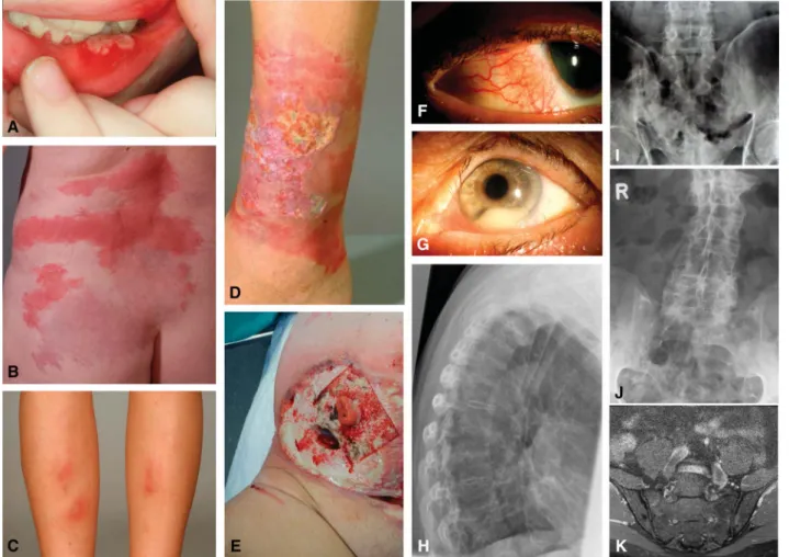
FIGURE 1. A, Oral aphthous ulcers, (B) Sweet’s syndrome, (C) erythema nodosum, (D) pyoderma gangrenosum, (E) peristomal pyoderma gan- grenosum, (F) episcleritis, (G) uveitis with hypopyon and dilated iris vessels, (H) conventional x-ray of the lateral spine demonstrating syn- desmophytes (bamboo spine), (I) plane radiograph of the ileosacral joints with bilateral sacroiliitis, (J) plane radiography of the sacrum with bilateral ankylosis, (K) coronal magnetic resonance image of the sacroiliac joints with active inflammation mainly on the left side and chronic inflammatory changes on both sides.
Downloaded from https://academic.oup.com/ibdjournal/article-abstract/21/8/1982/4602969 by Semmelweis University user on 30 July 2019
of patients with CD.29 A higher risk for peripheral arthralgia/
arthritis is seen in patients with IBD with colonic involvement and in patients suffering from perianal disease, erythema nodo- sum, stomatitis, uveitis, and pyoderma gangrenosum.1,20
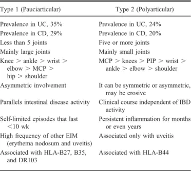
Peripheral arthralgia/arthritis has been classified into 2 entities (Table 2): type I (pauciarticular) arthralgie/arthritis usually affects less than 5 large joints, such as ankles, knees, hips, wrists, elbows, and shoulders and is often acute, asymmetrical, and migratory. The knee is commonly involved. Approximately, 20% to 40% of all patients have more than 1 episode of arthral- gia/arthritis. Pauciarticular arthralgia/arthritis is usually related to IBD activity and self-limiting with a maximum duration of up to 10 weeks.29Consequently, medical or surgical treatment of the underlying intestinal inflammation (i.e., colitis) is usually associ- ated with improvement of type I arthritis. Type II (polyarticular)
arthralgia/arthritis frequently is a symmetrical arthritis involving 5 or more small joints. It is not related with intestinal disease activ- ity and may precede IBD diagnosis. Type II arthropathy can persist for years (median of 3 yr).29 The metacarpophalangeal joint is most commonly involved. Type II arthritis is associated with an increased risk of uveitis but not erythema nodosum.29
The diagnosis/classification of type 1 and type 2 arthro- pathies is purely clinical, as imaging is most often normal with no evidence of significant inflammation or joint destruction.31Both types are seronegative (i.e., rheumatoid factor-negative), but may represent immunogenetically distinct entities. Type 1 peripheral arthropathy is associated with HLA-B27, HLA-B35, and HLA- DR103, whereas type 2 is associated with HLA-B44.25
As type II peripheral arthropathy usually occurs indepen- dently from intestinal activity and anti-inflammatory treatment may not be successful, physiotherapy and treatment of associated pain is the main treatment option in those cases.
Table 3 summarizes treatment options of EIM in IBD.
Other treatment modalities include rest, and intra-articular ste- roid injections. Use of sulphasalazine has been reported to improve peripheral arthropathies.45Therapy with nonsteroidal anti-inflammatory drugs (NSAIDs) in the management of IBD-associated peripheral arthropathies requires caution FIGURE 2. Chronology of EIM in patients with IBD. In one quarter of
patients with IBD, up to 4 EIM appeared before the time of IBD diagnosis. The median time before IBD diagnosis is 5 mo (range, 0–25 mo). In 75% of cases, the first EIM manifested after IBD diagnosis (median, 92 mo; range, 29–183 mo). Thirty years after diagnosis up to 50% of patients with IBD have suffered from at least 1 EIM.15
TABLE 1. Relationship Between EIM Activity and Intestinal Activity
EIM
Parallel Course of IBD
Separate Course of
IBD
May or May Not Parallel Disease Activity
Axial arthropathy ✓
Peripheral arthropathy ✓ ✓
(Type I) (Type II)
Erythema nodosum ✓
Pyoderma gangrenosum ✓
Sweet’s syndrome ✓
Oral aphthous ulcers ✓
Episcleritis ✓
Uveitis ✓
PSC ✓
Adapted from Trikudanathan et al.2
TABLE 2. Classification of Peripheral Arthropathy Associated with IBD
Type 1 (Pauciarticular) Type 2 (Polyarticular) Prevalence in UC, 35% Prevalence in UC, 24%
Prevalence in CD, 29% Prevalence in CD, 20%
Less than 5 joints Five or more joints Mainly large joints Mainly small joints Knee.ankle.wrist.
elbow.MCP. hip.shoulder
MCP.knees.PIP.wrist. ankle.elbow.shoulder Asymmetric involvement It can be symmetric or asymmetric,
may be erosive
Parallels intestinal disease activity Clinical course independent of IBD activity
Self-limited episodes that last ,10 wk
Persistent inflammation for months or even years
High frequency of other EIM (erythema nodosum and uveitis)
Associated only with uveitis Associated with HLA-B27, B35,
and DR103
Associated with HLA-B44
PIP, proximal interphalangeal joint; MCP, metacarpophalangeal joint.
Combined use of the Assessment of SpondyloArthritis International Society (ASAS) criteria for axial spondyloarthritis (SpA) and the ASAS criteria for peripheral SpA in the entire SpA population. In patients with predominantly axial involvement (back pain) with or without peripheral manifestations, the ASAS criteria for axial SpA6 are applied. In patients with peripheral manifestations only, the ASAS criteria for peripheral SpA are applied. In the entire ASAS population of 975 patients’sensitivity and specificity of the combined use of the 2 sets of criteria were 79.5% and 83.3%, respectively.
Adapted from Su et al,10Orchard et al,29and Rodriguez-Reyna et al.30
Downloaded from https://academic.oup.com/ibdjournal/article-abstract/21/8/1982/4602969 by Semmelweis University user on 30 July 2019
because of the reported association of exacerbation of IBD with NSAID use.46–50
In a study by Takeuchi et al, up to 25% of patients in remission experienced a disease flare when provided certain NSAIDs. It seems that COX-2 inhibitors may show a better safety profile and might be used with caution in patients with IBD suffering from peripheral arthropathies.49,51–54
Whether a discrimination of type I and type II arthropathy is clinically useful and meaningful has never been studied in detail. In most large IBD centers, this discrimination is not used. In fact, affection of small joints may disappear as well with the treatment of the underlying disease, whereas inflammation of the large joints may also occur as side effect of anti-TNF therapy. A careful documen- tation of joint affections, which is standard in rheumatology, certainly would be helpful and could improve the outcome of patients with IBD. This should be requested as a standard in IBD centers.
Axial Arthropathies
Axial arthropathies are less frequent than peripheral arthralgia/arthritis in patients with IBD, occurring in 3% to 5%
of patients although frequencies of up to 25% have been reported.9–13,55Males are more frequently affected than females.
In contrast to peripheral arthralgia/arthritis (at least in contrast to type I arthropathy), axial arthropathies are usually independent of the intestinal IBD activity. Axial arthropathies can be categorized into ankylosing spondylitis and sacroiliitis. Ankylosing spondyli- tis in patients with IBD occurs in 5% to 10% of patients and is mainly HLA-B27–positive.10,56,57Patients with ankylosing spon- dylitis often experience severe onset of back pain at young age, usually associated with morning stiffness or pain exacerbation by periods of rest. Physical examination reveals limited spinalflexion (Schober’s test) and reduced chest expansion. Radiographs in early stages may be normal or show only minimal sclerosis.
The disease course is usually progressive, resulting in permanent skeletal damage. Patients with IBD with advanced ankylosing spondylitis may show squaring of vertebral bodies, marginal syn- desmophytes, bone proliferation, and ankylosis, features classi- cally described as bamboo spine.
Sacroiliitis is observed radiographically in up to 25% of patients.6,48Most patients with sacroiliitis are HLA-B27–negative TABLE 3. Treatment Options of EIM in IBD
EIM Organ Specific EIM First-line Therapy Second-line Therapy References
Joints Peripheral arthritis Intraarticular/oral steroids,
sulfasalazine, immunomodulators, COX-2 inhibitors; treatment of IBDflare (type 1)
IFX, adalimumab Generini et al32
Type 1 (large joints) Herfarth et al33
Type 2 (small joints) Atzeni et al34
Axial arthropathies Physiotherapy, COX-2 inhibitors, MTX, sulfasalazine
IFX, adalimumab Sarzi-Puttini et al35
Ankylosing spondylitis Kaufmann et al36
Sacroileitis Generini et al32
Skin Pyoderma gangrenosum Oral steroids, cyclosporine, immunosupressives
IFX, adalimumab Brooklyn et al37 Kaufmann et al36 Regueiro et al38
Erythema nodosum Treatment of IBDflare IFX, adalimumab In Bechet’s disease
Tanida et al39
Sweet’s syndrome Topical/systemic steroids IFX Vanbiervliet et al40
Aphthous ulcers Treatment of IBDflare, topical steroids, oral steroids, topical lidocaine
IFX Kaufman et al35
Liver PSC Endoscopic retrograde cholangiography
for dilatation of dominant strictures, UDCA up to 15 m/kg, controversial for high dose
Transplantation Singh et al41
Eyes Uveitis Topical/systemic steroids, cyclosporine IFX Fries et al42
Hernandez Garfella43 In Bechet’s disease
Episcleritis Treatment of IBDflare, topical steroids Lakatos44
Adapted from Lakatos et al.44
Downloaded from https://academic.oup.com/ibdjournal/article-abstract/21/8/1982/4602969 by Semmelweis University user on 30 July 2019
and do not progress to ankylosing spondylitis. Patients with the radiographic finding of bilateral sacroiliitis are more likely to progress to ankylosing spondylitis.58
Treatment options of peripheral arthralgia/arthritis and axial arthropathies in IBD are summarized in Table 3. Therapeutic agents for axial arthropathies that have been reported include sulfasalazine, mesalamine, methotrexate (MTX), azathioprine, thalidomide, and anti-tumor necrosis factor therapy.32,59–61 TNF-antibodies such as infliximab and adalimumab have shown an improvement of axial arthropathies in several studies in patients with IBD and should be used especially in refractory cases.31,32,44,61–64
Axial arthropathies can impact work ability and cause an additional burden for patients with IBD. The diagnosis of axial arthropathies is followed by an access to medications that frequently are not approved for the treatment of patients with IBD. The treatment of axial arthropathies frequently is initiated by rheumatologists. However, it needs to be highlighted that this has to happen in close collaboration with the gastroenterologist.
Enbrel, which has been found not to be effective in IBD, is not a good treatment option for ankylosing spondylitis in patients with IBD.
EIM OF THE SKIN
The diagnosis of cutaneous EIM in IBD is based on the clinical picture and on their characteristic features and the exclusion of other specific skin disorders. Cutaneous disorders associated with IBD occur in up to 15% of patients.11,14
Erythema Nodosum
Erythema nodosum occurs in up to 15% of patients with CD and 10% of patients with UC.11 Other publications report a considerably lower frequency.4,14,65 Preponderance in female patients has been suggested.65,66Furthermore, erythema nodosum is frequently associated with eye and joint involvement, isolated colonic involvement, and pyoderma gangrenosum.65
Erythema nodosum is usually easily recognized as raised, tender, red, or violet inflammatory subcutaneous nodules of 1 to 5 cm in diameter, typically on the anterior extensor surface of the lower extremities but rarely on the face and trunk.11 It shows preponderance in females and patients with CD.14,66Unpublished data from the Swiss IBD Cohort Study indicate that location of erythema nodosum does not differ significantly between patients with CD and UC. Figure 3 illustrates the distribution of erythema nodosum lesions in male and female patients with IBD. The diag- nosis is established based on clinical judgment, and skin biopsies are rarely required. Erythema nodosum usually heals without scars. Its onset coincides with acute flares of IBD and is fre- quently self-limiting or improves with treatment of the underlying IBD.13Mild cases may be treated with leg elevation, use of an- algesics, potassium iodine, systemic corticosteroids, and compres- sion stockings.67
In severe or refractory cases, alternative causes of erythema nodosum should be investigated such as infections with
Streptococcus, Yersinia pseudotuberculosis, Yersinia enterocoli- tica, syphilis, sarcoidosis, Behcet’s disease, and use of oral contra- ceptives or other medication. After exclusion of other causes, severe cases may require systemic corticosteroids or immunosup- pressive therapy or TNF antibodies. Only a few case reports high- light the benefit of infliximab and adalimumab for erythema nodosum.68–71Please refer to Table 3 for an overview of treatment options of EIM in IBD.
Pyoderma Gangrenosum
Pyoderma gangrenosum is a much rarer, more severe, debilitating EIM, more common in UC than in CD. It affects women more frequently than men72,73and is associated with black African origin, familial history of UC, and pancolitis as the initial location of IBD, permanent stoma, eye involvement, and ery- thema nodosum.65The prevalence of pyoderma gangrenosum in IBD is 0.4% to 2%.4,6,14,66,74Vice versa, up to 50% of patients with pyoderma gangrenosum have underlying IBD.1The lesions are usually preceded by a trauma (even many years earlier) through a phenomenon known as pathergy. This trauma can even be minimal such as venous puncture or biopsy. Figure 4 shows the distribution of location of pyoderma gangrenosum lesions in male and female patients suffering from IBD in the SIBDCS.
Patients with severe disease and colonic involvement are most likely to develop this complication.75The disease course is unpredictable. Pyoderma gangrenosum usually begins as an ery- thematous pustule or nodule that spreads rapidly to the adjacent skin and develops into a burrowing ulcer with irregular violaceous edges.76 The deep ulcerations often contain purulent material, which is sterile on culture. These ulcers can be solitary or multi- ple, unilateral, or bilateral, and can range in size from several centimeters to an entire limb. The most common sites includes extensor surfaces of the legs (shins) and adjacent to a postsurgical stoma but can occur anywhere on the body, including the genita- lia.75Peristomal pyoderma gangrenosum is seen occasionally as a complication in patients with IBD. One study followed 20 con- secutive peristomal pyoderma gangrenosum patients and reported only limited success with local enterostomal care, debridement, and/or stomal revision but responded to a variety of medical therapies.77All these peristomal ulcers healed completely within a median of 11.4 months (range, 1–42 mo). Another smaller study reported on 7 patients with peristomal pyoderma gangrenosum,78 4 of which occurred in patients with IBD. Intravenous cyclospor- ine and infliximab were tried in some of those patients with success.
Cases of pyoderma gangrenosum evolving from preceding erythema nodosum also have been reported.79 The diagnosis is made clinically, although wound swabs and a skin biopsy may be needed to exclude other conditions. There are no pathognomonic histological features, generally revealing only diffuse neutrophil infiltration and dermolysis. Pyoderma gangrenosum has typically no association to the clinical activity of the underlying intestinal disease; however, pyoderma gangrenosum may resolve with treat- ment of the IBD. Up to 36% to 50% of patients with pyoderma
Downloaded from https://academic.oup.com/ibdjournal/article-abstract/21/8/1982/4602969 by Semmelweis University user on 30 July 2019
gangrenosum have IBD.80Pyoderma gangrenosum may resolve with treatment of the underlying IBD (Table 3). Mild cases usu- ally respond to local and topical therapy, including intralesional corticosteroid injections, moist treatment with hydroactive dress- ings, and topical sodium cromoglycate.76,81,82Effective systemic agents include oral sulfasalazine, dapsone, corticosteroids, and immunomodulators such as azathioprine, cyclophosphamide, cyclosporine, methotrexate, tacrolimus, and mycophenolate mofetil.67,76,81,83,84
Rapid healing of these lesions should be the therapeutic aim because pyoderma gangrenosum can be a debilitating skin disorder. However, response to therapy varies, and many patients with pyoderma gangrenosum have a disease course that is refractory to these agents. Adalimumab and infliximab are efficient treatment options in severe pyoderma gangrenosum cases and have been reported in several case reports and case series.35–37,64,77,78,85–105 For an overview on TNF-antibody thera- pies in EIM, please see Vavricka et al.64
PG is initially sometimes treated by surgical debridement. A surgical intervention typically worsens PG. If there is any doubt about the nature of an ulcer in patients with IBD, surgical debridement should be avoided until a PG is excluded. It has been discussed whether a maintenance treatment also is necessary for PG.
Sweet ’ s Syndrome
Sweet’s syndrome, or acute febrile neutrophilic dermatosis, is a rare dermatologic manifestation associated with CD and
UC.106,107Besides IBD, Sweet’s syndrome may also be associated with other systemic diseases such as malignancy. The cutaneous lesion of Sweet’s syndrome manifests as tender or papulosqua- mous exanthema or nodules involving the arm, legs, trunk, hands, or face. Other characteristic features of Sweet’s syndrome are leukocytosis and histologic findings of a neutrophilic infiltrate.
Associated systemic manifestations include arthritis, fever, and ocular symptoms, such as conjunctivitis. Its association with IBD usually parallels the gastrointestinal disease activity but may precede the diagnosis of IBD.108 The use of azathioprine has been implicated in the development of Sweet’s syndrome in a patient with IBD.109,110Table 3 reports on the treatment options of EIMs in IBD. Most cases of Sweet’s syndrome respond to topical or systemic corticosteroid therapy111 and heal without scarring. Metronidazole has been reported to be effective in 1 case report.108
Oral Lesions
The oral cavity is frequently affected in patients with IBD, especially the ones suffering from CD. Periodontitis and other lesions such as aphthous stomatitis and, in more severe cases, pyostomatitis vegetans are found in up to 10% of patients with IBD.10,14,112 Both diseases follow the course of the underlying IBD. Aphthous lesions are typically located on the labial and buccal mucosa but may also affect the tongue and oropharynx.
Pyostomatitis vegetans manifests as multiple pustular sometimes hemorrhagic eruptions anywhere on the oral mucosa with FIGURE 3. Unpublished data from the Swiss IBD cohort study.15Location of erythema nodosum in male and female patients suffering from IBD.
Downloaded from https://academic.oup.com/ibdjournal/article-abstract/21/8/1982/4602969 by Semmelweis University user on 30 July 2019
a cobblestone pattern. Therapy includes antiseptic mouthwashes and topical steroids (Table 3).67,105
OCULAR MANIFESTATIONS
Beside joints and skin, the eye is the third major tissue type predisposed to immune-mediated EIMs. Nearly, 2% to 5% of patients with IBD experience ocular manifestations,10,13,113particu- larly associated with concomitant musculoskeletal manifestations.4 Ocular manifestations are reported more frequently in patients with CD (3.5%–6.3%) than patients with UC (1.6%–4.6%) and include episcleritis and uveitis.9,11,13,14,55Patients older than 40 years have more likely iritis/uveitis than those younger than 40 years.6
Episcleritis is defined as painless hyperemia of the conjunc- tiva and sclera without changes of virus and often parallels the activity of the underlying IBD. Besides episcleritis, anterior uveitis is the most common ocular manifestations of IBD. The different types of uveitis are divided as follows: (1) anterior uveitis has its primary site of inflammation in the anterior chamber, (2) interme- diate uveitis with its primary site of inflammation being the vitreous, (3) posterior uveitis with its primary site of inflammation being the retina and the choroid, and (4) panuveitis with its primary site of inflammation including anterior chamber, vitreous, retina, and choroid. Uveitis occurs independently of disease activity and is defined as inflammation of the middle chamber of the eye. Uveitis occurs acutely or subacutely and is usually very painful. Anterior
uveitis is also referred to as iritis, which typically presents as pain, photophobia, and red eye and can be associated with blurry vision orfloaters. Diagnosis is confirmed by slit-lamp examination. An increasing number of case reports and pilot studies exist on the therapy of uveitis and episcleritis; however, only few reports focus on patients with IBD.42,114–124
Episcleritis and Scleritis
Episcleritis is more common in CD than in UC.1It is char- acterized by acute hyperemia, irritation, burning, and tenderness.
Episcleritis usually does not need specific treatment other than those for the underlying disease. Scleritis affects the deeper layers of the eye and can cause visual impairment if not diagnosed early.
Patients often complain of severe pain associated with tenderness to palpation.125Recurrent scleritis can lead to scleromalacia, ret- inal detachment, or optic nerve swelling. If therefore mandates aggressive treatment. Disease-specific treatment and topical ste- roid therapy usually provide prompt relief of symptoms (Table 3).
In case of impairment of vision, the presence of scleritis must be suspected, and prompt referral to an ophthalmologist is mandatory to avoid vision loss.
Uveitis
Uveitis is less common than episcleritis and occurs in 0.5%
to 3% of patients with IBD.4 When associated with UC, it is FIGURE 4. Unpublished data from the Swiss IBD cohort study.15Location of pyoderma gangrenosum in male and female patients suffering from IBD.
Downloaded from https://academic.oup.com/ibdjournal/article-abstract/21/8/1982/4602969 by Semmelweis University user on 30 July 2019
frequently bilateral, insidious in onset, and long-lasting.4 It presents as ocular pain, blurred vision, photophobia, and head- aches. In contrast to episcleritis, the temporal correlation of uveitis with IBD is less predictable, and its occurrence may precede the diagnosis of IBD. On slit-lamp examination, uveitis presents as a perilimbic edema and inflammatoryflare in the anterior cham- ber.125Prompt diagnosis and treatment with topical and systemic corticosteroids is necessary to prevent progression to blindness.
Steroid refractory cases are treated with cyclosporine A (Table 3).
Successful use of infliximab for IBD-associated uveitis was dem- onstrated in a patient with CD with uveitis and sacroileitis.42
HEPATOBILIARY EIM
Up to 50% of patients with IBD are affected by hepato- biliary manifestations during the course of their disease.5PSC, small-duct PSC, fatty liver disease, granulomatous hepatitis, auto- immune liver and pancreas disease, cholestasis, gallstone forma- tion, and liver injury are hepatobiliary manifestations of IBD.126
PSC Is the Most Frequent Biliary Manifestation of IBD
It is more common in patients with UC than in CD.
Approximately, 2.4% to 7.5% of patients with UC are diagnosed with PSC.127,128 Conversely, 75% of patients with PSC suffer from IBD, typically UC.129,130PSC manifests with inflammation and fibrosis of the biliary system that presents clinically with a chronic cholestatic disease. A cholestatic biochemical profile is seen, and characteristic features are frequently found on chol- angiography. These include multifocal bile duct strictures and segmental dilatation. PSC can precede the diagnosis of IBD; how- ever, some patients are even diagnosed with PSC several years after proctocolectomy due to UC.3
Patients with PSC should undergo colonoscopy to evaluate concomitant IBD. Extensive involvement of the colon with rectal sparing, backwash ileitis in UC, and predominance in male patients are typical features of PSC.3,130Patients with PSC can develop bouts of acute cholangitis and ultimately progress to cirrhosis, portal hypertension, and acute decompensation.131Inter- estingly, the diagnosis of PSC seems to influence the course of IBD, as patients with both PSC and UC are suggested to have a milder course of their colitis with less histological inflammation of the colon than patients without PSC.132Nevertheless, the pres- ence of PSC is an independent risk factor for the development of colorectal dysplasia and/or cancer in patients with IBD, leading to the recommendation for annual surveillance colonoscopies in affected patients from the time of first diagnosis of IBD.133–135 The natural course of PSC is independent of IBD, and the bile duct damage is irreversible and nonresponsive to medication. Ur- sodeoxycholic acid is used widely in patients with PSC; however, only limited effect has been shown. Ursodeoxycholic acid is re- ported to improve liver enzymes; however, the disease course of PSC is not changed.136Some patients with dominant strictures on endoscopic retrograde cholangiography might improve with
dilatation. The majority of patients with PSC ultimately require liver transplantation.
CONCLUSIONS
IBD is a systemic disease, and EIM are the proof that IBD is not only limited to the gut. Those EIM may affect multiple organs beyond the intestine. Sometimes, these EIM can even be more debilitating than the intestinal disease. Careful screening for EIMs in these patients and early appropriate diagnosis are imperative to prevent morbidity. In EIM responding to the underlying IBD, sufficient IBD therapy and careful monitoring of the EIM is often enough to improve symptoms of the EIM. In EIM not after the activity of the underlying IBD, a multidisciplin- ary approach is often needed. Clinicians who care for patients with IBD must recognize those various systemic manifestations, as failure to diagnose and treat them early may result in major morbidity.
REFERENCES
1. Rothfuss KS, Stange EF, Herrlinger KR. Extraintestinal manifestations and complications in inflammatory bowel diseases.World J Gastroen- terol.2006;12:4819–4831.
2. Trikudanathan G, Venkatesh PG, Navaneethan U. Diagnosis and thera- peutic management of extra-intestinal manifestations of inflammatory bowel disease.Drugs. 2012;72:2333–2349.
3. Navaneethan U, Shen B. Hepatopancreatobiliary manifestations and complications associated with inflammatory bowel disease. Inflamm Bowel Dis.2010;16:1598–1619.
4. Orchard TR, Chua CN, Ahmad T, et al. Uveitis and erythema nodosum in inflammatory bowel disease: clinical features and the role of HLA genes.Gastroenterology. 2002;123:714–718.
5. Danese S, Semeraro S, Papa A, et al. Extraintestinal manifestations in inflammatory bowel disease.World J Gastroenterol.2005;11:7227–7236.
6. Bernstein CN, Blanchard JF, Rawsthorne P, et al. The prevalence of extraintestinal diseases in inflammatory bowel disease: a population- based study.Am J Gastroenterol.2001;96:1116–1122.
7. Ricart E, Panaccione R, Loftus EV Jr, et al. Autoimmune disorders and extraintestinal manifestations infirst-degree familial and sporadic inflam- matory bowel disease: a case-control study.Inflamm Bowel Dis.2004;
10:207–214.
8. Mendoza JL, Lana R, Taxonera C, et al. [Extraintestinal manifestations in inflammatory bowel disease: differences between Crohn’s disease and ulcerative colitis [Article in Spanish]. Med Clin (Barc). 2005;125:
297–300.
9. Rankin GB, Watts HD, Melnyk CS, et al. National Cooperative Crohn’s Disease Study: extraintestinal manifestations and perianal complications.
Gastroenterology. 1979;77:914–920.
10. Su CG, Judge TA, Lichtenstein GR. Extraintestinal manifestations of inflammatory bowel disease. Gastroenterol Clin North Am.2002;31:
307–327.
11. Greenstein AJ, Janowitz HD, Sachar DB. The extra-intestinal complica- tions of Crohn’s disease and ulcerative colitis: a study of 700 patients.
Medicine. 1976;55:401–412.
12. Farmer RG, Hawk WA, Turnbull RB Jr. Clinical patterns in Crohn’s disease: a statistical study of 615 cases. Gastroenterology. 1975;68:
627–635.
13. Veloso FT, Carvalho J, Magro F. Immune-related systemic manifesta- tions of inflammatory bowel disease. A prospective study of 792 pa- tients.J Clin Gastroenterol.1996;23:29–34.
14. Vavricka SR, Brun L, Ballabeni P, et al. Frequency and risk factors for extraintestinal manifestations in the Swiss inflammatory bowel disease cohort.Am J Gastroenterol.2011;106:110–119.
15. Vavricka SR, Gantenbein C, Spoerri M, et al. Chronological order of appearance of extraintestinal manifestations relative to the time of IBD
Downloaded from https://academic.oup.com/ibdjournal/article-abstract/21/8/1982/4602969 by Semmelweis University user on 30 July 2019
diagnosis in the Swiss Inflammatory Bowel Disease Cohort.Inflamm Bowel Dis.2015;21:1794–1800.
16. Ardizzone S, Puttini PS, Cassinotti A, et al. Extraintestinal mani- festations of inflammatory bowel disease.Dig Liver Dis. 2008;40 (suppl 2):S253–S259.
17. Biancone L, Mandal A, Yang H, et al. Production of immunoglobulin G and G1 antibodies to cytoskeletal protein by lamina propria cells in ulcerative colitis.Gastroenterology. 1995;109:3–12.
18. Das KM, Sakamaki S, Vecchi M, et al. The production and character- ization of monoclonal antibodies to a human colonic antigen associated with ulcerative colitis: cellular localization of the antigen by using the monoclonal antibody.J Immunol.1987;139:77–84.
19. Geng X, Biancone L, Dai HH, et al. Tropomyosin isoforms in intestinal mucosa: production of autoantibodies to tropomyosin isoforms in ulcer- ative colitis.Gastroenterology. 1998;114:912–922.
20. Bhagat S, Das KM. A shared and unique peptide in the human colon, eye, and joint detected by a monoclonal antibody. Gastroenterology.
1994;107:103–108.
21. Das KM, Vecchi M, Sakamaki S. A shared and unique epitope(s) on human colon, skin, and biliary epithelium detected by a monoclonal antibody.Gastroenterology. 1990;98:464–469.
22. Satsangi J, Grootscholten C, Holt H, et al. Clinical patterns of familial inflammatory bowel disease.Gut. 1996;38:738–741.
23. Roussomoustakaki M, Satsangi J, Welsh K, et al. Genetic markers may predict disease behavior in patients with ulcerative colitis.Gastroenter- ology. 1997;112:1845–1853.
24. Ott C, Scholmerich J. Extraintestinal manifestations and complications in IBD.Nat Rev Gastroenterol Hepatol.2013;10:585–595.
25. Orchard TR, Thiyagaraja S, Welsh KI, et al. Clinical phenotype is related to HLA genotype in the peripheral arthropathies of inflammatory bowel disease.Gastroenterology. 2000;118:274–278.
26. Mallas EG, Mackintosh P, Asquith P, et al. Histocompatibility antigens in inflammatory bowel disease. Their clinical significance and their asso- ciation with arthropathy with special reference to HLA-B27 (W27).Gut.
1976;17:906–910.
27. Smale S, Natt RS, Orchard TR, et al. Inflammatory bowel disease and spondylarthropathy.Arthritis Rheum.2001;44:2728–2736.
28. Gravallese EM, Kantrowitz FG. Arthritic manifestations of inflammatory bowel disease.Am J Gastroenterol.1988;83:703–709.
29. Orchard TR, Wordsworth BP, Jewell DP. Peripheral arthropathies in inflammatory bowel disease: their articular distribution and natural his- tory.Gut. 1998;42:387–391.
30. Rodriguez-Reyna TS, Martinez-Reyes C, Yamamoto-Furusho JK. Rheu- matic manifestations of inflammatory bowel disease.World J Gastro- enterol.2009;15:5517–5524.
31. Brakenhoff LK, van der Heijde DM, Hommes DW. IBD and arthropa- thies: a practical approach to its diagnosis and management.Gut. 2011;
60:1426–1435.
32. Generini S, Giacomelli R, Fedi R, et al. Infliximab in spondyloarthrop- athy associated with Crohn’s disease: an open study on the efficacy of inducing and maintaining remission of musculoskeletal and gut manifes- tations.Ann Rheum Dis.2004;63:1664–1669.
33. Herfarth H, Obermeier F, Andus T, et al. Improvement of arthritis and arthralgia after treatment with infliximab (Remicade) in a German pro- spective, open-label, multicenter trial in refractory Crohn’s disease.Am J Gastroenterol.2002;97:2688–2690.
34. Atzeni F, Ardizzone S, Bertani L, et al. Combined therapeutic approach:
inflammatory bowel diseases and peripheral or axial arthritis.World J Gastroenterol.2009;15:2469–2471.
35. Sarzi-Puttini P, Antivalle M, Marchesoni A, et al. Efficacy and safety of anti-TNF agents in the Lombardy rheumatoid arthritis network (LORHEN).Reumatismo.2008;60:290–295.
36. Kaufman I, Caspi D, Yeshurun D, et al. The effect of infliximab on extraintestinal manifestations of Crohn’s disease.Rheumatol Int.2005;
25:406–410.
37. Brooklyn TN, Dunnill MG, Shetty A, et al. Infliximab for the treatment of pyoderma gangrenosum: a randomised, double blind, placebo con- trolled trial.Gut. 2006;55:505–509.
38. Regueiro M, Valentine J, Plevy S, et al. Infliximab for treatment of pyoderma gangrenosum associated with inflammatory bowel disease.
Am J Gastroenterol.2003;98:1821–1826.
39. Tanida S, Inoue N, Kobayashi K, et al. Adalimumab for the treatment of Japanese patients with intestinal Behcet’s disease.Clin Gastroenterol Hepatol.2015;13:940–948.
40. Vanbiervliet G, Anty R, Schneider S, et al. Sweet’s syndrome and ery- thema nodosum associated with Crohn’s disease treated by infliximab [Article in French].Gastroenterol Clin Biol.2002;26:295–297.
41. Singh S, Khanna S, Pardi DS, et al. Effect of ursodeoxycholic acid use on the risk of colorectal neoplasia in patients with primary sclerosing cholangitis and inflammatory bowel disease: a systematic review and meta-analysis.Inflamm Bowel Dis.2013;19:1631–1638.
42. Fries W, Giofre MR, Catanoso M, et al. Treatment of acute uveitis associated with Crohn’s disease and sacroileitis with infliximab.Am J Gastroenterol.2002;97:499–500.
43. Calvo-Rio V, Blanco R, Beltran E, et al. Anti-TNF-alpha therapy in patients with refractory uveitis due to Behcet’s disease: a 1-year follow-up study of 124 patients. Rheumatology (Oxford). 2014;53:
2223–2231.
44. Lakatos PL, Lakatos L, Kiss LS, et al. Treatment of extraintestinal manifestations in inflammatory bowel disease.Digestion. 2012;86(suppl 1):
28–35.
45. Clegg DO, Reda DJ, Abdellatif M. Comparison of sulfasalazine and placebo for the treatment of axial and peripheral articular manifestations of the seronegative spondylarthropathies: a Department of Veterans Af- fairs cooperative study.Arthritis Rheum.1999;42:2325–2329.
46. Bjarnason I, Hayllar J, MacPherson AJ, et al. Side effects of nonsteroidal anti-inflammatory drugs on the small and large intestine in humans.
Gastroenterology. 1993;104:1832–1847.
47. Kaufmann HJ, Taubin HL. Nonsteroidal anti-inflammatory drugs acti- vate quiescent inflammatory bowel disease.Ann Intern Med.1987;107:
513–516.
48. Miner PB Jr. Factors influencing the relapse of patients with inflamma- tory bowel disease.Am J Gastroenterol.1997;92:1S–4S.
49. Kefalakes H, Stylianides TJ, Amanakis G, et al. Exacerbation of inflam- matory bowel diseases associated with the use of nonsteroidal anti- inflammatory drugs: myth or reality?Eur J Clin Pharmacol.2009;65:
963–970.
50. Takeuchi K, Smale S, Premchand P, et al. Prevalence and mechanism of nonsteroidal anti-inflammatory drug-induced clinical relapse in patients with inflammatory bowel disease.Clin Gastroenterol Hepatol.2006;4:
196–202.
51. Sandborn WJ, Stenson WF, Brynskov J, et al. Safety of celecoxib in patients with ulcerative colitis in remission: a randomized, placebo-controlled, pilot study.Clin Gastroenterol Hepatol.2006;4:
203–211.
52. Reinisch W, Miehsler W, Dejaco C, et al. An open-label trial of the selective cyclo-oxygenase-2 inhibitor, rofecoxib, in inflammatory bowel disease-associated peripheral arthritis and arthralgia.Aliment Pharmacol Ther.2003;17:1371–1380.
53. Mahadevan U, Loftus EV Jr, Tremaine WJ, et al. Safety of selective cyclooxygenase-2 inhibitors in inflammatory bowel disease.Am J Gas- troenterol.2002;97:910–914.
54. El Miedany Y, Youssef S, Ahmed I, et al. The gastrointestinal safety and effect on disease activity of etoricoxib, a selective cox-2 inhib- itor in inflammatory bowel diseases.Am J Gastroenterol.2006;101:
311–317.
55. Monsen U, Sorstad J, Hellers G, et al. Extracolonic diagnoses in ulcer- ative colitis: an epidemiological study.Am J Gastroenterol.1990;85:
711–716.
56. Fornaciari G, Salvarani C, Beltrami M, et al. Muscoloskeletal manifes- tations in inflammatory bowel disease.Can J Gastroenterol.2001;15:
399–403.
57. Brewerton DA, James DC. The histocompatibility antigen (HL-A 27) and disease.Semin Arthritis Rheum.1975;4:191–207.
58. Braun J, Sieper J. The sacroiliac joint in the spondyloarthropathies.Curr Opin Rheumatol.1996;8:275–287.
59. Breban M, Gombert B, Amor B, et al. Efficacy of thalidomide in the treatment of refractory ankylosing spondylitis.Arthritis Rheum.1999;42:
580–581.
60. Keyser FD, Mielants H, Veys EM. Current use of biologicals for the treatment of spondyloarthropathies.Expert Opin Pharmacother.2001;2:
85–93.
Downloaded from https://academic.oup.com/ibdjournal/article-abstract/21/8/1982/4602969 by Semmelweis University user on 30 July 2019