Painting and the eye
ZZ NAGY1,2*
1Faculty of General Medicine, Department of Ophthalmology, Semmelweis University, Budapest, Hungary
2Faculty of Health Sciences, Department of Clinical Ophthalmology, Semmelweis University, Budapest, Hungary (Received: December 9, 2017; accepted: February 26, 2018)
This article reviews various ophthalmic diseases in the most famous historical paintings. Sometimes, the ophthalmic diseases might also substantially affect the vision of artists and we can deduct from their paintings what ophthalmic problems could have influenced these painters. They have their own painting, which is greatly influenced not only by their brain but also by their actual visual acuity. The most important ophthalmic diseases, which might affect vision and styles of painting, are refractive errors, cataract, glaucoma, ocular inflammations like uveitis; corneal opacities; haemorrhage in the vitreous, macular, and retinal degenerations; and retinal detachment.
In the history of the painting, the different styles can be recognised and very often the question raised is whether this style is influenced by the visual acuity of the artists or visual acuity has nothing to do with the different styles.
The aim of this study is to demonstrate how ophthalmic problems might have influencedfinal arts during the past centuries.
Keywords:ophthalmic diseases, paintings, visual acuity, vision, quality of vision
INTRODUCTION
For an artist, vision is very important. We very seldom think about the importance of visual acuity of an artist. For a painter, vision is much more than simply seeing a landscape or an object. They have their own painting style, which is greatly influenced by their brain and to a great extent by their actual visual acuity. If we consider the history of painting, different styles can be recognised. Very often, the question raised is whether this style is influenced possibly by the visual acuity of the artists or visual acuity has nothing to do with the different styles. The answer is certainly yes, regarding the vision. Unfortunately, we have had reliable medical records only since the last century and we have no official data before the 19th century. Yet from the paintings, we can deduct what ophthalmic problems could have influenced the most famous painters. During the past century, ophthalmology had developed with quick and huge steps. During the past three decades, we have moved from using drops and operating cataracts at a mature phase when patients have only light perception to operating at a much earlier stage. The reason is that ophthalmology has developed into a modern surgical and pharmacological subject and today we can restore perfect visual acuity with the new surgical methods, which was unimaginable 30 years ago. We are proud of it, but let us see how ophthalmic problems might have affectedfinal arts during the past centuries.
In ophthalmology, the most important refractive errors are myopia, hyperopia, and astigmatism. Myopia means short-sightedness, the person sees perfectly in the short distance, but his/her vision is blurred in the far distance.
Myopia can be compensated with biconcave lenses. In hyperopia, on the contrary, patients see quite well in the far distances and not so well in the short distances, but after the age of 45, the person has no sharp visual acuity for either short or long distances due to aging processes within the crystalline lens. Hyperopia can be compensated with bicon- vex lenses. Astigmatism originates from the Greek word stigma, which means “point-like”in English. Astigmatism means that the image of a point is not point-like. An astigmatic eye sees a point like a distorted line and can be compensated with toric or cylindrical lenses (the lens has refractive power in a certain angle, and has no refractive power when it is 90° away). Spectacle correction goes back to the 13th century, when talented glass blowers in Venice (Murano) invented the biconvex lenses for correcting pres- byopia. At that time, only the church was able to pay for this, so presbyopia correction was a privilege of the rich people.
For myopic correction, people had to wait another two centuries, and for astigmatic correction even more, until the laws of optics were discovered and described by Newton and Descartes.
The most important ophthalmic diseases, which might affect vision are refractive errors, cataract, glaucoma, ocular inflammations like uveitis; corneal opacities; haemorrhage in the vitreous, macular, and retinal degenerations; and retinal detachment.
* Corresponding address: Zoltán Zsolt Nagy; Faculty of General Medicine, Faculty of Health Sciences, Department of Ophthalmology, Department of Clinical Ophthalmology, Semmelweis University, Budapest, Hungary; E-mail:dekan@se‑etk.hu
This is an open-access article distributed under the terms of theCreative Commons Attribution-NonCommercial 4.0 International License, which permits unrestricted use, distribution, and reproduction in any medium for non-commercial purposes, provided the original author and source are credited, a link to the CC License is provided, and changes–if any–are indicated.
DOI: 10.1556/2066.1.2018.02 First published online May 18, 2018
OPHTHALMIC DISORDERS IN HISTORICAL PAINTINGS
Among medieval painters,El Greco(1541–1614) is one of the most astonishing painters. He was born in Crete, which was part of the Republic of Venice at that time. At the age of 27 years, he moved to Venice, where he spent 3 years learning painting. Titian, Tintoretto, and Veronese were his masters. He worked in the studio of Tintoretto. In 1570, he moved to Rome, where he observed the paintings of Raphael and Michelangelo. About in 1575, he left Rome and moved to Spain. The possible cause to leave Rome was the plague epidemic, which killed Titian as well. Then, he lived in Spain in Toledo (near Madrid) and received the name El Greco, which means The Greek. In his works, he mainly painted vertically elongated shapes in a mystical, religious atmosphere (Figure1).
His style was shocking at his time. Ophthalmologists assume that he had an uncorrected astigmatism [1]. At that time, no astigmatic correction was available, but he became world famous due to his specific vision and style. In his religious pictures, the shapes were interestingly even more elongated as they would have floated into heaven. No one knows for sure whether he really had astigmatism, but it is very certain because his pictures are very different from the style of his contemporaries and it is not necessary to mention that the effect was shocking for the people [2,3].
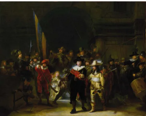
Rembrandt van Rijn (1606–1669) was a famous Dutch painter, whose name is almost equal tofine arts. He did not live too long, and his late works and change in style led critics and ophthalmologists to the conclusion that he developed eye problems, had cataract, and possibly macular
degeneration. He started his career in Leiden, and then he moved to Amsterdam where he became famous in a quite short time. By 1640, he had a large house in the center of Amsterdam, a beautiful aristocratic wife, and a lot of students learning from him. His wife died in 1642 possibly due to tuberculosis and left him with four children, but only one daughter survived until adult age. In 1656, plague appeared in Amsterdam and 18,000 people died. In the same year, Rembrandt had to declare bankruptcy, but he still had good contacts with middle-class merchants and physi- cians, and from these wealthy people, he continuously received orders for the rest of his life. Presbyopia was the first condition, which struck him around the age of 50. On a portrait, his mother wears reading spectacles, which was already available at his time [4]. In his late works, he was still able to paint the small details, so one can be sure that he possessed a presbyopic correction, which was unusual in his time. In his self-portraits, it seems that one eye may turn a bit outwards with a mild degree; therefore, some authors hypothesised that he might have had strabismus, but others questioned this finding [3,5]. At later age, he might have developed a nuclear-type (central) cataract, causing mild loss of visual acuity and change in colour discrimination.
His paintings became darker compared with his pictures created at younger ages (Figure2) [6]. Rembrandt is a good example that although he might have had some eye pro- blems, which were mainly age-related, he was able to continue his art until his death [1, 7]. Tonality of his paintings changed, but he was able to paint thefine details as well, even in his elder years.
One of the greatest Spanish painters was Goya y Lucientes (1746–1828), who was hit by a mysterious dis- ease in his midlife. He experienced loss of vision and hearing, ringing in the ears, vertigo, and weakness on one side of his body, abdominal pain, confusion, and malaise.
He got recovered during the next couple of months, but the deafness remained permanent. He was of course distressed by the poor visual acuity for months and was not able to paint. Thereafter, he continued painting but was not able to communicate with his students due to his permanent deaf- ness. It was presumed that he acquired syphilis based on the clinical signs and symptoms, such as loss of vision and hearing, motor disability, and behavioural changes. It is thought that it was a late-acquired syphilis, which was quite common those times. Contrary to the diagnosis of syphilis, he lived a long life. His symptoms started in 1792 when he was 46 years old, and he died in 1828 when he was 82 years old. According to others, Goya’s disease might have been an immunologic disorder, such as the Vogt–Koyanagi–Harada syndrome [8]. Typical symptoms are inflammation within the eye, hearing disturbances, and loss of pigment in the skin and hair. There is no evidence that Goya had loss of pigmentation in the skin or hair. Other possible cause might have been Cogan syndrome [8,9], which is characterised by inflammation of the cornea, vertigo, ringing phenomena in the ear, deafness, malaise, and weight loss. The hearing problem might be permanent and severe. Another physician raised a provocative hypothesis that Goya might have had lead toxicity. Lead toxicity might cause mental and physical problems, which Goya also had. The base of paints in Goya’s time was lead. In his time, paint pigments came in Figure 1.El Greco: St. Andrew and St. Francis (cc. 1575).
Source:https://commons.wikimedia.org/wiki/File%3AEl_Greco_- _St_Andrew_and_St_Francis_-_WGA10500.jpg
Painting and the eye
a solid form and had to be ground into powder before mixing with oil. Goya mixed the paint for himself and presumably might have acquired lead toxicity. Goya might also have had malaria infection and due to this he might have taken quinine, which could cause quinine toxicity. It can cause temporary blurred vision, colour vision defects, ear ringing, and vertigo. If the central nervous system is also affected, headaches, confusion, delirium, seizures, and coma may appear. Digestive symptoms include abdominal pain, nausea, vomiting, and diarrhea. If Goya presumably had malaria, it is likely that he was taking quinine for a longer period. The exact diagnosis of his midlife illness still remains unclear, but he was never the same after his severe midlife health problem [1]. His paintings were much darker and frequently contained psychotic elements (Figure 3).
Not only the colours were dark, but the theme was also horrific sometimes, e.g., Saturn Devouring His Son [10].
The cause of Goya’s near death illness experience is still a subject of speculation. The diagnosis of acquired syphilis is not well supported. Speculation about seizures, stroke, and schizophrenia also lacks proof. The arguments on an immune-mediated illness are not strong enough either. A more likely cause could be an infectious disease, such as malaria, meningitis, or encephalitis, which might be exag- gerated by quinine toxicity [8–11]. At the age of 82, in 1825, he developed urinary paralysis and hardness of the bladder, and a tumour was found in his perineum. At that time, it was not a treatable condition, so he died at the age of 82, leaving a great heritage for us.
From the second half of the 19th century, a new style, impressionism emerged. This style was considered shock- ing, revolutionary, and radical. Brushstrokes were quick and spontaneous, thick, and usually coarse. Instead of religious scenes, artists painted the real life, such as bridges, parks, locomotives, industry, and everyday people. Impressionism
was named after Claude Monet (1840–1926) titled his paintingImpression, Sunriseat an exhibition. His followers were called the Impressionists. He lived long enough to develop cataract, and the course of cataract can be followed Figure 2.Rembrandt van Rijn: The Night Watch (1642). Source:https://commons.wikimedia.org/wiki/File:Lundens_-_Nachtwache-Kopie.jpg
(Gerrit Ludens: Copy of The Night Watch by Rembrandt)
Figure 3.Goya: The Bewitched Man (1798). Source:https://
commons.wikimedia.org/wiki/File%3AFrancisco_de_Goya_y_
Lucientes_-_The_Bewitched_Man_-_WGA10039.jpg
in his artwork. When he became 66 years old, he noticed the first signs of cataract, although he was diagnosed with cataract only 4 years later [12,13]. The subtle differences in colour were difficult for him to differentiate due to the lens opacities. By September 1922, his vision had decreased dramatically, but he resisted surgery. With pupil dilation, he was still able to work, but it was not enough for the long run.
Later, he agreed to the cataract surgery. At that time, cataract surgery was quite complex and rudimentary compared with the methods we use today. No intraocular lenses existed, so visual rehabilitation was poor. Patients received very thick glasses, which caused visualfield and colour discrimination disturbances. The postoperative visual acuity was certainly not enough for a painter at that time. He had of course many problems after surgery, developed secondary cataract, and had to be reoperated. He had depression for a longer period due to his poor vision and wrote complaining letters to his surgeon Dr. Charles Coutela. By 1923, he achieved a 20/30 acuity (60% visual acuity compared with a person with normal vision), but he had to wear a +10.0+4.0×90°
spectacle correction. Due to the huge diopter difference, he was not able to see with both eyes together. He refused to have his left eye operated on. Eventually, he received a Zeiss glass later, but he still complained of problems with colour discrimination. Later, he was able to accept the changed visual acuity and the thick glasses, and Monet was able to continue paintings until his death [1, 14]. He was able to finish the water lily series (Figure4), which was ordered by the French state and can be seen today at the Orangerie in Paris. His last spectacle is displayed at the Musee Marmottan in Paris. In his later paintings, one can observe the change in tonality, all pictures are darker and fine details are rough.
After the cataract surgery, colours became lighter andfine details certainly better [15,16].
Vincent van Gogh(1853–1890) lived a very short life, as he died at the age of 37. He was born in the Netherlands just 1 year after his stillborn brother. He received the same name.
During his life, he showed a bizarre behaviour, mental illness, and a special artistic technique. During his last 2 years, there were mentally clear periods mixed with episodes of severe mental disorders. His mental disturbances were attributed to schizophrenia, bipolar disorders, enzyme
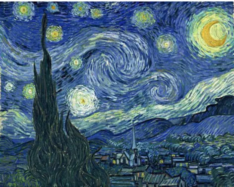
deficiency, epilepsy, Meniere’s disease, and chemical toxic- ity. Due to lack of precise medical records, it is not known what the real underlying cause was, but it is possible that he had several diseases during his lifetime. In his paintings, special haloes were painted, like inThe Starry Night(1889), therefore, some presume that he had hallucinations or a glaucomatous attack (angle closure glaucoma) causing those particular haloes around the stars, moon, and sun (Figure5).
Glaucoma has not been proved at all until now [1,3,15].
Even more interesting is his yellow period [Vase with Fifteen Sunflowers(1888) or self-portraits with straw hat].
In these pictures, the environment and the persons received a yellowish character (Figure 6).
Was it a conscious choice or was caused by a disease like xanthopsia [17]? During that time, Xanthopsia was described to be caused by substance abuse or chemical toxicity. Digitalis was widely used those times not only for cardiac diseases but also to treat melancholia, hypochon- dria, pain in the eyes, swelling of the eyelids, tearing, inflammation of the eyes, headache, vomiting, and nausea.
Van Gogh might have had maniac depression, a mental disorder for which no appropriate medication existed at the time. The other drug that could have caused xanthopsia was santonin. This drug was used for gastrointestinal distur- bances to treat intestinal parasites. Van Gogh was known to have digestive problems, which were treated with santonin.
He is also known to have had abnormal cravings (pica) for camphor as a sleep remedy (he could not sleep during the nights), and he also often consumed absinthe, which is a strong alcohol. Camphor might also have been in the background to cause xanthopsia. Whether his xanthopsia was chemically or drug-induced remains unclear [18].
Van Gogh was also known to experiment with different colours, so the yellow period might have been part of this.
He always wanted to be famous, but became fame only after his death. On July 27, 1890, he could not tolerate loneliness anymore, so during a depressive attack, he shot himself on a field in France where he often painted. He was able to go back to his room and he died 2 days later in the arms of his brother. He sold only one picture in his life, but later he became world famous. He always felt that fame will come after his death and he wrote in his letter:“Just as we take the Figure 4.Claude Monet: The Water Lily Pond (1919). Source:https://commons.wikimedia.org/wiki/File%3AWater-Lily_Pond_1919_
Claude_Monet_Metropolitan.jpg Painting and the eye
train to get to Tarascon or Rouen, we take death to go to a star. What’s certainly true in this argument is that while alive, we cannot go to a star, any more than once dead we’d able to take the train”(Arley, Monday, 9 or Tuesday, July 10, 1988) [19].
Edgar Degas (1834–1917) is a famous French painter and sculptor, whose style was characterised by hazy pastel indoor pictures with fresh colours and featureless faces with
usually off-center focal points. He experienced a progressive visual deterioration at an early age, already at 36 years, during the Franco-Prussian war, and he was not able to see clearly the target with the right eye while using a rifle. He wrote that this was possibly due to extreme weather con- ditions. However, it was due to his starting macular pro- blems. His problem was attributed to chorioretinitis, and to our present knowledge, he had a familiar macular degener- ation [1,3,15] and not an age-related one. He experienced a progressive loss of colour perception during the years. He possibly had a blue cone deficiency, which might explain the dominance of red colours in his pictures and the limited use of blue. His medical records are mostly lost, but his−1.5 diopter glasses are kept in Musee d’Orsay in Paris. They were dark-tinted gray glasses, which blocked 85% of the incoming light due to his light sensitivity. Later, he used an occluder (non-see-through) lens (could not see through) for the right eye and a stenopeic slit glass at 160° for the left one. Most probably, he had astigmatism, which was cor- rected this way [20]. The stenopeic slit was used to correct his irregular astigmatism and to reduce the incoming light and glare [14–16]. Certainly, he was not blind in the right eye and had a diminished visual acuity in the left eye, but with the aforementioned style, he was able to continue his art and left a great legacy for us.
Camille Pissarro(1830–1903) was also an outstanding member of the French impressionists. He is regarded with Claude Monet as a founder of impressionism. He was born in West Indies with a Portuguese origin, and was educated in France. Around 60 years of age, he developed a chronic inflammation around the right nasolacrimal duct, from which he could not get rid of until his death [1,3]. It did not disturb him in painting until his death, but rendered a torture during acute phases. He had several operations, but the problem was not solved. At that time, antibiotic therapy Figure 5.Vincent Van Gogh: The Starry Night (1889). Source:https://commons.wikimedia.org/wiki/File%3AVanGogh-
starry_night_ballance1.jpg
Figure 6.Van Gogh: Vase with Fifteen Sunflowers (1888).
Source:https://commons.wikimedia.org/wiki/File%
3AVincent_van_Gogh_-_Sunflowers_-_VGM_F458.jpg
was not available. Unfortunately, he developed several abscesses, had to use patches, homeopathic medications, and the abscess was incised several times, but none of them helped. He had probing, which caused complications such as false lacrimal passages, cellulitis (very painful inflamma- tion around the orbit with fever), and scarring. During acute abscess formation, he was unable to paint because of pain, patches, and fever. At that time, silver nitrate was the remedy of the nasolacrimal duct abscesses [21]. He com- plained a lot of his condition:“Parenteau (his ophthalmol- ogist) gave me silver nitrate drops to put in the eye and on the lids. This is hardly easy. Every morning there is more pus in the eye, so that I dare not venture a trip.”[22]. He could not visit his son in London, who was dying of tuberculosis. It was not until 1904 (1 year after his death), when Toti described the operation dacryocystorhinostomy, which could have solved the condition of Pissarro effec- tively much earlier [21,22].
Amadeo Modigliani(1884–1920) was an Italian sculptor and painter. He lived a very short life (36 years), mainly in Paris, and he survived during the First World War. He was a very emotional artist, who sought for beauty, passion, and cleanliness, and was influenced by the style of Sandro Boticelli. He also used elongated shapes in his paintings and sculptures, and was also possibly influenced by astig- matism [3,23]. In 1920, when living in Paris, he had kidney problems possibly caused by tuberculosis and shortly after he died in tuberculotic meningitis, when his wife (Jeanne Heubuterne) was pregnant with their second daughter. He became famous after his death and had a great influence on JenőBarcsay among the Hungarian artists.
Jules Cheret(1836–1932) was the founder of the modern lithographic poster. It was a type of commercial and artistic advertisement. He was a very successful illustrator and commercial graphic designer. Besides his posters, he also created portraits, sculptures, and also decorated the walls of the City Hall in Paris. His career came to an end due to an angle closure glaucoma attack in 1923 [24,25]. Ophthalmic treatment was ineffective, and he lost his vision. Claude Monet was his friend and Cheret lost his vision the same year when Monet had his cataract surgery. Five years before his death, he was completely blind with no light perception and he needed the help of another person while walking outside.
Francois Ravier(1814–1895) was famous for his roman- tic landscape paintings. Ravier had good health until the age of 65, when on an autumn day, he had a stroke and lost consciousness. During his treatment, he had to reduce physical exercise and had to take arsenic medication. At the age of 69, he experienced pain and loss of vision in his left eye. The diagnosis was a general word“ophthalmia.”He had haemorrhagic glaucoma and the pain was intractable, so the left eye had to be removed. He called himself humour- ously “one-eyed Cyclops”, and with the other eye, he continued creating outdoor paintings [24,25]. At the age of 74, he developed cataract on the right eye. There are no data how it affected his art, but he continued to work until the last year of his life, so certainly it was not a mature cataract, but rather a phacosclerosis.
Louis Valtat (1869–1952) was a French painter, who studied and lived in Paris. He studied from Henri de
Toulouse Lautrec as well. First, he was influenced by impressionism, later by pointillism, and then by the Nabis movement (from the Hebrew movement, “nabi” means prophet). He was influenced by Henri Matisse as well and was a personal friend of Renoir. Valtat painted landscapes with harsh colours, and he worked at the Mediterranean coast. He also met Renoir at the sea. At the age of 78, he was diagnosed with open-angle glaucoma [24,25]. His pictures were still consistent, meaning that his glaucoma did not affect his work for long. This type of glaucoma causes a peripheral visualfield defect, so central vision is maintained for long time. At that time, the only available drug was pilocarpine, which narrowed the pupil. He was able to paint, until glaucoma affected his central vision.
Roger Bissiere(1888–1964) was born in the Southwest of France and was educated in Bordeaux at the School of Fine Arts. After graduation, he moved to Paris, where he met Georges Braque, who together with Picasso founded cubism. In 1939, he noticed that his vision was deteriorat- ing. He was not able to see the top of his canvas, which corresponds with the upper visual field defect caused by primary open-angle glaucoma [24,25]. The loss of periph- eral vision cannot be detected in his paintings, because by moving his head, he was able to compensate this problem. In spite of this diagnosis and medical advice, he refused surgery for a decade. Over the next decade, his vision worsened and finally he agreed to have surgery. Due to successful surgery, the intraocular pressure was controlled and his visual deterioration slowed down. He was able to continue work. During the war years, he changed style, starting cubism in his works (non-representational art), using bright colours and short curved lines. He became famous in 1952, when the French government awarded him the Grand Prix National des Arts. Fortunately, surgery was able to prolong his painting career, unlike in case of his contemporaries, Cheret and Valtat.
Mary Cassatt(1844–1926) is the most well-known Amer- ican impressionist. She spent a long time in France, knowing the most outstanding French contemporaries. Mary Cassatt started to experience visual problems around 1900, but cataracts were not diagnosed until 1912 [1,3]. She also had diabetes mellitus, but since insulin therapy was not available until the 1920s, she had to undergo other treatments for that.
A rather bizarre therapy was with radium, which was dis- covered at the early years of the 20th century. There is no evidence that she was also treated with radium for her cataract, but in the prestigious American Journal of Ophthal- mology, there was an article about the useful effects of radium for the treatment of cataract in 1920 [26]. The harmful effects of radium were not discovered that time, even Marie Curie developed cataract from the experiments with radium in the lab, and she had to undergo cataract surgery on both eyes. Mary Cassatt was operated on the right eye in 1917 with an extracapsular technique. Naturally, she developed posterior capsule opacification and needed a second surgery to make a hole on the posterior capsule. She complained of bad vision after the secondary cataract was removed. In 1919, she had cataract removal on the left eye, and afterwards she had an endophthalmitis (had a bad tooth) and could not see at all with the left eye [27]. By 1919, she could not read anymore and could not paint. She died in 1926, the same Painting and the eye
year as Monet died. She was deprived of her art due to cataract and a severe diabetic retinal complication – much earlier than it should have happened.
Edvard Munch (1863–1944) was a Norwegian painter.
One of his most famous paintings isThe Scream. He was famous for the haunting images and psychological themes, and he used striking colours in his paintings. Munch’s father was a physician and a religious man. He was devastated after losing his wife in tuberculosis, when Munch was 5 years old. Death took her elder sister, due to tuberculosis again, when he was in his teens. He watched his sister dying and then he rejected religion. He visited Paris in 1885 and met contemporary artists. In his pictures, he depicted love, anxiety, and sex as well. Munch lived for a longer time in Paris and Berlin. Unfortunately, he had a nervous break- down in 1908 and in 1910 he moved back to Norway and continued to paint until his death in 1944 in Ekely (nearby Oslo). His left eye was weak (amblyopic for years and it is not clear whether trauma might have been the cause of it or it has nothing to do with it). In 1904, he was punched in the left eye in afight, but there is no record that the left eye would have been seriously injured. It was not until 1930, at the age of 67 years, when he suffered a haemorrhage on the right eye [28]. Because his vision was poor for both eyes, he was not able to work for months. Interestingly, he made a famous drawing of what he saw with the right eye. With six sketches, he documented what he saw and he also documented the course of the retinal/vitreous haemorrhage [29]. Professor Johan Reader, an eminent ophthalmologist in Oslo, advised him to have a complete rest, so that the eye could heal.
Hypertension could be a likely diagnosis, but there is no record that he had hypertension. Other cause could be retinal tear or vascular malformation, such as the age-related wet- type of macular degeneration [1,3]. Whatever was the cause, it healed with time, and Munch was able to continue paintings afterwards [30]. During the healing phase, he painted a certain sinister bird on his paintings, corresponding with the bleeding within the vitreous. These pictures resem- bled the so-called Rorschach test pictures. Munch effectively showed what he had experienced by the intraocular“debris” and incorporated his visions into artistic paintings. In another pastel, he portrayed himself in the bed, checking the vision of the right eye (covering the left one). On the right side of the picture, there is a dark spot resembling the death’s head at the foot of his bed. The shape of a sinister bird, which was mentioned earlier, appears in more pictures. In his works, there are some fantastic details: he painted scenes, the integral part of which lied within his eye, showing land- scapes with the style that no one else could have seen that way. He gave some very important clues about the disease progress. The sinister birds and scotomas disappeared by the year of 1931 from his paintings. He continued the full-scale painting until his death in 1944.
DISCUSSION AND CONCLUSIONS
In this article, some eye diseases were shown, which might have influenced the style of paintings. Myopia, presbyopia, amblyopia, colour deficiencies, visualfield defects, lentic- ular, and macular changes might influence the vision of an
artist. The cataract procedure, which was used in the times of impressionists, might seem barbaric and primitive for us. Thick lenses, with diminished visual field, changed colour discrimination, and the huge diopter difference between the operated and unoperated eyes made it some- times impossible for the artists to continue their work.
Today, we perform a 1-day surgery with a perfect surgical technique. The patients can go back to their normal life on the next day. In case of Monet, it took 6 months to be able to work again. Glaucoma and age-related macular degen- eration (AMD) are also treatable conditions. Glaucoma can be treated with drops and with different surgical techniques as well. Glaucoma differs much from cataract and retinal disorders, because the visual deterioration is gradual and painless (except for angle-closure glaucoma attack, which is very painful). The central macular fibers are preserved until the final stage of glaucoma. Therefore, painters are able to work with a constricted visualfield, and with the movement of the head, they are able to compensate for the visualfield defect. Although glaucoma is still consid- ered as the “thief of sight,” early detection helps to preserve vision for a much longer time than a few decades ago.
AMD can be effectively treated with anti-vascular endo- thelial growth factor intraocular injections and also with vitamins in case of dry macular degeneration.
The ocular complications of diabetes mellitus can also be effectively treated by the laser, vitrectomy, or cataract procedures. In our times, we are able to restore the visual acuity for a person with cataract operation with the optical result of seeing like a 20-year old. New generation of premium intraocular lenses, such as toric, multifocal, or multifocal-toric, makes vision perfect, if there is no other ocular pathology present. Yttrium aluminium garnet laser capsulotomy made posterior capsular opacification to be a problem that can be solved within a minute.
Psychological disorders can be treated by effective anti- psychotic drugs and psychotherapy as well. Spectacles became fashionable, especially sunglasses. Multifocal pro- gressive glasses provide a solution for presbyopia combined with all kinds of refractive errors. Contact lenses and refractive surgery provide solution for those who do not want to wear any spectacles. Patency of the lacrimal system is provided by surgery and helped by potent antibiotic treatment as well.
The last question is if the impressionists could have the most up-to-date ophthalmic surgical methods, would the impressionist movement have even occurred? And if yes, would it have been the same as it happened? We do not know the answer, but nowadays artists have these modern ophthalmic methods at their disposal, and we are keenly waiting for the new styles of paintings in our ages.
Funding sources:Nofinancial support was received for this study.
Conflict of interest: The author declares no conflict of interest.
REFERENCES
1. Marmor MF, Ravin JG. The Artist’s Eyes: Vision and the History of Art. London: Abrams; 2009.
2. Simunovic MP. “The El Greco fallacy” fallacy. JAMA Ophthalmol. 2014;132(4):491–94.
3. Trevor-Roper PD. The World Through Blunted Sight. 3rd ed.
London: Souvenir Press; 1997.
4. Marmor MF. Vision, eye disease, and art: 2015 Keeler Lec- ture. Eye (London). 2016;30(2):287–303.
5. Mondero NE, Crotty RJ, West RW. Was Rembrandt strabis- mic? Optom Vis Sci. 2013;90(9):970–79.
6. Marcus EL, Clarfield AM. Rembrandt’s late self-portraits:
psychological and medical aspects. Int J Aging Hum Dev.
2002;55(1):25–49.
7. Wiser I, Parnass AJ, Rachmiel R, Westreich M, Friedman T.
Rembrandt’s ocular pathologies. Ophthal Plast Reconstr Surg.
2016;32(4):305–9.
8. Ravin JG, Ravin TB. What alied Goya? Surv Ophthalmol.
1999;44(2):163–70.
9. Cawthorne T. Goya’s illness. Proc R Soc Med. 1962;55:213–17.
10. Guijarro-Castro C. Could neurological illness have influenced Goya’s pictorial style? Neurosci Hist. 2013;1(1):12–20.
11. Niederland WG. Goya’s illness: a case of lead encephalopa- thy? NY State J Med. 1972;72(3):413–18.
12. Ravin JG. Monet’s cataracts. JAMA. 1985;25(4):394–99.
13. Lanthony P. Cataract and the painting of Claude Monet. Points de Vue. 1993;2(9):12–25.
14. Marmor MF. Ophthalmology and art: simulation of Monet’s cataracts and Degas’ retinal disease. Arch Ophthalmol.
2006;124(12):1764–69.
15. Elliott DB, Skaff A. Vision of the famous: the artist’s eye.
Ophthal Physiol Opt. 1993;13:82–90.
16. Hamilton GH. The dying of the light: the late works of Degas, Monet and Cezanne. In: Rewald J, Weitzenhoffer F, eds.
Aspects of Monet. New York: Abrams; 1984. p. 218–41.
17. Arnold WN, Loftus LS. Xanthopsia and van Gogh’s yellow palette. Eye. 1991;5:503–10.
18. Lee TC. Van Gogh’s vision. Digitalis intoxication? JAMA.
1981;245(7):727–9.
19. van Gogh V. Further Letters of Vincent van Gogh 1886–1889.
London: Constable; 1929.
20. Ravin JG, Kenyon CA. Degas’loss vision: evidence for a diagnosis of retinal disease. Surv Ophthalmol. 1994;39:
57–64.
21. Ravin JG. Ophthalmology and the arts: Pissarro’s lacrimal problems. Ophthalmic Forum. 1984;2(1):54–55.
22. Ravin JG. Pissarro, dacryocystitis, and the development of modern lacrimal surgery. In: Henkes HE, ed. History of Ophthalmology, Vol 6. Dordrecht: Springer; 1994. p. 191–202.
23. Nathan J. The painter and handicapped vision. Clin Exp Optom. 2002;85(5):309–14.
24. Ravin JG. The visual difficulties of selected artists and limita- tions of ophthalmological care during the 19th and early 20th centuries (an AOS thesis). Trans Am Ophthalmol Soc.
2008;106:402–25.
25. Lanthony P. Art and Ophthalmology: The Impact of Eye Diseases on Painters. Piribebuy: Weyenborgh Publications;
2009.
26. Franklin WS, Cordes FC. Radium applicator for cataracts. Am J Ophthalmol. 1921;4(6):429–30.
27. Marmor MF. Vision loss and hearing loss in painting and musical composition. Ophthalmology. 2014;121(7):1480–5.
28. Marmor MF. A brief history of macular grids: from Thomas Reid to Edvard Munch and Marc Amsler. Surv Ophthalmol.
2000;44(4):343–53.
29. Meyer C. Max Linde, MD, a Luebeck ophthalmologist and patron of Edvard Munch. Surv Ophthalmol. 1999;43(6):
525–34.
30. Gruener A. Munch’s visions from within the eye. Br J Gen Pract. 2014;64(618):36–7.
Painting and the eye