The Nature of Infections Caused by Nuclear-Polyhedrosis Viruses
KEIO AIZAWA
Sericultural Experiment Station, Suginami-ku, Tokyo, Japan
I. Introduction 382
II. Symptomatology and Pathology 382
A. Lepidoptera 382 382
Β. Hymenoptera 384 384
C. Diptera 385 385
III. Mode of Virus Multiplication 385
A. Routes of Infection 385
B. Latent Period and Virus Multiplication 386 C. Virus Multiplication by Infection Experiments 388 D. Morphological and Histological Observations of Virus
Multiplication 391 391
E. Virus Multiplication by Means of Tissue Culture and
in Chick Embryo 393
IV. Cross Infection and Double Infection 394
A. Cross Infection 394
B. Double Infection 396
V. Susceptibility 397
V. 397
VI. Defense against Nuclear-Polyhedrosis Viruses 398
A. Defense against Infections 398
B. Resistance to Virus Disease 398
VII. Virulence, Infectivity, and Stability of Viruses 399
A. Virulence 399
B. Infectivity 400 400
C. Stability 400 400
VIII. Transmission of Virus 401
I X . Metabolic Changes in Virus Infection 402
References 403
381
12
I . INTRODUCTION
After nuclear-polyhedrosis viruses are injected into host insects, the viruses are adsorbed onto susceptible cells, invade the cells and, if con
ditions are suitable, will multiply in the cells. T h e multiplied viruses will be released from the infected cells, and noninfected cells will repeatedly be infected by such released viruses. T h e characteristic inclusion bodies, so-called polyhedra, are formed in the nuclei of cells infected with the virus. T h e polyhedra and nuclei increase in size and the infected cells eventually are destroyed by the accumulating polyhedra which may then be seen suspended in large numbers in the body cavity of host insects.
Owing to the virus multiplication and the formation of polyhedra, the external symptoms become clearly apparent, and the host insects finally die of the disease.
Nuclear-polyhedrosis viruses affecting nearly 200 species of Lepidop
tera, Hymenoptera, and Diptera have been reported (Hughes, 1957;
Martignoni and Langston, 1960). This type of virus disease occurs mainly in Lepidoptera. T h e general aspects of the infection caused by nuclear polyhedrosis have been discussed by a number of writers (Pail
lot, 1930; Letje, 1939; Bergold, 1943, 1953a, 1958a; Steinhaus, 1946, 1949a; Jahn, 1958; Smith, 1958, 1959; Krieg, 1961).
In this chapter, the process from the time of ingestion of nuclear- polyhedrosis viruses to that of death of host insects, and certain relation
ships between host insects and viruses will be discussed.
I I . SYMPTOMATOLOGY AND PATHOLOGY
A. Lepidoptera
1. Signs and Symptoms
In the case of the silkworm, Bombyx mori (Linnaeus), the infected larvae show no symptoms and no change in appetite during more than two-thirds of the incubation period. Usually, 5 to 7 days after infection, integumental membranes are swollen and larvae are impatient in move
ments. T h e normally clear hemolymph becomes turbid and the skin becomes very fragile. When the integument is ruptured, a milky hemo
lymph flows out, and the larvae eventually die. T h e dead larvae become flaccid. T h e period from the swelling of the segmental membranes to death is relatively short—from several hours to less than one day.
In the silkworm pupae, during the greater part of the incubation period, no external change is observed. But at the end of the disease, the skin is very easily ruptured on handling and the pupal body shows a homogenized appearance. Sometimes black markings are observed on the body surface around the time of death. When the virus is injected
12. N U C L E A R - P O L Y H E D R O S I S VIRUS I N F E C T I O N S 383 into pupae at a late pupal stage, virus-infected adults appear and the body becomes ruptured as in the pupae.
Representative nuclear polyhedroses in wild insects are those of the nun moth, Lymantria monacha (Linnaeus), and the gypsy moth, Por
thetria dispar (Linnaeus). In the former, the infected larvae show a loss of appetite and become flaccid. Before death, larvae show a ten
dency to move to tops of trees and hang in a characteristic way by their legs. Accordingly, this disease has been known as Wipfelkrankheit. In the dead larvae, a fluid of distintegrating internal tissues is liberated from the broken skin. T h e period from infection to death is 13 to 15 days in Lymantria monacha, and 4 to 24 days (mean 10 to 12 days) in Porthetria dispar. In the cabbage looper, Trichoplusia ni (Hübner), decrease of body weight and food consumption of virus-infected larvae were measured (Drake and McEwen, 1959).
2. Pathology
At an early stage of infection in the silkworm, chromatin aggregates and very small granules showing strong Brownian movement appear in the nuclear ring zone. T h e name "propolyhedra" was proposed by Smith and Xeros (1953b) for such small granules (0.2 to 0.4 μ in di
ameter) . These granules are early stages in the development of poly
hedra which grow around a dense central mass. Nuclei of infected cells increase in size (Smith and Xeros, 1953c; Yamafuji, 1952b). Nuclei which are filled with polyhedra occupy nearly all parts of the cells, and finally the latter are destroyed by the fully grown polyhedra. Sub
sequently they are suspended in large numbers in the hemolymph.
In the silkworm, the size of polyhedra shows very little variation in a given nucleus. Any variation in size from nucleus to nucleus seems to depend on the time and amount of infection of the individual cells.
Such a histopathological observation was reported by Steinhaus (1949b) in the nuclear polyhedrosis of the alfalfa caterpillar, Colias eurytheme Boisduval. T h e size of the polyhedra is usually relatively small when a considerable number of them are formed in a nucleus; however, the size is relatively large when only a few polyhedra are formed, at least in the silkworm. According to Smith and Xeros (1953b) polyhedra are formed all at once and subsequently only increase in size. There is no continuous production of new polyhedra in a given nucleus. But in the spruce budworm, Choristoneura fumiferana (Clemens), not only do polyhedra grow, but the number of them increases in the course of in
fection (Bird and Whalen, 1954b; Bird, 1959).
In the silkworm, and in other lepidopterous insects, polyhedra are formed in the nuclei of blood cells and cells of the fat body, tracheal
matrix, and epidermis. Generally polyhedra are not formed in glands or nerves; however, Aruga et al. (1957) observed the formation of poly
hedra in the middle and posterior portions of the silk glands in the silkworm.
Nuclear polyhedra have a diameter ranging from 0.5 to 15 μ. T h e size of polyhedra in the silkworm is generally 3 to 5 μ and the shape of polyhedra is usually hexagonal, but rarely tetragonal (Bergold, 1943;
Aizawa, 1955c). Tetragonal polyhedra, the corners of which are some
what rounded, are seen in silkworm pupae. Generally speaking, the shape and size of polyhedra in insects depend upon the species of insect virus concerned.
Polyhedra are not easily stained; however, pretreatment of polyhedra with dilute alkali or acid facilitates the staining with dyes. Gratia et al.
(1945), Iwasaki (1954), and Iwashita and Aruga (1957) reported his- tochemical observation of the infected cells. T w o days after infection, the ribonucleic acid (RNA) content of the cytoplasm and that of nu
cleoli increases accompanying the increase of the deoxyribonucleic acid (DNA) content of chromatin until polyhedra formation begins. Sub
sequently the nucleoli disintegrate and the R N A content in the cyto
plasm decreases. Polyhedra are Feulgen positive.
B. Hymenoptera
1. Signs and Symptoms
Nuclear polyhedroses have been found in hymenopterous insects such as the European spruce sawfly, Diprion hercyniae (Hartig), the Euro
pean pine sawfly, Neodiprion sertifer (Geoffroy), and the native jack- pine sawfly, Neodiprion pratti banksianae Rohwer; these polyhedroses have been well studied by Balch and Bird (1944), Bird (1952, 1953a, b, 1955, 1957), Bird and Elgee (1957), and Bird and Whalen (1953, 1954a). T h e diseased larvae show a faint yellow discoloration particu
larly on the third to fifth abdominal segments, become inactive and lose their appetite. T h e virus-infected larvae often exude a dark-brown fluid from the anus, and emit a milky white fluid. T h e period from infection to death, usually 6 to 16 days (mean 9.8 days), depends upon the tem
perature. Finally the larvae become flaccid, and the skin becomes fragile and, when broken, the liquid body contents are liberated.
2. Pathology
T h e outstanding characteristic of nuclear polyhedroses in Hymenop
tera is the formation of polyhedra in the nuclei of epithelium of the midgut. In the early stages of infection, nuclei and nucleoli swell and the coagulation of chromatin network may be observed. T h e nuclei
12. NUCLEAR-POLYHEDROSIS VIRUS INFECTIONS 385 then become filled with "immature" polyhedra and later with much larger polyhedra. Polyhedra are irregular in shape, and from 0.5 to 2 μ in size. T h e midgut of infected larvae is milky white and devoid of food.
Benz (1960) conducted the histochemical studies on the virus- infected midgut cells of Diprion hercyniae. Some nucleoli swell shortly after infection and their R N A content is released, whereas that of the cytoplasm is increased. Later on, the "virogenic stroma" (Xeros, 1956) in the chromatin develops, becomes denser and increasingly Feulgen positive; then the Feulgen-positive reaction decreases, and eventually polyhedra are formed.
C. Diptera
1. Signs and Symptoms
T h e only nuclear polyhedrosis known to occur in a dipteran insect has been found in the crane fly, Tipula paludosa (Meigen). T h i s dis
ease was found by Rennie (1923) and rediscovered by Smith and Xeros (1954a); it has been thoroughly studied by Smith (1955a, b, c ) . T h e infected larvae can be recognized by their unusual pallor, which in
creases as the disease progresses. At the last stage of the disease, larvae appear chalky white in color as contrasted with the earthy color in nor
mal larvae. When the integument is ruptured, milky white fluids pour out and many refractive crescent-shaped granules are seen in the fluids.
T h e incubation period is about 14 days but may be prolonged for a month or more during the winter.
2. Pathology
A chromatic mass forms in the enlarged nuclei of the blood cells and fat-body cells, and from this mass the chromatic material segregates into several spherical bodies. Later these granules are formed on the periphery of the nucleus, which occupies most of the cell. T h e highly refractive crescent-shaped polyhedra appear around the central mass and are finally accumulated in one hemisphere of the nucleus.
I I I . M O D E OF VIRUS MULTIPLICATION (See also Chapter 13) It may be said that the mechanism of virus multiplication concerns the nature of virus infection. T h e mode of virus multiplication has been investigated mainly on the basis of cytological observations, and from the quantitative estimation of virus infectivity after the infection.
A. Routes of Infection
T h e routes of infection in a nuclear polyhedrosis may include the following three: (1) subcutaneous infection, (2) oral infection, and
(3) infection through stigma. T h e first two routes are routinely used for experimental infection; artificial dissemination of nuclear-poly
hedrosis virus, and possible spread through the intestinal canal of birds (Franz et al., 1955), is based on oral infection. Infection through stigma has not yet been confirmed.
T h e hemolymph of insects infected with a nuclear-polyhedrosis virus contains two kinds of virus source, i.e., free virus particles and virus- containing polyhedra. Supernatants (7000 rpm for 10 to 20 minutes) of infected hemolymph contain infectious virus particles capable of causing infection through wounds in the insect integument. Infection may be caused by the administration of polyhedra contained in the hemolymph of infected insects and of those in dead cadavers. Infection does not take place by the injection of well-washed polyhedra into the insect body cavity. Polyhedra are phagocytosed by blood corpuscles, though the fate of them in the cells has not been clarified. However, such well-washed polyhedra cause the infection when administered orally (Glaser, 1928;
Ishimori, 1937). T h e polyhedra are dissolved by the alkaline gut juice, and the liberated virus particles penetrate through the gut epithelium and multiply in the cells of the blood and other tissues (Day et al., 1958;
Vago and Croissant, 1959; Bird, 1959).
Virus infectivity has not been demonstrated in the feces of typically virus-diseased larvae such as Lymantria monacha and Bombyx mori
(Escherich and Miyajima, 1911; Aizawa, 1953b). Apparently this is because of the action of the gut juice, which inactivates the virus (Kita- jima, 1932; Suzuki, 1937; Aizawa, 1953b, 1962a; Masera, 1954).
B. Latent Period and Virus Multiplication
It has been noticed that the latent period ( = incubation period) in the nuclear polyhedroses seems to have an intimate relationship with the temperature and the inoculum size of viruses. For example, Paillot
(1930) observed that silkworm larvae showed no symptoms 15 days after the virus infection at 16° to 17°C, whereas infected larvae showed symptoms in 6 days at 25 °C. Steinhaus (1949b) reported that the incuba
tion period of the nuclear polyhedrosis of the alfalfa caterpillar, Colias eury theme Boisduval, was an average of 7 days, whereas it took up to 3 weeks in the winter season in the laboratory.
T h e latent period of nuclear polyhedroses is affected by several fac
tors and is clearly concerned with virus multiplication. Aizawa (1953c) reported that the length of latent period following virus injection de
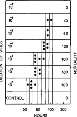
pended upon both the size of the inoculum anel the temperature. When the temperature is constant, the lower the virus concentration, the longer is the latent period (Fig. 1 ) . I f the virus concentration is constant, the
12. NUCLEAR-POLYHEDROSIS VIRUS INFECTIONS 387 higher the temperature, the shorter is the latent period (Fig. 2 ) . T h e latent period of virus-injected pupae is 30 to 36 days at 10°C, and no symptoms are observed after 36 days at 5°C.
10
-6
10 40
10
-4
10
:
100 40Ο ζ Ο ι- 3 ο
-3
10 ιδ2 10 -1
100 100 100
α:
lOONTROL
60 80 100 200 HOURS
FIG. 1. Effect of the size of the virus inoculum on the latent ( = incubation) period of silkworm larvae. Temperature after virus injection: 30°C. (From Aizawa, 1953c.)
15*C
·· ·· ·· ·· ··
20°C
·· ·· ·· ·· ··
lie I • • •
3fC •
• •
—I 1 1 1 I 1 1 1 1—
60
HOURS
FIG. 2. Effect of temperature on the latent (== incubation) period of silkworm larvae injected with nuclear-polyhedrosis virus. (From Aizawa, 1953c.)
Krieg (1955) adopted the median lethal time, L T5 0 (Sterbehalb
wertszeit) , which varied with the inoculum dose of polyhedra. In the nuclear polyhedrosis of the European pine sawfly, Neodiprion sertifer
(Geoffroy), the L T5 0, after inoculation with ΙΟ6, 104, and 102 polyhedra (per milliliter), was 7, 8.5, and 10 days, respectively. T h e L T5 0 at 29.6°, 20.8°, and 11.5°C was 4.5, 8, and 21 days, respectively. These results mean that the latent period depends upon the inoculum dose and the temperature after virus inoculation. However, the L T5 0 was not af
fected by the relative humidity.
Ayuzawa (1961b) observed no marked differences in the resistance to nuclear-polyhedrosis virus infection among silkworm strains. How
ever, the latent periods in pupae were different in various silkworm strains although the L D5 0 values were nearly the same in all strains examined. Accordingly, the latent period appears to vary according to the strain of the silkworm.
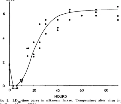
C. Virus Multiplication by Infection Experiments
T h e supernatant (3500 rpm for 15 minutes) of silkworm hemolymph infected with nuclear-polyhedrosis virus is very infectious and the L D5 0 is ordinarily 6.5 to 7.0 (log). T h e amount of virus in the supernatant
L D 5 0
6
4
2
0
0 20 4 0 6 0 8 0 H O U R S
FIG 3. L D5 0- t i m e curve in silkworm larvae. Temperature after virus injection:
25°C. (From Aizawa, 1959.)
12. N U C L E A R - P O L Y H E D R O S I S VIRUS I N F E C T I O N S 389 was estimated after virus injection and there was a decrease phase, an increase phase (logarithmic phase), and a stationary phase in the LDgo- time curve (Aizawa, 1959) (Fig. 3 ) . At the end of the increase phase of the LD5 0-time curve, polyhedra could be observed in the nuclei of blood cells at magnifications of 500 to 600 times (Aizawa, 1953c). When the temperature was lowered after virus injection, the gradient of the increase phase proved to be more gentle. However, its gradient was nearly identical at constant temperature with heavy and light doses of injected virus (Aizawa, 1953c, 1959) (Fig. 4 ) .
L D 5 0
H O U R S
FIG. 4. L Dg o- t i m e curve in silkworm pupae injected with heavy and light doses of virus. Temperature after virus injection: 27°C. (From Aizawa, 1959.)
T h e decrease phase appears to be dependent upon the adsorption of the virus onto the cells, as was observed in in vitro experiments. There was adsorption of the virus by the cells in pupae-extract broth, but in physiological saline solution, adsorption was not apparent. T h e virus was also not adsorbed by the tissues of chick embryos (Aizawa, 1959).
This phase could be caused by both the eclipse period and the adsorp
tion of virus. Yamafuji et al. (1954b) homogenized the virus-infected silkworm larvae with an alkaline solution and estimated qualitatively the infectivity of the supernatant, and they refer to the presence of an eclipse period. T h i s was later well described and substantiated by Krieg
(1958b). It is very interesting that a step multiplication of virus was indicated at the early period of infection in the silkworm polyhedrosis.
Such experiments may be said to be concerned with an estimation of relative total amount of virus in the larval body. Aizawa and Kawara- bata (unpublished data) estimated the virus amount in infected hemo
lymph and that in the supernatant of alkaline homogenates of the in
fected larval or pupal body according to the method of Yamafuji et al.
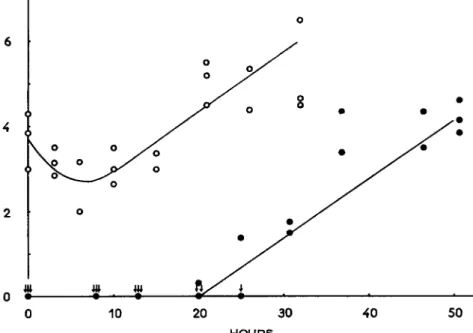
(1954b) and Krieg (1958b). T h e mode of the LD5 0-time curve in the infected body was nearly the same as in the hemolymph. This is differ-
Q
0 1 ι • • . . ι . , . , ι , . , , ι , , , , ι , 50 100 150 200 2 5 0
HOURS
FIG. 5. Number of liberated polyhedra in virus-infected hemolymph of silkworm larvae. Log number of polyhedra per cubic millimeter of hemolymph. (From Aizawa, 1953c.)
ent from the mode of the increase of free polyhedra in the hemolymph (Aizawa, 1953c), and it is similar to the results obtained in sigma virus of Drosophila melanogaster Meigen by Plus (1951, 1954, 1955).
T h e free polyhedra in the hemolymph increase logarithmically, and the gradient of the curve of increase depends upon the temperature
(Fig. 5 ) . This mode of increase is similar to that of the increase phase of the LD5 0-time curve (Aizawa, 1952a, 1953c). T h e increasing rate constant in the case of free polyhedra is 0.26 at 30°C and 0.18 at 20°C, and the constant at 5°C is calculated as 0.05 (Aizawa, unpublished data).
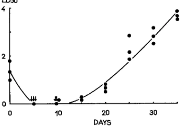
Actually the virus multiplication in silkworm pupae at 6° ± 1°C is very slow (Fig. 6 ) .
12. N U C L E A R - P O L Y H E D R O S I S VIRUS I N F E C T I O N S 391 Larvae of Neodiprion sertifer were inoculated with nuclear poly
hedra (50 ml of 1 χ 1 06/ m l ) and the total number of polyhedra formed in larvae after death was found to be 1.65 χ 1 01 1. Accordingly, poly
hedra were reproduced 3.3 χ 1 03 times the inoculated polyhedra. T h e number of polyhedra in a larva was about 1 08 (Krieg, 1955).
When silkworm larvae were injected with nuclear-polyhedrosis virus, and were starved after the virus injection, the mode of the LD5 0-time curve and the amount of virus in the infected hemolymph were very similar to those in fed larvae. T h i s result indicates that the virus mul
tiplies well in the starved larvae (Aizawa and Furuta, unpublished data). When the silkworm eggs were painted with nuclear polyhedra, the polyhedra were clearly formed in the newly hatched larvae which were starved (Takami and Kitazawa, 1959).
LD50
0 10 2 0 3 0 DAYS
FIG. 6. L D5 0- t i m e curve in silkworm pupae at 6° ± 1°C.
T h e use of human serum may act to promote virus infection in insects (Aizawa, 1954e). T h e supernatant of hemolyzed sheep red blood cells, hemoglobin, and chlorophyllin seem to have the action of a pro
tective colloid which promotes infection. However, it is not clear whether such substances are available for the adsorption and invasion of virus into host cells, or for protecting unadsorbed virus from inacti- vation in the insect body. KCN or N a N3 inhibit such an action of a protective colloid. Amino acids were not shown to effect virus multi
plication. Injection of metabolic inhibitors such as N a N3 and NaF seems to prolong the latent period (Aizawa, unpublished data).
D. Morphological and Histological Observations of Virus Multiplication Bergold (1950, 1952, 1953a, b, 1958a) postulated the following se
quence of the virus development. T h e development of the virus begins with one or several small spheres that grow within the developmental
membrane. These spheres elongate to kidney-shaped forms and then to V-shaped forms, and finally they change into straight rods. T h e virus rod with its intimate membrane is composed of several spherical subunits which are released from the intimate membrane and begin the cycle mentioned above. Sometimes the virus rod has a thin protrusion by which it attaches to the host cell.
According to Smith (1955c, 1959), Smith and Xeros (1953c), and Xeros (1953a, b, 1956), the nuclei of the cells swell and chromatin ag
gregates and forms a network. T h e virus rods seem to metamorphose from the material of chromatic network. Smith and Xeros (1954b) be
lieve that the acquisition of developmental or intimate membranes must take place as the virus rods become immersed in polyhedral protein.
T h e virus rods are pushed out into the ring zone where polyhedral protein accumulates, occluding the virus rods to form polyhedra.
Electron microscopic observations of virus-infected cells were made by Hughes (1953), Vago et al. (1955), Tomlin and Monro (1955), and Vago and Croissant (1960).
Xeros (1953b) observed the virus bundles in polyhedra which were reported by Komärek and Breindl (1924) (in their observations on the polyhedra of Wipfelkrankheit) by high magnification (1750 χ ) with the light microscope. Virus bundles are seen by pretreatment with 1 Ν HCl at 60°C for 10 minutes and can be stained with Giemsa solution.
T h e very smallest polyhedron may contain no, or only one, virus bundle.
Day et al. (1958) proposed a diagram of stages in the development of the nuclear-polyhedrosis virus of the larva of Pterolocera amplicornis Walker, from the time the polyhedra were ingested by the larva to that when the polyhedra were released from dead larvae, based on the results which had been previously reported by several workers together with their own observations. They postulate reinfection within a nucleus before and after the acquisition of the membrane which surrounds the virus rod, although such a reinfection has not yet been confirmed.
According to Bird (1957, 1959), in the early development of infec
tion a multitude of spheres from less than 20 to more than 250 ηΐμ in diameter appears in the ring zone between the chromatin and the nu
clear membrane. Many single virus rods appear without developmental membranes which attach to the central chromatic mass and to the nu
clear membrane. Bird (1959) proposed the following very important hypothesis:
Polyhedra are ingested by a larva and dissolve in the gut, liberating virus rods.
T h e virus rods attach to mid-gut cells and release infectious subunits. These pass through the mid-gut epithelium and enter a nucleus of susceptible cells.
12. N U C L E A R - P O L Y H E D R O S I S VIRU S I N F E C T I O N S 393
Multiplication take s plac e withi n th e nucleus , an d spherica l bodie s ar e formed . Each spher e contain s "germs " fo r a numbe r o f viru s particles , th e matur e for m of whic h i s rod-shaped . Bundle s o f rods , stil l containe d b y thei r developmenta l membranes, ar e surrounde d b y protei n whic h crystallize s t o for m polyhedra l inclusion bodies . Rod s fro m som e o f th e bundle s escap e fro m thei r develop - mental membranes , ar e neve r occlude d b y polyhedra l protein , an d attac h i n large numbe r t o th e nuclea r membran e an d als o t o othe r material s i n th e nucleus. The y releas e subunit s whic h penetrat e an d pas s ou t throug h th e nuclear membrane , an d infec t adjacen t cells .
Such sphere s o r subunit s (Bergold , 1950 , 1953a , b , 1958a ; Bird , 1959 ) should b e take n int o consideratio n wit h respec t t o developmenta l cycle s of viru s o r t o tha t o f a morphologica l uni t whic h retain s th e viru s infec - tivity becaus e i t shoul d b e remembere d tha t befor e th e establishmen t of th e morpholog y o f nuclear-polyhedrosi s viruse s b y Bergol d (1947 ) some purifie d "virus " particle s wer e considere d t o b e spherica l (Paillo t and Gratia , 1939 ; Glase r an d Stanley , 1943) .
£ . Viru s Multiplicatio n b y Mean s o f Tissu e Cultur e an d i n Chic k Embryo
T h e formatio n o f polyhedr a i n fibroblast s maintaine d i n tissu e cul - tures wa s observe d b y Trage r (1935) . T h e numbe r an d siz e o f polyhedr a increased, an d finall y th e fibroblasts wer e destroye d an d fre e polyhedr a were liberate d int o th e medium . T h e viru s multiplie d mor e tha n 100 0 times i n infectivit y 4 day s afte r infectio n an d reache d ove r 10,00 0 time s after 9 days . Aizaw a an d Vag o (1959a ) an d Vag o an d Chastan g (1960 ) also observe d th e formatio n o f polyhedr a i n fibroblasts . Martignon i an d Scallion (1961a , b ) maintaine d culture s o f hemocyte s o f th e variegate d cutworm, Peridroma saucia (Hübner) , an d i n the m observe d th e for - mation o f polyhedr a followin g inoculatio n o f th e virus .
Although attempt s t o cultur e th e separate d cell s fro m insec t tissue s have bee n mad e (Martignon i et al., 1958 ; Aizaw a an d Vago , 1959b) , a successful monolaye r cultur e ha s no t ye t bee n reported . Whe n thi s i s accomplished, th e plaqu e coun t wil l b e availabl e fo r th e viru s titration . In thi s connection , Da y an d Grac e (1961 ) hav e succeede d i n subcultur - ing fibroblast s o f Antheraea eucalypti (Scott) . T h i s accomplishmen t wil l contribute t o th e progres s o f solvin g th e problem s involvin g tissu e cul - ture an d viru s infection .
Attempts t o infec t chic k embryo s wit h nuclear-polyhedrosi s viruse s have bee n made ; however , definitel y positiv e result s hav e no t ye t bee n obtained (Steinhaus , 1951 ; Aizawa , 1961) .
IV. CROSS INFECTION AND D O U B L E INFECTION
A. Cross Infection
Bergold (1943) obtained, experimentally at least, positive results on cross infection between, and using the viruses of, Bombyx mori (Lin
naeus) , Porthetria dispar (Linnaeus), Lymantria monacha (Linnaeus), and Dendrolimus pint (Linnaeus); however, Bombyx mori was not susceptible to per os infection with polyhedra of Lymantria monacha or with those of Dendrolimus pint. Colias eurytheme Boisduval was susceptible to the virus of Colias lesbia (Fabricius) (Steinhaus, 1952).
T h e nuclear-polyhedrosis virus of Colias eurytheme and that of Pieris rapae (Linnaeus) are infectious for both host insects, and it is consid
ered that both viruses are identical (Tanada, 1954).
Smith (1953) and Smith and Xeros (1952, 1953a, b) studied cross-infec
tion and back-infection tests with about 30 species of insects, and they reported relatively easy success in obtaining positive results with nu
clear- and cytoplasmic-polyhedrosis viruses. Aglais urticae (Linnaeus), Vanessa to (Linnaeus), Phalera bucephala (Linnaeus), Arctia caja (Lin
naeus), and Sphinx populi (Linnaeus) are susceptible to the nuclear- polyhedrosis virus of Pyrameis cardui (Linnaeus). Sphinx ligustri is susceptible to the nuclear-polyhedrosis viruses of Panaxia dominula
(Linnaeus) and Telea polyphemus (Cramer). However, Porthetria dis
par is not susceptible to the nuclear-polyhedrosis viruses of Panaxia dominula and Lymantria monacha. Bombyx mori is not susceptible to the nuclear-polyhedrosis viruses of Porthetria dispar, Telea polyphemus, Sphinx ligustri, Lymantria monacha, Samia ricini (Boisduval), and Panaxia dominula.
T h e nuclear-polyhedrosis virus of Bombyx mori is successively trans
missible to Samia ricini (Aizawa, 1952b). Each virus is infectious for both host insects (Yamamasu et al., 1954, 1960). Choristoneura pinus
(Freeman) and Choristoneura fumiferana (Clemens) are susceptible to nuclear-polyhedrosis viruses of both insects (Stairs, 1960). T h e nuclear- polyhedrosis virus of Dictyoploca japonica (Butler) is infectious for Samia cynthia pry er i Butler but not for the silkworm (Ishikawa, 1960).
T h e nuclear-polyhedrosis viruses of Bombyx mori and Antheraea pernyi (Guerin) are infectious for Samia cynthia pryeri, while the former is not infectious for Antheraea yamamai (Guerin) (Tanaka, 1960). T h e virus of Samia cynthia pryeri is infectious for the silkworm (Ishikawa and Asayama, 1960).
Malacosoma americanum (Fabricius) is not susceptible to the viruses of Bombyx mori and Porthetria dispar, and the silkworm is resistant to infection by the viruses of Porthetria dispar and Lymantria monacha
12. NUCLEAR-POLYHEDROSIS VIRUS INFECTIONS 395 (Glaser, 1927). T h e nuclear-polyhedrosis virus of Pieris rapae is not infectious for Laphygma exigua (Hübner), Phryganidia californica Packard, and Prodenia praefica Grote (Tanada, 1954). Peridroma margaritosa (Haworth) is not susceptible to the virus of Colias euryth
eme (Steinhaus, 1960b). According to Vago (1953a), the nuclear-poly
hedrosis virus of Thaumetopoea pityocampa is not infectious for Bombyx mori, Pieris brassicae (Linnaeus), and Deilephila euphorbiae (Lin
naeus) . Galleria mellonella (Linnaeus), Bombyx mori, and Hyphan- tria cunea (Drury) are not susceptible to the virus of Euproctis flava
(Bremer) (Aizawa et al., 1957). Pseudaletia unipuncta (Haworth) is not susceptible to the nuclear-polyhedrosis virus of Spodoptera mauritia
(Boisduval) (Tanada, 1960). A positive cross infection was not ob
served among Barathra brassicae (Linnaeus), Hyphantria cunea, and Bombyx mori with their nuclear-polyhedrosis viruses (Aruga et al., 1960). T h e nuclear-polyhedrosis virus of Pseudaletia unipuncta is not infectious for Pieris rapae, Spodoptera mauritia, Hellula undalis (Fabri- cius), Laphygma exempta (Walker), Peridroma margaritosa, and Bom
byx mori (Tanada, 1959a).
Gershenson conducted cross-inoculation tests with 60 lepidopterous insect species and 6 different nuclear-polyhedrosis viruses. T h e shape and average size of polyhedra formed in an insect are always typical of those of the virus inoculated. For example, Aglais urticae infected with the virus of Bombyx mori shows large hexagonal polyhedra; small tri
angular polyhedra are formed when infected with the virus of Antheraea pernyi, and tetragonal polyhedra are formed when infected with the virus of Galleria mellonella. T h e characteristic shapes and mean sizes of polyhedra are determined exclusively by the properties of the virus, not by those of the host (Gershenson, 1955a, b, 1957). Double infection of a cell with an original and a mutant virus results in the development of two types of polyhedra within the cell nucleus (Gershenson, 1956a).
According to Bergold (1958a), each species of insect virus has its specific insect host; however, it may multiply in certain other insect hosts, particularly when large inocula are used. One of the most diffi
cult problems in the cross-inoculation experiments with insect viruses is the induction of the virus diseases. When an insect virus is inoculated into a nonspecific insect in which a nuclear polyhedrosis is known to appear, it is necessary to identify the viruses. T h e result in the cross- infection test should be carefully considered according to several cri
teria on the infection of polyhedroses. In this connection, Tanada (1954) performed a cross-infection test with the nuclear-polyhedrosis viruses of Colias eury theme and Pieris rapae, and the following evidence supports the positive results obtained: (1) Both viruses are transmissible
to either host insect. (2) Each virus is infectious to the original host after passage through the other host. (3) T h e size and shape of the polyhedra and virus particles of the two viruses are essentially the same.
(4) Each host exhibits its own symptoms when infected with either virus.
(5) T h e histopathologies within the two host species are similar. (6) Agglutination tests of polyhedra indicate a close relationship between the two viruses. Another example is shown in the GaZ/ma-adapted silk
worm nuclear-polyhedrosis virus. Galleria mellonella was injected with the nuclear-polyhedrosis virus of the silkworm, and serial passages through Galleria were conducted. This virus was infectious for both Galleria and Bombyx, and it was neutralized with the antiserum against the silkworm nuclear-polyhedrosis virus irrespective of test insect, Gal- leria or Bombyx. Accordingly, this virus was considered as the Galleria- adapted silkworm nuclear-polyhedrosis virus (Aizawa, 1962b).
B. Double Infection
In the silkworm, double infection with both nuclear and cytoplasmic polyhedroses is often seen in Japan. Smith and Xeros (1953a, b; Smith et al., 1953) reported the presence of both nuclear and cytoplasmic poly
hedroses in the same insect specimen in their laboratory in England.
Double infection with a nuclear-polyhedrosis virus and a granulosis virus was found in Euxoa segetum (Schiffermüller), Pieris rapae, Pseu- daletia unipuncta, and Nephelodes emmedonia (Cramer) (Steinhaus, 1957). T h e following insects have been reported as hosts to both nu
clear-polyhedrosis and granulosis viruses (Steinhaus, 1957): Peridroma margaritosa (Haworth), Junonia coenia (Hübner), Laphygma exigua
(Hübner), Laphygma frugiperda ( J . E. Smith), Sabulodes caberata (Guenee), Chorizagrotis auxiliaris (Grote), Euxoa segetum (Schiffer
müller) , Choristoneura fumiferana (Clemens), Estigmene acrea (Drury), Pieris rapae (Linnaeus), Autographa californica (Speyer), Cacoecia mu- rinana (Hübner).
It is very interesting that the virulence of the nuclear-polyhedrosis virus was found to increase in the double infection with polyhedrosis and granulosis viruses in Pseudaletia unipuncta (Tanada, 1956, 1959b, 1961). T h e heat-inactivated granulosis virus is still capable of enhancing the virulence of nuclear-polyhedrosis virus; however, heat-inactivated nuclear-polyhedrosis virus has no marked effect upon the virulence of granulosis virus (Tanada, 1959b). This increased virulence is consid
ered as an example of a synergism phenomenon.
Pseudaletia unipuncta fed a mixture of polyhedra and granular in
clusions, both from Pseudaletia unipuncta, died of nuclear polyhedrosis.
However, the nuclear polyhedra of Spodoptera mauritia were fed alone
12. NUCLEAR-POLYHEDROSIS VIRUS INFECTIONS 397 and in combination with the granular inclusions of Pseudaletia uni
puncta to larvae of Pseudaletia unipuncta; none of larvae died of nu
clear polyhedrosis. T h e nuclear polyhedra and granular inclusions of Pseudaletia unipuncta were fed, alone and combined, to Pieris rapae, Spodoptera mauritia, Bombyx mori, and Peridroma margaritosa; none of the larvae died from either virus disease (Tanada, 1959a, 1960).
In the double infection with nuclear-polyhedrosis and granulosis viruses in Choristoneura fumiferana, prior infection by one virus inter
feres with the infection by the second virus (Bird, 1959).
In double infections in the silkworm larvae with the nuclear-poly
hedrosis viruses of Bombyx mori and Samia cynthia pryeri, each type of polyhedra was formed in a cell nucleus, but both kinds of polyhedra were not observed in the same nucleus. However, large and triangular polyhedra, which were different from the two types of polyhedra in shape and size, were formed in a very few nuclei of cells (Ishikawa and Asayama, 1961). In the nuclear polyhedrosis of Hyphantria cunea, the virus of tetragonal polyhedra seemed to be less virulent than that of triangular polyhedra and interfered less with the latter virus (Aruga et al, 1961).
V . SUSCEPTIBILITY
In Pseudaletia unipuncta (Haworth), young larvae are more suscep
tible to infection with nuclear-polyhedrosis virus than older ones, and the susceptibility does not vary with the change of food. Synergistic action occurs by inoculation with both nuclear-polyhedrosis and gran
ulosis viruses and results in an increase of mortality, as mentioned before (Tanada, 1956, 1959a, b, 1961). T h e increased susceptibility in young larvae was also observed in Spodoptera mauritia (Boisduval) (Tanada,
1960). However, in Neodiprion sertifer, such a difference in suscepti
bility as related to the larval stage was not indicated when determined on the basis of L T5 0 (Krieg, 1955). In this case a relatively higher concentration of polyhedra (107/ml) was applied, and it would seem advisable to make a dosage-mortality analysis of the matter.
According to Bird (1953a) the prepupal stage of Diprion hercyniae (Hartig) is immune to infection with polyhedrosis virus. T h e prepupal midgut, a temporary gut, is composed entirely of embryonic cells and is not susceptible to infection.
T h e nuclear-polyhedrosis viruses of Trichoplusia ni (Hübner) and Heliothis zea (Boddie) are incapable of infecting their hosts through ingestion at temperatures of 39°C or above (Thompson, 1959).
V I . D E F E N S E AGAINST NUCLEAR-POLYHEDROSIS VIRUSES
A . Defense against Infections
Aizawa (1953a) immunized silkworm pupae with a vaccine made from infected hemolymph or partially purified virus. T h e effectiveness of vaccination was recognized although it was weak. However, the neu
tralizing antibody in the immune hemolymph was scarcely present, and an increase of agglutinins against polyhedra was not demonstrable
(Aizawa, 1954a, b, c ) . T h e effectiveness of immunization was not ex
plainable by principles of humoral immunity, and the immunological aspects of insect virus infections is greatly different from that in bac
terial infections.
After the virus injection, antiserum against silkworm nuclear-poly
hedrosis virus was injected into silkworm larvae at certain intervals.
This serum therapy was successful (Yamafuji et al., 1958a), although similar serum therapy was not successful in the case of silkworm pupae
(Aizawa, 1954c). In a system of Galleria mellonella larvae and the Ga//ena-adapted silkworm nuclear-polyhedrosis virus, serum therapy was successful (Aizawa, 1962b).
T h e silkworm nuclear-polyhedrosis virus is inactivated by heating at 50° or 60°C (Aizawa, 1953e), and the induction of nuclear polyhedrosis is not observed in the silkwOrm pupae by such heat treatment (Ishimori, 1940). After the virus injection, silkworm pupae were heated at 50°C for 15 minutes; however, such heat therapy was not successful in pre
venting the disease (Aizawa, 1954c).
Penicillin, streptomycin, Aureomycin, and Terramycin are not effec
tive for the control of virus infection (Bergold, 1953a; Aizawa, 1954c;
Krieg, 1957), but grasseriomycin which was produced from Streptomyces sp. has an effect upon the elongation of the incubation period (Ueda et al., 1955). Mortality decreased when purified virus suspension was mixed with 20 percent Kollidon (polyvinylpyrrolidone) and injected into Porthetria dispar (Bergold, 1948b). A higher percentage of silk
worm larvae pupated if sublethal doses of arsenic compounds to virus- infected larvae (Speyer, 1925).
Β. Resistance to Virus Disease
Some papers in this field have been published either on the point of resistance to virus induction or on the point of resistance to virus infec
tion. It has been claimed, and in some cases established, that poly
hedroses in the silkworm, Bombyx mori, can be induced by such treat
ments as low temperature and administration of certain chemicals.
Although various factors are involved in the induction of polyhedroses
12. NUCLEAR-POLYHEDROSIS VIRUS INFECTIONS 399 in insects (Vago, 1951, 1953b; Yamafuji, 1952a; Smith and Rivers, 1956;
Krieg, 1956, 1957, 1958a; Steinhaus, 1958; Aruga, 1959; Ayuzawa, 1961a), it is considered that some mechanism of resistance to virus induction is involved. However, Yamafuji et al. (1958b, c) could find no increasing resistance to virus induction in the silkworm. T h e existence or increase in resistance to virus infection was observed in Choristoneura fumiferana
(Bergold, 1951), Eucosma griseana (Hübner) (Martignoni, 1957), Di- prion hercyniae (Hartig) (Bird and Elgee, 1957), Pieris brassicae (Lin
naeus) (Rivers, 1958; Sidor, 1959; David and Gardiner, 1960), Pieris rapae (Linnaeus) (Martignoni and Schmid, 1961), and Phryganidia californica Packard (Martignoni and Schmid, 1961). On the contrary, Franz and Niklas (1954) and Bird (1955) reported no evidence of de
veloping resistance in Neodiprion sertifer (Geoffroy).
Aizawa et al. (1961) obtained a silkworm strain resistant to virus in
duction by means of cold treatment by selection through thirteen genera
tions. T h e susceptibility to virus infection among several silkworm strains, including both this strain resistant to virus induction and the nonselected (original) strain, were tested. From the results obtained, the resistance to virus induction is considered to be independent from that to virus infection. Ayuzawa (1961b) also examined the difference in sus
ceptibility to virus infection among silkworm strains, and he could not observe a clear difference; however, the latent, or incubation, period in the case of a few strains was longer than that of other strains although the L D5 0 was nearly the same with all the strains examined. Such an elongation of the incubation period seems to have some meaning in the resistance to virus infection.
V I I . VIRULENCE, INFECTIVITY, AND STABILITY OF VIRUSES
A. Virulence
Equal concentrations of nuclear polyhedra in Kotochalia junodi (Heylaerts) from different localities did not produce the same degree of killing (Ossowski, 1958, 1960), and the existence of virus strains with different virulence was suggested as an explanation for this. However, these results might be considered as reflecting differences in suscepti
bility to virus infection in the host insect.
A variant was obtained from the nuclear-polyhedrosis virus of Bom
byx mori, which forms hexagonal polyhedra by means of passages through chick embryo and silkworm pupae alternatively. T h i s variant virus forms tetragonal polyhedra in the silkworm larvae. However, the symptoms in the infected larvae are very much like those of the so- called "clear head" disease, which are quite different from the symptoms
of nuclear polyhedrosis. Virulence of this virus is nearly the same as that of the original virus, and it is neutralized with the antiserum against silkworm nuclear-polyhedrosis virus (Aizawa, 1961).
B. Infectivity
T h e L D5 0 dose in infection with polyhedra is 50 to 100 μg (2.5 χ 1 0 ~6 grams of virus) in Bombyx mori and 100 μg in Porthetria dispar and Malacosoma disstria (Hübner) (Bergold, 1958a). In Neodiprion sertifer,
the L D5 0 dose is 100 to 500 polyhedra (Bird and Whalen, 1953) and 50 to 100 polyhedra according to Krieg (1955).
T h e L D5 0 dose with nuclear-polyhedrosis virus is 4 to 8 χ 1 0- 1 3 grams of virus per larva (1000 to 2000 virus rods) in Bombyx mori, 1 χ 1 0- 1 0 grams of virus in Porthetria dispar, and 1.5 to 3.6 χ 1 0- 9 grams in Choristoneura fumiferana (Bergold, 1947, 1948a, 1951, 1953a,
1958a).
Extraction of infectious DNA has been tried. Gershenson (1956b) extracted protein-free DNA and DNA-free protein from polyhedra of Antheraea pernyi (Guerin), but obtained no infectivity with single prep
aration. Bergold (1958b, 1959) extracted infectious DNA from the silkworm nuclear-polyhedrosis virus by ^-aminosalicylate and phenol.
This DNA preparation is very viscous and contains a protein component which is serologically different from virus protein. However, the infec
tivity is very low (100 mg per larva) when compared with that of intact virus particles.
C. Stability
T h e stability of insect viruses is reviewed in detail by Bergold (1958a). T h e infectivity of virus held within polyhedra is rather stable;
for example, the infectivity of the silkworm nuclear polyhedra was shown to be retained well for at least twenty years, mostly at 4°C
(Steinhaus, 1954a, 1960a). Polyhedra of Diprion hercyniae (Hartig), however, lose virulence during storage, complete inactivation being reached after 12 years at 4.5°C (Neilson and Elgee, 1960). When kept at room temperature for 37 years, the infectivity of the silkworm nuclear polyhedra was lost completely (Aizawa, 1954d). Infectivity of poly
hedra is lost by heating (Bergold, 1943; Watanabe, 1951; Tanada, 1959a), and the infectivity of infected hemolymph is not shown after 30 minutes at 70°C. Dried infected silkworm hemolymph still keeps its infectivity after exposure to temperatures of 100° to 120°C for 1 hour, but loses it at 130°C for 30 minutes (Watanabe, 1951).
12. NUCLEAR-POLYHEDROSIS VIRUS INFECTIONS 401 T h e free virus in hemolymph retains its infectivity well at low temperatures. T h e infectivity does not decrease by holding the virus at
—25°C for 24 days, but it decreases slightly when virus is held at 5°C for 30 days. Heating at 50° or 60°C for 10 minutes destroys almost all infectivity (Aizawa, 1953d, e ) .
Virus infectivity is lost by ultrasonic irradiation for 20 minutes (Wa- tanabe, 1951) and by ultraviolet irradiation (Aizawa, 1955b). Inactiva- tion of the silkworm nuclear-polyhedrosis virus by chemicals such as phenol, mercuric chloride, formalin, potassium permanganate, antifor- min, alcohol, acid, alkali, and enzymes such as lipase, trypsin, and dia
stase have been studied by many investigators. Calcium and magnesium ions decrease the virus infectivity (Bergold, 1953a).
V I I I . TRANSMISSION OF VIRUS
Pupae and moths of the silkworm are easily infected with nuclear- polyhedrosis virus. As pointed out by Steinhaus (1954b) and Bergold
(1958a), there exist two possibilities as to methods by which the virus may be transmitted to the next generation. One is by the virus-contami
nated egg surface which newly hatched larvae ingest, and the other is by trans-ovum infection. T h e following phenomena indicate the pos
sible evidence of trans-ovum infection: Virus inclusions are formed in the embryo (Roegner-Aust, 1949; Umeya et al., 1955; Sager, 1960); field experiments indicate the possibility of trans-ovum infection of virus in Malacosoma fragile (Stretch) (Clark, 1955); and larvae 2 days after hatching showed typical symptoms of polyhedrosis in Telea polyphemus
(Cramer) (Smith and Wyckoff, 1951). Accordingly, Bergold (1958a) concluded that the virus is transmissible via the outside and/or the inside of host eggs.
Concerning latent and frank (acute) infections, Krieg (1958a) pro
posed the following hypothesis. Frank infection is caused by the appli
cation of a more-than-lethai dose, and either death or survival (toler
ance) results. When a less-than-lethal dose is applied, subacute infection is caused. Latent infection derives from such tolerant survival and sub
acute infection. Frank infection is provoked from latent infection (in
duction of virus disease), and there exists a possible elimination of latent infection. Such a hypothesis seems to cover the process concerning la
tent and frank infections, although it is felt that much more experi
mental evidence is necessary before definite conclusions can be made.
Martignoni (1957) emphasized the virus induction phenomenon be
cause of the sudden and simultaneous occurrence of the granulosis in
Eucosma griseana (Hübner) was not fully explained by transmission from diseased to healthy individuals and by trans-ovum passage of the infectious agent. There is still some doubt whether virus transmission through the eggs has been clearly recognized in the laboratory (O. J . Smith et al, 1956).
Umeya et al. (1955) and Aizawa et al. (1961) injected the silkworm nuclear-polyhedrosis virus into the pupae in a late stage, and eggs were obtained from heavily infected moths. T h e larvae that hatched from such eggs were reared normally or treated by low temperature ( 5 ° C , 24 hours). However, there was no marked difference in the induction rate between the normal and the virus-injected larvae. If the infectious virus is easily transmissible through the embryo, the formation of polyhedra in the embryo and the occurrence of polyhedrosis in the first stage of the next generation should be observed more frequently. If the infec
tious virus is transmissible in a latent state, the induction rate should be higher in virus-injected insects than in noninjected ones. From the results obtained, it would appear that infectious virus is not transmis
sible through embryos and that the lysogenization-like phenomenon, i.e., the change from infectious state to latent state, does not occur, at least in the silkworm.
It is necessary to have more detailed knowledge about the transmis
sion of the virus through insect eggs in relation to latent infection, a matter on which there is still some confusion.
I X . METABOLIC CHANGES IN VIRUS INFECTION
Amino acids of virus-infected silkworm hemolymph were investigated by Ishimori and Muto (1951), Yoshitake and Aruga (1950), Drilhon et al. (1951, 1952), and Koyanagi and Matsuoka (1954). Ishimori and Muto (1951) found a decrease of aspartic acid, cystine, glutamic acid, glutamine, threonine, tyrosine, and valine, and an increase of histidine in hemolymph from infected silkworms. Electrophoretic patterns of infected hemolymph indicated a change in the content of the protein components (Inagami, 1954; Kobayashi and Komatsu, 1956). In the hemolymph of Galleria mellonella infected with the Ga/tena-adapted silkworm nuclear-polyhedrosis virus, a protein component disappears which is different from the case of the virus-infected silkworm larvae
(Aizawa, 1955a; Aizawa and Murai, 1958; Aizawa, unpublished data).
Protein content of infected larvae is higher than that of normal larvae (Bergold and Friedrich-Freksa, 1947).
T h e total nucleic acids and protein in the fat body and hemolymph of infected larvae increase when symptoms appear (Shigematsu and
12. NUCLEAR-POLYHEDROSIS VIRUS INFECTIONS 403 Takeshita, 1958); however, Yamafuji et al. (1954a) found an increase of DNA and no marked change of R N A in diseased larvae.
Catalase activity decreases in infected larvae at the beginning of infection, but it increases rapidly at the time of polyhedra formation in the nuclei of infected cells (Ishimori and Iwasaki, 1941; Ishimori and Osawa, 1951). Hexokinase activity decreases when symptoms ap
pear (Shigematsu, 1958). Protease, ribonuclease, deoxyribonuclease (Yoshihara, 1959a, b ) , and dehydrogenase (Murai and Aizawa, 1957) in the virus-infected silkworms were investigated. Oxygen consumption of virus-infected larvae increases in the silkworm (Akune, 1951a). K20 content decreased in virus-infected silkworm larvae (Akune, 1951b), and the content of some fluorescent substances increased in virus-infected silkworm hemolymph (Drilhon, 1951; Drilhon et al., 1951, 1952).
REFERENCES
Aizawa, K. 1952a. Turbidity-time curve of the infected blood in the silkworm jaundice. Sansi-Kenkyü, 2, 78-80 (in Japanese).
Aizawa, K. 1952b. Preliminary note on the successive passage through Philosamia ricini Boisd. of the jaundice virus of Bombyx mori L . / . Sericult. Sei. Japan, 21, 170-172 (in Japanese with English summary).
Aizawa, K. 1953a. Sedimentation of the silkworm jaundice virus by the ultracen- trifuge. I. Effect of the vaccine made from the sediment. Japan. J. Appl. Zool., 18, 141-142.
Aizawa, K. 1953b. Silkworm jaundice virus in the excrements of the infected larvae.
Japan. J. Appl. Zool., 18, 143-144.
Aizawa, K. 1953c. Multiplication mode of the silkworm jaundice virus. I. On the multiplication mode in connection with the latent period and L D5 0- t i m e curve.
Bull. Sericult. Expt. Sta. (Tokyo), 14, 201-228 (in Japanese with English summary).
Aizawa, K. 1953d. Some methods of keeping the virus activity of the silkworm jaundice. Sansi-Kenkyü, 3, 75-77 (in Japanese).
Aizawa, K. 1953e. On the inactivation of the silkworm jaundice virus. Japan. J.
Appl. Zool., 17, 181-190 (in Japanese with English summary).
Aizawa, K. 1954a. Immunological studies of the silkworm jaundice virus. I.
Neutralization and adsorption test of the silkworm jaundice virus. Virus (Osaka), 4, 238-240 (in Japanese with English summary).
Aizawa, K. 1954b. Immunological studies of the silkworm jaundice virus. II.
Agglutination reaction of the polyhedral bodies. Virus (Osaka), 4, 241-244 (in Japanese with English summary).
Aizawa, K. 1954c. Immunological studies of the silkworm jaundice virus. III.
Experiments on the defence of infection in the silkworm jaundice. Virus (Osaka), 4, 245-248 (in Japanese with English summary).
Aizawa, K. 1954d. Dissolving curve and the virus activity of the polyhedral bodies of Bombyx mori L . , obtained 37 years ago. Sansi-Kenkyü, 8, 52-54 (in Japanese with English summary).
Aizawa, K. 1954e. Neutralization of silkworm jaundice virus by human serum.
Nature, 174, 748-749.
Aizawa, K. 1955a. Electrophoresis of the blood of silkworm, Bombyx mori, on the filter paper. / . Sericult. Sei. Japan, 24, 393-397 (in Japanese with English sum
mary).
Aizawa, K. 1955b. Inactivation of the silkworm jaundice virus by the ultraviolet irradiation. / . Sericult. Sei. Japan, 24, 398-399 (in Japanese with English summary).
Aizawa, K. 1955c. A preliminary note on the tetragonal polyhedra in the silkworm.
Sansi-Kenkyü No. 14(4), 11-13 (in Japanese with English summary).
Aizawa, K. 1959. Mode of multiplication of silkworm nuclear polyhedrosis virus.
II. Multiplication of the virus in the early period of the L D5 0- t i m e curve.
/. Insect Pathol., 1, 67-74.
Aizawa, K. 1961. Change in the shape of silkworm polyhedra by means of passage through chick embryo. Entomophaga, 6, 197-201.
Aizawa, K. 1962a. Antiviral substance in the gut-juice of the silkworm, Bombyx mori (Linnaeus). / . Insect Pathol., 4, 72-76.
Aizawa, K. 1962b. Infection of the greater wax moth, Galleria mellonella (Linnaeus), with the nuclear polyhedrosis virus of the silkworm, Bombyx mori (Linnaeus).
/. Insect Pathol, 4, 122-127.
Aizawa, K., and Murai, S. 1958. Electrophoresis of the blood of the silkworm, Bombyx mori on filter paper (2nd report). Phys.-Chem. Biol. (Tokyo), 4, 23-26 (in Japanese with English summary).
Aizawa, K., and Vago, C. 1959a. Sur l'infection ä Borrelinavirus en culture de tissus d'Insectes. Ann. inst. Pasteur, 96, 455-460.
Aizawa, K., and Vago, C. 1959b. Culture in vitro de cellules separees de tissus d'Insectes. Compt. rend. acad. sei., 249, 928-930.
Aizawa, K., Asahina, S., and Fukumi, H. 1957. Demonstration of the polyhedral diseases of Euproctis flava and Euproctis pseudoconspersa (Lepidoptera, Lymantrii- dae). Japan. J. Med. Set. & Biol, 10, 61-64.
Aizawa, K., Furuta, Y., and Nakamura, K. 1961. Selection of a resistant strain to virus induction in the silkworm, Bombyx mori. J. Sericult. Set. Japan, 30, 405-412 (in Japanese with English summary).
Akune, S. 1951a. Respiration and catalase action of the silk-worms attacked by polyhedral disease. / . Sericult. Set. Japan, 20, 30-32 (in Japanese).
Akune, S. 1951b. Studies on the potassium content of virus diseased bodies. Set.
Bull Fac. Agr. Kyushu Univ., 13, 149-153 (in Japanese with English summary).
Aruga, H. 1959. Mechanism of the resistance to virus diseases in insects. Recent Advances in Breeding (Japan), 1, 53-60 (in Japanese).
Aruga, H., Watanabe, H., Fukuhara, T., and Iwashita, Y. 1957. Mechanism of the virus resistance in the silkworm, Bombyx mori. I. On the formation of the polyhedral body in the nucleus of the silk-gland cell. / . Sericult. Sei. Japan, 26, 105-112 (in Japanese with English summary).
Aruga, H., Yoshitake, N., Watanabe, H., and Hukuhara, T . 1960. Studies on nuclear polyhedroses and their inductions in some Lepidoptera. Japan. J. Appl Entomol Zool, 4, 51-56 (in Japanese with English summary).
Aruga, H., Yoshitake, N., Watanabe, H., Hukuhara, T., Nagashima, E., and Kawai, T . 1961. Further studies on polyhedroses of some Lepidoptera. Japan. J. Appl En
tomol Zool, 5, 141-144 (in Japanese with English summary).
Ayuzawa, C. 1961a. On the induction of polyhedroses by the exposure to low temperature in the silkworm, Bombyx mori L . / . Sericult. Sei. Japan, 30, 101-108 (in Japanese with English summary).
12. NUCLEAR-POLYHEDROSIS VIRUS INFECTIONS 405 Ayuzawa, C. 1961b. On the resistance to infection of nuclear polyhedrosis in the
silkworm, Bombyx mori L . / . Sericult. Sei. Japan, 30, 413-419 (in Japanese with English summary).
Balch, R . E . , and Bird, F . T . 1944. A disease of the European spruce sawfly, Gilpinia hercyniae (Htg.), and its place in natural control. Set. Agr., 25, 65-80.
Benz, G. 1960. Histopathological changes and histochemical studies on the nucleic acid metabolism in the polyhedrosis-infected gut of Diprion hercyniae (Hartig).
/. Insect Pathol, 2, 259-273.
Bergold, G. 1943. Über Polyederkrankheiten bei Insekten. Biol Zentr., 63, 1-55.
Bergold, G. 1947. Die Isolierung des Polyeder-Virus und die Natur der Polyeder.
Z. Naturforsch., 2b, 122-143.
Bergold, G. 1948a. Bündeiförmige Ordnung von Polyederviren. Z. Naturforsch., 3b, 25-26.
Bergold, G. 1948b. Inaktivierung des Polyeder-Virus durch Kollidon. Z. Naturforsch., 3b, 300-301.
Bergold, G. H. 1950. T h e multiplication of insect viruses as organisms. Can. J.
Research, E 2 8 , 5-11.
Bergold, G. H. 1951. T h e polyhedral disease of the spruce budworm, Choristoneura fumiferana (Clem.) (Lepidoptera: Tortricidae). Can. J. Zool, 29, 17-23.
Bergold, G. H. 1952. Demonstration of polyhedral virus in blood cells of silkworms.
Biochim. et Biophys. Acta, 8, 397-400.
Bergold, G. H. 1953a. Insect viruses. Advances in Virus Research, 1, 91-139.
Bergold, G. H. 1953b. T h e multiplication of insect viruses. In "The Nature of Virus Multiplication," 2nd Symposium Soc. Gen. Microbiol. (P. Fildes and W . E . van Heyningen, eds.), pp. 276-283. Cambridge Univ. Press, London and New York.
Bergold, G. H. 1958a. Viruses of insects. In "Handbuch der Virusforschung" (C.
Hallauer and Κ. F . Meyer, eds.), Vol. 4, pp. 60-142. Springer, Vienna.
Bergold, G. H. 1958b. Some topics of insect virology. Trans. 1st Intern. Conf. Insect Pathol and Biol. Control Praha, 1958, pp. 191-195.
Bergold, G. H. 1959. Biochemistry of insect viruses. In "The Viruses" (F. M.
Burnet and W . M. Stanley, eds.), Vol. 1, pp. 505-523. Academic Press, New York.
Bergold, G., and Friedrich-Freksa, H. 1947. Zur Grösse und Serologie des Bombyx- raon-Polyedervirus. Z. Naturforsch., 2b, 410-414.
Bird, F . T . 1952. On the multiplication of an insect virus. Biochim. et Biophys.
Acta, 8, 360-368.
Bird, F . T . 1953a. T h e effect of metamorphosis on the multiplication of an insect virus. Can. J. Zool, 31, 300-303.
Bird, F . T . 1953b. T h e use of a virus disease in the biological control of the Euro
pean pine sawfly, Neodiprion sertifer (Geoffr.). Can. Entomologist, 85, 437-446.
Bird, F . Τ . 1955. Virus diseases of sawflies. Can. Entomologist, 87, 124-127.
Bird, F . T . 1957. On the development of insect viruses. Virology, 3, 237-242.
Bird, F . T . 1959. Polyhedrosis and granulosis viruses causing single and double infections in the spruce budworm, Choristoneura fumiferana Clemens. / . Insect Pathol, 1, 406-430.
Bird, F. T., and Elgee, D. 1957. A virus disease and introduced parasites as factors controlling the European spruce sawfly, Diprion hercyniae (Htg.) in central New Brunswick. Can. Entomologist, 89, 371-378.
Bird, F . T., and Whalen, Μ. M. 1953. A virus disease of the European pine sawfly, Neodiprion sertifer (Geoffr.). Can. Entomologist, 85, 433-437.