III./6.2.: Alzheimer's disease (AD)
AD is the most frequent cause of dementia. Its incidence (the number of new cases per year) is 0.5% at the age of 65, while 8% at the age of 85. The course of the disease is 8 to 12 years on average.
The disease was first described by Alois Alzheimer in 1906, but it became accepted as the most frequent cause of dementia only during the 80s.
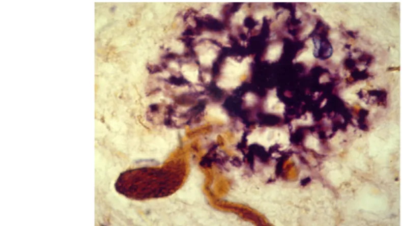
According to neuropathological studies, the characteristic histological changes of the disease in the brain appear several decades before the clinical manifestation of the disease. These changes include the neurofibrillary tangles (NFTs) in neuronal cell bodies, which are formed by the
hyperphosphorilated microtubule-binding protein tau, and the senile plaques, which are formed by the extracellularly accumulated -amyloid protein (Fig.
4).
Fig. 4: Histological changes in Alzheimer's disease. Neurofibrillary tangles are marked by brown, while senile plaques by purple colors. (Double immunhistochemical staining using
anti-tau (AT8) and anti-beta-amyloid antibodies).
It is widely argued which one of these histological hallmarks is the basic pathology in AD: according to the disciples of "taoism", NFTs are the core lesions ("taoism" comes from tau, the major constituent of NFTs), while disciples of "baptism" argue that senile plaques are the basic lesions in AD ("baptism" comes from beta-amyloid peptide which is the fragment of amyloid precursor protein [APP]). NFTs develop first in the medial parts of the temporal lobe and spread from here to limbic, and finally to neocortical areas.
According to genetic classification, AD is divided into early-onset (below the age of 65) autosomal dominant familial forms, and late-onset (above the age of 65) predominantly sporadic, but also rarely familial forms. Less than 5%
of AD is familial, and approximately half of these are caused by mutations of the presenilin-1 gene (on chromosome 14). The epsilon 4 allele of the
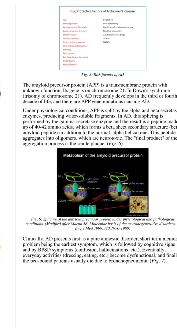
apolipoprotein E gene is the major genetic risk factor for sporadic AD. In addition to genetic factors, the main risk factors for AD are the following:
age (the most important risk factor), repeated head injuries, low level of education, and all vascular risk factors (hypertension, diabetes mellitus, atherosclerosis, smoking, etc.) (Fig. 5).
Fig. 5: Risk factors of AD
The amyloid precursor protein (APP) is a transmembrane protein with unknown function. Its gene is on chromosome 21. In Down's syndrome (trisomy of chromosome 21), AD frequently develops in the third or fourth decade of life, and there are APP gene mutations causing AD.
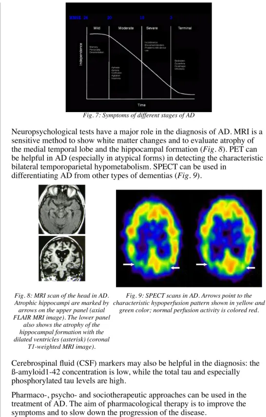
Under physiological conditions, APP is split by the alpha and beta secretase enzymes, producing water-soluble fragments. In AD, this splicing is
performed by the gamma-secretase enzyme and the result is a peptide made up of 40-42 amino acids, which forms a beta sheet secondary structure (beta- amyloid peptide) in addition to the normal, alpha helical one. This peptide aggregates into oligomers, which are neurotoxic. The "final product" of the aggregation process is the senile plaque. (Fig. 6)
Fig. 6: Splicing of the amyloid precursor protein under physiological and pathological conditions. (Modified after Martin JB. Molecular basis of the neurodegenerative disorders. N
Eng J Med 1999;340:1970-1980)
Clinically, AD presents first as a pure amnestic disorder, short-term memory problem being the earliest symptom, which is followed by cognitive signs and by BPSD symptoms (confusion, hallucinations, etc.). Eventually, everyday activities (dressing, eating, etc.) become dysfunctional, and finally the bed-bound patients usually die due to bronchopneumonia (Fig. 7).
Fig. 7: Symptoms of different stages of AD
Neuropsychological tests have a major role in the diagnosis of AD. MRI is a sensitive method to show white matter changes and to evaluate atrophy of the medial temporal lobe and the hippocampal formation (Fig. 8). PET can be helpful in AD (especially in atypical forms) in detecting the characteristic bilateral temporoparietal hypometabolism. SPECT can be used in
differentiating AD from other types of dementias (Fig. 9).
Fig. 8: MRI scan of the head in AD.
Atrophic hippocampi are marked by arrows on the upper panel (axial FLAIR MRI image). The lower panel
also shows the atrophy of the hippocampal formation with the dilated ventricles (asterisk) (coronal
T1-weighted MRI image).
Fig. 9: SPECT scans in AD. Arrows point to the characteristic hypoperfusion pattern shown in yellow and
green color; normal perfusion activity is colored red.
Cerebrospinal fluid (CSF) markers may also be helpful in the diagnosis: the ß-amyloid1-42 concentration is low, while the total tau and especially phosphorylated tau levels are high.
Pharmaco-, psycho- and sociotherapeutic approaches can be used in the treatment of AD. The aim of pharmacological therapy is to improve the symptoms and to slow down the progression of the disease.
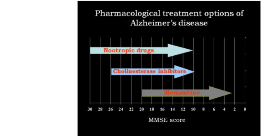
Symptomatic pharmacological therapy is helpful in alleviating both cognitive and BPSD symptoms. Treatment options are determined by the stage of the disease (Fig. 10): mild AD MMSE 21-26; moderate AD MMSE 10-20, severe AD MMSE 0-10.
Fig. 10: Drug classes used in the treatment of cognitive symptoms of AD in different stages of the disease
Nootropic drugs with diverse mechanism of action can be used in mild to moderate stages of AD.
The damage of the cholinergic system in AD was the first neurochemical abnormality described in this disease. Acetyl cholinesterase inhibitors (AChEI) were developed to treat the cholinergic deficit in AD. Treatment with AChEIs improves cognitive and BPSD symptoms, and slows disease progression. Available drugs include donepezil (Aricept), rivastigmine (Exelon), and galatamine (Reminyl). AChEI treatment is recommended in mild to moderate stages of the disease (MMSE scores of 26-10). Their side effects are mainly of cholinergic type, such as gastrointestinal and rarely cardiac effects.
In moderate to severe stages of AD, the N-metil-D-aspartate glutamate receptor non-competitive antagonist memantine (Ebixa) is used.
Drugs aimed at the proposed pathomechnism of AD are currently in
development. The following treatments have reached phase III clinical trials:
beta-amyloid immunization, inhibitors of beta-amyloid aggregation, and synthesis and tau-based therapies.