Review Article
Changes in Redox Signaling in the Skeletal Muscle with Aging
Péter Szentesi,1László Csernoch,1László Dux,2and Anikó Keller-Pintér 2
1Department of Physiology, Medical Faculty, University of Debrecen, Debrecen H-4002, Hungary
2Department of Biochemistry, Faculty of Medicine, University of Szeged, Szeged H-6720, Hungary
Correspondence should be addressed to Anikó Keller-Pintér; keller.aniko@med.u-szeged.hu
Received 22 June 2018; Revised 5 November 2018; Accepted 22 November 2018; Published 17 January 2019
Guest Editor: Christina Karatzaferi
Copyright © 2019 Péter Szentesi et al. This is an open access article distributed under the Creative Commons Attribution License, which permits unrestricted use, distribution, and reproduction in any medium, provided the original work is properly cited.
Reduction in muscle strength with aging is due to both loss of muscle mass (quantity) and intrinsic force production (quality).
Along with decreased functional capacity of the muscle, age-related muscle loss is associated with corresponding comorbidities and healthcare costs. Mitochondrial dysfunction and increased oxidative stress are the central driving forces for age-related skeletal muscle abnormalities. The increased oxidative stress in the aged muscle can lead to altered excitation-contraction coupling and calcium homeostasis. Furthermore, apoptosis-mediatedfiber loss, atrophy of the remainingfibers, dysfunction of the satellite cells (muscle stem cells), and concomitant impaired muscle regeneration are also the consequences of increased oxidative stress, leading to a decrease in muscle mass, strength, and function of the aged muscle. Here we summarize the possible effects of oxidative stress in the aged muscle and the benefits of physical activity and antioxidant therapy.
1. Introduction
With improved life quality conditions and the availability of treatments, life expectancy and consequently the number of elderly in the population have increased [1]. This change in population composition places increasing emphasis on the treatment of chronic, noncommunicable diseases as they have become major causes of death and disability worldwide, thus driving the need to understand the mechanism of aging andfind treatments for age-related diseases [2].
The skeletal muscle is the largest organ in the body com- prising~40% of its mass. It plays fundamental roles in move- ment, posture, and energy metabolism. The loss of skeletal muscle mass and function with age can have a major impact on quality of life and results in increased dependence and frailty. Age-related decline of skeletal muscle function (sarco- penia) results in strength loss [3]. This loss stems from two major sources, reductions in muscle mass (i.e., quantity) and decrease in its intrinsic capacity for producing force (i.e., quality). Both can be the consequence of several factors (Figure 1), including oxidative stress that is the result of the accumulation of reactive oxygen and nitrogen species (ROS/RNS). The free-radical theory of aging was established more than 60 years ago [4] and has become one of the most
studied theories to have been proposed. It is now accepted that this theory and its various spin-offs cannot alone explain the aging process [5, 6]. Nevertheless, huge amounts of data indicate that ROS-mediated aging phenotypes and age- related disorders exist [7, 8].
During physiological homeostasis the overall oxidative balance is maintained by the production of ROS/RNS from several sources and their removal by antioxidant systems, including endogenous or exogenous antioxidant molecules.
At physiological concentrations ROS/RNS play essential roles in a variety of signaling pathways. There is an optimal level of ROS/RNS to sustain both cellular homeostasis and adaptive responses, and both too low and too high levels of ROS/RNS are detrimental to cell functions [9]. The skeletal muscle consumes large quantities of oxygen and can generate great amounts of ROS and also reactive nitrogen species.
Mitochondria are one of the most important sources of ROS in the skeletal muscle; furthermore, NADPH oxidase (NOX) [10], xanthine oxidase [11], and phospholipase A2 (PLA2) [12, 13] are also involved in ROS production.
The origin of the increased ROS production and oxida- tive damage is mitochondrial dysfunction with aging [14], caused by age-related mitochondrial DNA mutations, deletions, and damage [15], as well as the impaired ability
Volume 2019, Article ID 4617801, 12 pages https://doi.org/10.1155/2019/4617801
of muscle cells to remove dysfunctional mitochondria [16].
Oxidative phosphorylation impairment can lead to decreased ATP production and further generation of ROS [4]. Interest- ingly, aging is associated not only with an increase in oxida- tive damage but also with an upregulation of antioxidant enzymes in the skeletal muscle [9]. Furthermore, the iron content of the mitochondria in the skeletal muscle increases with aging, amplifying the oxidative damage with the gener- ation of ROS [17]. Increased ROS production, mitochondrial DNA damage, and mitochondrial dysfunction was observed in aged muscles [18–20].
The skeletal muscle is highly plastic and shows sev- eral adaptations towards mechanical and metabolic stress [21, 22]. Oxidative stressors, like ROS, have long been taken into account as harmful species with negative effects in the skeletal muscle [23]. Proteins such as biomolecules are fre- quently affected by oxidation; thus, elevated ROS levels can cause reversible or irreversible posttranslational modification of cysteine, selenocysteine, histidine, and methionine. Oxida- tive posttranslational modifications of proteins are character- istic in the aged muscle, such as carbonylation which alters protein function [24]. The oxidative capacity of muscles is strongly associated with health and overall well-being.
Enhanced oxidative capacity in the skeletal muscle protects against several pathological phenomena (insulin resistance, metabolic dysregulation, muscle loss with aging, and increased
energetic deficits in myopathies) [25, 26]. These protective effects are largely associated with enhanced mitochondrial function and elevated numbers of mitochondria, which can protect against cellular stress.
Given the rapidly aging population, it is essential to better understand the development, progression, prevention, and treatment of age-related muscle diseases. The aim of this review is to discuss the possible effects of age-related oxida- tion on the skeletal muscle and highlight the benefits of phys- ical activity and intake of antioxidant compounds to protect from oxidative stress.
2. Oxidative Stress and EC-Coupling Machinery in Aging
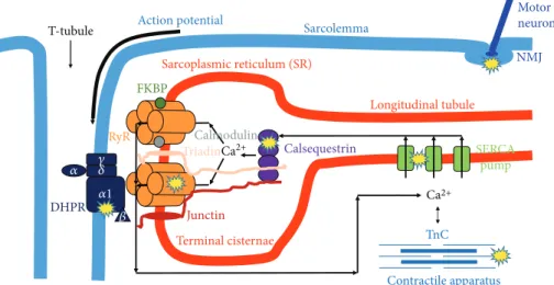
Ca2+, as a second messenger, is necessary for muscle con- traction. Ca2+ can originate from the extracellular space (the heart and smooth muscle) and from the intracellular store of the sarcoplasmic reticulum (SR) (in the skeletal mus- cle exclusively from SR). Excitation-contraction (EC) cou- pling, the steps from the trigger action potential to the development of force, starts with the activation of the voltage sensor dihydropyridine-sensitive, L-type Ca2+ channels (DHPRs). These can open the Ca2+release channel ryano- dine receptor (RyR) of SR [27]. The released Ca2+ freely diffuses into the intracellular space and, after binding with
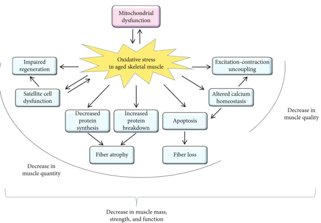
Mitochondrial dysfunction
Impaired regeneration
Satellite cell dysfunction
Decreased protein synthesis
Increased protein breakdown
Apoptosis
Altered calcium homeostasis Excitation-contraction
uncoupling
Fiber loss Fiber atrophy
Decrease in muscle quantity
Decrease in muscle mass, strength, and function
Decrease in muscle quality Oxidative stress
in aged skeletal muscle
Figure1: Schematic summary of the effects of oxidative stress in the aged skeletal muscle. The age-related increase in oxidative stress can result in mitochondrial dysfunction, and the dysfunctional mitochondria can further generate reactive oxygen species. The increased oxidative stress can lead to a decrease both in muscle quality and in muscle quantity. As a consequence of the increased oxidative stress, excitation-contraction uncoupling, altered calcium homeostasis, apoptosis-mediated fiber loss, atrophy of the remaining fibers, dysfunction of the satellite cells (muscle stem cells), and impaired muscle regeneration can be observed in the aged muscle leading to a decrease in muscle mass, strength, and function.
troponin-C (TnC), initiates muscle contraction. During relaxation, the Ca2+ is taken up by the sarco/endoplasmic reticulum calcium pump (SERCA) into the SR [28]. If any step in the EC-coupling pathway is disrupted, the voltage- induced Ca2+release from SR will be deficient and less cyto- plasmic Ca2+will be available to bind with TnC (Figure 2).
EC coupling has a unique structure in muscle fibers, called calcium release units (CRUs). They are formed by two membrane systems: the transverse- (t-) tubule, where the trigger action potential (depolarization) from the sarco- lemma goes into thefiber, and the calcium store SR terminal cisternae [29]. In a fully developed skeletal muscle fiber, a central t-tubule usually forms junctions with two SR forming a triad. The voltage sensor DHPRs localized in the t-tubule membrane [30] are in direct connection with the closely apposed calcium release channel RyRs in the SR membrane.
2.1. RyR. Mammalian RyR has three isoforms, which were originally identified in the skeletal muscle (RyR1), in the heart muscle (RyR2), and in the brain (RyR3). It is now known that some tissues express all three mammalian RyR isoforms [31–33]. Several cellular compounds (e.g., ATP, HCl, Ca2+, and Mg2+), specific proteins (phosphatases and kinases), and endogenous oxidative species can regulate RyR functions [34]. Abramson and Salama [35] were thefirst to propose a redox-dependent gating model of RyR, in which the channel pore opens after the oxidation and closes after the reduction of critical sulfhydryl moieties within the RyR complex. Gating transitions of the RyR channel are extremely fast; the open state usually lasts no longer than a few millisec- onds; thus, this hypothesis has been called into question because of its slow kinetic [36]. Additionally, isolated RyR1 reconstituted in an artificial lipid bilayer functioned similarly independently of the presence of cofactors to maintain the catalytic transfer of electrons [37].
Another possibility to control RyR1 gating is the trans- membrane redox potential of SR. In healthy mammalian cells, the redox potential of the cytosol is approximately -230 mV [38]. The majority of redox buffers within the cyto- sol of a muscle cell are based on the relative concentration of oxidized (GSSG) and reduced (GSH) glutathione or NADH and NAD+. In different nonmuscle cells, GSSG and GSH transporters have been found across the ER membrane [38, 39]. These transporters play an essential role in estab- lishing and maintaining the membrane redox potential gradient. It was shown that glutathione transport across SR/ER membranes is very fast and correlates with the expression of RyR1 in terminal cisternae [40]. These find- ings imply the presence of one or more transmembrane redox sensors in the RyR1 channel.
To study the redox regulation of RyR1 channel activity, Feng et al. [41] used artificial lipid bilayer membranes and precisely controlled the redox state by adjusting the [GSSG]/[GSH] ratio to change redox potentials on both the lumenal and cytoplasmic sides of the reconstituted channel.
Redox sensing may represent a widespread mechanism by which RyR1 channels respond to local changes in transmem- brane redox potential. As mentioned above, disulfide bond formation (sulfhydryl oxidation) in RyR1 usually takes place in the oxidizing environment of the SR lumen, not in the reducing environment of the cytosol.
Pessah et al. [42] demonstrated that RyR1 channel gating was accompanied by changes in the microenvironment of hyperreactive Cys residues. It was assumed that the localized redox potential could influence the domain with the redox sensor, which might change the stability of the closed state.
This means that the closed but not the open conformation of RyR1 senses redox changes. In this framework, the rapid gating transitions of RyR1 would not coincide with oxidation and reduction of disulfide bonds, and local changes in the
T-tubule Action potential
Sarcolemma
Motor neuron Sarcoplasmic reticulum (SR) NMJ
FKBP
Calmodulin
TriadinCa2+ Calsequestrin
Longitudinal tubule
SERCA pump
Ca2+
TnC
Contractile apparatus Junctin
Terminal cisternae DHPR
훼 훾 훿 훼1
훽 RyR
Figure2: Possible actions of age-related oxidative stress reducing skeletal muscle contraction. Accumulation of reactive oxygen and nitrogen species in the aged muscle results in protein modification and/or damage that could reduce muscle quality by altering musclefiber activation at the neuromuscular junction (NMJ), excitation-contraction (EC) coupling (DHPR, RyR, SERCA, calsequestrin), and cross-bridge cycling within the myofibrillar apparatus. DHPR: dihydropyridine receptor; FKBP: FK506 binding protein; RyR: ryanodine receptor; SERCA:
sarco/endoplasmic reticulum Ca2+pump; TnC: troponin-C.
redox environment would influence the overall operation of the channel.
With advancing age, RyR becomes increasingly oxidized and nitrosylated, which leads to leaky release channels.
RyR1 from aged mice was shown to be more oxidized and cysteine-nitrosylated compared to that from young animals.
Furthermore, these RyR channels lacked the stabilizing sub- unit FKBP12. Treating aged mice with the small molecule rycal drug S107 stabilized binding of FKBP12 to RyR1 reduc- ing intracellular Ca2+leakage, enhancing Ca2+release from SR, decreasing ROS, and improving muscle exercise [43].
Similarly, increased Ca2+ leakage from the SR, primarily through the RyRs, was found in type Ifibers of aged humans, and a reducing treatment with dithiothreitol inhibited RyR Ca2+ leakage, thus increasing net SR Ca2+ accumulation [44]. Other evidence of partially defective SR in the aged muscle is the decreased frequency of spontaneous Ca2+
release (spark) through RyR, observed by Park et al. [45]
and the authors of this review (unpublished data).
The expression of RyR also changes with age. Unpub- lished data of the authors of this review showed reduced RyR expression in aging mice. The whole tetramer was almost completely absent in the EDL muscle of old animals, and only a smaller amount of degraded RyR was found.
Interestingly, this was not the case in mice that did voluntary exercise throughout their entire life.
2.2. RyR-Associated Proteins. A lot of studies have inves- tigated the redox dependence of accessory proteins of RyR1, which contribute to the tight regulation of channel activity in the mammalian skeletal muscle. These proteins include the voltage sensor skeletal dihydropyridine recep- tor (L-type Ca2+ channel), calmodulin, triadin, junctin, FKBP12 (12 kDa FK506 binding protein), and calsequestrin in the SR lumen [34, 46].
To date there is no evidence that triadin and junctin have any role in the redox regulation of RyR1. On the other hand, reactive sulfhydryl groups within RyR1 chan- nels have been shown to help the binding of calmodulin, and functional responses of calmodulin to RyR1 may be redox regulated [47]. It was proposed that probably more than one class of sulfhydryl residues within the RyR1 channel complex suffer chemical modification, each con- tributing to a specific function. This question is still open because of the structural complexity of RyR1 and its asso- ciated proteins.
Calsequestrin-1 is a high-capacity Ca2+buffer, localized in the lumen of SR in close proximity to RyR1. It has been demonstrated that nNOS and NOX2 also colocalize with RyRs at the triad junctions, and the latter generate ROS, which stimulate Ca2+ release from the SR through RyR1 [48]. Recently, it was hypothesized that in musclefibers lack- ing Calsequestrin-1, the close positioning of either nNOS or NOX2 to RyR1 and the Ca2+-dependent activation of nNOS could be the consequence of increased production of ROS and RNS. This could finally lead to nitrosylation and glu- tathionylation of specific cysteine residues causing oxidative modifications that further increase the probability of leaky RyR1 channels [49].
RyR1 has four subunits to bind the small FK506 protein (FKBP12) [50]. FKBP12 associates mainly with the skeletal muscle isoform to regulate RyR1 function. Pharmacological removal of FKBPs causes uncoupling of RyR1 ion channels from their neighbors and thus activates Ca2+release from SR [51]. A recent study shows that the 1,4 benzoderivative S107 binds to multiple RyR1 sites with low affinity and stabi- lizes the RyR1-FKBP12 complex depending on the redox state of the calcium channel [52].
2.3. DHPR.DHPR is located in the t-tubules and plays a role in EC coupling as the voltage sensor triggering Ca2+release from the SR after an action potential. More than 20 years ago, Delbono et al. [53] recorded a significant reduction of maximum charge movement and L-type calcium current in musclefibers from biopsies of 65-75-year-old patients. This was accompanied with decreased Ca2+release from the SR.
Just a few years later it was shown that ROS may also target DHPRs, since ROS alter the dynamics of muscle K+contrac- tures [54]. A later study by the same research group using the mammalian diaphragm demonstrated an increase in tension after antioxidant application that is clearly dependent on DHPR function [55]. These results support the hypothesis that the DHPR redox state and RyR function are modulated in an interactive manner and modify contractility.
It was also shown that the expression of theα1 subunit of DHPR decreases with age and this is associated with the loss of skeletal muscle strength [56]. Thesefindings were ampli- fied by the fact that DHPR expression levels can be regulated by different mechanisms, independently from gene tran- scription or mRNA expression. In a very recent study, a novel finding was reported that cytoplasmic-located fast skeletal muscle troponin T3 (TnT3) regulates DHPR expression in skeletal musclefibers and calpain-induced cleavage of TnT3 is associated with DHPR downregulation in aged mice [57].
The reduced DHPR expression with aging increases the number of uncoupled RyR1s and, thus, decreases SR Ca2+
release which leads to EC uncoupling and finally decreased force production.
2.4. SERCA. The sarco/endoplasmic reticulum Ca-ATPase (SERCA) is the calcium pump that uptakes Ca2+from the cytosol to the SR during muscle relaxation. It has an impor- tant role in maintaining the resting intracellular Ca2+con- centration (around 100 nM). Evidence for NO inhibition of the Ca-ATPase was observed in the rabbit skeletal muscle, where sustained contractions led to significant (40–50%) inactivation of the pump [58]. One possible explanation could be the reactions with critical SH groups, since peroxynitrite treatment of Ca-ATPase from the rabbit skeletal muscle was correlated with oxidative and nitrosative modifications of cys- teines at several positions, of which one was deemed responsi- ble for enzyme inhibition [59]. Recently it was shown that SERCA1 is reversibly regulated via NO-dependent S- glutathiolation of specific cysteine residues which are embed- ded within the transmembrane domains of the pump. Some specific amino acid peroxides react selectively with a subset of cysteine residues of SERCA1, representing one of the targets for NO-dependent S-glutathiolation [60]. In a
parallel study it was also demonstrated that antioxidant treat- ment affects intracellular Ca2+concentration, increasing the maximum rates of ATP hydrolysis and uptake of Ca2+ by SERCA in the diaphragm [61].
The 53 kDa isoform of sarcalumenin, the major luminal glycoprotein associated with SERCA, was found to be down- regulated in the aged human muscle [62]. Interestingly this was accompanied with the upregulation of Calsequestrin-1.
In a recent study on humans, it was shown that these changes were reversed after 9 weeks of training by electrical stimula- tion [63] of the vastus lateralis muscle of sedentary senior volunteers. The decreased active SERCA, and thus insuffi- cient SR Ca2+content, can also be explained by thefindings of Boncompagni et al. [64]. Their electron microscopic study proved the presence of SERCA and Calsequestrin-1-rich tubular aggregates in the aging mouse skeletal muscle. They hypothesized that polymerization of SERCA induces its inactivity and this decreases the Ca2+ uptake capacity of SR. Similarly, the accumulated inactive Calsequestrin-1 in tubular aggregates is missing from SR and leads to reduced Ca2+storage capacity.
3. The Effects of Age-Dependent Structural Changes in the Skeletal Muscle
It has been suggested by Renganathan and colleagues [56]
that an uncoupling between DHPR and RyR1 in the CRUs (insufficient transmission of the sarcolemmal depolarization to the calcium release channel) with aging is one of the major determinants of the progressive decline in muscle strength.
This has been supported by transmission EM studies [65], which show a progressive disarrangement of triads in the aging human skeletal muscle. This results in a drastic reduc- tion in the overall number of CRUs available for releasing Ca2+to initiate the sliding of contractilefilaments and gener- ate force. Notwithstanding that the total number of CRUs is decreased, on average by more than 50% in the aging muscle, the decrease in the total amount of both DHPR and RyR1 was less, because the decrease in the total number of SR/t-tubule junctions is accompanied by an increase in the average size of RyR clusters which compensate for the loss of triads.
Besides Ca2+, ATP is also necessary to generate force, as well as for relaxation in the skeletal muscle. The main source of ATP is mitochondria. It was shown that mitochondria and CRUs are functionally linked to each other via ROS- and Ca2+-mediated cross-talk [66]. Furthermore, these two organelles are structurally connected by tethers, which pro- mote proximity and sufficient calcium signaling [67]. In the aged muscle, not only the ultrastructure, density, and dispo- sition of mitochondria and CRUs but also their reciprocal associations are altered. The density of CRUs and mitochon- dria is decreased in the aged muscle, with an increased num- ber of damaged mitochondria and mitochondria misplaced from their normal triadic position. A significant reduction in CRU-mitochondria pair density and their tethering was also observed in aged mice. These changes were accompanied with increased oxidative stress and with decreased mitochon- drial Ca2+ uptake and SR Ca2+ release [68]. These wrong
direction changes in the skeletal muscle can be prevented by regular exercise. The number of mitochondria is higher in athletic than in sedentary seniors, and furthermore, the number of CRU-mitochondria pairs is three times higher in senior sportsmen than in sedentary individuals. Since the correct association between CRUs and mitochondria is nec- essary for efficient ATP production, this can explain the sig- nificantly superior muscle performance in lifelong exercising seniors [68]. Similar results were obtained with mice that had access to running wheels for the second part of their lives (from 1 to 2 years of age) [69]. The authors of these studies concluded in their results that the huge age-dependent decrease affecting EC-coupling apparatuses and mitochon- drial functions in the skeletal muscle of humans and mice can be partly associated with inactivity in old age.
4. Oxidative Stress and Satellite Cell Dysfunction with Aging
The skeletal muscle has the remarkable ability to regenerate in response to injury. This regenerative capacity is due to the muscle stem cells (MuSCs), also called satellite cells that reside between the muscle fiber and its surrounding basal lamina [70]. The satellite cells are mitotically and physiolog- ically in a quiescent state (a G0 reversible arrest state) in the healthy muscle and express the Pax7 transcription factor.
They are stimulated upon muscle injury to enter the cell cycle and proliferate extensively and form myoblasts that will sub- sequently differentiate and fuse to form musclefibers. The differentiated myocytes are capable of fusing together and, with the preexisting myofibers, restore the muscle tissue. A small subset of the expanding satellite cells does not commit to terminal differentiation but self-renews to restore the qui- escent satellite cell pool for further needs [71]. The regenera- tive function of satellite cells declines with age [72, 73]
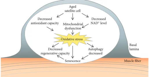
(Figure 3). At advanced geriatric age, this decline is maximal owing to transition from a normal quiescence into an irre- versible senescence (a G0 irreversible arrest).
The age-related deficits in muscle regeneration have been linked to changes in the satellite cell environment (such as inflammatory status) and/or satellite cell-intrinsic mecha- nisms [74]. Both the number and the functionality of satellite cells decrease with age [75–79], switching from quiescence to a senescent state [76]. The satellite cells are unequally distrib- uted among the differentfiber types and differ between mus- cles. In the rat extensor digitorum longus (EDL) muscle, satellite cells are observed most frequently on type IIAfibers and at approximately equal frequencies on type IIB and type I fibers [80]. Interestingly, the soleus contains a considerably higher percentage of satellite cells than the EDL [80]. In the young adults, satellite cell content did not differ between type I and type II musclefibers [81]. Aging is associated with a switch from fast to slow fiber type [82]. The abundance of resident satellite cells declines with age in myofibers from both fast- and slow-twitch muscles in mice [83], and a decrease in FGF signaling as a possible limiting factor of sat- ellite cell function during muscle aging has been identified [83]. In contrast, satellite cell content is reported to be specif- ically reduced in type II skeletal musclefibers in the elderly,
but not in type Ifibers [84]. This decline in satellite cell con- tent might be an important factor in the etiology of type II musclefiber atrophy, which accompanies the loss of skeletal muscle with age [81, 84].
The function of satellite cells is altered by oxidative stress with aging. In the aged muscle, the satellite cells exhibit a reduced capacity to proliferate and self-renew. The decrease in the self-renewing muscle stem cell pool can lead to decreased regenerative capacity of the muscle. The quiescent satellite cells have a low metabolic rate and display only a few active mitochondria and therefore are exposed to low levels of oxidative stress [85]. Gene expression studies have also indicated differences in the transcriptional profile of aged versus young satellite cells, e.g., changes in genes associated with mitochondrial function [86]. The ROS production was higher in isolated satellite cells from the aged muscle [87].
Furthermore, a decline in antioxidant capacity in satellite cells was also observed with age, diminishing satellite cell function with increased ROS levels [88]. It was reported that the antioxidant activity of catalase and glutathione transferase is reduced in aged satellite cells [89]. Several redox-dependent signaling pathways are deregulated in aged satellite cells;
decreased Notch [90], increased Wnt (wingless/integrated) [91], increased p38/MAPK (mitogen-activated protein kinase) [77], and JAK-STAT3 (Janus kinase-signal transducer of activation) [92] signaling were observed.
Mitochondrial dysfunction can result from decreased NAD+(nicotinamide adenine dinucleotide) levels of the cells.
Stem cells are thought to rely predominantly on glycolysis to yield energy, decreasing NAD+ concentration [93]. The reduction of the cellular NAD+level and its effect on mito- chondrial activity was shown to be a pivotal switch to modu- late satellite cell senescence [78]. Treatment with the NAD+ precursor, nicotinamide riboside, induced the mitochondrial unfolded protein response and synthesis of prohibitin pro- teins, rejuvenated the muscle stem cells in aged mice, and enhanced life span [78].
The activities of the ubiquitin-proteasome system, autophagy, and chaperones appear to decline with age [94].
During myogenesis and regeneration, an increase in protein synthesis and removal of misfolded proteins can be observed [73]. Oxidative stress can influence muscle satellite cells by altering their protein homeostasis. Basal autophagy is essen- tial to maintain the stem cell quiescent state [95]. Autophagy was shown to be essential to maintain the stemness of sat- ellite cells by preventing the senescence caused by mito- chondrial dysfunction and oxidative stress associated with aging [95].
5. Age-Related Muscle Loss and Oxidative Stress Sarcopenia, the age-associated generalized and progressive reduction in muscle mass, increases the susceptibility to mus- cle injury, serious falls, obesity, and diabetes [96], predicting frailty, disability, poor quality of life, and mortality in the elderly [97–101]. The prevalence of low muscle mass is esti- mated between 8 and 40% depending on the population stud- ied and the methods used to identify sarcopenia; it ranges from 15% at 65 years to 50% at 80 years [100, 101]. Progres- sive muscle loss starts at approximately the age of 40 years; it is estimated at about 8% per decade until the age of 70 years and then it increases to 15% per decade [102]. Reduction in muscle mass is usually combined with an increase in body fat mass; the accumulation of fat can be observed within the musclefibers. The high levels of ROS in the aging muscle can induce the transition of satellite cells into an adipogenic phenotype. This muscle-to-fat transition can explain the increased intramuscular adipose tissue associated with sarcopenia [103, 104].
Age-related muscle atrophy was shown to be associated with a decrease in the total number of muscle fibers and a simultaneous decrease in the size of the individual fibers. It was reported that age-related muscle loss in rodents [105]
and humans [82] can occur due to the loss of musclefibers
Aged satellite cell
Decreased NAD+ level Mitochondrial
dysfunction Decreased
antioxidant capacity
Oxidative stress Decreased
regenerative capacity
Autophagy decreased Senescence
Basal lamina Muscle fiber
Figure3: Age-related alterations in satellite cells. Mitochondrial dysfunction and decreased antioxidant capacity of aged satellite cells can lead to increased oxidative stress. The satellite cell dysfunction results in decreased regenerative capacity of the muscle. As a consequence of increased oxidative stress, a decrease in autophagy can lead to senescence.
and a decrease in the cross-sectional area of the remaining fibers. Several factors were reported to contribute to muscle atrophy with aging. The role of reduced protein synthesis, declines in neural function, hormonal deficits, chronic low- grade inflammation, loss of mitochondrial function, nuclear apoptosis, reduced function of satellite cells, and oxidative stress was reported [96, 106].
A relationship was observed between oxidative stress and muscle mass [107–110]. The disruption of signaling path- ways involving skeletal muscle reactive oxygen species has received increasing attention [6]. Age-associated accumula- tion of nitrotyrosine in muscle proteins was reported [108].
The accumulation of mitochondrial and nuclear DNA damage leads to the loss of skeletal muscle fibers [111].
Mitochondria-mediated apoptosis represents a central pro- cess driving age-related muscle loss [112]. Mitochondrial dysfunction is related not only to the loss of its capacity to generate ATP but also to the activation of apoptotic pathways leading to the irreversible cell loss that is characteristic of sarcopenia [112].
Further studies have shown that ROS accumulation can increase proteolysis leading to loss of muscle mass; increased ROS production activates the ubiquitin-proteasome path- way. Aging is associated with greater proteasome content and activity [113], increased expression of the ubiquitin ligase MuRF1 (Muscle RING-finger protein-1) and atrogin-1 [114], and increased calpain activity [115]; however, further studies are required to explore the role of oxidative stress in these age-related alterations. The potential role of age- dependent mitochondrial dysfunction and cumulative oxi- dative stress as the underlying cause of age-associatedfiber atrophy remains controversial; the pharmacological attenu- ation of age-related mitochondrial redox changes failed to rescue the age-associated muscle fiber atrophy, implying that the muscle mitochondrial redox environment is not a key regulator of fiber atrophy during sarcopenia [3].
Recently it was reported that sedentary humans display an age-related decline in the mitochondrial protein optic atrophy 1 (OPA1) that is associated with muscle loss [116].
FoxOs (Forkhead box proteins) are master regulators of autophagy and the ubiquitin-proteasome system [117] and are activated by oxidative stress and Akt inhibition. Impor- tantly, the acute inhibition of OPA1 results in an increased oxidative stress, andin vivo inhibition of FoxOs was suffi- cient to reduce muscle atrophy inOpa1−/−mice [117].
6. Antioxidant Therapies and Effects of Exercise on the Aged Muscle
The effects of exercise on aging in the skeletal muscle are very controversial. There is widespread agreement that oxidation could increase during exercise. Early studies have suggested that ROS play important roles in the inflammatory response to high-intensity or long-lasting exercise [118]. On the other hand, it has also long been known that moderate exercise increases the antioxidant capacity of the skeletal muscle by mitochondrial remodeling [119]. A more recent study sug- gested that endurance training stimulates mitochondrial remodeling which leads to an increase in mitochondrial
content and function [120]. Unfortunately recent rodent models suggest that exercise-induced mitochondrial remod- eling is defective in the aged muscle [121]. In contrast, as mentioned above, moderate exercise can improve the num- ber of CRU-mitochondria pairs and thus provide more ATP and Ca2+ for contraction [68]. Furthermore, resistance-type exercise training represents an effective strat- egy to increase satellite cell content and reverse type II muscle fiber atrophy in humans [81].
Another target of exercise against oxidative stress is the increased activity of enzymatic antioxidants (i.e., glutathione peroxidase, catalase, and superoxide dismutase) accompany- ing the exercise-induced ROS generation. For example, skeletal muscle-specific manganese superoxide dismutase- deficient mice, which showed reduced exercise activity without atrophy, presented significantly improved exercise activity of the skeletal muscle after a single administration of an antioxidant [122].
Numerous investigations have aimed to explore the effects of antioxidant treatment on skeletal muscle perfor- mance. Some of them also studied old muscles and found positive effects of such a treatment. For example, hydroxytyr- osol, which has high free-radical-scavenging capabilities, caused increasedin vivoforce in aged rats [123]. Recent stud- ies showed positive effects of resveratrol [124], some plant extracts (Rhus coriaria [125] and Rosmarinus officinalis [126]), and vitamins (vitamin C [127]). The increasing number of similar studies nowadays shows the importance and topicality of finding good antioxidant treatment for the aged muscle.
As discussed above, several studies report an elevation in levels of oxidized protein and DNA in the older skeletal mus- cle. To date, RyR is the only key protein in EC coupling for which lifelong voluntary training was investigated and found to improve its expression level in aged mice (unpublished data of the authors). The data showed a beneficial antioxi- dant effect of selenium supplementation on skeletal muscle performance in old animals. However, as the authors could not prove the direct effects of antioxidant treatment on ROS production, there are several key proteins in EC cou- pling which could be positively altered and, thus, enhance force production.
The effect of antioxidant compounds on aged satellite cells has already been reported. Tocotrienol is a vitamin E analogue bearing high antioxidant activity. The tocotrienol- rich fraction (TRF) replenished the regenerative capacity of the human senescent satellite cells [128]; furthermore, TRF is able to ameliorate antioxidant defence mechanisms and improve replicative senescence-associated oxidative stress in human satellite cells [129]. The vitamin E analogue trolox treatment prevented the appearance of senescence markers, restored the expansion, and rescued the proliferative and regenerative defect of geriatric satellite cells [72]. The effect of resveratrol was studied in the mouse myoblast cell and showed protection against ROS by improving Sirt1 (Sirtuin1) levels, increasing antioxidant production, and reducing apo- ptotic signaling and cell death [130]. Interestingly, the effect of exercise on the oxidative stress of satellite cells has not yet been investigated in the literature. The protective effects
of exercise, resveratrol, and their combination was shown to increase muscle mass in rats, probably associated with antia- poptotic signaling pathways through activation of AMPK (AMP-activated protein kinase)/Sirt1 [131]. In contrast, administration of the long-term mitochondria-targeted antioxidant, mitoquinone mesylate, failed to attenuate age- related oxidative damage or rescue the loss of muscle mass and function in the skeletal muscle of old mice [132].
7. Concluding Remarks
Lifelong maintenance of muscle mass and strength is a global health challenge. With an aging population, the problem of sarcopenia is becoming more and more important, and effec- tive strategies are required to improve muscle performance.
An average 30-year-old will lose about 25% of his or her mus- cle strength by age 70 and 50% of it by age 80. The improve- ment of mobility and independence is key for old people, and it relieves society from healthcare and social support costs.
Our knowledge about the signaling pathways mediating age-related muscle loss is still limited. Oxidative stress and subsequent alterations in signaling pathways could lead to different pathophysiological events at different stages of life, especially in old age. As was shown in this review, a lot of tar- gets in skeletal muscle could be altered by increased oxidative stress with aging. Some of them are targets of intrinsic fac- tors, but there are some which depend mainly on extrinsic actions. The effects of oxidative stress in muscles are so diverse that improving only one step is usually not enough to get better muscle performance. This means that only com- bined therapy could be effective, and continuous training will also allow musclefibers to incorporate higher levels of exog- enous antioxidants from dietary supplements.
In conclusion, the risks of oxidative stress-induced dam- age can be minimized with regular exercise, which has bene- ficial effects on physical and mental health. We have to emphasize that while it is never too late to begin exercise, an early start and regular practice throughout life would greatly improve outcomes in later years and slow down a body’s aging process. A lot of people try to start a training program late in life, when muscle performance is already diminished. It follows that muscle research has to promote the development of a new generation of physically active, healthy elderly citizens. To achieve this, and to minimize oxi- dative stress, the key could be a carefully developed exercise protocol combined with adequate antioxidant supplemen- tation. However, exercise can be restricted due to orthope- dic or cardiopulmonary limitations, which highlights the importance of the exploration of antioxidant therapy or nontraditional exercise. Regular exercise to maintain mus- cle function also has beneficial effects by reducing oxida- tive stress, not only in the muscle, but in all tissue, a fact that intrinsically would reduce/delay aging.
Conflicts of Interest
The authors declare that there is no conflict of interest regarding the publication of this paper.
Acknowledgments
This work was supported by a grant from the Hungarian National Research, Development and Innovation Office (NKFIH NK-115461), the GINOP-2.3.2-15-2016-00040 pro- ject, the EFOP-3.6.2-16-2017-00006, and the UNKP-17-4 New National Excellence Program of the Ministry of Human Capacities (Hungary). The project is cofinanced by the Euro- pean Union and the European Regional Development Fund.
References
[1] R. Suzman, J. R. Beard, T. Boerma, and S. Chatterji,“Health in an ageing world–what do we know?,” Lancet, vol. 385, no. 9967, pp. 484–486, 2015.
[2] J. L. Shadrach and A. J. Wagers,“Stem cells for skeletal mus- cle repair,”Philosophical Transactions of the Royal Society of London. Series B, Biological Sciences, vol. 366, no. 1575, pp. 2297–2306, 2011.
[3] G. K. Sakellariou, T. Pearson, A. P. Lightfoot et al., “Mito- chondrial ROS regulate oxidative damage and mitophagy but not age-related musclefiber atrophy,”Scientific Reports, vol. 6, no. 1, p. 33944, 2016.
[4] D. Harman, “Aging: a theory based on free radical and radiation chemistry,”Journal of Gerontology, vol. 11, no. 3, pp. 298–300, 1956.
[5] A. D. Romano, G. Serviddio, A. de Matthaeis, F. Bellanti, and G. Vendemiale, “Oxidative stress and aging,” Journal of Nephrology, vol. 23, Supplement 15, pp. S29–S36, 2010.
[6] J. Viña, C. Borras, K. M. Abdelaziz, R. Garcia-Valles, and M. C. Gomez-Cabrera,“The free radical theory of aging revis- ited: the cell signaling disruption theory of aging,”Antioxi- dants & Redox Signaling, vol. 19, no. 8, pp. 779–787, 2013.
[7] F. L. Muller, M. S. Lustgarten, Y. Jang, A. Richardson, and H. van Remmen,“Trends in oxidative aging theories,”Free Radical Biology & Medicine, vol. 43, no. 4, pp. 477–503, 2007.
[8] A. B. Salmon, A. Richardson, and V. I. Perez,“Update on the oxidative stress theory of aging: does oxidative stress play a role in aging or healthy aging?,”Free Radical Biology & Med- icine, vol. 48, no. 5, pp. 642–655, 2010.
[9] E. Le Moal, V. Pialoux, G. Juban et al.,“Redox control of skel- etal muscle regeneration,”Antioxidants & Redox Signaling, vol. 27, no. 5, pp. 276–310, 2017.
[10] S. K. Powers and M. J. Jackson,“Exercise-induced oxidative stress: cellular mechanisms and impact on muscle force pro- duction,”Physiological Reviews, vol. 88, no. 4, pp. 1243–1276, 2008.
[11] F. Derbre, B. Ferrando, M. C. Gomez-Cabrera et al.,“Inhibi- tion of xanthine oxidase by allopurinol prevents skeletal mus- cle atrophy: role of p 38 MAPKinase and E3 ubiquitin ligases,”PLoS One, vol. 7, no. 10, article e46668, 2012.
[12] D. Nethery, D. Stofan, L. Callahan, A. DiMarco, and G. Supinski, “Formation of reactive oxygen species by the contracting diaphragm is PLA(2) dependent,” Journal of Applied Physiology, vol. 87, no. 2, pp. 792–800, 1999.
[13] M. C. Gong, S. Arbogast, Z. Guo, J. Mathenia, W. Su, and M. B. Reid,“Calcium-independent phospholipase A2 modu- lates cytosolic oxidant activity and contractile function in murine skeletal muscle cells,”Journal of Applied Physiology, vol. 100, no. 2, pp. 399–405, 2006.
[14] J. Miquel, A. C. Economos, J. Fleming, and J. E. Johnson Jr.,
“Mitochondrial role in cell aging,”Experimental Gerontology, vol. 15, no. 6, pp. 575–591, 1980.
[15] E. Bua, J. Johnson, A. Herbst et al.,“Mitochondrial DNA- deletion mutations accumulate intracellularly to detrimental levels in aged human skeletal muscle fibers,” American Journal of Human Genetics, vol. 79, no. 3, pp. 469–480, 2006.
[16] H. N. Carter, C. C. Chen, and D. A. Hood,“Mitochondria, muscle health, and exercise with advancing age,”Physiology, vol. 30, no. 3, pp. 208–223, 2015.
[17] X. Xu, C. N. (. J.). Chen, E. A. Arriaga, and L. D. V. Thompson,
“Asymmetric superoxide release inside and outside the mitochondria in skeletal muscle under conditions of aging and disuse,”Journal of Applied Physiology, vol. 109, no. 4, pp. 1133–1139, 2010.
[18] C. M. Lee, L. E. Aspnes, S. S. Chung, R. Weindruch, and J. M.
Aiken, “Influences of caloric restriction on age-associated skeletal muscle fiber characteristics and mitochondrial changes in rats and micea,”Annals of the New York Academy of Sciences, vol. 854, 1 TOWARDS PROLO, pp. 182–191, 1998.
[19] J. Wanagat, Z. Cao, P. Pathare, and J. M. Aiken,“Mitochon- drial DNA deletion mutations colocalize with segmental elec- tron transport system abnormalities, muscle fiber atrophy, fiber splitting, and oxidative damage in sarcopenia,” The FASEB Journal, vol. 15, no. 2, pp. 322–332, 2001.
[20] R. Calvani, A. M. Joseph, P. J. Adhihetty et al.,“Mitochon- drial pathways in sarcopenia of aging and disuse muscle atro- phy,”Biological Chemistry, vol. 394, no. 3, pp. 393–414, 2013.
[21] M. A. Smith and M. B. Reid,“Redox modulation of contrac- tile function in respiratory and limb skeletal muscle,”Respi- ratory Physiology & Neurobiology, vol. 151, no. 2-3, pp. 229–241, 2006.
[22] J. Farup, F. de Paoli, K. Bjerg, S. Riis, S. Ringgard, and K. Vissing,“Bloodflow restricted and traditional resistance training performed to fatigue produce equal muscle hyper- trophy,” Scandinavian Journal of Medicine & Science in Sports, vol. 25, no. 6, pp. 754–763, 2015.
[23] J. M. Lawler, Z. Hu, and W. S. Barnes, “Effect of reactive oxygen species on K+ contractures in the rat diaphragm,” Journal of Applied Physiology, vol. 84, no. 3, pp. 948–953, 1998.
[24] M. A. Baraibar, M. Gueugneau, S. Duguez, G. Butler-Browne, D. Bechet, and B. Friguet,“Expression and modification pro- teomics during skeletal muscle ageing,” Biogerontology, vol. 14, no. 3, pp. 339–352, 2013.
[25] T. Wenz, F. Diaz, B. M. Spiegelman, and C. T. Moraes,
“Activation of the PPAR/PGC-1alpha pathway prevents a bioenergetic deficit and effectively improves a mitochondrial myopathy phenotype,” Cell Metabolism, vol. 8, no. 3, pp. 249–256, 2008.
[26] V. Ljubicic, P. Miura, M. Burt et al.,“Chronic AMPK activa- tion evokes the slow, oxidative myogenic program and trig- gers beneficial adaptations inmdxmouse skeletal muscle,”
Human Molecular Genetics, vol. 20, no. 17, pp. 3478–3493, 2011.
[27] G. Meissner and X. Lu,“Dihydropyridine receptor-ryanodine receptor interactions in skeletal muscle excitation- contraction coupling,” Bioscience Reports, vol. 15, no. 5, pp. 399–408, 1995.
[28] F. Wuytack, L. Raeymaekers, H. Smedt et al.,“Ca2+-transport ATPases and their regulation in muscle and brain,”Annals of the New York Academy of Sciences, vol. 671, 1 Ion-Motive AT, pp. 82–91, 1992.
[29] C. Franzini-Armstrong and A. O. Jorgensen,“Structure and development of E-C coupling units in skeletal muscle,” Annual Review of Physiology, vol. 56, no. 1, pp. 509–534, 1994.
[30] S. H. Yuan, W. Arnold, and A. O. Jorgensen,“Biogenesis of transverse tubules and triads: immunolocalization of the 1,4-dihydropyridine receptor, TS28, and the ryanodine receptor in rabbit skeletal muscle developing in situ,” The Journal of Cell Biology, vol. 112, no. 2, pp. 289–301, 1991.
[31] R. Coronado, J. Morrissette, M. Sukhareva, and D. M.
Vaughan,“Structure and function of ryanodine receptors,” American Journal of Physiology-Cell Physiology, vol. 266, no. 6, pp. C1485–C1504, 1994.
[32] T. Furuichi, D. Furutama, Y. Hakamata, J. Nakai, H. Takeshima, and K. Mikoshiba,“Multiple types of ryano- dine receptor/Ca2+ release channels are differentially expressed in rabbit brain,” The Journal of Neuroscience, vol. 14, no. 8, pp. 4794–4805, 1994.
[33] F. Mori, M. Fukaya, H. Abe, K. Wakabayashi, and M. Watanabe,“Developmental changes in expression of the three ryanodine receptor mRNAs in the mouse brain,”
Neuroscience Letters, vol. 285, no. 1, pp. 57–60, 2000.
[34] M. Fill and J. A. Copello,“Ryanodine receptor calcium release channels,”Physiological Reviews, vol. 82, no. 4, pp. 893–922, 2002.
[35] J. J. Abramson and G. Salama,“Critical sulfhydryls regulate calcium release from sarcoplasmic reticulum,”Journal of Bio- energetics and Biomembranes, vol. 21, no. 2, pp. 283–294, 1989.
[36] J. H. Shin, G. H. Yoo, C. J. Lee, and C. K. Suh,“Fast and slow gating types of SR ryanodine receptor/channel purified from canine latissimus dorsi muscle,” Yonsei Medical Journal, vol. 37, no. 1, pp. 72–80, 1996.
[37] I. N. Pessah, C. Beltzner, S. W. Burchiel, G. Sridhar, T. Penning, and W. Feng,“A bioactive metabolite of benzo[a]- pyrene, benzo[a]pyrene-7,8-dione, selectively alters micro- somal Ca2+ transport and ryanodine receptor function,” Molecular Pharmacology, vol. 59, no. 3, pp. 506–513, 2001.
[38] C. Hwang, A. Sinskey, and H. Lodish,“Oxidized redox state of glutathione in the endoplasmic reticulum,” Science, vol. 257, no. 5076, pp. 1496–1502, 1992.
[39] G. Bánhegyi, L. Lusini, F. Puskás et al.,“Preferential transport of glutathioneversusglutathione disulfide in rat liver micro- somal vesicles,” Journal of Biological Chemistry, vol. 274, no. 18, pp. 12213–12216, 1999.
[40] M. Csala, R. Fulceri, J. Mandl, A. Benedetti, and G. Bánhegyi,
“Ryanodine receptor channel-dependent glutathione trans- port in the sarcoplasmic reticulum of skeletal muscle,”Bio- chemical and Biophysical Research Communications, vol. 287, no. 3, pp. 696–700, 2001.
[41] W. Feng, G. Liu, P. D. Allen, and I. N. Pessah,“Transmem- brane redox sensor of ryanodine receptor complex,” The Journal of Biological Chemistry, vol. 275, no. 46, pp. 35902– 35907, 2000.
[42] I. N. Pessah, K. H. Kim, and W. Feng, “Redox sensing properties of the ryanodine receptor complex,”Frontiers in Bioscience, vol. 7, no. 1, pp. a72–a79, 2002.
[43] D. C. Andersson, M. J. Betzenhauser, S. Reiken et al.,“Ryano- dine receptor oxidation causes intracellular calcium leak and muscle weakness in aging,”Cell Metabolism, vol. 14, no. 2, pp. 196–207, 2011.
[44] C. R. Lamboley, V. L. Wyckelsma, M. J. McKenna, R. M.
Murphy, and G. D. Lamb,“Ca(2+) leakage out of the sarco- plasmic reticulum is increased in type I skeletal musclefibres in aged humans,”The Journal of Physiology, vol. 594, no. 2, pp. 469–481, 2016.
[45] K. H. Park, N. Weisleder, J. Zhou et al., “Assessment of calcium sparks in intact skeletal muscle fibers,”Journal of Visualized Experiments, no. 84, article e50898, 2014.
[46] J. J. Mackrill, S. O'Driscoll, F. A. Lai, and T. V. McCarthy,
“Analysis of type 1 ryanodine receptor-12 kDa FK506- binding protein interaction,” Biochemical and Biophysical Research Communications, vol. 285, no. 1, pp. 52–57, 2001.
[47] J. Z. Zhang, Y. Wu, B. Y. Williams et al.,“Oxidation of the skeletal muscle Ca2+release channel alters calmodulin bind- ing,” American Journal of Physiology-Cell Physiology, vol. 276, no. 1, pp. C46–C53, 1999.
[48] C. Hidalgo, G. Sánchez, G. Barrientos, and P. Aracena-Parks,
“A transverse tubule NADPH oxidase activity stimulates cal- cium release from isolated triads via ryanodine receptor type 1 S -glutathionylation,” Journal of Biological Chemistry, vol. 281, no. 36, pp. 26473–26482, 2006.
[49] A. Michelucci, S. Boncompagni, M. Canato, C. Reggiani, and F. Protasi,“Estrogens protect calsequestrin-1 knockout mice from lethal hyperthermic episodes by reducing oxidative stress in muscle,”Oxidative Medicine and Cellular Longevity, vol. 2017, Article ID 6936897, 15 pages, 2017.
[50] C. Franzini-Armstrong and F. Protasi,“Ryanodine receptors of striated muscles: a complex channel capable of multiple interactions,”Physiological Reviews, vol. 77, no. 3, pp. 699– 729, 1997.
[51] G. P. Ahern, P. R. Junankar, and A. F. Dulhunty,“Subcon- ductance states in single-channel activity of skeletal muscle ryanodine receptors after removal of FKBP12,”Biophysical Journal, vol. 72, no. 1, pp. 146–162, 1997.
[52] Y. Mei, L. Xu, H. F. Kramer, G. H. Tomberlin, C. Townsend, and G. Meissner, “Stabilization of the skeletal muscle ryanodine receptor ion channel-FKBP12 complex by the 1,4-benzothiazepine derivative S107,” PLoS One, vol. 8, no. 1, article e54208, 2013.
[53] O. Delbono, K. S. O'Rourke, and W. H. Ettinger,“Excitation- calcium release uncoupling in aged single human skeletal musclefibers,” The Journal of Membrane Biology, vol. 148, no. 3, pp. 211–222, 1995.
[54] J. M. Lawler and S. K. Powers,“Oxidative stress, antioxidant status, and the contracting diaphragm,” Canadian Journal of Applied Physiology, vol. 23, no. 1, pp. 23–55, 1998.
[55] J. M. Lawler, J. H. Kim, H. B. Kwak, and W. S. Barnes,“Redox modulation of diaphragm contractility: interaction between DHPR and RyR channels,”Free Radical Biology & Medicine, vol. 49, no. 12, pp. 1969–1977, 2010.
[56] M. Renganathan, M. L. Messi, and O. Delbono,“Dihydropyr- idine receptor-ryanodine receptor uncoupling in aged skele- tal muscle,” The Journal of Membrane Biology, vol. 157, no. 3, pp. 247–253, 1997.
[57] T. Zhang, A. S. Pereyra, Z. M. Wang et al.,“Calpain inhibition rescues troponin T3 fragmentation, increases Cav1.1, and
enhances skeletal muscle force in aging sedentary mice,” Aging Cell, vol. 15, no. 3, pp. 488–498, 2016.
[58] B. M. Klebl, A. T. Ayoub, and D. Pette,“Protein oxidation, tyrosine nitration, and inactivation of sarcoplasmic reticulum Ca2+-ATPase in low-frequency stimulated rabbit muscle,” FEBS Letters, vol. 422, no. 3, pp. 381–384, 1998.
[59] R. I. Viner, T. D. Williams, and C. Schoneich,“Peroxynitrite modification of protein thiols: oxidation, nitrosylation, and S-glutathiolation of functionally important cysteine residue (s) in the sarcoplasmic reticulum Ca-ATPase,” Biochemis- try, vol. 38, no. 38, pp. 12408–12415, 1999.
[60] E. S. Dremina, V. S. Sharov, M. J. Davies, and C. Schöneich,
“Oxidation and inactivation of SERCA by selective reaction of cysteine residues with amino acid peroxides,” Chemical Research in Toxicology, vol. 20, no. 10, pp. 1462–1469, 2007.
[61] A. R. Tupling, C. Vigna, R. J. Ford et al.,“Effects of buthio- nine sulfoximine treatment on diaphragm contractility and SR Ca2+ pump function in rats,”Journal of Applied Physiol- ogy, vol. 103, no. 6, pp. 1921–1928, 2007.
[62] M. Gueugneau, C. Coudy-Gandilhon, O. Gourbeyre et al.,
“Proteomics of muscle chronological ageing in post- menopausal women,” BMC Genomics, vol. 15, no. 1, p. 1165, 2014.
[63] S. Mosole, S. Zampieri, S. Furlan et al.,“Effects of electrical stimulation on skeletal muscle of old sedentary people,”Ger- ontology and Geriatric Medicine, vol. 4, p. 233372141876899, 2018.
[64] S. Boncompagni, F. Protasi, and C. Franzini-Armstrong,
“Sequential stages in the age-dependent gradual formation and accumulation of tubular aggregates in fast twitch muscle fibers: SERCA and calsequestrin involvement,”Age, vol. 34, no. 1, pp. 27–41, 2012.
[65] S. Boncompagni, L. d'Amelio, S. Fulle, G. Fano, and F. Protasi, “Progressive disorganization of the excitation- contraction coupling apparatus in aging human skeletal mus- cle as revealed by electron microscopy: a possible role in the decline of muscle performance,”The Journals of Gerontology Series A: Biological Sciences and Medical Sciences, vol. 61, no. 10, pp. 995–1008, 2006.
[66] V. Eisner, G. Csordas, and G. Hajnoczky, “Interactions between sarco-endoplasmic reticulum and mitochondria in cardiac and skeletal muscle–pivotal roles in Ca2+and reac- tive oxygen species signaling,” Journal of Cell Science, vol. 126, no. 14, pp. 2965–2978, 2013.
[67] S. Boncompagni, A. E. Rossi, M. Micaroni et al.,“Mitochon- dria are linked to calcium stores in striated muscle by devel- opmentally regulated tethering structures,” Molecular Biology of the Cell, vol. 20, no. 3, pp. 1058–1067, 2009.
[68] S. Zampieri, L. Pietrangelo, S. Loefler et al.,“Lifelong physical exercise delays age-associated skeletal muscle decline,” The Journals of Gerontology Series A: Biological Sciences and Medical Sciences, vol. 70, no. 2, pp. 163–173, 2015.
[69] L. Csernoch, J. Fodor, D. al-Gaadi et al.,“Modified calcium homeostasis in aged mouse skeletal muscle,” Biophysical Journal, vol. 112, no. 3, p. 99a, 2017.
[70] A. Mauro,“Satellite cell of skeletal musclefibers,”The Journal of Biophysical and Biochemical Cytology, vol. 9, no. 2, pp. 493–495, 1961.
[71] N. A. Dumont, C. F. Bentzinger, M. C. Sincennes, and M. A.
Rudnicki,“Satellite cells and skeletal muscle regeneration,” Comprehensive Physiology, vol. 5, no. 3, pp. 1027–1059, 2015.