Elsevier Editorial System(tm) for European Journal of Pharmaceutical Sciences
Manuscript Draft
Manuscript Number: EJPS-D-18-00198R1
Title: Preparation of novel tissue acidosis- responsive chitosan drug nanoparticles: characterization and in vitro release properties of Ca2+
channel blocker nimodipine drug molecules Article Type: Research Paper
Keywords: chitosan; hydrophobically modified; tissue acidosis- responsive; nimodipine; pH- sensitive drug release
Corresponding Author: Dr. László Janovák, Ph.D.
Corresponding Author's Institution: University of Szeged First Author: László Janovák, Ph.D.
Order of Authors: László Janovák, Ph.D.; Árpád Turcsányi; Éva Bozó;
Ágota Deák; László Mérai; Dániel Sebők, PhD; Ádám Juhász; Edit Csapó, PhD; Mohamed M Abdelghafour; Eszter Farkas, PhD; Imre Dékány , DSc;
Ferenc Bari, DSc
Manuscript Region of Origin: HUNGARY
Abstract: The pH- responsive intelligent drug release facility of
hydrophobically modified chitosan nanoparticles (Chit NPs) (d=5.2 ± 1.1 nm) was presented in the case of poorly water soluble Ca2+ channel blocker nimodipine (NIMO) drug molecules. The adequate pH- sensitivity, i.e. the suitable drug carrier properties of the initial hydrophilic Chit were achieved by reductive amination of Chit with hexanal (C6-) and
dodecanal (C12-) aldehydes. The successful modifications of the
macromolecule were evidenced via FTIR measurements: the band appearing at 1412 cm-1 (C-N stretching in aliphatic amines) in the cases of the
hydrophobically modified Chit samples shows that the C-N bond
successfully formed between the Chit and the aldehydes. Hydrophobization of the polymer unambiguously led to lower water contents with lower intermolecular interactions in the prepared hydrogel matrix: the initial hydrophilic Chit has the highest water content (78.6 wt.%) and the
increasing hydrophobicity of the polymer resulted in decreasing water content (C6-chit.: 74.2 wt.% and C12-chit.: 47.1 wt.%). Furthermore, it was established that the length of the side chain of the aldehyde
influences the pH- dependent solubility properties of the Chit.
Transparent homogenous polymer solution was obtained at lower pH, while at higher pH the formation of polymer (nano)particles was determined and the corresponding cut- off pH values showed decreasing tendency with increasing hydrophobic feature (pH= 7.47, 6.73 and 2.49 for initial Chit, C6- chit and C12-chit, respectively).
Next the poorly water soluble NIMO drug was encapsulated with the C6- chit with adequate pH- sensitive properties. The polymer-stabilized NIMO particles with 10 wt.% NIMO content resulted in stable dispersion in aqueous phase, the formation of polymer shell increased in the water solubility/ dispersibility of the initial hydrophobic drug. According to the drug release experiments, we clearly confirmed that the encapsulated
low crystallinity NIMO drug remained closed in the polymer NPs at normal tissue pH (pH=7.4, PBS buffer, physiological condition) but at pH <6.5 which is typical for seriously ischemic brain tissue, 93.6% of the
available 0.14 mg/ml NIMO was released into the buffer solution under 8 h release time. According to this in vitro study, the presented pH-
sensitive drug carrier system could be useful to selectively target ischemic brain regions characterized by acidosis, to achieve
neuroprotection at tissue zones at risk of injury, without any undesirable side effects caused by systemic drug administration.
Dr. Jelena Filipovic-Grcic May 17, 2018 Section Editor
European Journal of Pharmaceutical Sciences
Ms. No. EJPS-D-18-00198
Dear Jelena Filipovic-Grcic,
Uploaded, please find our revised manuscript with a title: Preparation of novel tissue acidosis- responsive chitosan drug nanoparticles: characterization and in vitro release properties of Ca2+ channel blocker nimodipine drug molecules by László Janovák, Árpád Turcsányi, Éva Bozó, Ágota Deák, László Mérai, Dániel Sebők, Ádám Juhász, Edit Csapó, Mohamed M. Abdelghafour, Eszter Farkas, Imre Dékány and Ferenc Bari.
Please find our detailed answers to the suggestions and comments of the reviewers.
The modifications are rooted in the comments.
Finally, let us thank the reviewer for the careful reading and thorough review of our work, and their positive criticism, helping us in improving the manuscript to a large extent.
Hopefully, the revised version meets the standard of the journal and can be accepted for publication.
Best wishes, and sincerely, yours:
Dr. László Janovák
Cover Letter
Preparation of novel tissue acidosis- responsive chitosan drug nanoparticles:
characterization and in vitro release properties of Ca2+ channel blocker nimodipine drug molecules
László Janováka,*, Árpád Turcsányia, Éva Bozóa, Ágota Deáka, László Méraia, Dániel Sebőka, Ádám Juhásza,b, Edit Csapóa,b, Mohamed M. Abdelghafourc, Eszter Farkasd, Imre Dékánya,b,
Ferenc Barid
aDepartment of Physical Chemistry and Materials Science, University of Szeged, H-6720, Szeged, Rerrich B. tér 1, Hungary
bMTA-SZTE Biomimetic Systems Research Group, University of Szeged, H-6720, Szeged, Dóm tér 8, Hungary
cDepartment of Chemistry, Faculty of Science, Zagazig University, Zagazig, Egypt
dDepartment of Medical Physics and Informatics, Faculty of Medicine & Faculty of Science and Informatics, University of Szeged, H-6720 Szeged, Korányi fasor 9, Hungary
* Corresponding authors. Tel.: +36 62 544 210; Fax: +36 62 544 042.
E-mail address: janovakl@chem.u-szeged.hu (L. Janovák)
Abstract
The pH- responsive intelligent drug release facility of hydrophobically modified chitosan nanoparticles (Chit NPs) (d=5.2 ± 1.1 nm) was presented in the case of poorly water soluble Ca2+ channel blocker nimodipine (NIMO) drug molecules. The adequate pH- sensitivity, i.e.
the suitable drug carrier properties of the initial hydrophilic Chit were achieved by reductive amination of Chit with hexanal (C6-) and dodecanal (C12-) aldehydes. The successful modifications of the macromolecule were evidenced via FTIR measurements: the band
*Abstract
appearing at 1412 cm-1 (C-N stretching in aliphatic amines) in the cases of the hydrophobically modified Chit samples shows that the C-N bond successfully formed between the Chit and the aldehydes. Hydrophobization of the polymer unambiguously led to lower water contents with lower intermolecular interactions in the prepared hydrogel matrix:
the initial hydrophilic Chit has the highest water content (78.6 wt.%) and the increasing hydrophobicity of the polymer resulted in decreasing water content (C6-chit.: 74.2 wt.% and C12-chit.: 47.1 wt.%). Furthermore, it was established that the length of the side chain of the aldehyde influences the pH- dependent solubility properties of the Chit. Transparent homogenous polymer solution was obtained at lower pH, while at higher pH the formation of polymer (nano)particles was determined and the corresponding cut- off pH values showed decreasing tendency with increasing hydrophobic feature (pH= 7.47, 6.73 and 2.49 for initial Chit, C6- chit and C12-chit, respectively).
Next the poorly water soluble NIMO drug was encapsulated with the C6- chit with adequate pH- sensitive properties. The polymer-stabilized NIMO particles with 10 wt.% NIMO content resulted in stable dispersion in aqueous phase, the formation of polymer shell increased in the water solubility/ dispersibility of the initial hydrophobic drug. According to the drug release experiments, we clearly confirmed that the encapsulated low crystallinity NIMO drug remained closed in the polymer NPs at normal tissue pH (pH=7.4, PBS buffer, physiological condition) but at pH <6.5 which is typical for seriously ischemic brain tissue, 93.6% of the available 0.14 mg/ml NIMO was released into the buffer solution under 8 h release time.
According to this in vitro study, the presented pH-sensitive drug carrier system could be useful to selectively target ischemic brain regions characterized by acidosis, to achieve neuroprotection at tissue zones at risk of injury, without any undesirable side effects caused by systemic drug administration.
Keywords: chitosan, hydrophobically modified, tissue acidosis- responsive, nimodipine, pH- sensitive drug release
Point-by-point response to the Reviewers' comments
The Reviewers’ comments are always followed by our response highlighted in yellow.
Reviewers' comments:
Reviewer #1:
We would like to thank the valuable discussions on this manuscript. Your comment was very useful.
Please find our answer below:
With respect, László Janovák
The paper subject is interesting addressing the use of a chitosan hydrophobic derivativative to obtain the pH dependent release of a poorly soluble drug. I have however some observations/requests 1.) The introduction seems a bit too long, and the part concerning release kinetics should be shortened.
We appreciate the comment of Reviewer#1, the mentioned part of the introduction has been re-edited to a shorter form (the number of words in the relating text has been reduced from 1138 to 678) (see the cancelled sentences in pages 4-6)
2.) The centrifugation to determine the swelling is not too clear: after addition of NaOH polymer NPs are formed (nanoparticles? which dimensions?. Are these NP centrifuged? under which conditions (time, rpm, g)?
During the study of the pH- dependent solvation/ swelling properties of the C6- chit sample, the conformation properties of the polymer chains were varied from expanded state (acidic pH) to collapsed state (slightly basic pH). This conformation changes of the macromolecule chains is also can be seen on Fig.4A, at acidic pH (<~6.7) transparent polymer solution was obtained, while at higher pH (>~7) the turbidity of the sample was increased duo to the precipitation/ contraction of the polymer chains (see page 13, lines 5-16).
This pH- dependent solvation/ swelling properties of the C6- chit sample was also studied by gravimetric swelling measurements. The water uptake tests were carried out in buffer solutions with 0.9 wt% NaCl content at 37 °C. The pH values of the buffer solutions (pH= 5.0, 6.0, 6.5, 7.0, 8.0) were set by means of 0.2 M phosphate or 0.1 M citrate buffer system using a Metrohm 827 pHlab pH meter.
After 24 h of immersion time, the obtained polymer samples with pH- dependent swollen (expanded macromolecules) or precipitated (collapsed state) polymer chains were concentrated by centrifugation (20 mins; 14500 rpm; Eppendorf miniSpin Instrument), the clear supernatants were removed and the water contents of the obtained polymer contained sediment were determined (see page 9, lines 25- 32).
3.) I have some concerns about the meaning of fickian / non fickian diffusion behaviour from 5 nm nanoparticles: do the authors really think that this analysis is relevant for the applications of their
carriers? what's the practical meaning?
We accept and appreciate the questions of Reviewer#1, according to the diffusion process. When interpreting the data obtained during the fitting of the dissolution profiles, the size of the carrier actually avoided our attention. In general case the diffusion of the solute, swelling of the polymeric matrix and material degradation are suggested to be the main driving forces for solute transport from drug containing polymeric particles. Fickian diffusion refers to the solute transport process while the
*Response to Reviewers
polymer relaxation time is larger than the solvent diffusion time. When polymer relaxation- and solvent diffusion time are nearly equal the macroscopic drug release becomes anomalous or non-Fickian. In this way these considerations relate to a system where the macroscopic dimension allows to distinguish well-defined relaxation times. Thanks to the constructive and thought-provoking questions of the Reviewer#1, the relating part of the manuscript has been modified or added as follows.
“The observed release index value (n = 0.69 for NIMO/ C6-chitosan composite at pH=6.00) suggest the existing of an anomalous non Fickian transport where combination of diffusional and relaxational transport results the anomalous process. Despite this fair mathematical result, due to the extremely small diameter of the carrier particles, meaningful physical meaning of the last mentioned transport phenomenon is highly questionable.” (see page 15, lines 4-10; page 16, lines 1-12).
Reviewer #2:
First of all, we would like to thank the Reviewer for his very positive evaluation of the manuscript, and for the insightful comments which truly improved the quality of the manuscript.
With respect, László Janovák
The manuscript report a new intelligent drug release system in order to administrate selectively drugs in acidic enviroments. The topic is interesting and the description of the material properties is
exhaustive but paper is not so strong in biological aspects. I would like that authors clarify some aspects
As regards the biological aspects, a comprehensive in vivo study is under progress at the moment at the Department of Medical Physics and Informatics, University of Szeged. These experiments rely on a rodent global forebrain ischemia model, in which tissue pH in the cerebral cortex shifts to the acidic range. The pH shift is expected to initiate drug release from the nanoparticle suspensions washed directly on the brain surface. The outcome of nanoparticle-delivered drug treatment is being compared to direct drug-effect to confirm successful drug release from the nanoparticles. Tissue pH is monitored live, synchronous with ischemia induction and treatment. The preliminary data are encouraging. Taken the large scale and significance of the in vivo study, the results will be published in a subsequent manuscript.
1) Chitosan have similar behaviour respect to pH that the modify polymer. Authors indicate the advantages to encapsulate NIMO in the new polymer due its hydrophobicity but I think it would be useful to include the swelling and release experiments with NIMO encapsulated in chitosan in order to justify the convenience of the chemical modification of the polymer and indicate the efficacy of encapsulation. Sometimes, drug can be included as a suspension the drug release profile is interesting
The release kinetic of drug from unmodified chit was also measured in order to verify the adequate effect of the polymer modification on drug release properties. The difference of release profiles of C6- chit and unmodified chit at normal tissue pH (=7.4) was obvious (Fig. 8B). At this pH the unmodified chit particles were partially opened (see Fig. 4A.) and thus, ~50% of encapsulated NIMO was released (Fig. 8B). However, the release was negligible in the case of C6- chit particles due to the modified pH-responsive properties and the closed, undissolved polymer structure at this pH (see Fig.
4A.). (see page 16, lines 13-19 and Fig. 8B and 4A.)
2) It would be interesting to indicate the number of replicates in each assays and include the error bars in graphics.
In the case of the TG, DSC and swelling measurements, as well as drug release experiments, the measurements were carried out triplicate and average values are reported. Error bars refer to the standard deviation. (see page 9, lines 12-14; page 10, lines 2-4 and page 10, lines 19-20; Figures 3., 4C and 8A.)
3) In figure 8 release has been expressed as %. It is difficult to interpret the graphics this way. Could represent the release depending on the amount of drug that has been encapsulated in the np?
Beside the percentage release profile of the drug, the absolute concentration of NIMO content was also presented in the manuscript. Fig. 8 presents the absolute (A.) and percentage (B.) drug release profiles which were carried out over a period of 8 hours and at body temperature (37 °C) and in physiological saline, while Fig. 9 shows the calculated (ct/c) NIMO concentration values as a function of release time with the fitted curves obtained by different kinetic models (see page 15, lines 4-7 and
Figure 8.).
4) It would be useful to include a stability study although it was short to demonstrate the viability of this formulation for biological applications in addition to a toxicity study if possible.
It is also important to note that the drug release experiments were carried out under physiological conditions (37 °C; 0.9 wt.% NaCl content) and the nanodispersion was stable at pH=7.4 through the studied time interval (8h) without any aggregation and sedimentation. This is certainly due to the presence of the electrostatic repulsion among the polymer nanoparticles and indicates that the modified C6-chit possess positive charge at pH=7.4. Note that the pKa value of the initial chit is about 6.5 (Rinaudo, 2006; Prabaharan, 2018) (see page 15 lines 24-29). The results of toxicity study will be published in our next paper (see answer for first question).
5) Authors propose the system as an alternative for isquemic treatment so, it will be useful to propose a formulation and administration way. Parenteral administration requires isotonicity and it will be necessary to study the stability of the nanoparticles in the isotonic environment
See previous answer.
Graphical abstract
*Graphical Abstract (for review)
1 Preparation of novel tissue acidosis- responsive chitosan drug nanoparticles:
characterization and in vitro release properties of Ca2+ channel blocker nimodipine drug molecules
László Janováka,*, Árpád Turcsányia, Éva Bozóa, Ágota Deáka, László Méraia, Dániel Sebőka, Ádám Juhásza,b, Edit Csapóa,b, Mohamed M. Abdelghafourc, Eszter Farkasd, Imre Dékánya,b,
Ferenc Barid
aDepartment of Physical Chemistry and Materials Science, University of Szeged, H-6720, Szeged, Rerrich B. tér 1, Hungary
bMTA-SZTE Biomimetic Systems Research Group, University of Szeged, H-6720, Szeged, Dóm tér 8, Hungary
cDepartment of Chemistry, Faculty of Science, Zagazig University, Zagazig, Egypt
dDepartment of Medical Physics and Informatics, Faculty of Medicine & Faculty of Science and Informatics, University of Szeged, H-6720 Szeged, Korányi fasor 9, Hungary
* Corresponding authors. Tel.: +36 62 544 210; Fax: +36 62 544 042.
E-mail address: janovakl@chem.u-szeged.hu (L. Janovák)
Abstract
The pH- responsive intelligent drug release facility of hydrophobically modified chitosan nanoparticles (Chit NPs) (d=5.2 ± 1.1 nm) was presented in the case of poorly water soluble Ca2+ channel blocker nimodipine (NIMO) drug molecules. The adequate pH- sensitivity, i.e.
the suitable drug carrier properties of the initial hydrophilic Chit were achieved by reductive amination of Chit with hexanal (C6-) and dodecanal (C12-) aldehydes. The successful modifications of the macromolecule were evidenced via FTIR measurements: the band
*Manuscript
Click here to view linked References
2 appearing at 1412 cm-1 (C-N stretching in aliphatic amines) in the cases of the hydrophobically modified Chit samples shows that the C-N bond successfully formed between the Chit and the aldehydes. Hydrophobization of the polymer unambiguously led to lower water contents with lower intermolecular interactions in the prepared hydrogel matrix:
the initial hydrophilic Chit has the highest water content (78.6 wt.%) and the increasing hydrophobicity of the polymer resulted in decreasing water content (C6-chit.: 74.2 wt.% and C12-chit.: 47.1 wt.%). Furthermore, it was established that the length of the side chain of the aldehyde influences the pH- dependent solubility properties of the Chit. Transparent homogenous polymer solution was obtained at lower pH, while at higher pH the formation of polymer (nano)particles was determined and the corresponding cut- off pH values showed decreasing tendency with increasing hydrophobic feature (pH= 7.47, 6.73 and 2.49 for initial Chit, C6- chit and C12-chit, respectively).
Next the poorly water soluble NIMO drug was encapsulated with the C6- chit with adequate pH- sensitive properties. The polymer-stabilized NIMO particles with 10 wt.% NIMO content resulted in stable dispersion in aqueous phase, the formation of polymer shell increased in the water solubility/ dispersibility of the initial hydrophobic drug. According to the drug release experiments, we clearly confirmed that the encapsulated low crystallinity NIMO drug remained closed in the polymer NPs at normal tissue pH (pH=7.4, PBS buffer, physiological condition) but at pH <6.5 which is typical for seriously ischemic brain tissue, 93.6% of the available 0.14 mg/ml NIMO was released into the buffer solution under 8 h release time.
According to this in vitro study, the presented pH-sensitive drug carrier system could be useful to selectively target ischemic brain regions characterized by acidosis, to achieve neuroprotection at tissue zones at risk of injury, without any undesirable side effects caused by systemic drug administration.
Keywords: chitosan, hydrophobically modified, tissue acidosis- responsive, nimodipine, pH- sensitive drug release
1. Introduction
Over the past few decades, stimuli-sensitive (bio)polymers have become an important class of polymers and have received much attention for their biomedical applications, leading to an explosive growth in the related research fields (Hoffman, 1995; Vancsó, 2003). Also termed
3
‘stimuli-responsive’, ‘intelligent’, ‘environmental-sensitive’, or ‘smart’, these polymers undergo extensive conformational changes in response to only small changes in the environmental factors (e.g., pH, temperature, light, magnetic field, reduction/oxidation potentials, enzymes, ionic strength, ultrasound, etc.), which often accompany significant molecular, microscopic, and macroscopic changes in the physico-chemical and biological properties of the polymers (Kutnyanszky et al., 2012). In particular, stimuli-sensitive polymers have been often utilized in the fields of controlled and self-regulated drug delivery to demonstrate an optimum drug release according to a physiological need (Song et al., 2013).
Much attention has been directed in recent years to polyelectrolyte hydrogels that show different swelling behaviors in response to small variation in solution conditions such as temperature and pH (Caykara et al., 2000; Nonaka et al., 1998; Lee et al., 1996). These hydrogels are used in variety of novel applications, including controlled drug delivery (Horiike et al., 2000), immobilized enzyme system (Putman & Kopecek, 1995) and separation processes (Karadag et al., 1995). Much of the fundamental on the swelling behaviors of the hydrogels were investigated since Tanaka (Tanaka, 1978) has suggested the swelling theory that clarified the relation between the swelling behaviors and temperature of the swelling medium.
The pH-sensitive polymers are probably the most commonly studied class of environmentally sensitive polymer systems in drug delivery research. A pH-responsive polymer consists of chemically or physically cross-linked polymeric backbones which bear ionic pendant groups (Khalid et al., 2009; Flory, 1953). When these ionic pendant groups are ionized and develop charges on the polymeric networks, the hydrogel swells due to the generated electrostatic repulsion forces (Bucholz & Graham, 1997). From the viewpoint of potential applications, pH-sensitive polymers seem to be of significance as the physiological pH range is from 1.2 (stomach) to 7.4 (duodenum) and different body parts have special pH surroundings. Various pH-sensitive polymers have been developed for biomedical applications and recently attracted much attention for pH targeting nanomedicine (Kutnyanszky et al., 2012). Hydrophobically modified pH-responsive polymers have a sensitive balance between charged repulsion and hydrophobic interactions (Liu et al., 2007). When ionizable groups are protonated and electrostatic repulsion forces disappear within the polymer network, hydrophobic properties dominate, introducing hydrophobic effects that cause aggregation of the polymer chains from the aqueous environment (Aoki et al., 1994).
4 Among the biopolymers, chitin and its derivative chitosan (Chit) have been receiving increasing attention. Due to its biocompatibility, biodegradability and non-toxicity, chitosan has increasingly been used in the biomedical and pharmaceutical fields (Kumar, 2000). Chit is a linear copolymer of β(1→4) linked 2-acetamido-2-deoxy-β-D-glucopyranose and 2-amino- 2-deoxy-β-D-glycopyranose. It is easily obtained by the deacetylation of chitin, the second most abundant polysaccharide found in nature as a component of exoskeletons of crustaceans and insects (Rinaudo, 2006; Prabaharan, 2008). A typical process of extraction of chitin consists of a first step of deproteinization followed by a second step of demineralization (Dutta et al., 2004; Aranaz et al., 2009). Chitin and Chit are structurally similar to cellulose, except for an acetamido or amine group at C-2 instead of a hydroxyl group (cellulose) at this position (Dutta et al., 2004). Chemical modifications have been used to prepare Chit derivatives with enhanced biological and physicochemical properties. The pKa of the amine groups in Chit is ~6.5 and therefore it can be solubilized by diluted acids through protonation of its amine groups on the C-2 position of the D-glucosamine repeat unit, rendering the corresponding chitosan salt in solution (Rinaudo, 2006; Prabaharan, 2018). According to this polyelectrolyte nature, Chit exhibits pH- dependent solubility properties (Croisier & Jérôme, 2013).
The pH- responsive polymers are highly applicable for drug delivery system in those areas, where pH difference can be observed between the normal and abnormal function of a tissue.
Recently some paper was reported for example that, in ischemic tissue, acidosis augmented by spreading depolarization is a relevant signal that could be utilized for the initiation of drug release (Mutch & Hansen, 1984; Menyhárt et al., 2017). The accumulation of lactate has a major contribution to transient tissue acidosis during spreading depolarization, and the duration of the acidic shift is determined by the efficacy of pH buffering systems involving carbonic anhydrase enzymes. Indeed, the duration and degree of tissue acidosis together determine the survival of neurons, therefore marked tissue acidosis has been traditionally considered as a damaging component of cerebral ischemia (Nedergaard et al., 1991). Based on significant pH differences between ischemic (pH<6.5) and normal tissues (pH= 7.4), the development of pH-sensitive carriers that can respond to slightly acidic condition could be useful in targeted pH- mediated drug delivery.
The applied chemical condition (solvent, ionic strength, hydrogen ion activity, temperature etc.) may cause significant changes in the solubility, the diffusivity and the dissolution properties of the drug molecules from polymeric network. Analysis of in vitro drug dissolution profiles helps to find the suitable release kinetic model and thereby obtaining
5 information on the mechanism of dissolution process (Csapó et al., 2016). Numerous mathematical models (zero-order, first-order, Weibull, Hixone-Crowell, Korsmeyere-Peppas, etc) have been developed to describe the release properties of the drug molecules. The mechanistic theories interpret the dissolution on the basis of the physical and chemical processes that determinate the release rate, such as diffusion, swelling, erosion, precipitation and/ or degradation. Widely applied semi-empirical assumptions of drug release is the Korsmeyere-Peppas power-law, while an ordinarily used mechanistic scheme is the Higuchi square root of time law. The empirical/semi-empirical models can be auspiciously used to evaluate experimental dissolution profiles considering the appropriate underlying mechanism, but mechanistic models need to relate these results to describe the drug delivery system. Until now it has not appeared general mathematical model in the literature that takes into account all the determining effects, in this way we use multiple models at the same time that are widely used in literature.
The integrated rate law of the first order reaction kinetic equation is a typically used formula which states that the change in concentration with respect to change in time is dependent only on the drug concentration (Benkő et al., 2015).
(1)
where C0 is the initial concentration of drug in the formulation, Ct is the concentration of drug in the drug formulation at time t, and k is the first-order release constant with units of reciprocal time.
Many important biological reactions, can be described using second order kinetics. To describe the change of the rate of a second-order reaction with concentration of products the overall differential rate equation is used as well as the integrated rate equation.
(2)
where C0 is the initial concentration of drug in the carrier, Ct is the concentration of residual drug in the drug formulation at time t, and k2 is the second-order release constant with units of concentration multiplied by reciprocal time.
Nowadays, numerous authors utilize the semi-empirical power low equation that was proposed by Korsmeyer and Peppas (Peppas & Merrill, 1997). The formula was developed to expressly modelling the dissolution of a drug molecule from a polymeric network using the following relationship:
(3)
6 where C0 is the initial concentration of drug in the drug formulation, Ct is the concentration of released drug at time t, km is the kinetic constant and n the release index, indicating the way of the drug dissolution. The release mechanism is a function of the diffusion exponent n, thereby n = 0.5 suggests a Fickian diffusion; 0.5 < n < 1.0 supports an anomalous non Fickian transport (both diffusional and relaxational transport); for n = 1.0, the release mechanism is represented by a case-II, relaxational transport, time-independent, zero-order model. The n values refer to the geometries of the particles; in the diffusion-controlled release if the value of n is between 0.45 and 0.43, the geometries are slab, cylinder or sphere, respectively.
Hopfenberg model (Fu & Kao, 2010) is applied for the cases of release from eroding polymer surface in which surface area keep nearly constant value during the dissolution process. For arbitrary geometry following equation express the mentioned model:
(4)
where Ct is the concentration of dissolved component in time t, C is the total concentration of drug dissolved when the storage amount is exhausted, ke is the erosion rate constant, C0 is the initial concentration of drug in the matrix and a0 is the initial radius for a sphere, cylinder or the half-thickness for a slab. The value of N is 1, 2 and 3 for a slab, cylinder and sphere, respectively. The erosion rate constant (ke), the initial concentration of drug in the matrix (C0) and is the initial radius for a sphere (a0) can be merged into a zero order rate constant (k0).
Many times the drug release process can be described with the Fick's diffusion relationship or with the simplified Higuchi expression (Siepmann & Peppas, 2011). Mathematical formula for drug dissolution from matrix systems was not reported until 1961 by Higuchi. The most familiar form of the Higuchi model is the simplified Higuchi equation, which relates drug concentration to the square root of time. This expression is valid for the delivery system where the initial drug concentration in the matrix is much higher than the solubility of the drug.
(5)
where Ct is the concentration of drug in the drug matrix at time t and kH is the Higuchi dissolution constant.
A general empirical equation reported by Weibull (Costa & Sousa Lobo, 2003) was adapted to the release process and this model can be successfully describe to almost all kinds of dissolution profiles. When applied to drug dissolution from pharmaceutical dosage forms, the Weibull equation expresses the accumulated fraction of the drug (Ct), in solution at time, t, by
7
(6)
where ‘a’ is the scale and ‘b’ is the shape parameter which defines the time scale of the process, Ti is the location parameter, represents the lag time before the onset of the dissolution or release process and in most of the cases will be zero. The shape parameter, b, characterizes the curves as either exponential (b=1), S-shaped (b>1) or parabolic (b<1).
To evaluate the value of kinetic constants of the applied release kinetic models, the sum of the square of differences between the measured and predicted dissolution profiles have been minimalized using a self-developed spreadsheet software based application for nonlinear parameter estimation (Juhász et al., 2016).
In this paper we designed and constructed intelligent, tissue acidosis- responsive drug delivery NPs. According to the in vitro experiments carried out under physiological condition, the developed NPs were released relevant drugs to the site of evolving ischemic injury (pH<6.9).
The drug candidate to be associated with the pH- sensitive drug delivery system was the L- type voltage sensitive Ca2+ channel blocker NIMO, which can inhibit the occurrence of spreading depolarization (Dreier et al., 2002; Richter et al., 2012).
2. Materials and methods 2.1. Reagents
For the synthesis of hydrophobically modified biopolymers high molecular weight (310-375 kDa) Chit (C12H24N2O9, degree of deacetylation: 75-85%, Sigma-Aldrich), hexanal (C6H12O, 98%, Sigma-Aldrich), and dodecanal (C12H24O, 92%, Sigma-Aldrich) were used as initial biopolymer and hydrophobizing agents, respectively. Sodium cyanoborohydride (NaCNBH3, 95%, Sigma-Aldrich) was the reducing agent during the reaction. The NIMO (C21H26N2O7, 98%,), as model drug was purchased from Alpha Aesar. The additional reagents, such as ethanol (C2H6O, 99.99%, MOLAR Chemicals Ltd.), acetic acid (C2H4O2, 99.5 %, Reanal), pentasodium triphosphate (Na5P3O10, 100 %, Merck KGaA), di-sodium hydrogen phosphate dodecahydrate (Na2HPO4×12 H2O, 100 %, Molar), citric acid (C6H8O7, 100 %, Molar), sodium chloride (NaCl, 100 %, Molar), sodium hydroxide (NaOH, 100 %, Reanal) and hydrochloric acid (HCl, 36.5 % aqueous solution, Molar) were used as received without further purification.
8 2.2. Synthesis of hexanal and dodecanal aldehydes modified Chit
During the synthesis of hydrophobic Chit derivatives 4 g powdered Chit was dissolved in 96 ml 5 vol% aqueous acetic acid solution. After the complete dissolution of the polymer 0.4 and 0.2% diluted solutions were prepared from the viscous aqueous stock solution for the synthesis of hexanal (C6-chit) and dodecanal (C12-chit) modified Chit samples, respectively.
Next, 4.19 ml hexanal and 7.75 ml dodecanal were added to 250 and 500 ml 0.4 and 0.2%
polymer solutions, respectively. According to this amounts, 60% of the available nominal amine pendant groups of Chit was reacted with the aldehyde molecules. After 5 min stirring 17.5 ml 1 M freshly prepared sodium cyanoborohydride solution was added dropwise to complete the reaction. The solutions were stirred overnight then the obtained hydrophobized Chit polymer were separated from the media with spray drying technique using a Büchi Mini Spray Dryer B-290 device (Tinlet= 170 °C, Toutlet= 100 °C, Aspirator: 100%).
2.3. Preparation of NIMO loaded C6-chit NPs
The NIMO containing polymer NPs were prepared by precipitation method using ethanol as precipitating agent. First, 0.1 g of C6-chit was dissolved in 10 ml 5% acetic acid and the pH was adjusted to 5.5 with 10 M NaOH solution. Next, 2 mg powdered NIMO was dissolved in 2 ml ethanol in order to get a 0.1% of homogeneous solution. In the second step the 1.0 % aqueous C6-chit solution was added dropwise to the ethanol based NIMO solution under vigorously stirring at 25°C. The procedure was repeated with pure distilled water without Chit content in order to prepare bare NIMO (micro)particles without polymer shell. The formation of particles, i. e. the appearing turbidity was monitored with an Ocean Optics USB2000 (Ocean Optics Ins., United States) diode array spectrophotometer at λ = 500 nm using a 1 cm quartz cuvette.
2.4. Methods of sample characterization
The Fourier transform infrared (FTIR) spectroscopy measurements were performed by a Biorad FTS-60A FT-IR spectrometer by accumulation of 256 scans at a resolution of 4 cm−1 between 4300 and 300 cm−1. All spectral manipulations were performed using Thermo Scientific GRAMS/AI Suite software.
9 The X-ray diffractograms of the powdered NIMO and polymer coated NIMO were recorded on a Philips X ray diffractometer (XRD) with CuK (= 0.1542 nm) as the radiation source at ambient temperature in the 2–35° (2Θ) range applying 0.02° (2Θ) step size.
The morphology and the particle size of the prepared biopolymer coated NIMO NPs and the bare NIMO were examined by a transmission electron microscope (TEM). The investigation was performed using a Tecnai G2 20 X-Twin type instrument (FEI, Hillsboro, OR, USA), operating at an acceleration voltage of 200 kV. For TEM measurements the samples were dropped on a copper mounted lacy carbon film (200 Mesh) and dried.
The thermal behavior of the initial Chit and its modified derivatives with various hydrophobicity were investigated with differential scanning calorimetry (DSC) and thermogravimetric (TG) measurements. The polymer samples were swelled in 5% acetic acid solution for the thermoanalytic measurements. From each samples 1 wt% solutions in acidic media were prepared and the systems were stirred for 48 h to reach the swelling/ dissolution equilibrium. Following this, solutions were centrifuged (15 mins; 14500 rpm; Eppendorf miniSpin Instrument), then the resulting supernatant was isolated and the obtained sediment was measured. The water content of the prepared concentrated hydrogel sample was determined by TG measurements: the samples were heated from 30 to 130 °C at a heating rate of 2.5°C/min (Mettler-Toledo TGA/SDTA 851e Instrument). The value of desorption enthalpy for each water content of the gels was measured by DSC. The measurements were performed using a Mettler-Toledo 822e differential scanning calorimeter in the temperature range of 30- 130 °C at a heating rate of 2.5 °C/min. Both the TG and DSC measurements were carried out triplicate, and average water content and desorption enthalpy values are reported. Error bars refer to the standard deviation.
2.5 pH-dependent solubility properties of the Chit and its modified derivatives
The solubility properties of the samples as the function of pH was monitored with an Ocean Optics USB2000 (Ocean Optics Ins., United States) diode array spectrophotometer at λ = 500 nm using a 1 cm quartz cuvette. During the measurements 1 M NaOH solution was added dropwise to 5 ml 1% polymer solution dissolved in 5% acetic acid, under vigorously stirring at 25°C and the absorbance of the polymer solution was continuously measured. The evolved
10 turbidity of the dispersion during the dropwise addition of the NaOH solution was indicated the formation of polymer NPs.
The pH- dependent solvation/ swelling properties of the C6- chit sample was also studied by gravimetric swelling measurements. The water uptake tests were carried out in buffer solutions with 0.9 wt% NaCl content at 37 °C. The pH values of the buffer solutions (pH=
5.0, 6.0, 6.5, 7.0, 8.0) were set by means of 0.2 M phosphate or 0.1 M citrate buffer system using a Metrohm 827 pHlab pH meter. After 24 h of immersion time, the obtained polymer samples with pH- dependent swollen (expanded macromolecules) or precipitated (collapsed state) polymer chains were concentrated by centrifugation (20 mins; 14500 rpm; Eppendorf miniSpin Instrument), the clear supernatants were removed and the water contents of the obtained polymer contained sediment were determined. Water uptake (g/g) of a samples was calculated as follows: Swelling= (wt - wd)/wd, where wd and wt are the weights of dry and wet samples, respectively. The experiments were carried out triplicate, and average swelling values are reported. Error bars refer to the standard deviation.
2. 6. In vitro drug release experiments
The in vitro experiments of the NIMO were carried out using a cellulose membrane (Munktell&Filtrak GmbH, grade: 390). The amount of released NIMO from the C6-chit NPs was measured spectrophotometrically by following the intensity change of the characteristic absorbance peak of NIMO at λ = 360 nm. Based on the previously determined calibration curve, within the range of 0-0.145 mg/ml the concentration of NIMO was directly proportional to the absorbance maximum values measured at 360 nm [cNIMO (mg/ml) = (Aat
360nm -0.0556)/4.763; R2 = 0.99572]. NIMO-loaded NPs suspensions (10 ml) (corresponding to 0.145 mg/ ml of NIMO) were placed in cellulose membrane, closed, and dropped into 100 ml of a buffer solution media with pH=7.4 (PBS buffer solution) and pH=6.0 (37 °C; 0.9 wt.% NaCl content). NIMO is a poorly water-soluble drug with an equilibrium solubility of a 2.55- 3.86 g/ml in pure aqueous media (Hu et al., 2003; Zhao et al., 2014). Therefore, the release curves of samples were tested under sink condition by adding 0.5% sodium dodecyl sulfate (SDS) into the dissolution media. At selected time intervals, 3 ml of aqueous solution was withdrawn from the release media and the concentration of the NIMO content was measured. The release experiments were also carried out triplicate, and average values are reported with the calculated standard deviations.
11 3. Results and discussion
3.1. Structural characterization and swelling properties of the synthetized hydrophobic Chit samples
In order to achieve the appropriate pH- responsive property and the cut- off pH the initial Chit was modified with aldehydes with varied chain- length (Figure 1.) (Janovák et al., 2014).
According to Figure 1. our final procedure of reductive amination was to split the formation of chitosan – aldehyde derivatives into imine and amine Chit derivative (Ahmadi et al., 2015). Figure 2. illustrates the FTIR spectra of the synthesized hydrophobic C6-chit and C12- chit samples and the initial unmodified Chit, as well. The absorbance of the bands at 2920 and 2855 cm-1 (CH2 symmetric and asymmetric stretching (Tipson, 1968)) increased in the case of C6-chit and more significantly in the case of C12-chit. This indicates that the hydrophobic alkyl chains are present in the hydrophobically modified polymers, and the difference in intensity shows that the C12-chit contains more methylene groups. The absence of bands in the 2830-2695 cm-1 region (C-H stretching in the H-C=O group of aldehydes (Tipson, 1968)) and in the 1740-1720 cm-1 region (C=O stretching (Tipson, 1968) shows that no residual aldehydes were remained in the samples after the washing process. In the case of pure Chit the band at 1650 cm-1 (amide I peak (Subramanian & Vijayakumar, 2012)) was shifted to 1623 and 1626 cm-1, respectively and the absorbance decreased, which shows that the amide groups of pure Chit disappeared due to the effect of sodium cyanoborohydryde, and primary amine groups formed which explains that the absorbance maximum is near 1620 cm-1 (N-H deformation for primary amine group in D-glucosamine (Tipson, 1968)). This band can also be attributed to the -C=N- stretching (1624-1635 cm-1 (Hoffmann et al., 2009)) which shows that the chitosan primary amine groups and the aldehydes successfully reacted and formed an imine, as shown in Figure 1. The band appearing at 1412 cm-1 (C-N stretching in aliphatic amines (Tipson, 1968)) in the cases of the hydrophobically modified Chit samples shows that the C-N bond successfully formed between the Chit and the aldehydes. They can be derived from the hydrogenation of the previously formed imines, and also explains why the intensity of peaks at 1623 and 1626 cm-1 were so weak. Moreover, the peak at 1576 cm-1 (pure Chit, amide II. peak (Tipson, 1968; Subramanian & Vijayakumar, 2012)) shifts to 1562 cm-1 (N-H deformation in secondary amines (Tipson, 1968) in the case of hydrophobically modified samples and its absorbance increases, which further proves that the reaction was successful. The further bands present in all samples are: 3363 and 3295 cm-1 (N-H stretching for primary and secondary amines (Hoffmann et al., 2009)), overlapped with a broad band for O-H stretching. The band at 2875 cm-1 can be attributed to C-H stretching (probably CH3
12 (Rahmi et al., 2015)) and the band at 1380 cm-1 is for C-H deformation in CH3 group (Rahmi et al., 2015). Amide peaks (Subramanian & Vijayakumar, 2012): I. at 1650, II. at 1576, III. at 1326 cm-1, and C-O-C deformation at 1060 cm-1.
After the structural characterization, the swelling of the samples were also examined in order to quantify, by thermoanalytical methods, the different hydrophilicities of the samples. Figure 3. shows the thermoanalytical (TG and DSC) curves of the initial Chit and the hydrophobically modified polymers. Before the TG measurements, the diluted 1 wt.%
polymer solutions were concentrated by centrifugation, the supernatants were removed and the water contents of the obtained hydrogel based sediments were measured. Figure 3.A.
represents the TG curves of the prepared Chit hydrogel samples. In all cases, a significant weight loss can be observed between 30 and 150 °C. This is obviously due to the water evaporation of the sample. The inserted grey columns show the determined water content values of the gels calculated from the corresponding weight (water) losses. The results indicate that the initial hydrophilic Chit has the highest water content (78.6 wt.%) and the increasing hydrophobicity of the polymer resulted in decreasing water content (C6-chit: 74.2 wt.% and C12-chit: 47.1 wt.%), The value of desorption enthalpy for each water content of the gels was measured by DSC (Figure 3.B.). In all cases endothermic peak was observed after the evaporation of the water content of the gels. In the course of the evaluation of DSC curves, values of enthalpy of vaporization (Hw) were calculated from the areas under the peaks and normalized to water contents determined from TG values (Janovák et al., 2009).
The diverse hydrophilicities of the polymers swollen under identical conditions also manifest themselves in the course of DSC measurements: the most hydrophilic initial Chit produced the highest peak, followed by C6- and C12- chi samples. The corresponding desorption enthalpy values were 32.85, 30.15 and 24.39 kJ∙mol-1, respectively. Furthermore, the minima of DSC peaks are shifted to lower temperatures with increasing hydrophobicity: the minima of initial Chit, C6- and C12- chit are at 112.8, 106.5 and 96.3 °C, respectively. It can therefore be established that the hydrophobicity of the initial Chit can be controlled via modification with various carbon chain lengths aldehydes.
3.2. pH- dependent solubility properties of the modified Chit
After the solubility properties of the polymers the pH- dependent behaviors were also studied.
Figure 4A. represents the solvation properties of the initial and the hydrophobically modified
13 chitosan derivatives as a function of pH. As it can be seen, transparent homogenous polymer solutions exist at lower pH, while the appearing turbidity at higher pH indicates the formation of polymer (nano)particles. This pH- dependent solubility of Chit is known from the literature (Croisier & Jérôme, 2013) and this is due to the polycationic nature of the polymer: the amino groups are protonated at low pH and deprotonated at high pH. However, it also can be seen, that the turbidity, i.e. the solubility properties of the polymers are strongly depended on the side chain length of the hydrophobizing agent. Figure 4B. shows the calculated cut- off pH of the polymers determined from the inflexion points of the precipitation curves. The determined cut- off pH values show decreasing tendency with increasing hydrophobic nature of the polymer. The explanation is that in polycationic Chit supplemented with hydrophobic side chains, a three-dimensional lattice is formed as a result of associative interactions between hydrophobic parts; furthermore the hydrophobic regions partially inhibit the dissociation of the hydrophilic amino groups (Janovák et al., 2014). In case of C6-chit, the presence of the polymer NPs resulted in a turbid opal dispersion above pH=7, however, up until pH=6 the measured absorbance values were almost decreased to zero. This pH- dependent solvation properties of the C6-chit was also characterized by water uptake measurements under physiological conditions. During the study of the pH- dependent solvation/ swelling properties of the C6- chit sample, the conformation properties of the polymer chains were varied from expanded state (acidic pH) to collapsed state (slightly basic pH). This conformation changes of the macromolecule chains is also can be seen on Figure 4A, at acidic pH (<~6.7) transparent polymer solution was obtained, while at higher pH (>~7), the turbidity of the sample was increased duo to the precipitation/ contraction of the polymer chains. According to the swelling studies, the water uptake of copolymers starts to increase below pH = 7.0 (Figure 4C.). Below the normal nervous tissue pH (<7.1-7.4), the measured swelling values are high (>13.2 g/g) and this high water content means increasing solvation of the polymer chains which causes the dissolution of the polymer particles. This is in good agreement with the presented particle formation/ precipitation properties of the polymer (Figure 4A.). The appearing low absorbance value below pH=6.0 is obviously due to the complete dissolution of the polymer NPs which was occurred in a narrow pH range (pH=~0.6-0.8). This pH change- driven NPs formation or disintegration can be preferably applied for pH- induced drug release, more precisely for the treatment of spreading depolarization. According to the literature data the tissue acidosis accompanying the spreading depolarization causes that the normal (~7.4) tissue pH decreases to lower value due to the accumulation of lactate (Menyhárt et al., 2017).
14 3.3. Characterization of NIMO loaded C6- chit NPs
NIMO, a Ca2+ channel blocker, has low oral bioavailability due to poor solubility and insufficient dissolution rate (Kumar et al., 2010). However, the formation of water- soluble polymer shell on the surface of poor water soluble drug NPs can be solved this problem.
Furthermore, the advantageous pH sensitive properties of the outer polymer shell can be also used for the pH- responsive and controlled drug release of the encapsulated drugs (Janovák et al., 2014; Yoshida et al., 2013). Figure 5 shows that the dropwise addition of distilled water to the NIMO solution containing 0.1 % of ethanol results an opaque NIMO suspension due to the precipitation of the drug molecules. This was achieved when the ethanol (as good solvent) content was lower than 30 volume% and the appearance of the larger flakes/ aggregates indicates the formation of rough disperse heterogeneous system with low dispersibility. It should be also noted that the stability of the obtained suspension was very low, immediately after the formation of the particles significant sedimentation was experienced. However, when this precipitation procedure was repeated in the presence of C6-chit, a homogeneous and smooth dispersion was obtained, indicating the colloidal state of the particles with high dispersibility. The resulted polymer-stabilized NIMO particles (nominal NIMO content: 10 wt.%) exhibit kinetic stability in aqueous phase, because no aggregation or sedimentation was observed after the preparation. In other word, the polymer shell increased the water solubility/
dispersibility of the initial poorly water soluble NIMO drug.
After the precipitation of the NIMO molecules the morphological properties and the size of the dry precipitate were also studied. As the TEM pictures indicate, the bare NIMO molecules are formed micron- sized needle like crystals during the precipitation/ crystallization process (Figure 6A.). The crystalline phase of the obtained product was also evidenced via XRD measurements: the appearing sharp peaks are characteristic for the NIMO (Gorajana et al., 2011) and confirm the highly crystal state of the drug (Figure 7). However, the TEM images also show that in the presence of C6- chit well-defined spherical NIMO NPs with 5.2 ± 1.1 nm of average diameter are formed (Figure 6B). The corresponding XRD pattern of the NIMO loaded C6-chit NPs show lower intensity than the bare, uncoated NIMO which corresponds to the lower crystallinity of the drug (Figure 7). The lower crystallinity is advantageous, because the solubility of the drug molecules depends on the formation of intermolecular hydrogen bonds between the solvent and the solute molecules. The crystalline form is more stable than
15 the amorphous form and has a lower energy at the molecular level with stronger bonding (mostly ionic bonds) between molecules that require higher energy to break. So, higher solubility means higher dissolution rate and better bioavailability (Khadka et al., 2014; Jeong et al., 2003).
3.4. In vitro acidosis- induced drug release properties of NIMO from C6- chit nanoparticles Considering the goal of this study, the C6-chit NPs showed adequate pH- sensitive properties, because the hydrophobized macromolecules formed NPs at normal nervous tissue pH (=7.1- 7.4), but complete dissolution was occurred with a slight change (tissue acidosis) of the pH (pH=~0.6-0.8) at physiological conditions (Fig. 4.). In order to verify the pH- induced drug release properties of the polymer NPs, the release profile of NIMO were studied at the pH of normal tissue (pH=7.4, PBS buffer solution) and at ischemic tissue pH (=6.0), as well. The release study of the bare uncoated NIMO have also been performed at pH=7.4 as reference.
Fig. 8 presents the absolute and percentage drug release profiles which were carried out over a period of 8 hours and at body temperature (37 °C) and in physiological saline, while Fig. 9 shows the calculated (ct/c) NIMO concentration values as a function of release time with the fitted curves obtained by different kinetic models. In the case of the bare, uncoated NIMO, percentage amount of the released drug molecules were continuously increased up 210 min and after three and a half hours it was almost reached the 100% plateau value. In this case the following rate constant value (k) was provided: 4.48 · 10-3 mg cm-3 s-1 at pH=7.4 via the fitted (Fig. 9) second-order rate model (Eq. 2). As regarding the encapsulated NIMO sample, the drug release profile at pH= 6.0 exhibits a delayed drug release rate compared to the bare NIMO. This is because the kinetic of the release process is controlled by the disintegrating polymer shell, so the diffusion rate is decreased. Moreover, a significant difference can be seen on the release rate of NIMO between pH= 7.4 and 6.0 under the same experimental condition. Due to the continuous dissolution of the C6-chit shell at pH= 6.0 (ischemic tissue), the released amount of NIMO was almost the same (93.6%, 0.131 mg/ml), as the amount of the uncoated drug (98.1%, 0.137 mg/ml). However, at normal tissue pH (=7.4), the NPs were remained in undissolved/ closed state and thus, the release of the encapsulated NIMO was negligible under the studied time interval. This dissolved and undissolved state of the polymeric NPs at pH of ischemic (pH= 6.0) and normal (pH =7.4) tissue, respectively can also be seen in the inserted photos: at pH=6.0 a homogenous, transparent polymer solution
16 was obtained after the release process, while at pH=7.4 the dispersion was preserved its turbidity indicating the presence of NPs in the medium. It is also important to note that the drug release experiments were carried out under physiological conditions (37 °C; 0.9 wt.%
NaCl content) and the nanodispersion was stable at pH=7.4 through the studied time interval (8h) without any aggregation and sedimentation. This is certainly due to the presence of the electrostatic repulsion among the polymer nanoparticles and indicates that the modified C6- chit possess positive charge at pH=7.4. Note that the pKa value of the initial chit is about 6.5 (Rinaudo, 2006; Prabaharan, 2018). According to the results the drug release profile for NIMO/C6-chit at pH=7.40 was exhibited a completely retarded, while at pH=6.00 delayed drug release rate compared to the pure uncoated NIMO. This is because the kinetic of the release process is controlled by the C6-chit, so the diffusion rate is significantly decreased.
The determined k2 value was 9.05 · 10-3 mg cm-3 s-1 in this case.
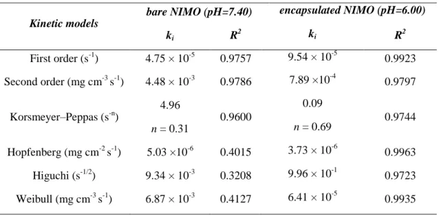
The corresponding half-life value (t1/2 = 2.52 inNIMO/ C6-chitosan compositeat pH=6.00) is near five times larger than the half-life of bare drug sample (t1/2 = 0.44 h for NIMO at pH=7.40). The kinetic constants, release indexes and correlation coefficients (R2) values, which are summarised in Table 1 indicate what processes are controlled by the suggested release mechanism. Based on the values of correlation coefficients release of the NIMO from the polymer network was well described by the Weibull and Hopfenberg equations, while the drug release from concentrated solutions agreed with the second-order reaction kinetic model.
The observed release index value (n = 0.69for NIMO/ C6-chitosan compositeat pH=6.00) suggest the existing ofan anomalous non Fickian transport where combination of diffusional and relaxational transport results the anomalous process. Despite this fair mathematical result, due to the extremely small diameter of the carrier particles, meaningful physical meaning of the last mentioned transport phenomenon is highly questionable.
Finally, the release kinetic of drug from unmodified chit was also measured in order to verify the adequate effect of the polymer modification on drug release properties. The difference of release profile of C6-chit and unmodified chit at normal tissue pH (=7.4) was obvious (Fig.
8B). At this pH the unmodified chit particles were partially opened (see Fig. 4A.) and thus
~50% of encapsulated NIMO was released. However, the release was negligible in the case of C6- chit particles due to the modified pH-responsive properties and the closed, undissolved polymer structure at this pH (see Fig. 4A.).
17 Thus, it can be concluded, that the adequate pH sensitive properties of the C6-chit NPs were also expressed in the drug release behaviors and according to the in vitro experimental results the prepared NPs could be used for the treatment of ischemic injury.
Table 1. Interpretation of the release experiments using different kinetic models
Kinetic models
bare NIMO (pH=7.40) encapsulated NIMO (pH=6.00)
ki R2 ki R2
First order (s-1) 4.75 × 10⁻⁵ 0.9757 9.54 × 10⁻⁵ 0.9923 Second order (mg cm-3 s-1) 4.48 × 10⁻³ 0.9786 7.89 ×10⁻⁴ 0.9797
Korsmeyer–Peppas (s-n)
4.96
0.9600
0.09
0.9744
n = 0.31 n = 0.69
Hopfenberg (mg cm-2 s-1) 5.03 ×10⁻⁶ 0.4015 3.73 × 10⁻⁶ 0.9963 Higuchi (s-1/2) 9.34 × 10⁻³ 0.3208 9.96 × 10⁻¹ 0.9723 Weibull (mg cm-3 s-1) 6.87 × 10⁻³ 0.4127 6.41 × 10⁻⁵ 0.9935 4. Conclusion
Based on significant pH differences between seriously ischemic (pH<6.5) and normal (pH=7.1-7.4) nervous tissue, the development of pH-sensitive carriers that can respond acidic pH could be useful in targeted pH- mediated drug delivery. In this paper we designed and constructed intelligent, tissue acidosis- responsive drug delivery NPs (d=5.2 ± 1.1 nm).
According to the in vitro experiments the developed hydrophobized Chit NPs with C6-side chains were released the Ca2+ channel blocker NIMO drug at the pH of ischemic tissue (=6.0), while at normal pH (=7.4) the drug molecules was remained closed in polymer shell.
At pH=6.0 93.6% (0.131 mg/ml) of the available NIMO was released into the environment, while at pH= 7.4 the amount of drug was negligible.
Beside the relevant pH- dependent drug release properties, it was also presented, that the modified Chit samples possess changed wetting properties: increasing hydrophobicity of the polymer resulted decreasing water content (C6-chit.: 74.2 wt.% and C12-chit.: 47.1 wt.%) compared to the initial Chit (78.6 wt.%). According to the TEM and XRD measurements, the bare NIMO molecules were formed micron- sized needle like crystals with well- developed crystal phase, however, in the presence of C6-chit spherical NIMO NPs were formed with lower crystallinity. This lower crystallinity of the poorly water soluble NIMO is advantageous, because the solubility of the drug molecules depends on the crystallinity and