O F C A R C I N O G E N E S I S B Y THE A M I N O A Z O D Y E S * ELIZABETH C. MILLER AND JAMES A. MILLER
McArdle Memorial Laboratory, Medical School, University of Wisconsin, Madison, Wisconsin
I. Introduction 287 II. Structural Requirements for Carcinogenicity 288
III. Metabolism 292 A. Over-all Metabolism 293
B. N-Demethylation and N-Methylation 293 C. Reduction of the Azo Linkage 295 D. Formation of Protein-Bound Dye 296 IV. Biochemical Alterations of the Liver following Ingestion of the Dyes 301
V. On the Mechanism of Carcinogenesis 302
References 305
I. Introduction
One aspect of the resistance of tissues to chemicals is the adaptive change of certain cells to variants upon which the drug has little effect.
One such type of variant appears to be the tumor cell, which, through its additional capacity for relatively unlimited growth, may lead to the embarrassment and eventual death of the host. It seems to us that a valuable approach to the therapy and prophylaxis of cancer is the deter
mination of the key alterations leading to the induction of these tumor cells. Such investigations have been pursued ever since the means of inducing tumors in experimental animals were first discovered. But the last 15 years, with their rapid developments in biochemistry, have seen a gratifying surge of advances. Many of these investigations have been directed toward an elucidation of the metabolism of the chemical carci
nogens and toward determining the biochemical differences between normal tissues, tissues under the influence of carcinogens, and tumor tissues. Often it has been difficult, if not impossible, to determine the
* The work of the authors in this field has been supported by grants from the National Cancer Institute, Public Health Service, the American Cancer Society, the Jane Coffin Childs Memorial Fund for Medical Research, and the Alexander and Margaret Stewart Trust Fund.
287
relevance of the findings to the problem and to determine whether the differences between normal, "precancerous," and tumor tissue are cause or effect. In spite of these problems, our knowledge of the changes that occur during carcinogenesis has increased considerably in the past few years and there are hints about which alterations may be of critical importance.
Since it seems reasonable to expect that most of the basic reactions involved in carcinogenesis will be similar regardless of the carcinogen involved, we have chosen to study the induction of liver tumors by 4-dimethylaminoazobenzene (DAB) and related compounds. In this
discussion we will first review some of the observations made on this carcinogenic process and then attempt to extrapolate these findings and the resulting ideas to other systems.
This carcinogenic system appears to have a number of advantages for studies on mechanism of action. When DAB is fed to rats at a level of 0.06 per cent in an appropriate diet for about four months, 50 to 100 per cent of the animals will develop one or more hepatic tumors within six months from the start of dye feeding. Tumor induction is specific for the liver. The incidence and rate of appearance of the tumors can be either increased or decreased by feeding DAB in appropriately altered diets, by varying the level of dye, or by using derivatives of DAB with greater or lesser carcinogenic activities. The dye is relatively specific for the rat; it induces tumors only slowly in the mouse and not at all in six other species that have been tested. These factors, together with the size and accessibility of the liver, permit correlations to be made between biochemical changes occurring in the liver at early pe
riods and the probability of finding gross tumors at a later date. In addition, the liver has the advantage of being better characterized' biochemically than most other tissues of the animal body.
II. Structural requirements for carcinogenicity
In order to elucidate the structural features required for activity, a rather large number of compounds related to DAB have been assayed for carcinogenicity. From these studies it appears that in order to have appreciable activity a compound of this series must have or be able
to acquire in vivo (a) an azo linkage joining two aromatic rings, (b) at least one N-methyl group, together with the proper second substi- tuent on the amino group, and (c) either no ring substitutent or only certain ones, preferably in the 3' position if other than fluorine (Miller and Miller, 1953).
The requirement for at least one IV-methyl group for activity (Miller and Baumann, 1945; Miller and Miller, 1948; Sugiura et al., 1945), the reversible demethylation of DAB to 4-monomethylaminoa- zobenzene (MAB) in vivo (Miller et al., 1945) and their equal carci
nogenic activities (Miller and Baumann, 1945; Sugiura et al., 1945), and the largely irreversible demethylation of MAB to the noncarci- nogenic dye 4-aminoazobenzene (AB) (Miller and Miller, 1952; Miller et al., 1945) suggest that MAB is a key intermediate in the carcinogenic process either directly or through conversion to DAB or another meta
bolite. This idea is further substantiated by the inactivity of N-benzyl- methyl- and 2V-/?-hydroxyethylmethyl-AB, both of which are dealkylated at extremely slow rates in vivo compared with the rapid dealkylation of the active dyes (Miller and Miller, 1948). Other evidence comes from studies on the in vivo methylation of AB and its 4'-fluoro and 3'-methyl derivatives (Miller and Miller, 1952). Although only traces could be detected in the livers of rats fed AB, appreciable amounts of the IV-methyl derivatives (chiefly monomethyl) were found in the livers of rats fed 4'-fluoro- and 3'-methyl-AB. Unlike AB, each of these dyes has a readily demonstrable, though low, carcinogenic activity.
Information on the structure of the carcinogenically active deriv
ative of DAB has also been obtained from the studies on various ring- substituted derivatives (Table 1). Thus, the inactivity of the 2-, 2'-, 3'-, and 4'-hydroxy derivatives largely eliminates these compounds and their major metabolites from consideration (Miller and Miller, 1948;
Miller et al., 1949). This is of importance since at least one compound (4'-hydroxy-DAB) is a known metabolite and the others are suspect (Mueller and Miller, 1948). Synthesis of the 3-hydroxy derivative has not yet been achieved, but assay of this compound is desirable in view of the implication of 2-amino-l-naphthol as the carcinogenic metabolite of ß-naphthylamine (Bonser et al., 1951, 1952) and the weak, though definite, carcinogenic activity of 3,3'-dihydroxy-benzidine (Baker, 1953).
Walpole et al. (1952) have suggested, though not demonstrated, that o-hydroxylation may be of importance in the activity of the aminobi- phenyls. However, incomplete tests of 3-hydroxy-4-aminobiphenyl and 3-hydroxy-4-acetylaminobiphenyl in our laboratory indicate that they
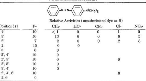
. N ( C H3)2
Relative Activities (unsubstituted dye = 6 )
Position (s) Υ CH3- HO- CFa- α N02-
4' ΙΟ < 1 0 0 ϊ 0
3' 10 10 0 0 6 5
2' 7 2 0 0 2 3
2 10 0 0
3 6 0
2', 4' 10 0
2', 5' 10 0 0
3', 5' 10 0
3', 4' 10 0
2', 4', 6' 10 0
are much less carcinogenic toward the mammary gland of the rat than 4-acetylaminobiphenyl, if they have any activity at all (Miller et al, 1954).
All the mono- and polyfluoro derivatives of DAB with the substi- tuents on the prime ring have proved more active than the parent compound (Miller et al., 1949, 1953, 1954). These very high activities provide strong positive evidence that neither the 4' position nor either of the two pairs of equivalent positions, (3',5') and (2',6'), can be directly involved in the carcinogenic process. Thus, if reactions- essen
tial to the carcinogenic process occurred at these sites in vivo, the si
multaneous substitution of fluorine in each member of a pair of equiv
alent positions should have prevented, or at least strongly inhibited, the formation of tumors. The possibility that all or most of the prime positions could function equally well in a hypothetical carcinogenic reaction involving the ring seems remote. On the other hand, the data obtained to date on the substitution of fluorine in the other ring, while less complete, do not rule out reactions at these sites as being important in carcinogenesis. Thus, although 2-fluoro-DAB is at least twice as active as DAB (Miller et al, 1949) 2,6-difluoro-DAB, 2,6,3', 5'-tetrafluoro-DAB, and 2,6,2',4',6'-pentafluoro-DAB were all inactive in our tests (Miller et al, 1954). This indicates that at least one mem
ber of the pair (2,6) must be unsubstituted for the compound to be active. Whether this requirement is dependent on "enzyme fit" or
TABLE 1. The Relative Carcinogenicities of Various Ring-Substituted Derivatives of 4-Dimethylaminoazobenzene
chemical substitution is not known. 3-Fluoro-DAB has approximately the same activity as DAB (Miller et al.y 1954); the synthesis of 3-5-
difluoro-DAB has not yet been achieved.
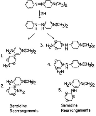
The activities of these fluoro dyes has also eliminated both possible benzidine rearrangement products and two of the three possible semi- dine rearrangement products from consideration as carcinogenic inter
mediates (Fig. 1) (Miller et al, 1953). The inactivity of 2,6-difluoro-
1
2HBenzidine Semidine Rearrangements Rearrangements Ο = positions blocked in ^»e'-trifluoro-DAB
FIG. 1. The possible rearrangement products of 4-dimethylaminohydrazobenzene.
DAB, however, leaves open the possibility that 2-amino-5-dimethylami- nodiphenylamine could be involved in the process, although there is not other supporting evidence and data on the protein-bound dyes
(vide infra) suggest that the carcinogenic derivative is an azo dye.
However, this compound is now being synthesized for a direct test.
III. M e t a b o l i s m
Studies on the metabolism of chemical carcinogens should ulti
mately lead not only to a knowledge of the effects of the tissue on the carcinogen, but also to the discovery of the direct biochemical attack of the carcinogen and its metabolites on the target tissue. However, the decisive metabolic processes leading to the formation of tumor cells must occur relatively early in the experimental period, but gross tumors cannot be found until at least three months after the beginning of the dye-feeding period. Thus, assessment of the importance of specific re
actions can be made only by correlating the occurrence and intensity of various reactions in the early stages with the time of appearance
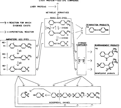
LIVER P R O T E I N — A Z O DYE COMPOUNDS
LIVER PROTEINS
- > = REACTION FOR WHJCH EVIDENCE E X I S T S
- > = HYPOTHETICAL REACTION
AMPHOTERIC AZQ D Y E S
METABOLIC DERIVATIVES T
B A S I C A Z O D Y E S
l = / N
[C H3] - > 1 I; - > H C H O
J j >HCHO
DE AMINATION PRODUCTS
* * < X >
COMPOUND s
—>
—>
—>
REARRANGEMENT PRODUCTS
eg CH3
4^
[demethyloted products
/ < * 3 Ί
t H 3 MONOPHENYL A M I N E S
FIG. 2. The present knowledge of the metabolism of D A B and its derivatives in the rat.
and the incidence of gross tumors. This necessitates comprehensive studies on each phase of metabolism, so that the critical reactions can be pointed out both by positive correlative evidence for their impor
tance and by strong evidence against the importance of other reaction mechanisms.
A. OVER-ALL METABOLISM
Studies on the metabolites excreted in the urine and on the dyes occurring in the liver and other tissues have demonstrated that at least four types of reactions are involved in the metabolism of these dyes by the rat—(a) IV-demethylation and N-methylation, (b) reductive cleav
age of the azo linkage, (3) ring hydroxylation, especially in the 4' posi
tion, and (d) binding to certain liver proteins (Fig. 2) (Miller and Miller, 1953). The first three reactions account for the metabolism of the major share of the ingested dye. The sequence in which they occur is not definitely established and probably differs from molecule to mole
cule, but N-demethylation appears to be the major first reaction (Miller et al., 1945). The fourth reaction, binding to liver proteins of an azo dye metabolite, probably occurs with only a small share of the ingested dye, but it is this reaction which appears to be of importance in the carcinogenic reaction.
B. N-DEMETHYLATION AND N-METHYLATION
N-Demethylation is one of the major reactions affecting both DAB and MAB, as evidenced by the presence of both free MAB and AB in the liver, AB in the red blood cells, and high levels of conjugated p-phenylenediamine in the urine (Miller and Miller, 1946; Miller et al., 1945; Stevenson, et al., 1942). Likewise N-methylation is apparently a major reaction affecting MAB, since the levels of free DAB and MAB in the liver are essentially the same when either dye is fed. N-Methyl- ation of AB, on the other hand, occurs to a very limited extent (Miller and Miller, 1952). Dealkylation also occurs when azo dyes bearing N-ethyl groups are administered, but N-benzyl and Ν-β-hydroxyethyl substituents are metabolized very slowly (Kensler et al., 1950; Miller and Miller, 1948).
While small amounts of transmethylation of the methyl groups of DAB or MAB to other compounds cannot be excluded, the major meta
bolic pathway appears to be an oxidative one. Thus, when DAB, MAB, or certain of their ring-methyl derivatives labeled with C1 4 in the N-methyl groups were fed to rats, C1 402 was rapidly expired; 70 to
80 per cent of the dose could be accounted for within 48 hours (Bois- sonnas et al, 1949; MacDonald et al, 1953; Miller et al, 1952). Anal
yses of the tissues indicated that small amounts of radioactivity were incorporated into the ß-carbon of serine and the N-methyl groups of choline (MacDonald et al, 1953; Miller et al, 1952), in a manner sim
ilar to that obtained after the administration of formaldehyde-C14 (Jons- son and Mosher, 1950; Sakami and Welch, 1950; Siegel and Lafaye, 1950; du Vigneaud et al, 1951). Even when DAB-N-methyl-C14 was fed for up to 13 weeks in a choline-free, methionine-low diet, only 1.4 per cent of the methyl groups of the choline were derived from those of the dye (MacDonald et al, 1954). In a similar type of experiment du Vigneaud and associates (1941) found that at least 80 per cent of the methyl groups of choline came from labeled methionine. The source of the methyl groups for the methylation of MAB has not been ascer
tained, but it seems logical to expect that this reaction is a trans
methylation.
For a study of the N-demethylation reaction with rat liver homo- genates 3-methyl-MAB has been used as substrate (Mueller and Miller, 1953). This dye was chosen since in vitro demethylation of the second
ary aminoazo dyes occurs much more rapidly than with the tertiary aminoazo dyes. Further, the 3-methyl group almost completely hinders the reductive cleavage of the azo linkage so that the demethylation reaction can be studied independently of other reactions. This system requires oxygen, triphosphopyridine nucleotide, diphosphopyidine nu
cleotide, adenosine triphosphate, and magnesium ions for maximum ac
tivity. When the reaction is carried out in the presence of semicarbazide, formaldehyde and 3-methyl-AB are recovered in amounts accounting stoichiometrically for the 3-methyl-MAB metabolized. This result, to
gether with the in vivo studies on the C1 4-labedel dyes, indicates that an IV-methylol derivative is an intermediate in the N-demethylation of DAB and its derivatives.
Recent studies have shown that the activity of the demethylation system is greater in mouse than in rat liver and that the activity with eiher species is dependent on the diet (Brown et al, 1954a). The other tissues of either species have little or no activity (Mueller and Miller, 1954). This is also true of the induced liver tumors in the rat. The activity of mouse liver can be simulated two to three times by the in
corporation of 0.05 to 0.2 per cent of certain pure cyclic peroxides (ascaridole, 9,10-dimethyl-l,2-benzanthracene photooxide, or pinane hydroperoxide) or of certain poly cyclic aromatic hydrocarbons (pyrene,
phenanthrene, 20-methylcholanthrene, 3,4-benzpyrene, or 1,2-ben- zanthracene) in a synthetic diet for one week prior to assay. The type of compounds capable of similarly activating rat liver have been studied in less detail, but a two- to threefold activation can be routinely ob
tained 24 hours after intraperitoneal injection of 500 ^g. of 20-methyl- cholanthrene (Miller and Miller, 1954). With the livers of both species enzymes in both the particulate and supernatant fractions are needed for appreciable activity, and it is the enzyme system in the particulate fractions that is activated by these hydrocarbons and peroxides.
C. REDUCTION OF THE AZO LINKAGE
The findings that rats fed DAB, MAB, or AB excreted conjugated p-phenylenediamine and p-aminophenol in the urine in amounts equiv
alent to about one-half the dye fed demonstrated that reduction of the azo linkage is a major metabolic reaction (Miller and Miller, 1946).
The dyes are also rapidly reduced in vitro. With either homogenate or slice techniques kidney preparations have only one-third the activity of those from liver, and other tissues, including the induced liver tumors, are inactive (Kensler, 1948; Mueller and Miller, 1949). Various ring- substituted derivatives of DAB are also reductively cleaved, but there is no correlation between the rate of reduction and carcinogenic activity
(Kensler and Chu, 1950; Mueller and Miller, 1954). In the case of DAB the dye destroyed can be accounted for stoichiometrically as ]V,IV-dimethyl-p-phenylene diamine and aniline (Mueller and Miller, 1949). With homogenates the maximum rate of reduction is obtained in an anaerobic system fortified with diphosphopyridine nucleotide, triphosphopyridine nucleotide, magnesium ions, and an oxidizable sub
strate (Mueller and Miller, 1949). A requirement for flavin-adenine di- nucleotide can also be demonstrated with carbon dioxide-treated homo
genates (Mueller and Miller, 1950), and evidence of a riboflavin co
enzyme has also been obtained in experiments with liver slices (Kens
ler, 1948, 1949). Since the amines formed by reductive cleavage of the azo dyes have little or no carcinogenic activity (Kinosita, 1940; Miller and Baumann, 1945; Miller and Miller, 1948; White et al, 1948), a major share of the protective action of riboflavin (Miller and Miller, 1953) against carcinogenesis by DAB probably results from its par
ticipation in this reaction.
As with the demethylation system enzymes in both the particulate and supernatant fractions of liver are needed for activity (Mueller and
Miller, 1949), and the activity of the system in vivo can be markedly stimulated by administration of certain peroxides or hydrocarbons (Miller and Miller, 1954). However, at least in the case of oral or intra- perintoneal administration of 20-methylcholanthrene, activation of the reduction system requires larger amounts of hydrocarbon and longer latent periods than does the activation of the demethylation system.
The activities of both the reduction and demethylation systems are markedly reduced by the feeding of carcinogenic dyes such as DAB or its 3'-methyl derivative (Miller et al, 1952). When low levels of 20-methylcholanthrene or certain other polycyclic hydrocarbons are fed simultaneously with 3'-methyl-DAB, the activities of the enzyme sys
tems are maintained at the level found in rats on the basal dye-free diet, and liver tumor induction is strongly inhibited. Apparently, the livers of hydrocarbon-treated animals destroy the dye more rapidly than those of rats fed dye alone, so that the amount of dye available for carcinogenic reactions is reduced. This situation would appear to be an example of how resistance to a drug can be increased by the adminis
tration of a second drug that stimulates the metabolism of the first to inactive products.
D. FORMATION OF PROTEIN-BOUND DYE
When DAB is fed, protein-bound derivatives can be detected in the liver within a few days; the level rises to a maximum at about four weeks and then gradually falls to a half-maximal level by the time gross tumors appear at three to four months (Miller and Miller, 1947). While this reaction is quantitatively less important than the reduction or de
methylation reactions, qualitatively it appears to be much more signifiT cant. Contrary to the findings with the reactions just discussed, correla
tions can be made between the presence and amount of bound dye and the probability of tumor formation under various conditions; these correlations indicate that the bound dye may be intimately involved in the carcinogenic reaction induced by the azo dyes (Miller and Miller,
1947, 1952; Miller et al, 1949).
The bound dye cannot be released from the protein by extraction with boiling solvents or with hot trichloroacetic acid (which removes the nucleic acids) or by dialysis, but it is liberated by alkaline or tryptic digestion at rates that approximate the rates of hydrolysis of the protein.
Of the dye liberated by alkaline hydrolysis, about 10 per cent is a mix
ture of MAB and AB, and the remainder is strongly polar and extract- able from the alkaline hydrolyzate only by a polar solvent mixture such
as ethyl ether-ethanol (Miller and Miller, 1947). When any of the ring- methyl derivatives of DAB are fed, a comparable picture is obtained except that the level of bound dye and the time at which the maximum is reached differ with the various dyes (Miller et al., 1949).
The spectra of the polar dyes from DAB and its 2-, 2'-, 3'-, and 4'-methyl derivatives in acid suggest that they are Ν,Ν-dialkyl-substi- tuted aminoazo dyes (Miller et al., 1948). The spectra of the polar dyes from 3-methyl-MAB and 3-methyl-DAB, on the other hand, correspond to that of the monomethyl dye. Since the 3-methyl group strongly hin
ders the methylation of 3-methyl-MAB either in vivo or in chemical methylations (Miller and Miller, 1948, 1954), very little dimethyl dye, compared with the amount of monomethyl dye, may be available for reaction with protein in this case. This suggests that the bound dyes may be mixtures of derivatives of the dimethyl and monomethyl dyes and that in most cases the dimethyl derivatives predominate.
Reduction of the polar dyes from DAB or its ring-methyl derivatives yields aniline or the appropriate toluidine and an unidentified polar amine (Miller et al., 1949). Thus the dyes must be bound to the protein at some point on the "diamine" ring or to a substituent on this ring.
We have favored the N-methyl groups or, more likely, a metabolic de
rivative such as the N-methylol as the site of binding. Thus, only N- methyl aminoazo dyes or those capable of being N-methylated in vivo form appreciable amounts of bound dye. N-methylol derivatives are known to react readily with compounds possessing reactive hydrogens;
the Mannich bases thus formed should be alkali-stable if a —N-CH2-C—
ι
grouping is formed (as in the case of a reactive =CH— of tyrosine or
I I
histidine) and alkali-labile when a —N-CH2-N— grouping is formed
I
(e.g., by reaction with a —NH of an amide or guanido group) (Fraen- kel-Conrat and Olcott, 1948). This type of reaction could, then, explain the liberation of both polar and nonpolar dyes, although it is also pos
sible that the MAB and AB are bound through amide linkages. In the test tube protein-bound derivatives of AB and MAB can be made by reaction between the dye, formaldehyde, and any of a number of native or denatured proteins, presumably through the intermediate formation of IV-hydroxmethyl dyes (Miller and Miller, 1954). Hydrolysis of these bound dyes yields polar and nonpolar fractions very similar to those
obtained on hydrolysis of the liver proteins from dye-fed rats. On the other hand, Nye and Luck (1953) have suggested on the basis of spec
troscopic evidence alone the presence of a substituent in the 2-position.
Although further data are needed, it is interesting that the necessity for a reaction at the 2-position would explain the inactivity of the dyes with fluoro substituents in the 2 and 6 positions. The possibility that reactions at two sites are involved must also be considered.
The properties of crude and partially purified preparations of the polar dye liberated by alkaline hydrolysis of liver protein from rats fed 3'-methyl-DAB suggest the presence of amino acid residues (Brown et al., 1954b). Thus, the major share of the dye is insoluble between pH 2 and pH 9 and soluble outside this range and is precipitable by alkaline mercuric acetate. These preparations are strongly contaminated by ninhydrin-reactive material and difficultly freed from it. On acetyla
tion the dye loses much of its polar property. The acetylated product has essentially the same absorption spectrum in acid, in particular the high maximum at 525 ταμ, as the parent dye; apparently the 4-amino group of the dye is not attacked and there is an acetylatable amino group elsewhere on the molecule. Furthermore, certain synthetic amino acid derivatives of 3'-methyl-MAB (y-(m-tolylazo-p-methylanilino)-«-amino- butyric acid and less definitely characterized dyes obtained by the reaction of 3'-methyl-MAB, formaldehyde, and acetylated tryptophane or tyrosine and subsequent hydrolysis) have similar Rf's on chromatog
raphy to those of certain fractions of the polar dye from rat liver. On the other hand, one fraction, obtained by chromatography on silica and puri
fied by repeated chromatography on paper, contained less than 0.1 mole of ninhydrin-reactive material per mole of dye. However, since this fraction accounted for only 5 to 10 per cent, the results cannot be extra
polated to the bulk of the dye.
The evidence that the protein-bound dyes play a causal role in the induction of tumors by the aminoazo dyes consists of a series of correla
tions between the presence or amount of bound dye after various periods of dye-feeding and the probability of tumor formation (Miller and Miller, 1947, 1952; Miller et al., 1949). Thus, the azo dyes are specific carcinogens for the liver of the rat, and the protein-bound dyes have been found only in the liver and, to a small extent, in the blood plasma of dye-fed rats.
Mice develop liver tumors only slowly, and the livers of the other species tested (chickens, cotton rats, hamsters, guinea pigs, rabbits, and chip
munks ) are resistant; likewise, the livers of mice contain only a low level of bound dye while none can be detected in the livers of the other
species. Two dietary conditions are known that cause a lowering in the level of bound dye in the liver and a reduction in tumor incidence. These are the feeding of high levels of dietary riboflavin (Miller and Miller, 1953) or low levels of certain polycyclic hydrocarbons (Richardson et ah, 1952). In both cases the rats receiving the inhibitory diet consume more diet and dye than the control animals, but they also maintain higher hepatic levels of the enzymes responsible for the metabolism of the dye to inactive products. Thus, the lower levels of bound dye and the de
creased tumor incidence in these cases may be related to a decrease in the amount of dye available in the liver cells. The level of bound dye in the livers of rats fed different levels of DAB can also be correlated with the tumor incidences obtained with these diets.
Further correlations come from studies on the levels of bound dye and the tumor incidences of rats fed various derivatives of DAB. With the exception of 3'-methyl- and 4'-fluoro-AB (Miller and Miller, 1952), only dyes with at least one N-methyl group are bound to the liver protein to an appreciable extent; likewise, with these same exceptions, at least one iV-methyl group is needed for significant carcinogenic activity. These dyes
j N- N3 ' - M e - D A B (10-12)
WEEKS
FIG. 3. The levels of protein-bound dye in the livers of rats fed D A B or certain of its ring-methyl derivatives.
further emphasize the importance of the N-methyl groups, since each is unusual in being N-methylated to an appreciable extent in vivo. An
other correlation was found on analysis of the livers of rats fed the various ring-methyl derivatives of DAB (Miller et al., 1949). These isomers vary in carcinogenic activity from 0 to 12, and there is an approximately inverse correlation between the time of dye feeding before the maximum levels of bound dye are reached in the liver and the carcinogenicity of the derivative. With DAB and the 2'-, 3'-, and 4'-methyl derivatives there is also a correlation with the level of bound dye during the first three to four weeks of dye feeding (Fig. 3 ) . With the 2- and 3-methyl derivatives, however, the levels of bound dye are higher than anticipated on the basis of their low carcinogenic activities (Fig. 4 ) . This can be explained only in part by the lower rates of turnover of the dye-protein combinations with these dyes (Miller and Miller, 1952).
3-Me-DABJ_<l]__ =_-^*r
/ χ^ ' 2- M e - D A B (0)
WEEKS
FIG. 4. The levels of protein-bound dye in the livers of rats fed DAB, 2-methyl- DAB, or 3-methyl-MAB.
An intriguing observation is that the protein-bound dyes cannot be detected in the azo dye-induced tumors even when the dyes are fed continuously (Miller and Miller, 1947; Price et al., 1949). This difference between liver and tumor appears to be an intrinsic property of the tissues,
although the experiments and interpretations are complicated by the ex
clusively arterial blood supply of the liver tumors as compared to the portal-arterial supply of the liver (Breedis and Young, 1949). Likewise, the soluble proteins of liver tumors contain only about one-tenth as much of a slowly moving electrophoretic fraction, designated h, as the soluble proteins of liver (Sorof and Cohen, 1951). Since it is this h protein that carries 80 to 90 per cent of the bound dye in the soluble protein fraction (Sorof et al., 1951), it is provocative to consider that the reaction of the protein with dye was instrumental in its loss from the induced liver tumor cells. It should be noted that, though the major share (50 to 60 per cent) of the bound dye is combined with the soluble proteins, it is also present in all the particulate fractions (Price et al., 1948, 1949, 1950). Thus, the combination of the h protein with dye and its subsequent loss from the cells may be duplicated with other proteins in other cell fractions. The character of the resulting cells would depend on the nature of the pro
teins lost or altered. More information is needed to determine the relative importance of the protein-dye combinations in the various liver cell fractions.
IV. Biochemical alterations of the liver following ingestion of the dyes
One approach to determining the changes associated with the induc
tion of tumors is a comparison of the biochemical composition of normal liver, livers from rats fed carcinogenic and noncarcinogenic dyes, and the tumors induced by the dyes. These studies are complicated by the alter
ations in the histology of the liver during dye feeding and by the con
troversy concerning the cells of origin of the tumors. The finding of Price and associates (1952) that the disappearance of hyperplastic bile duct
like cells, especially marked after three to four weeks of feeding of 3'-methyl-DAB, was apparently due to their conversion to parenchyma
like cells and that most or all of the induced tumors had a common origin in areas of cholangiofibrosis has simplified this problem. Yet, it must be recognized that analyses on the livers of dye-fed rats represent averages for cells of different types and sizes, the relative proportions of which differ with the dye fed and the time of feeding. In spite of these diffi
culties, such analyses, especially when combined with histological studies, have proved useful in elucidating the pattern of changes accompanying carcinogenesis by the dyes.
In a comparison of the livers of rats fed DAB, its ring-methyl deriva-
tives, and its 4'-fluoro derivative, Price et al. (1948,1949,1950) and Potter et al. (1950) showed that the ingestion of the carcinogenic dyes resulted in large alterations from normal in the contents of nucleic acids, ribo
flavin, and protein in certain of the liver cell fractions, while little change was usually found when the noncarcinogenic dyes were fed. Other studies, particularly with respect to the mitochondria, have been made with certain of these dyes by Schneider et al. (1953), Striebich et al.
(1953) and Allard et al. (1952, 1953). The general picture that has emerged is that in the livers of rats fed carcinogenic dyes the amount of deoxypentosenucleic acid per cell nucleus remains fairly constant, while the increased cellular density, particularly after one to two months of dye feeding, causes an increased content of this nucleic acid per gram of tissue. The number of mitochondria per cell and per gram of tissue decreases markedly, but the amounts of pentosenucleic acid, protein, and of certain enzymes per mitochondrion remain essentially normal. The changes from normal in the livers of rats fed the carcinogenic dyes are generally even more exaggerated in the induced tumors.
These studies have also pointed to the remarkable doubling in the number of mitochondria in the liver cells of rats fed 2-methyl-DAB.
These mitochondria contain essentially normal amounts of protein, ribo
flavin, and succinoxidase, but reduced amounts of pentosenucleic acid, cytochrome c reductase, uricase, octanoxidase, nucleases, and oxalacetic oxidase.
V . O n the mechanism of carcinogenesis
In the Introduction we posed the question of how normal cells are converted to tumor cells by the aminoazo dyes. The correlations between the presence or amount of bound dye under various conditions and the probability of tumor formation have led us to suggest that the combina
tion between a derivative of the dye fed (possibly a N-hydroxymethyl dye) and certain liver proteins may be one of the initial steps in this carcinogenic reaction. The further observation that the soluble protein that combines quite specifically with the dye is present in very low amounts in the induced tumors suggests that the binding with dye mediates the loss of this protein from the cell. Thus, if the dye prevents or reduces the synthesis of the proteins, cells would eventually be formed with less and finally none of the proteins originally attacked by the dye.
In the case of a self-duplicating protein the dye can be conceived as interfering with the replication of the protein to which it is attached;
with other proteins, if the dye is bound during the synthesis of the protein, the protein-dye combination might dissociate less readily than the normal protein from the site of synthesis. One needs, then, only to make the additional assumption that the loss of the soluble protein or similar losses of other proteins binding with the dye in other parts of the cell so alter the metabolism of the cell that it can grow and divide independently of the normal controlling factors.
Although in most cases the level of bound dye can be correlated with the expected tumor incidence, the levels of bound dye in the livers of rats fed 2-methyl-DAB or 3-methyl-MAB are greater than would be expected on the basis of their low activities, even when allowance is made for the lower rates of turnover of the protein-bound dyes. Investi
gations with 2-methyl-DAB have shown that, as with the other carcino
genic and noncarcinogenic dyes studied, most of the bound dye in the soluble protein fraction of the liver is combined with the h proteins. It is possible, however, that the bound dye in the particulate fractions of the livers from rats fed the 2- or 3-methyl dyes is attached to different proteins than when other dyes (especially carcinogenic ones) are admin
istered, and that it is one of these proteins that is critical to the carcin
ogenic process. Another possible explanation is that the combination of the dye with certain proteins is a necessary, but not sufficient, step in the induction of tumors. Thus, if the protein bearing the dye still has enzymatic function, the fit between the protein and its substrate or coenzymes may depend on the structure of the dye. Or, a second reaction (covalent or hydrogen bonding) between the dye and the same or a second cellular component necessary for carcinogenesis might be hin
dered in some dyes capable of undergoing the initial binding reaction.
In this respect it is pertinent that, while high levels of bound dye have been encountered in these two situations, which do not yield high tumor incidences, the converse has not been observed. In each case where tumors are induced in signficant numbers high levels of protein- bound dye have been found in the liver.
Although the azo dyes are instrumental in the induction of these liver tumors, they apparently play no role in the persistence and growth of the tumor cells. Gross tumors continue to grow and kill the hosts whether or not the rats are maintained on diets containing the dye.
Likewise, tumors frequently are found in the livers of rats that were grossly tumor-free at the end of dye feeding. No bound dye can be detected in the tumors at any time, and the free dye disappears soon after transfer to a dye-free diet.
The hypothesis that tumors are induced by a process that involves the binding of a carcinogen with one or more proteins of the suscep
tible tissues and the ultimate loss of these proteins from some cells can also be extended to carcinogenesis by other chemicals. Thus, protein- bound derivatives of certain polycyclic hydrocarbons (3,4-benzpyrene and 1,2,5,6-dibenzanthracene (Heidelberger and Weiss, 1951; Miller, 1951; Weist and Heidelberger, 1953a, b) and of 2-acetylaminofluorene (Miller and Miller, 1952; Weisburger et al., 1953) have been found in tissues susceptible to their action. In the case of the hydrocarbons cor
relations have been found between the amounts of protein-bound deriva
tives under various conditions or in different tissues and the likelihood of tumor formation. The bound derivatives of 2-acetylaminofluorene are widely distributed in the tissues of rats ingesting the carcinogen (Miller and Miller, 1954). Since 2-acetylaminofluorene induces tumors in a wide variety of tissues whose relative susceptibilities depend on the experi
mental conditions, it is possible that the initial protein-binding reaction of the carcinogen takes place in many tissues (Bielschowsky, 1944;
Cantarow et al., 1948; Engel and Copeland, 1951; Miller et al., 1949;
Wilson et al., 1941). Other factors must then be limiting in the tissues not giving rise to tumors.
In a wider sense the theory that tumors are induced by a process of protein (or enzyme) deletion may apply to other forms of carcinogen
esis. Thus, physical carcinogens such as the radiations from radioactive elements or ultraviolet light may damage in some cells key proteins required for the control of growth but not those essential for growth itself. Carcinogenic viruses could be considered to compete so effectively for cell nutrients that cells deficient in certain proteins would be formed;
some of these cells would presumably retain the ability to grow although lacking certain of the normal controls. From this viewpoint a carcino
genic virus, like carcinogenic chemicals and radiations, would be an
"initiating" but not a "continuing" cause of cancer.
Of course, the protein pattern leading to neoplastic growth need not be the same in the induction of tumors by different carcinogens or even in the induction of various tumors by a given carcinogen. In each case, however, the composition of the resulting cells must be one that permits continued proliferation that is free of at least some of the normal growth control systems. It is also evident that in most of these processes large numbers of cells in the target tissues are damaged by the carcinogenic agent. Some of the damaged cells may die because they lack enzymes vital to life. Others? though deficient in some respects, may survive and
reproduce, but at a rate no greater than that of the more normal cells.
Only a few of the damaged cells may be altered in just the right ways to make them capable of vigorous and relatively uncontrolled growth.
In an appropriate environment these cells could proliferate to yield tumor masses.
References
Allard, C , de Lamirande, G., and Cantero, A. (1952). Can. J. Med. Set. 30, 543.
Allard, C , de Lamirande, G., and Cantero, A. (1953). Can. J. Med. Set. 31, 103.
Baker, R. K. (1953). Cancer Research 13, 137.
Bielschowsky, F. (1944). Brit. J. Exptl. Pathol. 25, 1.
Boissonnas, R. Α., Turner, R. Α., and du Vigneaud, V. (1949). /. Biol. Chem. 180, 1053.
Bonser, G. M., Clayson, D. B., and Juli, J. W. (1951). Lancet 241, 286.
Bonser, G. M., Clayson, D. B., Juli, J. W., and Pyrah, L. N. (1952). Brit. J. Cancer 6, 412.
Breedis, C , and Young, G. (1949). Federation Proc. 8, 351.
Brown, R. R., Miller, J. Α., and Miller, E. C. (1954a). /. Biol. Chem. 209, 211.
Brown, R. R., Miller, J. Α., and Miller, E. C. (1954b). Unpublished.
Cantarow, Α., Stasney, J., and Paschkis, Κ. E. (1948). Cancer Research 8, 412.
du Vigneaud, V., Cohn, Μ., Chandler, J. P., Schenck, J. R., and Simonds, S. (1941).
/. Biol. Chem. 140, 625.
du Vigneaud, V., Verly, W. G. L., Wilson, J. E., Rachele, J. R., Ressler, C , and Kinney, J . M. (1951). /. Am. Chem Soc. 73, 2782.
Engel, R. W., and Copeland, D. H. (1951). Cancer Research 11, 180.
Fraenkel-Conrat, H. L., and Olcott, H. S. (1948). /. Biol. Chem. 174, 827.
Heidelberger, C , and Weiss, S. M. (1951). Cancer Research 11, 885.
Jonsson, S., and Mosher, W. A. (1950). /. Am. Chem. Soc. 72, 3316.
Kensler, C. J . (1948). Cancer 1, 483.
Kensler, C. J. (1949). /. Biol. Chem. 179, 1079.
Kensler, C. J., and Chu., W. C. (1950). Arch. Biochem. 25, 66.
Kensler, C. J., Magill, J . W., and Sugiura, K. (1947). Cancer Research 7, 95.
Kinosita, R. (1940). Yale J. Biol, and Med. 12, 287.
MacDonald, J . C , Miller, J. Α., and Miller, E . C. (1954). Proc. Am. Assoc. Cancer Research 1 ( 2 ) , 30.
MacDonald, J. C , Plescia, A. M., Miller, E. C , and Miller, J. A. (1953). Cancer Research 13, 292.
Miller, E . C. (1951). Cancer Research 11, 100.
Miller, E . C , and Miller, J. A. (1947). Cancer Research 7, 468.
Miller, E . C , and Miller, J. A. (1952). Cancer Research 12, 547.
Miller, E . C , Miller, J. Α., and Brown, R. R. (1952). Cancer Research 12, 282.
Miller, E. C , Miller, J. Α., Sandin, R. Β., and Brown, R. K. (1949). Cancer Research 9, 504.
Miller, E. C , Miller, J. Α., Sapp, R. W., and Weber, G. M. (1949). Cancer Research 9, 336.
Miller, E . C , Plescia, Α. M., Miller, J. Α., and Heidelberger, C. (1952). J. Biol.
Chem. 196, 863.
Miller, J. Α., and Baumann, C. A. (1945). Cancer Research 5, 227.
Miller, J. Α., and Miller, E . C. (1946). Cancer Research 6, 39.
Miller, J. Α., and Miller, E. C. (1948). /. Exptl. Med. 87, 139.
Miller, J. Α., and Miller, E . C. (1952). Cancer Research 12, 283.
Miller, J. Α., and Miller, E . C. (1953). Advances in Cancer Research 1, 339.
Miller, J . Α., and Miller, E . C. (1954). Unpublished.
Miller, J. Α., Miller, E . C , and Baumann, C. A. (1945). Cancer Research 5, 162.
Miller, J. Α., Miller, E . C , and Finger, G. C. (1953). Cancer Research 13, 93.
Miller, J. Α., Miller, E . C., and Finger, G. C. (1954). Unpublished.
Miller, J. Α., Miller, E . C., and Sandin, R. B. (1954). Unpublished.
Miller, J . Α., Sapp, R. W., and Miller, E . C. (1948). /. Am. Chem. Soc. 70, 3458.
Miller, J. Α., Sapp, R. W., and Miller, E . C. (1949). Cancer Research 9, 652.
Mueller, G. C., and Miller, J. A. (1948). /. Biol. Chem. 176, 535.
Mueller, G. C., and Miller, J. A. (1949). J. Biol Chem. 180, 1125.
Mueller, G. C., and Miller, J . A. (1950). /. Biol Chem. 185, 145.
Mueller, G. C., and Miller, J. A. (1953). /. Biol Chem. 202, 579.
Mueller, G. C , and Miller, J. A. (1954). Unpublished.
Nye, W., and Luck, J. M. (1953). Abstract Chicago Meeting of the Division of Bio
logical Chemistry Am. Chem. Soc. 11c.
Potter, V. R., Price, J . M., Miller, E . C , and Miller, J. A. (1950). Cancer Research 10, 28.
Price, J. M., Harman, J. W., Miller, E . C., and Miller, J. A. (1952). Cancer Research 12, 192.
Price, J. M., Miller, E . C., and Miller, J . A. (1948). /. Biol Chem. 173, 345.
Price, J. M., Miller, E . C., Miller, J. Α., and Weber, G. M. (1949). Cancer Research 9, 398.
Price, J. M., Miller, E . C., Miller, J. Α., and Weber, G. M. (1950). Cancer Research 10, 18.
Price, J . M., Miller, J. Α., Miller, E . C., and Weber, G. M. (1949). Cancer Research 9, 96.
Richardson, H. L., Stier, A. R., and Borsos-Nachtnebel, E . (1952). Cancer Research 12, 356.
Sakami, W., and Welch, A. D. (1950). /. Biol Chem. 187, 379.
Schneider, W. C., Hogeboom, G. H., Shelton, E., and Striebich, M. J. (1953). Can
cer Research 13, 285.
Siegel, I., and Lafaye, J. (1950). Proc. Soc. Exptl. Biol. Med. 74, 620.
Sorof, S., and Cohen, P. P. (1951). Cancer Research 11, 378.
Sorof, S., Cohen, P. P., Miller, E . C , and Miller, J. A. (1951). Cancer Research 11, 383.'
Stevenson, E . S., Dobriner, K., and Rhoads, C. P. (1942). Cancer Research 2, 160.
Striebich, M. J., Shelton, E., and Schneider, W. C. (1953). Cancer Research 13, 279.
Sugiura, K., Halter, C. R., Kensler, C. J., and Rhoads, C. P. (1945). Cancer Research 5, 235.
Walpole, A. L., Williams, H. C , and Roberts, D. C. (1952). Brit. J. Ind. Med. 9, 255.
Weisburger, Ε . K., Weisburger, J . H., and Morris, H. P. (1953). Arch. Biochem.
and Biophys. 42, 474.
White, F . R., Eschenbrenner, A. B., and White, J. (1948). Acta Unto. Intern, contra cancrum 6, 75.
Wiest, W. G., and Heidelberger, C. (1953a). Cancer Research 13, 250.
Wiest, W. G., and Heidelberger, C. (1953b). Cancer Research 13, 255.
Wilson, R. H., De Eds, F., and Cox, A. J . (1941). Cancer Research 1, 595.