MicroRNA-155 Deficiency Attenuates Liver Steatosis and Fibrosis without Reducing Inflammation in a Mouse Model of
Steatohepatitis
Timea Csak1☯, Shashi Bala1☯, Dora Lippai2, Karen Kodys1, Donna Catalano1, Arvin Iracheta-Vellve1, Gyongyi Szabo1*
1Department of Medicine, University of Massachusetts Medical School, Worcester, Massachusetts, United States of America,22nd Department of Medicine, Semmelweis University, Budapest, Hungary
☯These authors contributed equally to this work.
*gyongyi.szabo@umassmed.edu
Abstract
Background & Aim
MicroRNAs (miRs) regulate hepatic steatosis, inflammation and fibrosis. Fibrosis is the con- sequence of chronic tissue damage and inflammation. We hypothesized that deficiency of miR-155, a master regulator of inflammation, attenuates steatohepatitis and fibrosis.
Methods
Wild type (WT) and miR-155-deficient (KO) mice were fed methionine-choline-deficient (MCD) or -supplemented (MCS) control diet for 5 weeks. Liver injury, inflammation, steato- sis and fibrosis were assessed.
Results
MCD diet resulted in steatohepatitis and increased miR-155 expression in total liver, hepa- tocytes and Kupffer cells. Steatosis and expression of genes involved in fatty acid metabo- lism were attenuated in miR-155 KO mice after MCD feeding. In contrast, miR-155
deficiency failed to attenuate inflammatory cell infiltration, nuclear factorκbeta (NF-κB) acti- vation and enhanced the expression of the pro-inflammatory cytokines tumor necrosis fac- tor alpha (TNFα) and monocyte chemoattractant protein-1 (MCP1) in MCD diet-fed mice.
We found a significant attenuation of apoptosis (cleaved caspase-3) and reduction in colla- gen andαsmooth muscle actin (αSMA) levels in miR-155 KO mice compared to WTs on MCD diet. In addition, we found attenuation of platelet derived growth factor (PDGF), a pro- fibrotic cytokine; SMAD family member 3 (Smad3), a protein involved in transforming growth factor-β(TGFβ) signal transduction and vimentin, a mesenchymal marker and indirect indi- cator of epithelial-to-mesenchymal transition (EMT) in miR-155 KO mice. Nuclear binding of
OPEN ACCESS
Citation:Csak T, Bala S, Lippai D, Kodys K, Catalano D, Iracheta-Vellve A, et al. (2015) MicroRNA-155 Deficiency Attenuates Liver Steatosis and Fibrosis without Reducing Inflammation in a Mouse Model of Steatohepatitis. PLoS ONE 10(6):
e0129251. doi:10.1371/journal.pone.0129251
Academic Editor:Rafael Aldabe, Centro de Investigación en Medicina Aplicada (CIMA), SPAIN Received:November 12, 2014
Accepted:May 6, 2015 Published:June 4, 2015
Copyright:© 2015 Csak et al. This is an open access article distributed under the terms of the Creative Commons Attribution License, which permits unrestricted use, distribution, and reproduction in any medium, provided the original author and source are credited.
Data Availability Statement:All relevant data are within the paper.
Funding:This work was supported by National Institute on Alcohol Abuse and Alcoholism-AA020744 to G.S. (http://www.niaaa.nih.gov), and National Institute on Alcohol Abuse and Alcoholism had no role in the study design, collection, analysis, or interpretation of data.
Competing Interests:The authors have declared that no competing interests exist.
CCAAT enhancer binding proteinβ(C/EBPβ) a miR-155 target involved in EMT was signifi- cantly increased in miR-155 KO compared to WT mice.
Conclusions
Our novel data demonstrate that miR-155 deficiency can reduce steatosis and fibrosis with- out decreasing inflammation in steatohepatitis.
Introduction
MicroRNAs (miRNAs) are small non-coding RNAs that regulate the expression of numerous target genes at the transcriptional or translational level and play important roles in liver dis- eases [1]. Altered miRNA profiles were reported in patients with non-alcoholic steatohepatitis (NASH) compared to healthy controls [2,3], as well as in various animal models of NASH [4,5]. NASH is characterized by steatosis, inflammation, hepatocyte death and at later stages fi- brosis, cirrhosis, and the development of hepatocellular carcinoma (HCC) [5]. All of these pro- cesses can be regulated by miRNAs [1].
A clinically relevant challenge in NASH research is to define factors that lead to progression of steatosis to steatohepatitis and fibrosis. Increasing evidence suggests the role of innate im- munity, pattern recognition receptors, including TLR4 and TLR9, stimulated by various micro- bial and endogenous danger molecules in the development of steatohepatitis and fibrosis [6,7].
miRNA-155 (miR-155) is a master regulator of inflammation that affects both innate and adaptive immunity [8]. miR-155 is induced by Toll-like receptor (TLR) ligands and it enhances the translation of tumor necrosis factor alpha (TNFα), a pro-inflammatory cytokine identified in the pathogenesis of the metabolic syndrome and steatohepatitis [9]. Increased miR-155 has been found in the liver in a mouse model of alcoholic liver disease (ALD) in hepatocytes [10]
and in Kupffer cells [11]. Furthermore, miR-155 is enhanced TNFαproduction in Kupffer cells in ALD [11]. Alcohol increased miR-155 in macrophages via NF-κB activation, and up-regula- tion of miR-155 was induced by the TLR4 ligand, lipopolysaccharide (LPS) in ALD [11,12]. In- creased gut permeability, elevated serum endotoxin, and increased TNFαproduction by liver macrophages are causally related in the pathogenesis of both alcoholic [11] and non-alcoholic steatohepatitis [13]. There are several models of nonalcoholic steatohepatitis, with substantial differences [14]. Increased miR-155 expression has been reported in both the choline-defi- cient-amino acid defined (CDAA) and the high fat diet (HFD) NASH models [5,15]. However, its role in the methionine-choline deficient (MCD) model, particularly in inflammation and in- nate immune responses awaits investigation. To study inflammation and fibrosis, the MCD model has some advantages compared to other models, despite of the lack of peripheral insulin resistance. While HFD induces steatosis, the inflammation is less prominent, and there is no or minimal fibrosis compared to the MCD diet. The degree of necroinflammatory changes and fi- brosis is more severe and rapid in MCD-steatohepatitis making it more suitable for studying the progression of NASH.
Persistent and excessive liver damage leads to chronic inflammation and fibrosis [16]. Im- pairment in the pathways involved in inflammation, tissue repair, and excessive deposition of extracellular matrix leads to liver fibrosis. Recruited inflammatory cells and resident macro- phages, and Kupffer cells produce cytokines, including IL-1β, TGFβ, etc., that contribute di- rectly or indirectly to the activation of hepatic stellate cells (HSCs) and therefore liver fibrosis [17].
Here we hypothesized that miR-155 has a role in the development and progression of nonal- coholic steatohepatitis and fibrosis. Our novel data show that miR-155 deficiency promotes in- flammation, and increases some inflammatory cytokines/chemokines such as TNFαand MCP1 in MCD-steatohepatitis. This shows the complex role of miR-155 in the inflammatory pathways and also emphasizes the importance of its negative regulatory role in inflammation.
Our findings also revealed that despite of the significant inflammation, miR-155 deficiency at- tenuates steatosis and fibrosis in NASH suggesting that miR-155 regulates fibrosis, at least par- tially, independent of inflammation in the liver.
Materials and Methods
Animal studies
This study was approved by the University of Massachusetts Medical School Institutional Ani- mal Use and Care Committee. Six-to-eight week-old female C57Bl/6 wild type (WT) mice were fed with methionine-choline-deficient (MCD) diet for 3, 6 or 8 weeks; controls received a DL-methionine (3 g/kg) and choline bitartrate (2 g/kg) supplemented (MCS) diet(Dyets Inc., Bethlehem,PA,USA); n = 5–10. miR-155 deficient (knock out/KO) mice with the appropriate control groups were fed with MCD or MCS diet for 5 weeks (n = 6–10). miR-155 KO mice were purchased from Jackson laboratory(Bar Harbor,Maine,USA)and breeding colony was maintained in the animal facility of UMMS.
Liver cell isolation
Primary murine hepatocytes, liver mononuclear cells (LMNCs) and Kupffer cells (KCs) were isolated from MCS or MCD diet-fed mice by an enzyme-based tissue digestion method as de- scribed previously [18,11]. LMNCs and KCs were plated and stimulated or not with 100ng/ml LPS for 6 hours; TNFαprotein levels were measured in the supernatants using ELISA(BD Bio- sciences,San Jose,CA,USA), while cellular miR-155 expression was evaluated by qPCR as de- scribed [11].
Biochemical analysis and cytokine measurements
Serum alanine aminotransferase (ALT) was determined using a kinetic method(D-TEK,Ben- salem,PA,USA). Liver triglyceride levels were assessed using the L-Type Triglyceride H kit (Wako Chemicals USA Inc.,VA,USA). Serum and liver TNFα(BioLegend Inc.,San Diego CA, USA), IL-1β(R&D Systems,Minneapolis,MN,USA)and MCP1(BioLegend Inc.,San Diego CA, USA)levels were determined by ELISA as described by manufactures.
Histopathological analysis
Sections of formalin-fixed livers were stained with hematoxylin-eosin and scored for steatosis, necrosis and lobular and portal inflammation by a pathology expert using the scoring system described by Kleiner DE, Brunt EM et al. [19]. Fibrosis was assessed by Sirius Red staining and quantification of the Sirius Red positive area using Image J program. Oxidative stress was eval- uated by 4-Hydroxynonenal (4-HNE) immunohistochemistry (IHC). The slides were analyzed under light microscopy at 100X and 200X.
RNA analysis
Total RNA was extracted using the RNeasy kit(Qiagen Sciences,Maryland,USA). cDNA was transcribed with Reverse Transcription System(Promega Corp.,Madison,WI). Real-time quantitative polymerase chain reaction was performed using iCycler(Bio-Rad Laboratories
Inc.,Hercules,CA)and SYBR Green. Primer sequences are available upon request. All results were normalized to 18S mRNA expression.
For miRNA analyses total RNA was isolated using Direct-zol RNA MiniPrep with on col- umn DNA digestion(Zymo Research Corp.,Irvine,CA,USA)and RT-qPCR were performed using TaqMan miRNA assays (Ambion, Austin, TX, USA); all results were normalized to snoRNA202 expression.
Western blot
Whole cell lysates were extracted from liver. Samples with equal amounts of protein were sepa- rated in a polyacrylamide gel, transferred and identified on nitrocellulose membrane with spe- cific primary antibodies followed by HRP–labeled secondary antibodies. The following antibodies were applied:αSMA(Abcam,Cambridge,MA,USA); PDGF(Abcam,Cambridge, MA,USA);; caspase-3(Cell Signaling,Danvers,MA), Smad2/3(Cell Signaling,Danvers,MA).
Beta actin or beta tubulin(Abcam,Cambridge,MA,USA)were used as loading controls.
Electrophoretic Mobility Shift Assay (EMSA)
Liver nuclear proteins were isolated as described [20] and 5μg nuclear protein was subjected to EMSA using consensus, double-stranded HRE oligonucleotide specific for NFκB and C/EBP (Santa Cruz Biotechnology,Santa Cruz,CA,USA). C/EBPβnuclear binding was detected by EMSA supershift using C/EBPβspecific antibody(Santa Cruz Biotechnology,Santa Cruz,CA, USA)30 min prior to the labeled oligonucleotide.
Statistical analysis
Statistical significance was determined using the non-parametric Kruskal-Wallis and Mann- Whitney tests. Data are shown as mean±standard error and were considered statistically signif- icant at p<0.05.
Results
MCD diet-induced steatohepatitis is associated with increased miR-155 expression in parenchymal and non-parenchymal cells in the liver
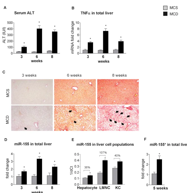
In previous studies, the miRNA profile of steatohepatitis in various animal models [4,5] and in human non-alcoholic steatohepatitis (NASH) [2,3] has been investigated, but little is known about their functional role in the pathogenesis of the disease. The methionine-choline deficient model of steatohepatitis results in early lipid accumulation, prominent necro-inflammation and later fibrosis [21], as shown inFig 1(A: serum ALT, B: liver TNFαmRNA, C: Sirius Red staining). Therefore, despite its disadvantages, such as lack of peripheral insulin resistance, it is a useful tool to study the progression of steatohepatitis. MicroRNA-155 (miR-155), a master regulator of inflammation, enhances the translation of TNFα, a pro-inflammatory cytokine in- duced during innate immune responses by Toll-like receptor (TLR) ligands [8,9]. Increased miR-155 expression has been reported in choline-deficient-amino-acid-defined (CDAA) and in high fat diet models of steatohepatitis [5,15], but little is known in MCD diet-induced steato- hepatitis. Here, we show increased miR-155 expression in the livers of MCD diet-fed mice throughout the progression of the disease, with a peak at 6 weeks (Fig 1D). miR-155 is abun- dantly expressed in immune cells [8], however, low expression is also present in hepatocytes [5,10]. Thus, we evaluated the cell-specific expression of miR-155 in isolated hepatocytes, liver resident macrophages (Kupffer cells [KCs] and liver mononuclear cells (LMNCs), the latter containing monocytes, lymphocytes and dendritic cells. The purity of these cell populations
Fig 1. MCD diet-induced steatohepatitis is associated with increased miR-155 expression in parenchymal and non-parenchymal cells.C57Bl/6 mice were fed with methionine-choline deficient (MCD) or supplemented (MCS) control diet for 3, 6 and 8 weeks. Serum alanine aminotransferase levels (A) and TNFαmRNA expression (B) were determined (n = 5-8/group). Liver fibrosis was assessed by Sirius Red staining (100x; n = 3-6/group), representative slides are shown (C). miR-155 expression was detected by qPCR in total livers (n = 5-8/group) (D). Primary murine hepatocytes (n = 7/group), Kupffer cells (n = 6, pooled data, 2 datapoints/group) and liver mononuclear cells (LMNC; n = 3-4/group) were isolated from a subset of mice after 6 weeks of MCS or MCD diet feeding and cell-specific miR-155 expression was determined and represented as 1/dCt (E). miR-155*expression was determined in total livers (F). (*) indicates p<0.05 MCS vs. corresponding MCD group. Statistics was performed on fold change data.
doi:10.1371/journal.pone.0129251.g001
was previously reported [18]. We found increased miR-155 expression in LMNCs (107% in- crease over MCS) and in hepatocytes (36% increase over MCS) in MCD diet-induced steatohe- patitis (Fig 1E). There was a 40% increase in miR-155 expression in KCs as well, but statistical significance could not be calculated due to pooled samples resulting in a small sample size (Fig 1E). The complementary strand miR-155expression was also increased in the livers of MCD diet-fed mice (Fig 1F).
miR-155 deficiency attenuates liver steatosis but does not prevent liver injury in MCD-induced steatohepatitis
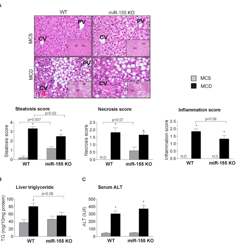
To study the biological role of miR-155 in steatohepatitis in vivo we used miR-155 deficient mice. We found attenuated hepatic steatosis in the miR-155 deficient mice compared to the WTs after MCD diet feeding at 5 weeks (Fig 2A: liver histology steatosis score, 2B: liver triglyc- eride levels). In contrast, in the MCS diet-fed control mice miR-155 deficiency enhanced the fat deposition compared to WTs (Fig 2A: liver histology steatosis score, 2B: liver triglyceride levels). Liver inflammation and injury indicated by histology inflammation score, necrosis score (Fig 2A), and serum ALT (Fig 2C) were not significantly attenuated by miR-155 deficien- cy in MCD diet-induced steatohepatitis. Notably, serum ALT levels were increased at an inter- im time point (2 weeks; data not shown) similar to that found in and earlier high fat diet feeding [15].
The different effect of miR-155 deficiency on steatosis between MCD and MCS diet-fed control mice suggests that miR-155 might have different role/s in fat accumulation in normal and diseased conditions, and its effect might depend on the major pathogenetic steps of steato- sis, such as increased fatty acid influx and / or synthesis vs.“lipid trap”. The latter is involved in the development of MCD-steatohepatitis due to the decreased phosphatidylcholine and very low density lipoprotein (VLDL) synthesis [14].
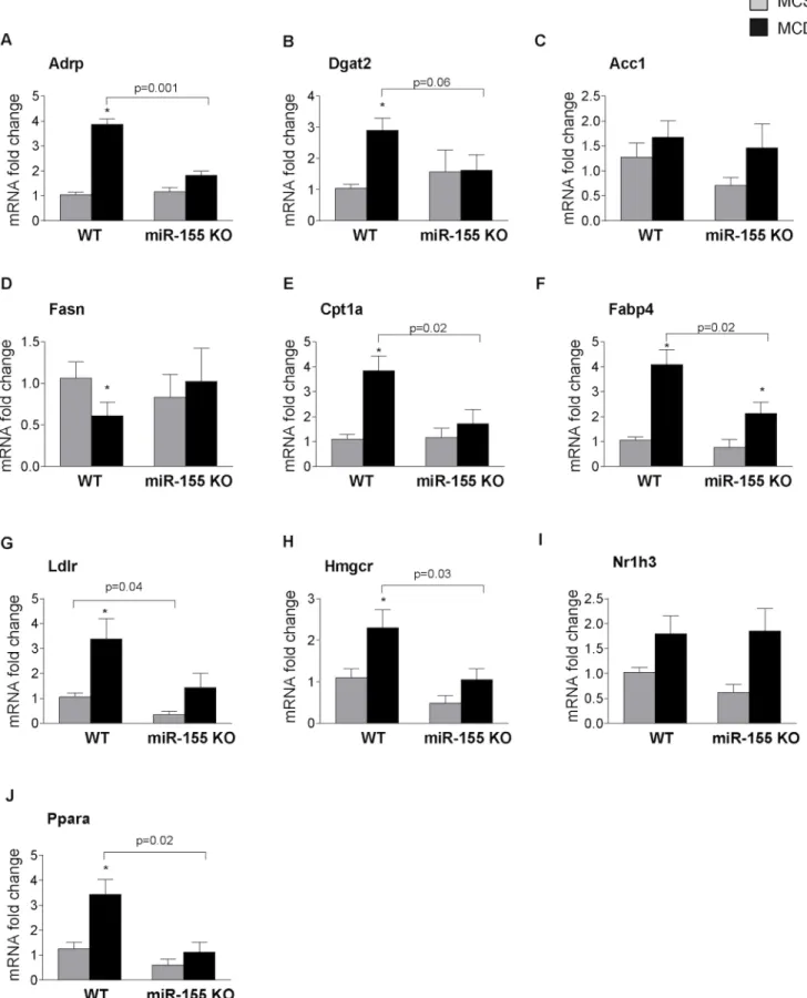
Therefore next we analyzed the expression of genes involved in (1) fatty acid uptake (adi- pose differentiation-related protein / Adrp); (2) triglyceride synthesis (diacylglycerol O-acyl- transferase 2 / Dgat2); (3) fatty acid synthesis (acetyl-Coenzyme A carboxylase / Acc1; fatty acid synthase / Fasn); (4) fatty acid oxidation (carnitine palmitoyl transferase 1a / Cpt1a); (5) fatty acid binding protein (Fabp4) that links lipid metabolism with inflammation; (6) cholester- ol metabolism (low density lipoprotein receptor / Ldlr; 3-hydroxy-3-methylglutaryl-CoA-re- ductase / Hmgcr); and (7) transcription factors involved in lipid metabolism (nuclear receptor subfamily 1, group H, member 3 / Nr1h3 / LXRα; peroxisome proliferator activated receptor alpha / Ppara).
Adrp, a lipid droplet protein that promotes fatty acid storage in form of triglycerides and in- hibits VLDL secretion [22] and Dgat2, another protein facilitating triglyceride synthesis [23]
were increased in WT MCD fed mice and this increase was prevented in miR-155 deficient mice (Fig 3A and 3B). While we found no significant difference between genotypes (WT and miR-155 KO) in the expression of Acc1 (Fig 3C) (a gene involved in fatty acid synthesis) the MCD diet induced significant reduction of Fasn expression that was rescued by mir-155 defi- ciency (Fig 3D). In concordance with previous studies, Cpt-1a, a key rate-limiting enzyme in the fatty acid oxidation [24] was increased in MCD-steatohepatitis in WT mice and not in miR-155 KO mice (Fig 3E). Fabp4, an adipokine that links lipid metabolism and inflammation [25], was increased by MCD diet in WT mice, and was attenuated by miR-155 deficiency (Fig 3F). Ldlr, a cell surface receptor responsible for the cellular uptake of low density lipoprotein (LDL) molecule [26], was significantly increased in MCD diet-fed mice compared to MCS con- trols. miR-155 deficiency attenuated Ldlr expression in both MCS and MCD groups, however in the latter, it did not reach statistical significance (p = 0.07) (Fig 3G). Similarly, Hmgcr, the
rate limiting enzyme in cholesterol synthesis was increased in the MCD diet-fed WT mice, and miR-155 deficiency attenuated Hmgcr expression, which reached statistical significance in the
Fig 2. miR-155 deficiency does not prevent liver injury, but attenuates liver steatosis in MCD-steatohepatitis.Wild type (WT) and miR-155 deficient (KO) mice were fed with methionine-choline deficient (MCD) or supplemented (MCS) control diet for 5 weeks. Liver histology was evaluated by hematoxilin- eosin staining (200x, inserts 100x; n = 5/group), representative slides are shown (A). Steatosis, necrosis, and lobular inflammation, were scored by a pathology expert (A). Liver triglyceride (B) and serum ALT (C) levels were determined (n = 6-8/group). (*) indicates p<0.05 MCS vs. corresponding MCD group. N.D. = not detectable or score = 0.
doi:10.1371/journal.pone.0129251.g002
Fig 3. miR-155 deficiency alters expression of genes in the lipid metabolism.Wild type (WT) and miR-155 deficient (KO) mice were fed with methionine- choline deficient (MCD) or supplemented (MCS) control diet for 5 weeks. mRNA expression of Adrp (A), Dgat2 (B), Acc1 (C), Fasn (D), Cpt1a (E), Fabp4 (F), Ldlr (G), Hmgcr (H), Nr1h3 (I) and Ppara (J) was measured in the livers (n = 6-8/group). (*) indicates p<0.05 MCS vs. corresponding MCD group.
doi:10.1371/journal.pone.0129251.g003
MCD diet-fed group (Fig 3H) [27]. In contrast to the report on miR-155 deficiency in HFD [15], we did not find an increased expression of the transcription factor Nr1h3 (LXRα) in miR- 155 KO mice (Fig 3I). This might explain the different findings on some LXR-reponsive genes (Fasn–Fig 3D, CD36- data not shown). PPARα, another transcription factor involved in lipid metabolism [28], was highly induced by MCD diet in WT mice, and the increase was complete- ly prevented in the miR-155 KOs (Fig 3J).
In summary, miR-155 deficiency in MCD diet-fed mice affected Adrp, Dgat2, Fasn, Cpt1a, Fabp4, Hmgcr and Ppara expression, while in MCS diet-fed controls Ldlr expression was sig- nificantly attenuated. The decreased Ldlr levels might result in impaired Ldl clearance and as a consequence of this, higher cholesterol levels [26]. Since cholesterol levels correlate with the intrahepatic fat content [29], it is tempting to speculate that the higher cholesterol levels might potentially contribute to the development of steatosis in the miR-155 deficient MCS diet-fed mice.
miR-155 deficiency attenuates liver fibrosis in MCD-induced steatohepatitis
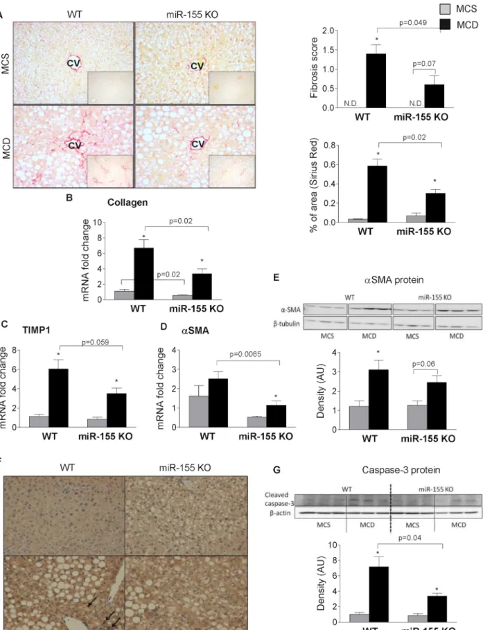
MCD diet results in liver fibrosis; therefore, next we evaluated the effect of miR-155 deficiency on fibrosis. Despite comparable liver injury and inflammation, (Fig 2A) we found significantly reduced liver fibrosis in the miR-155 deficient mice after MCD diet feeding indicated by the Sirius Red staining and fibrosis score (Fig 4A). Genes related to fibrosis, such as collagen 1a (Fig 4B), tissue inhibitor of metalloproteinase 1 (TIMP-1) (Fig 4C) andαSMA (Fig 4D) were reduced in the miR-155 deficient mice compared to the WTs after MCD feeding. The SMA protein expression was significantly increased only in WT mice and not in miR-155 KO mice after MCD diet (Fig 4E). Previously, others and we have shown the role of oxidative stress in the development of fibrosis in non-alcoholic steatohepatitis [6,30]. Here, we found that MCD diet resulted in increased 4-HNE staining, a marker of oxidative stress, in both genotypes com- pared to the MCS control groups (Fig 4F). Furthermore, it appears that MCD diet-fed WT ani- mals had more 4-HNE adducts (black arrows) than the MCD diet-fed miR-155KOs suggesting a potential role of oxidative stress in fibrosis. Oxidative stress also plays a pivotal role in cell death (apoptosis); a link to stellate cell activation [31]. Here, we found increased cleaved (ac- tive) caspase-3 expression in the MCD diet-fed WT mice compared to MCS controls and it was significantly attenuated in the miR-155 KOs on MCD diet (Fig 4G). This suggests less apo- ptotic cell death in the miR-155 KO animals, despite the overall comparable liver injury.
miR-155 deficiency does not prevent inflammation in MCD-induced steatohepatitis
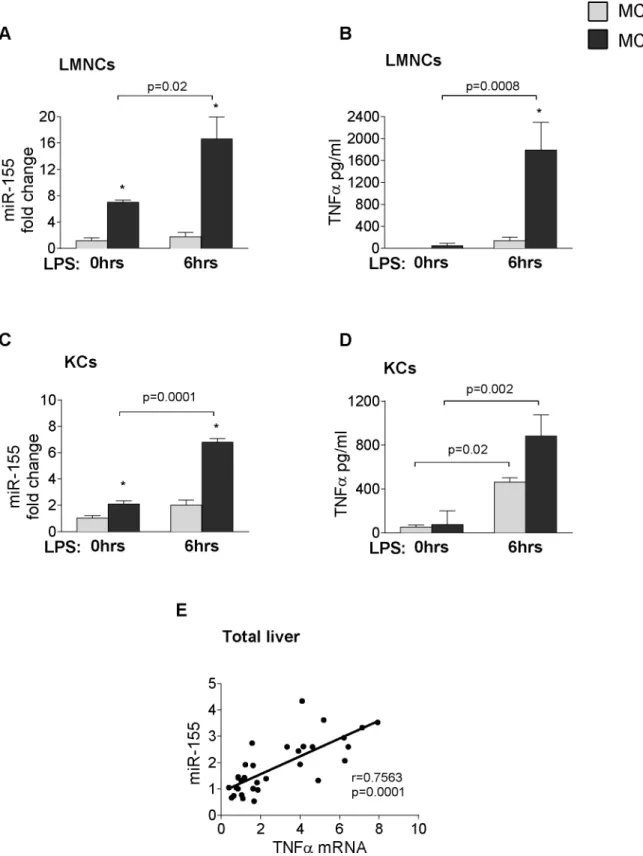
Liver fibrosis is preceded by inflammation in NASH [16]. miR-155, a master regulator of in- flammation, is induced by TLR ligands and enhances the translation of TNFα[11]; a pro-in- flammatory cytokine identified in the pathogenesis of metabolic syndrome and steatohepatitis [6]. Thus next we studied hepatic inflammation and evaluated the role of TLR activation in miR-155 and TNFαexpression. An in vitro challenge with the TLR4 ligand LPS, induced a sig- nificantly higher miR-155 expression and TNFαsecretion in isolated liver mononuclear cells (LMNCs) or Kupffer cells (KCs) in MCD-steatohepatitis compared to MCS controls (Fig 5A:
LMNCs–miR-155,Fig 5B: LMNCs–TNFα;Fig 5C: KCs–miR-155,Fig 5D: KCs–TNFα). Hepat- ic miR-155 expression also showed a positive correlation with TNFαmRNA in the WT livers (Fig 5E).
However, despite the positive correlation between miR-155 and TNFαlevels, we found that overall, hepatic inflammation was not attenuated in the miR-155 deficient mice (Fig 2).
Fig 4. miR-155 deficiency attenuates liver fibrosis in MCD-steatohepatitis.Wild type (WT) and miR-155 deficient (KO) mice were fed with methionine- choline deficient (MCD) or supplemented (MCS) control diet for 5 weeks. Liver fibrosis was assessed by Sirius Red staining (200x; n = 3-5/group),
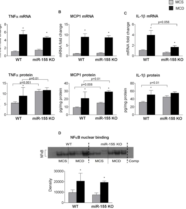
miR-155 deficiency did not reduce TNFαmRNA or protein levels (Fig 6A, mRNA upper panel, protein lower panel), and TNFαprotein levels were higher in the miR-155 KO mice (Fig 6Alower panel). Monocyte chemoattractant protein (MCP1), one of the key chemokines was also comparable between the genotypes at the mRNA level, and higher protein levels were found in the miR-155 deficient mice (Fig 6B, mRNA upper panel, protein lower panel). IL-1β, a pro-inflammatory and pro-fibrotic cytokine that is a putative miR-155target, was reduced at mRNA level in miR-155 KOs compared to WTs on MCD diet (Fig 6Cupper panel). MCD diet induced increased IL-1βprotein (total) only in WT mice, not in the miR-155 KOs (Fig 6C lower panel) compared to MCS controls. This might be related to the baseline IL-1βlevel in the miR-155 KO MCS control group.
Similarly to the cytokines, NF-κB nuclear binding was increased in both WT and miR-155 KO mice after MCD feeding (Fig 6D) suggesting a comparable level of inflammation in both genotypes.
miR-155 regulates Smad3 and C/EBPβ activation in steatohepatitis induced liver fibrosis
While inflammation can promote fibrosis, some studies have shown the divergence of these processes [21]. As a next step, we aimed to investigate how miR-155 affects fibrosis without sig- nificant attenuation of inflammation. Transforming growth factorβ(TGFβ) and Platelet de- rived growth factor (PDGF), released by macrophages and liver sinusoidal endothelial cells, regulate hepatic stellate cell activation [17]. Previous studies have suggested a role for TGFβin the development of fibrosis in NASH [6,7]. In our experiments there was no significant differ- ence in TGFβmRNA expression between WT and miR-155 KO mice both in the control and MCD groups (Fig 7A). However, PDGF, another pro-fibrogenic factor, was significantly atten- uated in miR-155 deficient mice compared to WT mice both at the mRNA and protein levels (Fig 7B: mRNA andFig 7C: protein) suggesting a potential role in the fibrosis development.
While TGFβmRNA levels were comparable between genotypes, there are several genes in TGFβsignaling that are putative mir-155 targets. Thus, next we evaluated some of the down- stream signaling molecules of TGFβsuch as the miR-155 target Smad2 [32] and Smad3 (www.
microrna.org). Interestingly, we found no difference in Smad2 protein levels between geno- types (Fig 7D). However, we found a drastic reduction of Smad3 protein levels in the miR-155 KO animals (Fig 7D) suggesting that dysfunctional TGFβsignaling might contribute to the at- tenuated fibrosis in miR-155 KO mice.
We also evaluated markers of epithelial-mesenchymal transition (EMT), including E-cad- herin and the mesenchymal marker, vimentin, as another possible mechanism of fibrosis.
While there was no significant difference in the E-cadherin levels between the genotypes (data not shown), vimentin mRNA expression was significantly attenuated in the miR-155 KO mice compared to WTs after MCD diet feeding (Fig 7E). C/EBPβis a target of miR-155 and recent studies have suggested a role of C/EBPβin EMT [33,34]. We found enhanced C/EBP nuclear binding in the livers of miR-155 KO compared to WT mice after MCD feeding (Fig 7F). C/EBP exits in various forms,α,β,γ,δ[35], therefore next we determined the nuclear binding of C/
EBPβusing a super shift assay. We found consistently enhanced C/EBPβnuclear binding in
representative slides are shown. Fibrosis was scored by an expert pathologist as well as quantified using Image J program (A, right panels). Liver collagen- 1a (B), TIMP-1 (C) andαSMA (D) mRNA expression was measured by qPCR (n = 6-8/group).αSMA protein expression was detected by Western blot from whole liver lysates (E, top panel: Western blot; E, bottom panel: densitometry; n = 3/group). Oxidative stress was evaluated by 4-HNE IHC (200x; n = 3-5/
group), representative slides are shown (F). Caspase-3 protein expression was assessed by Western blot (G, top panel: Western blot; G, bottom panel:
densitometry; n = 3/group). (*) indicates p<0.05 MCS vs. corresponding MCD group. N.D. = not detectable or score = 0.
doi:10.1371/journal.pone.0129251.g004
Fig 5. LPS induces miR-155 and TNFαexpression in hepatic immune cells.Isolated LMNCs and Kupffer cells from C57Bl/6 WT mice were stimulated or not with 100ng/ml LPS for 6 hours in vitro. miR-155 expression (A: LMNCs, C: KCs) and TNF-αprotein secretion (B: LMNCs, D: KCs) were measured in the cells and in the supernatant, respectively (n = 8-10/group). RNA was isolated from C57Bl/6 WT mice fed with methionine-choline deficient (MCD) or supplemented (MCS) control diet for 3, 6 and 8 weeks. miR155 and TNFαmRNA expression was determined (n = 5-6/group, total 32) and correlation was plotted (E); correlation coefficient is shown. (*) indicates p<0.05 MCS vs. corresponding MCD group.
doi:10.1371/journal.pone.0129251.g005
Fig 6. miR-155 deficiency does not attenuate hepatic inflammation in MCD-steatohepatitis.Wild type (WT) and miR-155 deficient (KO) mice were fed with methionine-choline deficient (MCD) or supplemented (MCS) control diet for 5 weeks. Liver TNFα(A top panel: mRNA, A bottom panel: protein), MCP1 (B top panel: mRNA, B bottom panel: protein) and IL1-β(C top panel: mRNA, C bottom panel: protein) mRNA and protein levels were measured by qPCR and ELISA, respectively (n = 6-8/group). NF-κB nuclear binding was evaluated by EMSA using liver nuclear extracts (D, top panel: representative blot, bottom panel: densitometry showing cumulative data of n = 6/group). (*) indicates p<0.05 MCS vs. corresponding MCD group.
doi:10.1371/journal.pone.0129251.g006
miR-155 KO mice compared to WT mice after MCD feeding (Fig 7G). This data suggests that the lack of miR-155 expression promotes C/EBPβactivation in steatohepatitis.
Discussion
Due to the increasing prevalence of obesity and metabolic syndrome, non-alcoholic fatty liver disease (NAFLD) [which includes simple steatosis (NAFL) and steatohepatitis (NASH)] is one of the most common liver diseases worldwide [36]. NASH is characterized by steatosis, hepato- cellular injury and necroinflammation that might lead to fibrosis, cirrhosis and potentially he- patocellular carcinoma [37]. microRNAs, small non-coding RNAs, regulate lipid metabolism, inflammation, cell proliferation and regeneration at the transcriptional or translational level [1]. An altered miRNA profile has been reported in steatohepatitis in humans [2,3], as well as in animal models [4,5,38].
Here we report novel findings on the dual role of miR-155 on regulating steatosis and fibro- sis in the methionine-choline deficient (MCD) model of steatohepatitis. We show that there is an increased miR-155 expression in both parenchymal and non-parenchymal cells in MCD diet-fed livers and we demonstrate that miR-155 deficiency attenuates steatosis in the MCD model. Our findings reveal that miR-155 regulates fibrosis at least partially independent of in- flammation in NASH, since miR-155 deficiency attenuated MCD diet-induced fibrosis despite of sustained significant inflammation and liver injury. Finally, we propose that miR-155 affects fibrosis at multiple levels, via direct and indirect targets, including PDGF, Smad3 and C/EBPβ in NASH.
In the HFD model, hepatic steatosis was significantly enhanced by miR-155 deficiency [15].
Interestingly, here we found attenuated hepatic steatosis in the miR-155 deficient mice after the MCD diet. In contrast, mice on the MCS diet showed enhanced fat deposition similar to the previous study on HFD [15]. The different effect of miR-155 deficiency on steatosis in the MCD and MCS diets and the discrepancy between the MCD and HF diets suggest that miR- 155 might have different roles in fat accumulation in healthy livers and in the various stages of diseased conditions. We found decreased steatosis at 5 weeks in MCD diet-fed miR-155 KO mice where liver fibrosis was also present. In contrast, the HFD model results in early steatosis with minimal or no fibrosis. Clinical observations suggest that the stage of NASH influences steatosis since reduced fat accumulation has been found in the fibrotic stage of NASH [39].
Increased fatty acid uptake, increased fatty acid synthesis, impaired fatty acid oxidation or impaired VLDL secretion might all contribute to hepatic steatosis [14]. While in a HFD model, one of the major mechanisms is the increased fatty acid influx, in MCD-steatohepatitis the me- thionine and choline deficiency leads to decreased phosphatidylcholine, and therefore VLDL synthesis [14], resulting a“lipid trap”. Here we found that miR-155 deficiency attenuated PPARαexpression, a transcription factor involved in lipid metabolism [28]. Genes related to fatty acid uptake, storage and VLDL synthesis, including Adrp and Dgat2, as well as Cpt1a and Fabp4, all under potential PPARαregulation [40,41,42,43], were significantly reduced in miR- 155 KO mice on MCD diet compared to WTs. Adrp promotes fatty acid storage in the form of triglycerides and inhibits VLDL secretion [22], and Dgat2 facilitates triglyceride synthesis [23].
Fig 7. miR-155 deficiency attenuates Smad3 and vimentin expression and enhances C/EBPβnuclear binding in MCD-steatohepatitis.Wild type (WT) and miR-155 deficient (KO) mice were fed with methionine-choline deficient (MCD) or supplemented (MCS) control diet for 5 weeks. Liver TGFβ(A) and PDGF (B) mRNA expression was measured by qPCR (n = 6–8). Liver PDGF (C) Smad2 and Smad3 protein expression (D) were analyzed by Western blot (top panels: blot, bottom panels: densitometry; n = 3-4/group). Vimentin (E) mRNA expression was measured by qPCR (n = 6-8/group). C/EBP nuclear binding was measured by EMSA using liver nuclear extracts (F, left panel: representative blot; right panel: densitometry, n = 6-8/group). C/EBPβnuclear binding was assessed by EMSA supershift using C/EBPβantibody (G, left panel), densitometry of the shifted band is graphed (G, right panel; n = 2/group).
(*) indicates p<0.05 MCS vs. corresponding MCD group.
doi:10.1371/journal.pone.0129251.g007
Therefore, our findings suggest that miR-155 targets critical steps in the fatty acid uptake/TG synthesis/VLDL secretion, most likely indirectly, rather than the fatty acid synthesis, since we did not find significant differences between genotypes in the latter one. Notably, genes in fatty acid synthesis, such as Fasn, are under LXRαregulation [15], which was not affected by miR- 155 deficiency in our model.
Beyond fatty acid metabolism, dietary cholesterol has also been shown to contribute to the development of fatty liver and steatohepatitis [29,44,45]. NAFLD is associated with dyslipi- demic profile, including high large VLDL, small dense LDL and low HDL concentration [29].
Key molecules in cholesterol metabolism, Ldlr (LDL uptake / clearance) and Hmgcr (rate-lim- iting enzyme in cholesterol synthesis) were attenuated by miR-155 deficiency in HFD model [15]. Here, we found increased Ldlr and Hmgcr mRNA expression in MCD diet-fed WT mice compared to MCS controls. miR-155 deficiency significantly attenuated the Ldlr levels in the MCS control group, and to a lesser extent in the MCD diet-fed mice. We did not the have op- portunity to measure cholesterol levels, but the reduced Ldlr expression might result in im- paired LDL clearance and therefore higher cholesterol levels [26]. Therefore, it is tempting to speculate that the higher cholesterol levels are due to the impaired clearance, and play role in the augmented steatosis in the MCS control group. Following the logic of this hypothesis, the changes in Hmgcr might be compensatory. PPARαcan regulate Ldlr transcription [28], and here we found that the MCD diet induced a significant increase in Ppara expression, which was prevented by miR-155 deficiency. Other genes that were affected by mir-155 deficiency in the HFD model (CD36, Cebpa, Pck1), have not changed in our MCD model.
Overall these results suggest that miR-155 targets lipid metabolism via multiple mechanisms and it might vary depending on the model of steatohepatitis. Further studies are needed to clar- ify the exact pathomechanism via which miR-155 targets genes in the lipid metabolism.
Hepatic steatosis is a risk factor; a preceding step for nonalcoholic steatohepatitis and its progression to fibrosis according to the two-hit hypothesis model [46]. However, here we found attenuation of steatosis, but not liver injury or inflammation in the miR-155 KO ani- mals. Previous studies suggest that triglyceride accumulation could be protective against pro- gressive liver damage, since DGAT2 inhibition resulted in reduced triglyceride synthesis but enhanced hepatic free fatty acids and oxidative stress [47]. While here we found reduced DGAT2 expression in miR-155 KO mice, there was no increase in oxidative stress compared to the WTs making this a less likely explanation in our model. MCD diet-fed WT mice seemed to have slightly more 4-HNE adducts. Oxidative stress plays pivotal role in cell death, a link to stellate cell activation [31]. Parallel with the MCD diet-induced enhanced oxidative stress, we found evidence of increased apoptosis in both WT and miR-155 KO mice on the MCD diet.
However, there was a significant reduction in cleaved, active, caspase-3 levels in the miR-155 KOs compared to WTs on MCD diet, suggesting an attenuation of apoptosis by miR-155 defi- ciency despite the comparable overall liver injury (ALT levels). Cell death, including apoptosis is a feature of chronic liver diseases, and is associated with fibrosis [31]. Therefore, our data supports the hypothesis that the mechanisms of hepatocyte death (eg. apoptosis vs. necrosis) rather than simply the extent of it determine the fibrogenic response as it was suggested by Witek et al. [48].
Steatohepatitis, and in general any type of chronic inflammation and cell death, tissue dam- age of the liver, potentially leads to liver fibrosis. One of the proposed mechanisms of the fibro- sis development is the monocyte/macrophage recruitment/activation and inflammatory and fibrogenic cytokine production [49,50]. The miR-155 target TNFαhas been shown to enhance hepatic stellate cell activation and promote fibrosis in some studies [51]. However, here we found comparable inflammatory cell infiltration between genotypes and the TNFαand MCP1 protein levels were higher in the miR-155 KO mice. In some ways, this data is not surprising
since miR-155 targets several negative and positive regulators of the inflammatory pathways [16]. In other ways though, it suggests the detachment of inflammatory cell infiltration and liver injury from fibrosis. Our observation is in concordance with some previous reports show- ing divergence of hepatic inflammation, injury and fibrosis [21,48].
According to previous studies IL-1βand TNFαinfluences fibrosis via hepatic stellate cell survival and not activation [52], while other cytokines, including TGFβand PDGF, released by macrophages and liver sinusoidal endothelial cells, regulate hepatic stellate cell activation [17].
We found no significant difference in the TGFβmRNA levels between genotypes either in the control or the MCD group. TGFβis synthesized as a long precursor polypeptide that is cleaved to mature protein and Latency Associated Polypeptide (LAP). The bioactivity of mature TGFβ is dependent on its release from LAP. The measurement of bioactive TGFβlevel in tissues is challenging. However, the drastic reduction of Smad3, a miR-155 target and downstream sig- naling protein of TGFβsuggests impaired TGFβsignaling, rather than impaired TGFβlevels in the miR-155 KO mice. Similarly, PDGF expression was also attenuated in the miR-155 KO ani- mals. Overall, these suggest that miR-155 might contribute to liver fibrosis via activation of he- patic stellate cells in our model. The role of NADPH oxidase complex-dependent oxidative stress has also been reported in the activation of hepatic stellate cells [53]. Based on our data it appears that MCD diet induced oxidative stress, indicated by the 4-HNE staining, might be slightly higher in the WT animals compared to miR-155 KOs.
Stellate cells and their transdifferentiation into extracellular matrix producing myofibro- blasts is the central event in fibrosis, however, other contributors of fibrogenic cells exist too, including portal fibroblasts, bone marrow-derived mesenchymal cells and EMT [17]. During EMT, epithelial cells undergo morphological changes to acquire a fibroblast-like phenotype with down-regulation of adhesion molecules and up-regulation of mesenchymal markers such as vimentin [54]. Reports are somewhat contradictory in terms of hepatocyte EMT [54,55].
Our data on the increased expression of vimentin raise the possibility of EMT. More impor- tantly, we found a significantly decreased vimentin expression in the miR-155 KO mice. While vimentin is not a direct target of miR-155, C/EBPβ, a well-established miR-155 target [5] plays a role in EMT in various cancers [33,34]. Since a loss of C/EBPβpromotes EMT in mammary epithelial cells [33], it is tempting to speculate that augmentation of C/EBPβin miR-155 KO mice, as we have found, might also contribute to an attenuation of fibrosis in our model. The regulation of fibrosis by miR-155 likely occurs at multiple levels, and it is not restricted to NASH-fibrosis; for example, we also found a reduction of carbon-tetrachloride induced-fibro- sis in miR-155 deficient mice [56]. In addition to miR-155, numerous other microRNAs can af- fect liver fibrosis, including miR-122. We recently reported a direct link between miR-122 and vimentin expression [57].
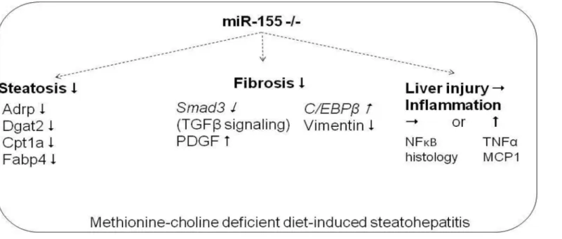
In conclusion, here we show novel data that miR-155 deficiency attenuates liver steatosis and fibrosis, but not liver injury and inflammation in the MCD model of steatohepatitis (Fig 8). The regulation of steatosis by miR-155 varies depending on the steatohepatitis model; in- volving genes in TG and VLDL synthesis in methionine-choline deficiency. The regulation of fibrosis is independent of overall liver injury in MCD-steatohepatitis and to a certain extent is detached from inflammation, since miR-155 deficiency did not attenuate hepatic injury, in- flammatory cell infiltration or TNFαproduction. The potential fibrotic mechanisms identified in our model include regulation of apoptosis (caspase-3), Smad3, PDGF and C/EBPβby miR- 155. Finally, our data also underline the importance of the negative regulatory role of miR-155 in the inflammation pathways in NASH.
Acknowledgments
The authors greatly appreciate Dr. Jin-Kyu Park and Dr. Benedek Gyongyosi help in evaluation and scoring of the histology slides.
Author Contributions
Conceived and designed the experiments: TC SB GS. Performed the experiments: TC SB DL KK DC AIV. Analyzed the data: TC SB GS. Contributed reagents/materials/analysis tools: TC SB DL KK DC GS. Wrote the paper: TC SB GS.
References
1. Szabo G, Bala S. MicroRNAs in liver diseases. Nat Rev Gastroenterol Hepatol. 2013; 10: 542–552.
doi:10.1038/nrgastro.2013.87PMID:23689081
2. Cheung O, Puri P, Eicken C, Contos MJ, Mirshani F, Maher JW, et al. Nonalcoholic steatohepatitis is associated with altered hepatic microRNA expression. Hepatology 2008; 48:1810–1820. doi:10.1002/
hep.22569PMID:19030170
3. Estep M, Armistead D, Hossain N, Elarainy H, Goodman Z, Baranova A, et al. Differential expression of miRNAs in the visceral adipose tissue of patients with non-alcoholic fatty liver disease. Aliment Phar- macol Ther. 2010; 32: 487–497. doi:10.1111/j.1365-2036.2010.04366.xPMID:20497147
4. Dolganiuc A, Petrasek J, Kodys K, Catalano D, Mandrekar P, Velayudham A, et al. MicroRNA expres- sion profile in Lieber-DiCarli diet-induced alcoholic and methionine choline deficient diet-induced nonal- coholic steatohepatitis models in mice. Alcohol Clin Exp Res. 2009; 33: 1704–1710. doi:10.1111/j.
1530-0277.2009.01007.xPMID:19572984
5. Wang B, Majumder S, Nuovo G, Kutay H, Volinia S, Patel T, et al. Role of microRNA-155 at early stages of hepatocarcinogenesis induced by choline-deficient and amino acid-defined diet in C57BL/6 mice.
Hepatology 2009; 50: 1152–1161. doi:10.1002/hep.23100PMID:19711427
6. Csak T, Velayudham A, Hritz I, Petrasek J, Levin I, Lippai D, et al. Deficiency in myeloid differentiation factor-2 and toll-like receptor 4 expression attenuates nonalcoholic steatohepatitis and fibrosis in mice.
Am J Physiol Gastrointest Liver Physiol. 2011; 300: G433–G441. doi:10.1152/ajpgi.00163.2009 PMID:21233280
Fig 8. Summary figure: Role of miR-155 in experimental MCD induced steatohepatitis.Putative direct miR-155 targets are in italics.
doi:10.1371/journal.pone.0129251.g008
7. Miura K, Kodama Y, Inokuchi S, Schnabl B, Aoyama T, Ohnishi H, et al. Toll-like receptor 9 promotes steatohepatitis by induction of interleukin-1 beta in mice. Gastroenterology 2010; 139: 323–34.e7. doi:
10.1053/j.gastro.2010.03.052PMID:20347818
8. Tili E, Michaille JJ, Costinean S, Corce CM. MicroRNAs, the immune system and rheumatic disease.
Nat Clin Pract Rheum. 2008; 4: 534–541.
9. Tili E, Michaille JJ, Cimino A, Costinean S, Dumitru CD, Adair B, et al. Modulation of miR-155 and miR- 125b levels following lipopolysaccharide/TNF-alpha stimulation and their possible roles in regulating the response to endotoxin shock. J Immunol. 2007; 179, 5082–5089. PMID:17911593
10. Bala S, Szabo G. MicroRNA Signature in Alcoholic Liver Disease. Int J Hepatol. 2012; 2012: 498232.
doi:10.1155/2012/498232PMID:22518321
11. Bala S, Marcos M, Kodys K, Csak T, Catalano D, Mandrekar P, et al. Up-regulation of microRNA-155 in macrophages contributes to increased tumor necrosis factor {alpha} (TNF{alpha}) production via in- creased mRNA half-life in alcoholic liver disease. J Biol Chem. 2011; 286: 1436–1444. doi:10.1074/
jbc.M110.145870PMID:21062749
12. O'Connell RM, Taganov KD, Boldin MP, Cheng G, Baltimore D. MicroRNA-155 is induced during the macrophage inflammatory response. Proc Natl Acad Sci USA. 2007; 104: 1604–1609. PMID:
17242365
13. Miele L, Valenza V, La Torre G, Montalto M, Cammarota G, Ricci R, et al. Increased intestinal perme- ability and tight junction alterations in non-alcoholic fatty liver disease. Hepatology 2009; 49: 1877– 1887. doi:10.1002/hep.22848PMID:19291785
14. Imajo K, Yoneda M, Kessoku T, Oqawa J, Maeda S, Sumida Y, et al. Rodent models of nonalcoholic fatty liver disease/ nonalcoholic steatohepatitis. Int J Mol Sci. 2013; 14: 21833–21857. doi:10.3390/
ijms141121833PMID:24192824
15. Miller AM, Gilchrist DS, Nijjar J, Araldi E, Ramirez CM, Lavery CA, et al. MiR-155 has a protective role in the development of non-alcoholic hepatosteatosis in mice. PLoS One 2013; 8: e72324. doi:10.
1371/journal.pone.0072324PMID:23991091
16. Seki E, Schnabl B. Role of innate immunity and the microbiota in liver fibrosis: crosstalk between the liver and gut. J Physiol. 2012; 590 (Pt3): 447–458.
17. Elpek GO. Cellular and molecular mechanisms in the development of liver fibrosis: An update. World J Gastroenterol. 2014; 20: 7260–7276. doi:10.3748/wjg.v20.i23.7260PMID:24966597
18. Csak T, Ganz M, Pespisa J, Kodys K, Dolagniuc A, Szabo G. Fatty acid and endotoxin activate inflam- masomes in mouse hepatocytes that release danger signals to stimulate immune cells. Hepatology 2011; 54: 133–144. doi:10.1002/hep.24341PMID:21488066
19. Kleiner DE, Brunt EM, Natta MV, Behling C, Contos MJ, Cummings OW, et al. Design and validation of a histological scoring system for nonalcoholic fatty liver disease. Hepatology 2005; 41: 1313–1321.
PMID:15915461
20. Nath B, Levin I, Csak T, Petrasek J, Mueller C, Kodys K, et al. Hepatocyte-specific hypoxia-inducible factor-1αis a determinant of lipid accumulation and liver injury in alcohol-induced steatosis in mice.
Hepatology 2011; 53: 1526–1537. doi:10.1002/hep.24256PMID:21520168
21. Velayudham A, Dolganiuc A, Ellis M, Petrasek J, Kodys K, Mandrekar P, et al. VSL#3 probiotic treat- ment attenuates fibrosis without changes in steatohepatitis in a diet-induced nonalcoholic steatohepati- tis model in mice. Hepatology 2009; 49: 989–997. doi:10.1002/hep.22711PMID:19115316
22. Magnusson B, Asp L, Boström P, Ruiz M, Stillemark-Billton P, Linden D, et al. Adipocyte differentiation- related protein promotes fatty acid storage in cytosolic triglycerides and inhibits secretion of very low density lipoproteins. Atheroscler Thromb Vasc Biol. 2006; 26: 1566–1571. PMID:16627799
23. Zammit VA. Hepatic triacylglycerol synthesis and secretion: DGAT2 as the link between glycaemia and triglyceridaemia. Biochem J. 2013; 451: 1–12. doi:10.1042/BJ20121689PMID:23489367
24. Rinella ME, Elias MS, Smolak RR, Fu T, Borensztajn J, Green RM. Mechanism of hepatic steatosis in mice fed a lipogenic methionine-choline deficient diet. J Lipid Res. 2008; 49: 1068–1076. doi:10.1194/
jlr.M800042-JLR200PMID:18227531
25. Thumser AE, Moore JB, Plant NJ. Fatty acid binding proteins: tissue-specific functions in health and disease. Curr Opin Clin Nutr Metab Care. 2014; 17: 124–129. doi:10.1097/MCO.0000000000000031 PMID:24500438
26. Ai D, Chen C, Han S, Ganda A, Murphy AJ, Haeusler R, et al. Regulation of hepatic LDL receptors by mTORC1 and PCSK9 in mice. J Clin Invest. 2012; 122(4): 1262–1270. doi:10.1172/JCI61919PMID:
22426206
27. Patel DD, Knight BL, Wiggins D, Humpreys SM, Gibbons GF. Distrubances in the normal regulation of SREBP-sensitive genes in PPAR-alpha deficient mice. J Lipid Res. 2001; 42(3): 328–337. PMID:
11254743
28. Zeng L, Tang WJ, Yin JJ, Zhou BJ. Signal transductions and nonalcoholic fatty liver: a mini-review.
World J Gastroenterol. 2014; 7(7): 1624–1631.
29. FonTacer K, Rozman D. Nonalcoholic fatty liver disease: Focus on lipoprotein and lipid deregulation. J Lipids. 2011; ID:783976.
30. Dattaroy D, Pourhoseini S, Das S, Alhasson F, Seth RK, Nagarkatti M, et al. Micro RNA 21 inhibition of SMAD7 enhances fibrogenesis via leptin mediated NADPH oxidase in experiemental and human non- alcoholic steatohepatitis. Am J Physiol Gastrointest Liver Physiol. 2014 In press; 2014 Dec 11:
ajpgi.00346.2014. doi:10.1152/ajpgi.00346.2014
31. Chakraborty JB, Oakley F, Walsh MJ. Mechanisms and Biomarkers of Apoptosis in Liver Disease and Fibrosis. Int J Hepatol. 2012; 2012: 648915. doi:10.1155/2012/648915PMID:22567408
32. Louafi F, Martinez-Nunez RT, Sanchez-Elsner T. MicroRNA-155 targets Smad2 and modulates the reponse of macrophages to transforming growth factor-β. J Biol Chem. 2010; 285: 41328–41336. doi:
10.1074/jbc.M110.146852PMID:21036908
33. Johansson J, Berg T, Kurzejamska E, Pang MF, Tabor V, Jansson M, et al. MiR-155-mediated loss of C/EBPβshifts the TGF-βresponse from growth inhibition to epithelial-mesenchymal transition, invasion and metastasis in breast cancer. Oncogene 2013; 32: 5614–5624. doi:10.1038/onc.2013.322PMID:
23955085
34. Li J, Shan F, Xiong G, Chen X, Guan X, Wang JM, et al. Epidermal Growth Factor-induced C/EBPbeta participates in EMT by dampening miR203 in esophageal squamous cell carcinoma. J Cell Sci. 2014;
127(Pt17): 3735–3744.
35. Lekstrom-Himes J, Xathopoulos KG. Biological role of the CCAAT/Enahncer binding protein family of transcription factors. J Biol Chem. 1998; 273: 28545–28548. PMID:9786841
36. Michelotti GA, Machado MV, Diehl AM. NAFLD, NASH and liver cancer. Nat Rev Gastroenterol Hepa- tol. 2013; 10: 656–665. doi:10.1038/nrgastro.2013.183PMID:24080776
37. Chalasani N, Younossi Z, Lavine JE, Diehl AM, Brunt EM, Cusi K, et al. The diagnosis and manage- ment of non-alcoholic fatty liver disease: Practice guideline by the American Association of the Study for Liver Diseases, American College of Gastroenterology, and the American Gastroenterological As- sociation. Hepatology 2012; 55: 2005–2023. doi:10.1002/hep.25762PMID:22488764
38. Poqribny IP, Starland-Davenport A, Tryndyak VP, Han T, Ross SA, Rusyn I, et al. Difference in expres- sion of hepatic microRNAs miR-29c, miR-34a, miR-155, and miR-200b is associated with strain-specif- ic susceptibility to dietary nonalcoholic steatohepatitis in mice. Lab Invest. 2010; 90: 1437–1446. doi:
10.1038/labinvest.2010.113PMID:20548288
39. Adams LA, Lymp JF, Sauver J, St, Sanderson SO, Lindor KD, Feldstein A, et al. The natural history of nonalcoholic fatty liver disease: a population-based cohort study. Gastroenterology 2005; 129: 113– 121. PMID:16012941
40. Edvardsson U, Ljungberg A, Linden D, William-Olsson L, Peilot-Sjögren J, Ahnmark A, et al. PPARal- pha activation increases triglyceride mass and adipocyte differentiation-related protein in hepatocytes.
J Lipid Res. 2006; 47(2): 329–340. PMID:16282640
41. Paland N, Gamliel-Lazarovich A, Coleman R, Fuhrman B. Urokinase-type plasminogen activator (uPA) stimulates triglyceride synthesis in Huh7 hepatoma cells via p38-dependent upregulation of DGAT2.
Atherosclerosis 2014; 237(1): 200–207. doi:10.1016/j.atherosclerosis.2014.09.003PMID:25244504 42. Feingold KR, Wang Y, Moser A, Shigenaga JK, Grunfeld C. LPS decreases fatty acid oxidation and nu-
clear hormone receptors in the kidney. J Lipid Res. 2008; 49(10): 2179–2187. doi:10.1194/jlr.
M800233-JLR200PMID:18574256
43. Rakhshandehroo M, Knoch B, Muller M, Kersten S. Peroxisome Proliferator-Activated Receptor Alpha Target Genes. PPAR Research 2010; ID: 612089.
44. Matsuzawa N, Takamura T, Kurita S, Misu H, Ota T, Ando H, et al. Lipid-induced oxidative stress causes steatohepatitis in mice fed an atherogenic diet. Hepatology 2007; 46(5): 1392–1403. PMID:
17929294
45. Kainuma N, Fujimoto M, Sekiya N, Tsuneyama K, Cheng C, Takano Y, et al. Cholesterol-fed rabbit as a unique model of nonalcoholic, nonobese, non-insulin-resistant fatty liver disease with characteristic fi- brosis. J Gastroenterol. 2006; 41(10): 971–980. PMID:17096066
46. Day CP, James OF. Steatohepatitis”a tale of two“hit”? Gastroenterology 1998; 114: 842–845. PMID:
9547102
47. Yamaguchi K, Yang L, McCall S, Huang J, Yu XX, Pandey SK, et al. Inhibiting triglyceride synthesis im- proves hepatic steatosis but exacerbates liver damage and fibrosis in obese mice with nonalcoholic steatohepatitis. Hepatology 2007; 45: 1366–1374. PMID:17476695
48. Witek RP, Stone WC, Karaca FG, Syn WK, Pereira TA, Agboola KM, et al. Pan-caspase inhibitor VX- 166 reduces fibrosis in an animal model of steatohepatitis. Hepatology 2009; 50(5): 1421–1430. doi:
10.1002/hep.23167PMID:19676126
49. Yang L, Seki E. Toll like receptors in liver fibrosis: cellular crosstalk and mechanisms. Front Physiol.
2012; 3: Article 138.
50. Yang LL, Liu JQ, Bai XZ, Fan L, Han F, Jia WB, et al. Acute downregulation of miR-155 at wound sites leads to a reduced fibrosis through attenuating inflammatory response. Biochem Biophys Res Com- mun. 2014; 453(1): 153–159. doi:10.1016/j.bbrc.2014.09.077PMID:25264197
51. Connolly MK, Bedrosian AS, Mallen-St Clair J, Mitchell AP, Ibrahim J, Stroud A, et al. In liver fibrosis, dendritic cell govern hepatic inflammation in mice via TNFα. J Clin Invest. 2009; 119: 3213–3225. doi:
10.1172/JCI37581PMID:19855130
52. Pradere JP, Kluwe J, De Minicis S, Jiao JJ, Gwak GY, Dapito DH, et al. Hepatic macrophages but not dendritic cells contribute to liver fibrosis by promoting the survival of activated hepatic stellate cells in mice. Hepatology 2013; 1461–1473.
53. Bataller R, Schwabe RF, Choi WY, Paik YH, Lindquist J, Qian T, et al. NADPH oxidase signal trans- duces angiotensin II in hepatic stellate cells and is critical in hepatic fibrosis. J Clin Invest. 2003; 112:
1383–1394. PMID:14597764
54. Cicchini C, Amicone L, Alonzi T, Marchetti A, Macone C, Tripodi M. Molecular mechanisms controlling the phenotype and the EMT/MET dynamics of hepatocyte. Liv Int. 2014; In Press, 2015 Feb;35(2):302– 10. doi:10.1111/liv.12577
55. Taura K, Miura K, Iwaisako K, Osterreicher CH, Kodama Y, Penz-Osterreicher M, et al. Hepatocytes do not undergo epithelial-mesenchymal transition in liver fibrosis in mice. Hepatology 2010; 51: 1027– 1036. doi:10.1002/hep.23368PMID:20052656
56. Bala S, Csak T, Petrasek J, Catalano D, Kodys K, Szabo G. MiRNA-155 regulates CCl4-induced liver inflammation and fibrosis via targeting pro- inflammatory and pro-fibrotic genes. Hepatology 2012; 56 (4) Suppl: .:1107A (#1968) doi:10.1016/j.jvs.2012.04.010PMID:22818832
57. Csak T, Bala S, Lippai D, Satishchandran A, Catalano D, Kodys K, et al. microRNA-122 regulates hyp- oxia-inducible factor-1 and vimentin in hepatocytes and correlates with fibrosis in diet-induced steato- hepatitis. Liver Int. 2015; 35(2): 532–541. doi:10.1111/liv.12633PMID:25040043