CASE CONTROL STUDY
Gabor Lendvai, Zsuzsa Schaff, MTA-SE Tumor Progression Re- search Group, Semmelweis University, 1091 Budapest, Hungary Katalin Jármay, Gizella Karácsony, Tibor Wittmann, 1st Department of Medicine, University of Szeged, 6720 Szeged, Hungary
Tünde Halász, Zsuzsa Schaff, András Kiss, 2nd Department of Pathology, Semmelweis University, 1091 Budapest, Hungary Ilona Kovalszky, Kornélia Baghy, 1st Department of Pathology and Experimental Cancer Research, Semmelweis University, 1085 Budapest, Hungary
Author contributions: Jármay K, Schaff Z and Kiss A designed the study; Jármay K, Karácsony G, Kovalszky I, Wittmann T, Schaff Z and Kiss A chose and diagnosed the patient samples;
Lendvai G, Karácsony G and Halász T performed the research;
Lendvai G, Jármay K, Karácsony G, Kovalszky I, Baghy K, Schaff Z and Kiss A analyzed and interpreted the data; Lendvai G, Schaff Z and Kiss A wrote the paper; all authors critically reviewed and approved the manuscript.
Supported by Grants from the National Scientific Research Fund, No. OTKA K101435, No. K108548 and No. 105763 Correspondence to: Zsuzsa Schaff, MD, PhD, DSc, Profes- sor of Pathology, 2nd Department of Pathology, Semmelweis University, Ulloi 93, 1091 Budapest,
Hungary. schaff.zsuzsa@med.semmelweis-univ.hu Telephone: +36-1-215-6921 Fax: +36-1-215-6921 Received: February 11, 2014 Revised: May 7, 2014 Accepted: June 13, 2014
Published online: November 7, 2014
Abstract
AIM: To assess the expression of selected microRNAs (miRNA) in hepatitis C, steatotic hepatitis C, noninfect- ed steatotic and normal liver tissues.
METHODS: The relative expression levels of miR-21, miR-33a, miR-96, miR-122, miR-125b, miR-221 and miR-224 were determined in 76 RNA samples isolated from 18 non-steatotic and 28 steatotic chronic hepati- tis C (CHC and CHC-Steatosis, respectively) cases, 18 non-infected, steatotic liver biopsies of metabolic origin
(Steatosis) and 12 normal formalin-fixed paraffin-em- bedded liver tissues using TaqMan MicroRNA Assays.
All CHC biopsy samples were obtained prior to initiat- ing therapy. Patients’ serum biochemical values, which included glucose, triglyceride, cholesterol, alanine aminotransferase(ALT), aspartate aminotransferase (AST), gamma-glutamyl-transferase (GGT), alkaline phosphatase (AP), were obtained and correlated with relative miRNA expression.
RESULTS: When compared with control non-infected liver samples, miR-122 and miR-221 levels were re- duced in CHC-Steatosis (P < 0.03) and in CHC, CHC- Steatosis and Steatosis (P < 0.01). Alternatively, the expression of miR-33a and miR-224 were elevated in CHC-Steatosis and Steatosis in comparison to control tissue (P < 0.01). The levels of miR-33a and miR-224 in CHC-Steatosis (P < 0.02) and miR-224 in Steato- sis (P < 0.001) were increased in comparison to CHC samples. By contrast, the expression of miR-21 did not differ statistically between diseased and normal liver samples. Levels of miR-33a correlated negatively with serum AST and AP levels in Steatosis as well as with necroinflammatory grade in CHC, whereas miR-21 cor- related positively with AST in Steatosis and displayed negative correlation with triglyceride level in CHC-St- eatosis. In contrast, miRNA levels were not correlated with ALT, GGT, cholesterol levels or fibrosis stage.
CONCLUSION: Differences in miRNA expression were observed between CHC and steatotic CHC, CHC and steatotic liver, but not between steatotic CHC and stea- totic liver of metabolic origin.
© 2014 Baishideng Publishing Group Inc. All rights reserved.
Key words: Chronic hepatitis C; Steatosis; MicroRNA;
Expression; miR-33a; miR-224
Core tip: Chronic hepatitis C (CHC) and steatosis are DOI: 10.3748/wjg.v20.i41.15343 © 2014 Baishideng Publishing Group Inc. All rights reserved.
Elevated miR-33a and miR-224 in steatotic chronic hepatitis C liver biopsies
Gabor Lendvai, Katalin Jármay, Gizella Karácsony, Tünde Halász, Ilona Kovalszky, Kornélia Baghy, Tibor Wittmann, Zsuzsa Schaff, András Kiss
liver diseases that can progress into hepatocellular car- cinoma. In the current study, differences were found in expression of selected microRNAs in biopsy samples of steatotic liver, CHC-infected, and steatotic CHC-infected liver, compared to control samples. Interestingly, levels of miR-224, which are increased in hepatocellular car- cinoma, were elevated in both types of steatotic liver when compared with normal or CHC-infected only liver tissues, and may be an indicator of a precancerous state.
Lendvai G, Jármay K, Karácsony G, Halász T, Kovalszky I, Baghy K, Wittmann T, Schaff Z, Kiss A. Elevated miR-33a and miR-224 in steatotic chronic hepatitis C liver biopsies. World J Gastroenterol 2014; 20(41): 15343-15350 Available from: URL:
http://www.wjgnet.com/1007-9327/full/v20/i41/15343.htm DOI:
http://dx.doi.org/10.3748/wjg.v20.i41.15343
INTRODUCTION
Hepatitis C virus (HCV) infection is a significant health care problem worldwide. It is estimated that approxi- mately 170 million people are infected with HCV[1] and 3% of the world’s population is chronically infected with this virus[2]. Approximately 70% to 80% of acute infect- ed cases will develop into chronic hepatitis C (CHC) due to failed elimination of the virus; nearly 20% of CHC patients develop cirrhosis and 20% of the cirrhotic cases further progress to hepatocellular carcinoma (HCC)[3]. Treatment options for CHC include combination of pegylated interferon and ribavirin, however, the virus still fails to clear in a significant proportion of treated patients[4], which contributes to the numerous liver trans- plantations worldwide. A new triple therapy regimen with protease inhibitors holds great promise as clinical trials have ended with a significantly improved percent- age of patients reaching a sustained viral response[5].
In addition to cirrhosis, hepatic steatosis is also fre- quently observed in CHC in approximately 40%-86%
of CHC patients depending on the viral genotype[6-9]. In cultured Huh7 cells, for example, HCV genotype 3a core protein results in the highest level of triglyceride ac- cumulation[10,11]. The prevalence of steatosis in HCV-in- fected patients is two-folds higher than that observed in hepatitis B virus-infected patients, which demonstrates a correlation between CHC and nonalcoholic fatty liver disease (NAFLD)[12]. Hepatic steatosis occurs when fatty acids, delivered either from the circulation or synthesized de novo by the liver, exceed the liver’s capacity to me- tabolize fat by means of β-oxidation or to secrete fat as very-low-density lipoproteins (VLDL). This imbalance between delivery of fat and its subsequent secretion or metabolism leads to accumulation of lipid droplets con- taining triglycerides and cholesteryl esters, predominantly in hepatocytes[13]. In NAFLD, the development of stea- tosis is linked to obesity and metabolic disorders such as hyperlipidemia, insulin resitance and diabetes[14,15]. In
addition, steatosis is associated with higher alanine ami- notransferase (ALT) levels[8].
microRNAs (miRNA) are short RNA molecules con- sidered to negatively modulate gene expression[16] through fine-tuning gene expression involved predominantly in development, immunity, differentiation and homeosta- sis[17]. miRNAs act at the posttranscriptional level and in- duce translational arrest by binding to the 3’ untranslated region (UTR) of messenger RNAs, leading to a reduc- tion or blockage of protein synthesis[18]. In comparison to normal homeostatic conditions, altered miRNA ex- pression has been reported in cancers[19] and in several other pathologies including liver diseases[20,21]. Moreover, a few miRNAs are already suggested to be potential bio- markers for HCC and chronic hepatitis B infection[22,23].
In the present study, CHC-infected, steatotic CHC- infected and NAFLD-based steatotic liver biopsies were compared to non-infected, normal liver samples to assay differences in the expression of selected miR- NAs that previously have been associated with fibrosis (miR-21, miR-221), fat metabolism (miR-33a, miR-122) and hepatocarcinogenesis (miR-21, miR-122, miR-221, miR-224)[24-27].
MATERIALS AND METHODS
Patients
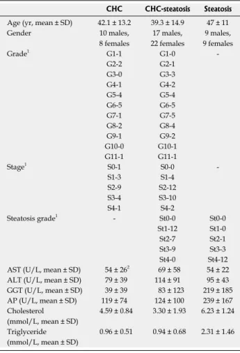
A total of 64 patients were enrolled in this study, from which 46 CHC-infected patients (genotype 1/b) were hospitalized at the 1st Department of Medicine at the University of Szeged. These patients were divided into two groups (CHC or CHC-Steatosis) according to the presence of steatosis in the liver samples, as diagnosed by experienced pathologists. Accordingly, 18 patients with CHC but without any apparent signs of steatosis were included in the CHC group, whereas 28 CHC pa- tients having either mild or severe steatosis were includ- ed in the CHC-Steatosis group (Table 1). An additional 18 patients with metabolic steatosis of varying degrees but no HCV infection were selected for the Steatosis group from the archives of the 2nd Department of Pa- thology at Semmelweis University. Twelve non-infected, normal liver samples served as controls and were ob- tained from deceased patients after organ donation, just prior to ligation of the abdominal aorta and reperfusion.
In addition, the following serum biochemical values were detected and recorded at the time of biopsy: glucose, triglyceride, cholesterol, ALT, aspartate aminotransfer- ase (AST), gamma-glutamyl-transferase (GGT), alkaline phosphatase (AP). The selected samples were analysed retrospectively with permission obtained from the local Ethical Committee based on the ethical guidelines of the 1975 Declaration of Helsinki. Antiviral treatment had not been initiated before obtaining the liver biopsy samples from the CHC patients.
Histology
Liver samples were processed according to routine pa- thology procedures. Briefly, liver tissues were submerged
Table 1 Clinical background of patients enrolled in this study
in 10% neutral buffered formalin (in phosphate-buffered saline, pH 7.0) immediately after removal and fixed for 24 h at room temperature. Following dehydration in an ethanol series and xylene, the formalin-fixed samples were embedded in paraffin (FFPE samples). Paraffin- embedded samples were cut into 3 to 5 μm thick sec- tions and staining with HE as well as with picrosyrius red for highlight connective tissue. Histological grade and stage were determined using the Ishak scoring sys- tem[28].
RNA isolation
RNA was isolated from several 3 to 4 μm thick sections using the RNeasy FFPE Kit (QIAGEN, Venlo, Nether- lands) according to the manufacturer’s instructions with modification for co-purification of miRNAs[29]. Traces of genomic DNA were removed using Turbo DNase digestion (Ambion of Thermo Fisher Scientific Inc., Waltham, United States).
Reverse transcription and quantitative real time PCR Expression of individual miRNAs was determined us-
ing the following TaqMan MicroRNA Assays (Applied Biosystems of Thermo Fisher Scientific Inc.): miR-21 (ID: 000397), miR-33a (ID: 000424, v9.2 where was called miR-33), miR-96 (ID: 000186), miR-122 (ID:
002245), miR-125b (ID: 000449), miR-221 (ID: 000524) and miR-224 (ID: 002099). These selected miRNAs are reported to be associated with fibrosis, fat metabolism and hepatocarcinogenesis[24-27]. Reverse transcription (RT) and quantitative real time PCR (qPCR) were performed according to the manufacturer’s instructions. Briefly, the RT reaction was carried out using a TaqMan MicroRNA Reverse Transcription Kit (Applied Biosystems) in a final volume of 7.5 μL containing 10 ng total RNA. qPCR was performed using the TaqMan Universal PCR Master Mix No AmpErase UNG (Applied Biosystems) in a final volume of 10 μL containing 0.65 μL RT product. Am- plification reactions were run on an ABI PRISM 7000 Sequence Detection System (Applied Biosystems). Rela- tive expression was calculated by the 2-∆∆Cq formula, ap- plying the average Cq value of miR-96 and miR-125b as the most stable reference determined by the NormFind- er application[30], and normalized to the median ∆Cq value of the normal liver samples.
Statistical analysis
The normalized relative expression data were analyzed with a nonparametric Kruskal-Wallis ANOVA and me- dian test using STATISTICA software, version 9.1 (Stat- Soft Inc., Tulsa, OK, United States). Correlation analysis between miRNA expression and histologic grades or serum biochemical values of patients was performed with a nonparametric Spearman rank correlation using GraphPad PRISM software, version 5.01 (GraphPad Software Inc, La Jolla, CA, United States). A P-value of 0.05 was set as the threshold for statistical significance.
RESULTS
Differences in miRNA expression
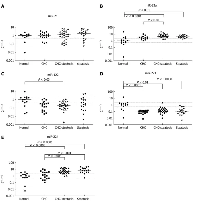
Deregulated miRNA expression was observed in the diseased liver samples as compared with normal control liver samples. Decreased levels of miR-221 were found in CHC, CHC-Steatosis and Steatosis samples (1/11-fold, P < 0.0001; 1/8-fold, P < 0.01 and 1/11-fold, P < 0.0008; respectively) and reduced expres- sion of miR-122 was detected in CHC-Steatosis sam- ples (1/5-fold, P < 0.03) when compared with normal liver (Figure 1). In contrast, increased levels of miR-33a and miR-224 were found in CHC-Steatosis (5-fold, P
< 0.0001 and 5-fold, P < 0.0003; respectively) and Ste- atosis (4-fold, P < 0.01 and 7-fold, P < 0.0001; respec- tively) samples in comparison with control normal liver samples. The expression of miR-21 was not statistically different between the diseased and normal liver samples.
Elevated levels of miR-33a and miR-224 were observed in CHC-Steatosis samples compared with CHC samples (2-fold, P < 0.02 and 4-fold, P < 0.003; respectively).
Furthermore, increased miR-224 expression was found
CHC CHC-steatosis Steatosis Age (yr, mean ± SD) 42.1 ± 13.2 39.3 ± 14.9 47 ± 11
Gender 10 males,
8 females
17 males, 22 females
9 males, 9 females
Grade1 G1-1 G1-0 -
G2-2 G2-1
G3-0 G3-3
G4-1 G4-2
G5-4 G5-4
G6-5 G6-5
G7-1 G7-5
G8-2 G8-4
G9-1 G9-2
G10-0 G10-1
G11-1 G11-1
Stage1 S0-1 S0-0 -
S1-3 S1-4
S2-9 S2-12
S3-4 S3-10
S4-1 S4-2
Steatosis grade1 - St0-0 St0-0
St1-12 St1-0
St2-7 St2-1
St3-9 St3-3
St4-0 St4-12
AST (U/L, mean ± SD) 54 ± 262 69 ± 58 54 ± 22 ALT (U/L, mean ± SD) 79 ± 39 114 ± 91 95 ± 43 GGT (U/L, mean ± SD) 39 ± 39 83 ± 123 219 ± 185 AP (U/L, mean ± SD) 119 ± 74 124 ± 100 239 ± 167 Cholesterol
(mmol/L, mean ± SD)
4.59 ± 0.84 3.30 ± 1.93 6.23 ± 1.24 Triglyceride
(mmol/L, mean ± SD)
0.96 ± 0.51 0.94 ± 0.68 2.31 ± 1.46
1Number of cases for each grade and stage are given. The histologic grade and stage were determined according to the Ishak scoring system; 2The numerical data are presented as the average ± SD. ALT: Alanine trans- aminase; AP: Alkaline phosphatase; AST: Aspartate transaminase; CHC:
Chronic hepatitis C; GGT: Gamma-glutamyl-transferase.
in Steatosis samples compared with CHC samples (five- fold, P < 0.001) (Figure 1).
Correlation between miRNA expression, serum biochemical values and histology grade
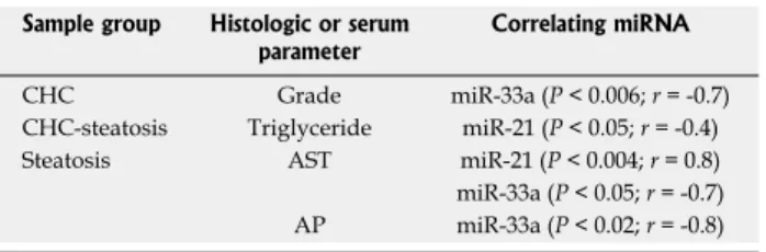
miR-33a levels showed a negative correlation with AP and AST values in Steatosis samples and also with the histology grade in CHC samples. In contrast, the expres- sion of miR-21 correlated positively with AST levels in Steatosis samples but showed a negative correlation with triglyceride levels in CHC-Steatosis samples (Table 2). The analyzed miRNA levels displayed no correlation with ALT, GGT, cholesterol levels or fibrosis stage in the liver disease samples. In addition, a correlation between miRNA expression and steatosis grade was not observed
in either the CHC-Steatosis or the Steatosis group.
DISCUSSION
In HCV infection, it is implied that the pathogenesis of steatosis is linked to both metabolic and viral fac-
tors[12,13,31-33]. HCV has been shown to contribute to st-
eatosis in many ways. The HCV core protein facilitates the accumulation of triglyceride-rich droplets through impaired lipid oxidation and inhibits microsomal trig- lyceride transfer protein activity, which ultimately re- duces the assembly and secretion of apolipoprotein B-containing VLDL[11,34]. In addition to perturbed fat metabolism, fat is reported to be required for the as- sembly of new HCVs[35]. In addition, HCV forms
10 1 0.1 0.01 0.001
2-△△Cq
Normal CHC CHC-steatosis steatosis miR-21
100 10 1 0.1 0.01 0.001
2-△△Cq
Normal CHC CHC-steatosis Steatosis miR-33a
P < 0.0001 P < 0.01
P < 0.02
10 1 0.1 0.01 0.001
2-△△Cq
Normal CHC CHC-steatosis Steatosis miR-122
P < 0.03
100 10 1 0.1 0.01 0.001
2-△△Cq
Normal CHC CHC-steatosis Steatosis miR-221
P < 0.0008 P < 0.01
P < 0.0001
100 10 1 0.1 0.01 0.001
2-△△Cq
Normal CHC CHC-steatosis Steatosis miR-224
P < 0.001 P < 0.003 P < 0.0003P < 0.0001
A B
C D
E
Figure 1 Normalized microRNA expression levels detected in liver samples. The horizontal lines indicate the median of the data set. The displayed P values are the results of Kruskal-Wallis ANOVA test. The upper dotted line indicates two-fold expressional elevation, the lower dotted line signifies a one-half reduction in expres- sion. CHC: Chronic hepatitis C; CHC-steatosis: Steatotic Chronic hepatitis C.
Table 2 Non-parametric Spearman's rank correlation be- tween serum biochemical values, histology grade and miRNA expression levels
lipoviral particles with host lipoproteins and these com- plexes are enriched in triglyceride, cholesterol, and sev- eral apolipoproteins. These structures, which resemble VLDLs, help the viruses avoid detection by the immune system[35]. Furthermore, HCV viral proteins (core, NS2 and NS4B) may contribute to transcriptional activation of lipogenic genes in hepatocytes, such as SREBPs and PPAR-α,γ, contributing to fat accumulation by elevating cholesterol biosynthesis and import from the blood and by deregulating fatty acid β-oxidation and insulin sensi- tivity, respectively[12].
It has been shown that low sterol conditions allow liver cells to protect themselves from additional sterol loss. SREBP-2, a transcription factor, is induced by low intracellular cholesterol level and activates lipogenic genes[25]. This also induces production of miR-33a, which is co-expressed with SREBP-2 from intron 16 of the SREBP-2 gene. miR-33a protects cells from sterol loss by downregulating the ABCA1 transporter needed for efflux of cholesterol to generate high density lipoprotein (HDL)[36]. It has also been demonstrated that activation of miR-33a results in reduced insulin signalling, cellular β-oxidation and cell proliferation by inhibiting CDK6 and cyclinD1, leading to cell cycle arrest in G1 phase[36]. Therefore, antagonism of miR-33a has been proposed as a therapeutic strategy for treating metabolic syndrome and NAFLD[25].
Results of the present study showing elevated miR- 33a levels in CHC-Steatosis samples as compared with CHC, and also in CHC-Steatosis and Steatosis samples when compared with normal liver samples are in accor- dance with previous findings. These findings indicate an elevated fat retention in samples having hepatic steatosis.
The lack of differences between CHC-Steatosis and Steatosis intriguingly suggests that the combination of HCV and steatosis does not further increase the level of miR-33a. Moreover, miR-33a did not correlate with the steatosis stage either in the CHC-Steatosis or Steatosis sample groups.
In contrast, the expression level of miR-122 did not differ between the examined liver disease groups and decreased expression was observed in CHC-Steatosis samples as compared with control liver samples. As in the case of miR-33a, the regulatory function of miR-122 is also connected to fat metabolism. It may act indi-
rectly on lipid metabolism as silencing of miR-122 is reported to decrease cholesterol level, VLDL and HDL fractions as well as fat accumulation in the liver; a target gene involved in fat metabolism, however, is yet to be identified[17]. Therefore, the role of miR-122 in lipid me- tabolism and the observed decreased expression of this miRNA in CHC-Steatosis samples appears contradic- tory. However, miR-122 is known to be a liver-enriched miRNA, composing 70% of total miRNAs in normal hepatocytes[37]. Upon liver injury, when normal hepato- cytic activity is compromised[38], miR-122 levels have been shown to be decreased. For example, miR-122 expression is downregulated in NAFLD[20,21], HCV-infected cases[24]
and HCC[17]. Accordingly, the observed reduction in miR-122 expression further suggests that it likely results from reduced hepatocyte activity due to liver injury.
In HCV infection, miR-122 has also been shown to bind to HCV RNA at two sites of the 5’ UTR, which promotes replication and stimulates translation in vitro[39]. In vivo, however, it is more likely that the role of miR-122 binding to the 5’ UTR is to protect HCV RNA from exo- nuclease degradation, as a correlation between miR-122 expression and intrahepatic or serum viral load has not been identified in vivo[40]. Similarly, our results did not demonstrate increased levels of miR-122 in CHC and CHC-Steatosis samples in the presence of HCV. Further- more, reduced miR-122 expression could also indicate a disease state preceding cancer, as the loss of miR-122 is a frequent finding in HCC and correlates with migration, invasion and in vivo tumorigenesis[41].
It is known that downregulation of a miRNA may upregulate target oncogenes, thus promoting tumor growth due to stimulation of proliferation. In contrast, upregulation of a miRNA may prevent expression of tumor suppressor genes, which normally function to inhibit progression of the cell cycle[19]. In hepatocar- cinogenesis, gradual increases in expression of miR-21, miR-221 and miR-224 are reported to occur as the liver transitions from normal liver through cirrhosis to fully developed HCC[42]. Therefore, we aimed to investigate whether CHC and steatosis - diseases that eventually lead to HCC - would show an elevated expression of these miRNAs. It was interesting to find that the ob- served levels were quite different: miR-224 was upregu- lated, miR-221 was downregulated and miR-21 showed no change.
miR-224 is reported to be an oncomiR that promotes proliferation, migration and invasion in HCC through activation of AKT signaling, and has been shown to function in liver carcinogenesis and progression[43]. Therefore, the observed elevation of miR-224 levels in CHC-Steatosis and Steatosis samples compared with both CHC and normal liver samples may indicate that steatotic CHC and steatosis could be disease states with molecular changes characteristic of a precancerous stage.
It has been suggested that an imbalance in the normal miRNA pattern leading to disease onset can be mea- sured long before the onset of a cancer[16]. miR-221, also a known oncomiR, was found to be elevated in HCC
Sample group Histologic or serum
parameter Correlating miRNA
CHC Grade miR-33a (P < 0.006; r = -0.7)
CHC-steatosis Triglyceride miR-21 (P < 0.05; r = -0.4) Steatosis AST miR-21 (P < 0.004; r = 0.8) miR-33a (P < 0.05; r = -0.7) AP miR-33a (P < 0.02; r = -0.8) AP: Alkaline phosphatase; AST: Aspartate transaminase; CHC: Chronic hepatitis C; miRNA: MicroRNA.
and in precancerous stages of human cirrhotic CHC and a nonalcoholic steatohepatitis mouse model[27]. miR-221 primarily targets tumor suppressors that inhibit the cell cycle. In contrast to the observed elevation in miR-224 levels, the reduced expression of miR-221 in our liver disease samples may indicate that miR-221 is not turned on as early in CHC, CHC-Steatosis or NAFLD, although increased miR-221 expression has been observed in choline and folate-deficient NAFLD animal models[44]. The oncomiR miR-21 has been implicated in promoting cell proliferation, migration, invasion and tumor growth by inducing epithelial-mesenchymal transition through the AKT/ERK pathway[45]. miR-21 is often overex- pressed in cancers, including HCC[27], and in the regener- ating liver[24]. Upregulation of miR-21 is also reported to be present in human liver biopsies of obese patients and rats that were fed high-fat diets. It has been shown that unsaturated fatty acids upregulate miR-21 levels, and this in turn downregulates PTEN and causes fatty acids to trigger steatosis[46]. In contrast, our results did not reveal elevated expression of miR-21 in the examined liver dis- ease groups. Variations in individual miR-21 levels have already been reported in CHC patients[24] and downregu- lation of miR-21 has also been found in obese mice and in steatotic cells[47,48].
Interestingly, our analysis revealed negative correla- tions of miR-33a expression with AST and AP levels and inflammation. A negative correlation was also ob- served when miR-21 levels were compared with triglyc- eride levels. Thus, it appears that fat metabolism may be influenced by the extent of necroinflammation in CHC and Steatosis samples, most likely through the release of miR-33a from damaged liver cells into serum. In addition, high fat is capable of influencing miR-21- regulated proliferation where it may affect liver regenera- tion in CHC-Steatosis. Nevertheless, necroinflammation seemed to correlate with the level of miR-21 in Steatosis samples, thus contributing to the stimulation of pro- liferation. In support of this finding, AST levels have been reported to correlate with miR-21 in a CCl4 mouse model for fibrosis[24].
In conclusion, deregulated expression of selected miRNAs (with the exception of miR-21) was observed in the CHC, CHC-Steatosis and Steatosis groups. The levels of miR-33a and miR-224 were increased in associ- ation with steatosis when compared with CHC-infected and normal livers. Nevertheless, CHC, as a dual effect together with steatosis, might also have contributed to the elevation of these miRNAs in the CHC-Steatosis group, in which the expression of miR-122 might be reduced for similar reasons. In contrast, the observed decrease in the expression of miR-221 was independent of etiology. Intriguingly, no difference in the expression of selected miRNAs was found between CHC-Steatosis samples and steatotic liver samples of metabolic origin, indicating that biological changes caused by steatosis are more profound at the molecular and cellular level than that of CHC alone; however further studies are war-
ranted on this topic.
COMMENTS
Background
Hepatic steatosis is a frequent occurrence in chronic hepatitis C (CHC) infec- tion, which develops due to an imbalance in the uptake and secretion/metabo- lism of fat by hepatocytes. Metabolic factors, as in the case of non-alcoholic fatty disease, and viral factors have been implicated in the cause of steatosis.
Therefore, steatotic CHC-infected liver may bear certain molecular changes that differ from CHC-infection alone and hepatic steatosis of metabolic origin.
Research frontiers
In cancers and in several other pathologies including liver diseases, altered expression of microRNAs (miRNA) has been reported and there is an intense drive to identify miRNAs that could serve as biomarkers to uniquely character- ize a disease or a disease state. With regard to liver diseases specifically, miRNAs have already been proposed as potential biomarkers for hepatocel- lular carcinoma and chronic hepatitis B infection. In addition, identification of miRNAs with altered expression levels and their affected targets will help us to understand the processes leading to the disease and to identify potential thera- peutic targets for treatment.
Innovation and breakthroughs
Altered expression of miRNAs in CHC infection and hepatic steatosis of meta- bolic origin has been reported. At the miRNA level, however, it is not known whether steatotic CHC is closer to CHC or to steatosis, or if it shows a unique miRNA expression pattern. Furthermore, the samples obtained in the present study were acquired prior to the initiation of therapy, thus providing pure dis- ease samples for analysis.
Application
The study results presented here suggest that the combined effect of CHC in- fection and steatosis on miRNA expression may be different from CHC infection alone and normal liver tissue but not from hepatic steatosis of metabolic origin.
This further indicates that biological changes caused by steatosis may be more profound than CHC infection alone at molecular and cellular levels.
Terminology
miRNAs are noncoding RNAs that are approximately 20 nucleotides in length and are known to interfere with gene expression at the posttranscriptional level, usually resulting in prevention of protein synthesis. miRNAs play an important role in many cellular processes, such as development, differentiation and homeostasis, by fine tuning the expression of genes involved. Thus, it is not surprising that their expression is changed in different disease states.
Peer review
This is an interesting study on the role of miRNAs in CHC infection and steato- sis, in which altered miRNA expression was found in steatotic CHC-infected and steatotic liver samples of metabolic origin in relation to CHC infection alone. Intriguingly, the levels of analyzed miRNAs were not altered between the steatotic CHC-infected and steatotic livers. The results are reflective of true disease states as the tissue samples were obtained prior to initiating therapy.
REFERENCES
1 Czepiel J, Biesiada G, Mach T. Viral hepatitis C. Pol Arch Med Wewn 2008; 118: 734-740 [PMID: 19202952]
2 Alter MJ. Epidemiology of hepatitis C virus infection. World J Gastroenterol 2007; 13: 2436-2441 [PMID: 17552026]
3 Aman W, Mousa S, Shiha G, Mousa SA. Current status and future directions in the management of chronic hepatitis C. Vi- rol J 2012; 9: 57 [PMID: 22385500 DOI: 10.1186/1743-422X-9-57]
4 Moriishi K, Matsuura Y. Exploitation of lipid components by viral and host proteins for hepatitis C virus infection.
Front Microbiol 2012; 3: 54 [PMID: 22347882 DOI: 10.3389/
fmicb.2012.00054]
5 Casey LC, Lee WM. Hepatitis C virus therapy update 2013.
Curr Opin Gastroenterol 2013; 29: 243-249 [PMID: 23563981 DOI: 10.1097/MOG.0b013e32835ff972]
6 Khan M, Jahan S, Khaliq S, Ijaz B, Ahmad W, Samreen B, Hassan S. Interaction of the hepatitis C virus (HCV) core
COMMENTS
with cellular genes in the development of HCV-induced ste- atosis. Arch Virol 2010; 155: 1735-1753 [PMID: 20842391 DOI:
10.1007/s00705-010-0797-7]
7 Hwang SJ, Lee SD. Hepatic steatosis and hepatitis C: Still unhappy bedfellows? J Gastroenterol Hepatol 2011; 26 Suppl 1:
96-101 [PMID: 21199519 DOI: 10.1111/j.1440-1746.2010.06542.x]
8 Jármay K, Karácsony G, Nagy A, Schaff Z. Changes in lipid metabolism in chronic hepatitis C. World J Gastroenterol 2005;
11: 6422-6428 [PMID: 16425410]
9 Giannini E, Ceppa P, Botta F, Fasoli A, Romagnoli P, Cresta E, Venturino V, Risso D, Celle G, Testa R. Steatosis and bile duct damage in chronic hepatitis C: distribution and rela- tionships in a group of Northern Italian patients. Liver 1999;
19: 432-437 [PMID: 10533803]
10 Hourioux C, Patient R, Morin A, Blanchard E, Moreau A, Trassard S, Giraudeau B, Roingeard P. The genotype 3-spe- cific hepatitis C virus core protein residue phenylalanine 164 increases steatosis in an in vitro cellular model. Gut 2007; 56:
1302-1308 [PMID: 17213339]
11 Abid K, Pazienza V, de Gottardi A, Rubbia-Brandt L, Conne B, Pugnale P, Rossi C, Mangia A, Negro F. An in vitro mo- del of hepatitis C virus genotype 3a-associated triglycerides accumulation. J Hepatol 2005; 42: 744-751 [PMID: 15826725]
12 Felmlee DJ, Hafirassou ML, Lefevre M, Baumert TF, Schus- ter C. Hepatitis C virus, cholesterol and lipoproteins--im- pact for the viral life cycle and pathogenesis of liver disease.
Viruses 2013; 5: 1292-1324 [PMID: 23698400 DOI: 10.3390/
v5051292]
13 El-Zayadi AR. Hepatic steatosis: a benign disease or a si- lent killer. World J Gastroenterol 2008; 14: 4120-4126 [PMID:
18636654]
14 Rosmorduc O, Fartoux L. HCC and NASH: how strong is the clinical demonstration? Clin Res Hepatol Gastroenterol 2012; 36:
202-208 [PMID: 22326764 DOI: 10.1016/j.clinre.2011.12.011]
15 Krawczyk M, Bonfrate L, Portincasa P. Nonalcoholic fatty liver disease. Best Pract Res Clin Gastroenterol 2010; 24: 695-708 [PMID: 20955971 DOI: 10.1016/j.bpg.2010.08.005]
16 Ciesla M, Skrzypek K, Kozakowska M, Loboda A, Jozkow- icz A, Dulak J. MicroRNAs as biomarkers of disease onset.
Anal Bioanal Chem 2011; 401: 2051-2061 [PMID: 21544542 DOI: 10.1007/s00216-011-5001-8]
17 Hu J, Xu Y, Hao J, Wang S, Li C, Meng S. MiR-122 in he- patic function and liver diseases. Protein Cell 2012; 3: 364-371 [PMID: 22610888 DOI: 10.1007/s13238-012-2036-3]
18 Carthew RW, Sontheimer EJ. Origins and Mechanisms of miRNAs and siRNAs. Cell 2009; 136: 642-655 [PMID: 19239886 DOI: 10.1016/j.cell.2009.01.035]
19 Shenouda SK, Alahari SK. MicroRNA function in cancer: on- cogene or a tumor suppressor? Cancer Metastasis Rev 2009; 28:
369-378 [PMID: 20012925 DOI: 10.1007/s10555-009-9188-5]
20 Kerr TA, Korenblat KM, Davidson NO. MicroRNAs and liver disease. Transl Res 2011; 157: 241-252 [PMID: 21420035 DOI: 10.1016/j.trsl.2011.01.008]
21 Lakner AM, Bonkovsky HL, Schrum LW. microRNAs:
fad or future of liver disease. World J Gastroenterol 2011; 17:
2536-2542 [PMID: 21633658 DOI: 10.3748/wjg.v17.i20.2536]
22 Borel F, Konstantinova P, Jansen PL. Diagnostic and thera- peutic potential of miRNA signatures in patients with he- patocellular carcinoma. J Hepatol 2012; 56: 1371-1383 [PMID:
22314424 DOI: 10.1016/j.jhep.2011.11.026]
23 Waidmann O, Bihrer V, Pleli T, Farnik H, Berger A, Zeuzem S, Kronenberger B, Piiper A. Serum microRNA-122 levels in different groups of patients with chronic hepatitis B virus infection. J Viral Hepat 2012; 19: e58-e65 [PMID: 22239527 DOI: 10.1111/j.1365-2893.2011.01536.x]
24 Marquez RT, Bandyopadhyay S, Wendlandt EB, Keck K, Hoffer BA, Icardi MS, Christensen RN, Schmidt WN, Mc- Caffrey AP. Correlation between microRNA expression lev- els and clinical parameters associated with chronic hepatitis C viral infection in humans. Lab Invest 2010; 90: 1727-1736
[PMID: 20625373 DOI: 10.1038/labinvest.2010.126]
25 Dávalos A, Goedeke L, Smibert P, Ramírez CM, Warrier NP, Andreo U, Cirera-Salinas D, Rayner K, Suresh U, Pas- tor-Pareja JC, Esplugues E, Fisher EA, Penalva LO, Moore KJ, Suárez Y, Lai EC, Fernández-Hernando C. miR-33a/b contribute to the regulation of fatty acid metabolism and in- sulin signaling. Proc Natl Acad Sci USA 2011; 108: 9232-9237 [PMID: 21576456 DOI: 10.1073/pnas.1102281108]
26 Ogawa T, Enomoto M, Fujii H, Sekiya Y, Yoshizato K, Ikeda K, Kawada N. MicroRNA-221/222 upregulation indicates the activation of stellate cells and the progression of liver fibrosis. Gut 2012; 61: 1600-1609 [PMID: 22267590]
27 Karakatsanis A, Papaconstantinou I, Gazouli M, Lyberopou- lou A, Polymeneas G, Voros D. Expression of microRNAs, miR-21, miR-31, miR-122, miR-145, miR-146a, miR-200c, miR-221, miR-222, and miR-223 in patients with hepatocel- lular carcinoma or intrahepatic cholangiocarcinoma and its prognostic significance. Mol Carcinog 2013; 52: 297-303 [PMID:
22213236 DOI: 10.1002/mc.21864]
28 Ishak K, Baptista A, Bianchi L, Callea F, De Groote J, Gudat F, Denk H, Desmet V, Korb G, MacSween RN. Histological grading and staging of chronic hepatitis. J Hepatol 1995; 22:
696-699 [PMID: 7560864]
29 Doleshal M, Magotra AA, Choudhury B, Cannon BD, Labourier E, Szafranska AE. Evaluation and validation of total RNA extraction methods for microRNA expression analyses in formalin-fixed, paraffin-embedded tissues. J Mol Diagn 2008; 10: 203-211 [PMID: 18403610 DOI: 10.2353/
jmoldx.2008.070153]
30 Andersen CL, Jensen JL, Ørntoft TF. Normalization of real- time quantitative reverse transcription-PCR data: a model- based variance estimation approach to identify genes suited for normalization, applied to bladder and colon cancer data sets. Cancer Res 2004; 64: 5245-5250 [PMID: 15289330]
31 Barba G, Harper F, Harada T, Kohara M, Goulinet S, Matsu- ura Y, Eder G, Schaff Z, Chapman MJ, Miyamura T, Bréchot C. Hepatitis C virus core protein shows a cytoplasmic local- ization and associates to cellular lipid storage droplets. Proc Natl Acad Sci USA 1997; 94: 1200-1205 [PMID: 9037030]
32 Ratziu V, Trabut JB, Poynard T. Fat, diabetes, and liver injury in chronic hepatitis C. Curr Gastroenterol Rep 2004; 6:
22-29 [PMID: 14720450]
33 Monto A, Alonzo J, Watson JJ, Grunfeld C, Wright TL. Ste- atosis in chronic hepatitis C: relative contributions of obesity, diabetes mellitus, and alcohol. Hepatology 2002; 36: 729-736 [PMID: 12198667]
34 Cross TJ, Rashid MM, Berry PA, Harrison PM. The impor- tance of steatosis in chronic hepatitis C infection and its management: A review. Hepatol Res 2010; 40: 237-247 [PMID:
20394672]
35 Bassendine MF, Sheridan DA, Bridge SH, Felmlee DJ, Neely RD. Lipids and HCV. Semin Immunopathol 2013; 35: 87-100 [PMID: 23111699 DOI: 10.1007/s00281-012-0356-2]
36 Cirera-Salinas D, Pauta M, Allen RM, Salerno AG, Ramírez CM, Chamorro-Jorganes A, Wanschel AC, Lasuncion MA, Morales-Ruiz M, Suarez Y, Baldan Á, Esplugues E, Fernán- dez-Hernando C. Mir-33 regulates cell proliferation and cell cycle progression. Cell Cycle 2012; 11: 922-933 [PMID:
22333591 DOI: 10.4161/cc.11.5.19421]
37 Lagos-Quintana M, Rauhut R, Yalcin A, Meyer J, Lendeckel W, Tuschl T. Identification of tissue-specific microRNAs from mouse. Curr Biol 2002; 12: 735-739 [PMID: 12007417]
38 Ding X, Ding J, Ning J, Yi F, Chen J, Zhao D, Zheng J, Liang Z, Hu Z, Du Q. Circulating microRNA-122 as a potential biomarker for liver injury. Mol Med Rep 2012; 5: 1428-1432 [PMID: 22427142 DOI: 10.3892/mmr.2012.838]
39 Shimakami T, Yamane D, Jangra RK, Kempf BJ, Spaniel C, Barton DJ, Lemon SM. Stabilization of hepatitis C virus RNA by an Ago2-miR-122 complex. Proc Natl Acad Sci USA 2012;
109: 941-946 [PMID: 22215596 DOI: 10.1073/pnas.1112263109]
40 Sarasin-Filipowicz M. Interferon therapy of hepatitis C:
molecular insights into success and failure. Swiss Med Wkly 2010; 140: 3-11 [PMID: 19950038]
41 Scisciani C, Vossio S, Guerrieri F, Schinzari V, De Iaco R, D’Onorio de Meo P, Cervello M, Montalto G, Pollicino T, Raimondo G, Levrero M, Pediconi N. Transcriptional regu- lation of miR-224 upregulated in human HCCs by NFκB inflammatory pathways. J Hepatol 2012; 56: 855-861 [PMID:
22178270 DOI: 10.1016/j.jhep.2011.11.017]
42 Pineau P, Volinia S, McJunkin K, Marchio A, Battiston C, Terris B, Mazzaferro V, Lowe SW, Croce CM, Dejean A.
miR-221 overexpression contributes to liver tumorigenesis.
Proc Natl Acad Sci USA 2010; 107: 264-269 [PMID: 20018759 DOI: 10.1073/pnas.0907904107]
43 Ma D, Tao X, Gao F, Fan C, Wu D. miR-224 functions as an onco-miRNA in hepatocellular carcinoma cells by activating AKT signaling. Oncol Lett 2012; 4: 483-488 [PMID: 23741247]
44 Tryndyak VP, Latendresse JR, Montgomery B, Ross SA, Beland FA, Rusyn I, Pogribny IP. Plasma microRNAs are sensitive indicators of inter-strain differences in the sever- ity of liver injury induced in mice by a choline- and folate- deficient diet. Toxicol Appl Pharmacol 2012; 262: 52-59 [PMID:
22561871 DOI: 10.1016/j.taap.2012.04.018]
45 Bao L, Yan Y, Xu C, Ji W, Shen S, Xu G, Zeng Y, Sun B, Qian H, Chen L, Wu M, Su C, Chen J. MicroRNA-21 suppresses PTEN and hSulf-1 expression and promotes hepatocellular carcinoma progression through AKT/ERK pathways. Can- cer Lett 2013; 337: 226-236 [PMID: 23684551 DOI: 10.1016/
j.canlet.2013.05.007]
46 Vinciguerra M, Sgroi A, Veyrat-Durebex C, Rubbia-Brandt L, Buhler LH, Foti M. Unsaturated fatty acids inhibit the expression of tumor suppressor phosphatase and tensin homolog (PTEN) via microRNA-21 up-regulation in hepato- cytes. Hepatology 2009; 49: 1176-1184 [PMID: 19072831 DOI:
10.1002/hep.22737]
47 Li S, Chen X, Zhang H, Liang X, Xiang Y, Yu C, Zen K, Li Y, Zhang CY. Differential expression of microRNAs in mouse liv- er under aberrant energy metabolic status. J Lipid Res 2009; 50:
1756-1765 [PMID: 19372595 DOI: 10.1194/jlr.M800509-JLR200]
48 Zheng L, Lv GC, Sheng J, Yang YD. Effect of miRNA-10b in regulating cellular steatosis level by targeting PPAR- alpha expression, a novel mechanism for the pathogenesis of NAFLD. J Gastroenterol Hepatol 2010; 25: 156-163 [PMID:
19780876 DOI: 10.1111/j.1440-1746.2009.05949.x]
P- Reviewer: Osna NA S- Editor: Ma YJ L- Editor: A E- Editor: Liu XM