Effects of Pituitary Adenylate Cyclase Activating Polypeptide on Human Sperm Motility
R. Brubel&P. Kiss&A. Vincze&A. Varga&A. Varnagy&
J. Bodis&L. Mark&E. Jambor&G. Maasz&
H. Hashimoto&Zs. Helyes&G. Toth&A. Tamas&
M. Koppan&D. Reglodi
Received: 30 March 2012 / Accepted: 3 May 2012
#Springer Science+Business Media, LLC 2012
Abstract Pituitary adenylate cyclase activating polypep- tide (PACAP), a neuropeptide with diverse effects, was originally isolated as a hypothalamo-hypophyseal pep- tide. Subsequent studies showed highest levels of PACAP in the testis after the brain, suggesting that it influences the development and functioning of sperma- tozoa. Indeed, it has been proven that PACAP has an effect on spermatogenesis, both locally and via influ- encing the hypothalamo-hypophyseal–gonadal axis. The aim of the present study was to determine whether PACAP has an effect on human sperm motility and whether it is present in the human seminal fluid.
Furthermore, the sperm head morphology was studied in mice lacking endogenous PACAP. Human samples were obtained from healthy adult volunteers and andro- logical patients. The effects of PACAP on the motility
of human sperm cells were investigated using a com- puter aided sperm analysis system. In cases where the motility was lower, addition of PACAP to the samples increased the motility and the ratio of rapid progressive and medium progressive sperm motility groups. The presence of PACAP could not be detected in human seminal fluid samples by means of mass spectrometry.
Investigating sperm head morphology with routine his- tology in PACAP deficient mice revealed that both the longitudinal and transverse diameters were significantly lower in PACAP deficient mice, without marked difference in the shape, as revealed by scanning electron microscopy.
Keywords Sperm motility . Semen . Mass spectrometry . Scanning electron microscopy
M. Koppan and D. Reglodi contributed equally to the present work.
R. Brubel (*)
:
P. Kiss:
A. Vincze:
A. Varga:
A. Tamas:
D. Reglodi
Department of Anatomy, PTE-MTA Lendulet PACAP Research Group, University of Pecs,
Pecs, Hungary
e-mail: brubelreka@gmail.com A. Varnagy
:
J. Bodis:
M. KoppanDepartment of Obstetrics and Gynaecology, University of Pecs, Pecs, Hungary
L. Mark
:
E. Jambor:
G. MaaszDepartment of Biochemistry and Medical Chemistry, University of Pecs,
Pecs, Hungary H. Hashimoto
Laboratory of Molecular Neuropharmacology,
Graduate School of Pharmaceutical Sciences, Osaka University, Osaka, Japan
H. Hashimoto
Center for Child Mental Development,
United Graduate School of Child Development, Osaka University, Kanazawa University and Hamamatsu University
School of Medicine, Osaka, Japan H. Hashimoto
Department of Molecular Pharmaceutical Science, Graduate School of Medicine, Osaka University, Osaka, Japan
Z. Helyes
Department of Pharmacology and Pharmacotherapy, University of Pecs,
Pecs, Hungary G. Toth
Department of Medical Chemistry, University of Szeged, Szeged, Hungary
DOI 10.1007/s12031-012-9806-5
Introduction
Pituitary adenylate cyclase activating polypeptide (PACAP) was originally isolated from the hypothalamus, based on its cAMP-increasing effect in pituitary cells (Arimura 2007;
Miyata et al. 1989). Although the last two decades since its discovery have revealed that the effects of PACAP reach beyond hypthalamo-hypophyseal effects, its functions in the endocrine system are still in focus of research (Counis et al.
2007; Sherwood et al.2000; Vaudry et al.2009). Regarding reproductive endocrinology, PACAP has been shown to play a role in the regulation of gonadotropin secretion (Counis et al.2007; Koves et al.2003; Szabo et al.2004), in fertility, receptivity, implantation and reproductive behav- ior (Apostolakis et al.2005; Sherwood et al.2007) and in placental functions (Reglodi et al. 2008). Relatively few human data are available on the functions of PACAP in reproductive endocrinology. It has been shown that PACAP infusion increased some pituitary hormone levels in men (Chiodera et al.1995). Recently, we have described a cor- relation between the number of retrieved oocytes and PACAP levels in the follicular fluid in women undergoing superovulation treatment (Koppan et al.2012).
In male animals, PACAP has been shown to influence the development and functioning of spermatozoa (Gozes et al.
1998; Li et al.2004). In peripheral organs, highest level of PACAP was detected in the testis in the earliest PACAP studies (Arimura et al. 1991). Subsequent studies have revealed that PACAP plays a role in the regulation of sper- matogenesis and testicular aging. Spermatogenesis is thought to be influenced by PACAP at several levels:
PACAP is expressed in immature sperm cells, the epididymis-derived PACAP may influence the final stages of spermiogenesis, PACAP regulates the activity of the supporting Sertoli cells, and the peptide acts at hormonal level, influencing testosterone synthesis by Leydig cells (Heindel et al.1992; El-Gehani et al.2000; Lacombe et al.
2006; Li et al. 2004; Shioda et al. 1994; Yanaihara et al.
1998; West et al.1995).
Previously, it has been described that the addition of PACAP7–27 hybrid antagonist results in a dose-dependent reduction in sperm motility in golden hamster (Gozes et al.
1998). This suggests that PACAP increases sperm motility.
A recent study has reported that PACAP indeed increases sperm motility and penetration to promote fertilization in mice (Tanii et al.2011). Given the lack of data in humans, the first aim of the present study was to investigate whether the addition of PACAP to the seminal fluid influences sperm motility in humans.
After obtaining positive results, we raised the question as to whether PACAP occurs in the human seminal fluid, which is a complex mixture of products of the male repro- ductive tract from the seminiferous tubules through the
epididymis and the accessory genital glands (Chalabi et al.
2002). Proteomic/peptidomic analysis of the seminal fluid has gained increasing interest not only for the elucidation of the biological roles played by different peptides and proteins but also in search for potential biomarkers in male infertility (Khan et al.1992). We have previously shown the presence of PACAP in human serum, breast milk and follicular fluid using MALDI and MALDI TOF/TOF mass spectrometry analysis (Borzsei et al. 2009; Brubel et al. 2011), so in search of PACAP in the seminal fluid we decided to apply this high throughput technique adapted to analyze peptide/
protein composition of biological fluids (Hu et al.2006).
Based on the inhibitory effects of the PACAP antagonist on sperm motility (Gozes et al. 1998), it is suggested that endogenous PACAP is needed for proper motility. We there- fore hypothesized that the lack of PACAP may result in abnormal sperm morphology leading to abnormal motility.
The normal structure of several organs and tissues has been shown to be morphologically intact at macroscopical and light microscopical level in PACAP knockout mice (Azuma et al. 2008; Ferencz et al. 2010; Reglodi et al. 2012;
Szabadfi et al.2012; Szakaly et al.2011), but the structure of sperms has not been investigated yet. Therefore, the third aim of the present study was to investigate sperm morphol- ogy in PACAP gene knockout mice using light and scanning electron microscopical examinations.
Materials and Methods
Sperm Motility Analysis
Seminal fluid samples were collected from healthy adult male volunteers (age between 20 and 25 years,n030) and patients with fertility problems (age between 25 and 35 years,n040) after at least 3 days of abstinence. Human sample collection was carried out according to a protocol approved by the institutional ethic committee (3117/2008, 3610/2009), and after obtaining written consent of the volunteers. Sperm mo- tility was determined by medeaLAB CASA, which is a well- established system for standardized sperm analysis, also sup- ported by the World Health Organisation (Youn et al. 2011;
Nöthling and Dos Santos 2012). We divided the sperms according to the Manual of the CASA into four groups based on the motility: groups A (rapid progressive), B (medium progressive), C (non-progressive) and D (immotile). After measuring the control motility (10μl of semen without any treatment), we treated the samples with saline (5μl of semen together with 5μl of saline) or PACAP1–38 (5μl of semen with 5μl of 100 nmol PACAP1–38) followed by a second motility measurement. Results of groups A and B with good motility were combined as well as those from groups C and D with worse motility.
Mass Spectrometry
Samples were obtained from healthy volunteers, similarly to the described motility analysis in the previous section (n040).
The samples were further processed for mass spectrometry analysis based on modifications of earlier descriptions (Schiller et al.2000). The peptidase inhibitor aprotinin was added to all samples (30μl/ml). The seminal fluid sample was vortexed, and 100μl was centrifuged at 4,500 rpm for 10 min.
In order to analyze the presence of PACAP in the seminal fluid, the supernatant was removed, desalted and cleaned using 0.1 % trifluoroacetic acid (TFA) solution with Zip- Tip18 pipette tips (Millipore Kft., Hungary). The purified proteins and peptides were eluted directly onto the MALDI target plate (MTP 384 massive target T, Bruker Daltonics, Bremen, Germany) by 3 μl of acetonitrile/0.1 % TFA (50/50, v/v) solution. One microliter of matrix solution (10 mg/ml) was prepared fresh every day by dissolving the matrix in acetonitrile/0.1 % TFA (1/2, v/v) and mixing with the purified solution. Detection of PACAP was attempted using three different matrices:α-cyano-4-hydroxycinnamic acid, sinapic acid and 2,5-dihydroxybenzoic acid. In order to analyze the presence of PACAP in the spermatozoa-rich fraction, the pellet was washed with 200μl TBS (tris-Na-Cl) and centrifuged at 4,500 rpm for 10 min. Supernatant was removed, and 100μl of lysis buffer (20 mM Hepes, 10 mM EGTA, 2 mM EDTA, pH07.5–8) was added to the pellet.
The solution was vortexed and ultrasounded and eluted directly onto the MALDI target plate (MTP 384 massive target T, Bruker Daltonics, Bremen, Germany) by 3μl of acetonitrile/0.1 % TFA (50/50, v/v) solution. One microliter of saturated matrix solution was prepared fresh every day by dissolving 3,5-dimethoxy-4-hydroxycinnamic acid in acetonitrile/0.1 % TFA (1/2, v/v) and was mixed with the purified solution (Schiller et al.2000). Finally, in order to confirm the detectability of PACAP in seminal fluid samples, we added extra amount of PACAP to measured samples in different concentrations between 100 ng and 10μg PACAP38 dissolved in 5μl TFA. The exogenously added PACAP38 was then detected using the above- described method.
For mass spectrometry, the ions were accelerated under delayed extraction conditions (200 ns) in positive ion mode with an acceleration voltage of 20.00 kV. The instrument uses a 337-nm pulsed nitrogen laser, model MNL-205MC (LTB Lasertechnik Berlin GmbH., Berlin, Germany). Exter- nal calibration was performed in each case using the Bruker Peptide Calibration Standard (#206195 Peptide Calibration Standard, Bruker Daltonics, Bremen, Germany). Protein masses were acquired with a range of m/z 1,000–10,000.
Each spectrum was composed by accumulating data from 200 consecutive laser shots for standard solution and 1,000 for seminal fluid samples. The Bruker FlexControl 2.4
software was used for control of the instrument and the Bruker Flexanalysis 2.4 software for spectra evaluation.
Light and Scanning Electron Microscopical Analysis of Sperm Heads in PACAP Deficient Mice
The study was performed using male PACAP−/−and control C57BL/6 mice. The maintenance and generation of the PACAP−/− mice was followed as described earlier by Hashimoto et al. (2001). Mice were bred and kept in the Laboratory Animal House of the Department of Anatomy of the University of Pecs. Animal housing, care and applica- tion of experimental procedures were in accordance with institutional guidelines under approved protocols (No.
BA02/2000-24/2011, University of Pecs). Animals were maintained under a 12-h light/dark cycle with free access to food and water. For sperm head morphological analysis, adult PACAP−/−and control mice were terminally anesthe- tized with an overdose of mixture of ketamine and xylazin.
Epididymis was dissected from each PACAP-deficient and wild-type animal (n05/group). For light microscopic evalu- ation, epididymal seminal fluid was dissolved in 1 ml stan- dard IVF solution (used during in vitro fertilization procedures), dropped to a gelatin coated slide and dispersed.
After drying on a 40°C plate, haematoxylin–eosin staining was performed. Sperms were photographed (Nikon FXA microphotograph), and both the longitudinal and transverse diameters of the sperm heads were measured (n>300) using Adobe Photoshop CS4 extended 11.0.1 program. Statistical analysis (non-parametric, Mann–Whitney U-test) was car- ried out with GraphPad Prism 5.0. For scanning electron microscopic evaluation, sperms were fixed in 2 % of formaldehyde and 2.5 % of glutaraldehyde fixative over- night in 4°C after dissection. Samples were washed and dehydrated. One drop of supernatant was dropped to a glass surface, and it was sputter coated with gold. The sample was examined and photographed with a Jeol scanning electron microscope.
Results
Sperm Motility Analysis
Our results show that saline administration did not influence the ratio of rapid progressive and medium progressive sperm cells (type A + B) or the ratio of the other groups (type C + D).
Compared to saline, 100 nmol PACAP1–38 significantly in- creased the ratio of type A + B sperm cells, while it signifi- cantly decreased the ratio of type C + D sperms (Fig.1). We found this motility-increasing effect only in samples where the percentage of group A was below 80 %. In cases where the motility was above the average (group A was over 80 %),
PACAP1–38 could not increase the ratio of good motility groups. Accordingly, the data obtained in these samples are omitted from the final results.
Mass Spectrometry
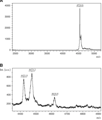
The sample preparation procedure optimized for each bio- logical fluid was suitable for measuring and identifying low molecular weight peptides by mass spectrometry. Based on our previous and current results, the sensitive and reproduc- ible identification of PACAP1–38 can be carried out by using linear MALDI TOF MS. The characteristic peak of PACAP1–38, 4534.6 Da, was verified in the PACAP stan- dard solutions (Fig. 2a). However, our mass spectrometry analysis could not reveal the presence of PACAP38 in the seminal fluid samples obtained either from healthy adult male volunteers or patients with fertility problems (Fig. 2b). In order to confirm that the inability to detect PACAP was not due to technical problems with the seminal fluid, we repeated these experiments with exogenously added PACAP in different concentrations and using differ- ent matrices. PACAP38 was easily detectable in seminal fluid samples when added exogenously to samples that measured negative beforehand (Fig.3).
Sperm Morphology in PACAP Deficient Mice
The main morphological parameters showed marked differences between the two groups (Fig.4). Sperm heads from PACAP deficient mice were smaller, and more abnormal-shaped heads could be observed than in their wild-type littermates (Fig.4a–c).
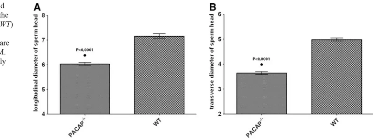
Morphological analysis showed that both longitudinal and trans- verse diameters of sperm heads from PACAP−/− mice were significantly smaller than those found in wild-type mice. Mean longitudinal diameter values were 6.04μm in PACAP−/−and 7.16μm in wild-type mice (Fig.5a). Mean transverse diameter values were 3.64μm in PACAP-/- and 4.98μm in wild-type
mice (Fig. 5b). The difference between the above-mentioned parameters was statistically significant (P<0.0001). Detailed ultrastructural surface analysis by scanning electron microscopy showed no marked alteration in the shape of sperms from wild- type and PACAP−/−mice (Fig.6).
Discussion
In the present study we showed that PACAP increases sperm motility in human spermatozoa. In addition, we provided evidence that the lack of endogenous PACAP leads to abnormal sperm morphology.
Although the exact role of PACAP in spermatogenesis is not yet known, several lines of evidence show that PACAP is involved in the development of spermatozoa. First, the testis contains the highest levels of PACAP after the nervous system (Arimura et al. 1991). Second, PACAP and its messenger RNA occur in developing spermatogonia and spermatocytes and in the wall of the epididymis (Daniel and Habener2000;
Hannibal and Fahrenkrug1995; Koh et al.2003; Kononen et al. 1994; Leung et al. 1998; Yanaihara et al. 1998). Third, PACAP has effects on the supporting cells of Sertoli, where the peptide increases cAMP production (Heindel et al.1992).
Fig. 1 Graph showing that PACAP1–38 significantly increased the ratio of rapid progressive and medium progressive sperm cells (type A + B) and decreased the ratio of non-progressive and immotile sperm cells (type C + D). *P<0.05 vs saline-treated group
A
4534.6
0 1000 2000 3000 4000
2500 3000 3500 4000 4500 5000
m/z
B
4473.2 4421.8
4618.8
200 400 600 800 Int. [a.u.]
4400 4500 4600 4700 4800 4900
m/z Fig. 2 aMALDI TOF spectrum of PACAP38 in positive ion mode using linear detection indicating the molecular weight at 4534.6 Da in PACAP38 standard. b MALDI TOF spectrum of a seminal fluid sample. The peptide peak characteristic for PACAP38 could not be identified
A
4053.6
4161.9 4317.5
4487.1 4776.5
5000.0 6274.0
2770.9
5805.4 3122.6
3244.1 2913.3
3809.0
1945.5
2480.7
1654.8 0 1 2 3 4 5 x104
x104
Intens. [a.u.]
2000 2500 3000 3500 4000 4500 5000 5500 6000
m/z
B
4826.8 4534.2
3243.2
2769.6
3964.8
3121.3 2909.1
3807.0
4314.1
2610.0 2333.1
3409.8
0.0 0.5 1.0 1.5 2.0
Intens. [a.u.]
2000 2500 3000 3500 4000 4500 5000 5500 6000
m/z
Fig. 3 A representative MALDI TOF spectrum of a seminal fluid sample in a wide molecular weight range.aNative seminal fluid sample.bSeminal fluid sample with exogenously added 0.5μg PACAP38 showing an extra peak at a molecular weight 4534m/z, identified as PACAP
Fig. 4 Representative microphotographs of haematoxylin–eosin-stained sperm heads obtained from wild-type mice (WT,a) and PACAP-deficient mice (PACAP−/−,bandc).aNormal sperm from WT mouse.b Sperm from PACAP−/−mouse, with smaller head diameter than WT.cSperm with abnormal head morphology in PACAP−/−
mice. Scale bar: 15μm
In addition, PACAP has been shown to have several other functions in the testis, such as stage-specific suppressive action on immature Leydig cell proliferation and stimulation of testosterone secretion in Leydig cells (El-Gehani et al.
1998, 2000; Matsumoto et al. 2008; Rossato et al. 1997).
Finally, PACAP is able to cross the blood-testis barrier (Banks et al.1993), and it is expressed in testicular blood vessels leading to vasodilatory effects in testicular and epididymal microvessels (Koh et al.2003; Lissbrant et al.1999).
As far as motility is concerned, PACAP antagonists have been shown to inhibit sperm motility, suggesting an endoge- nous role of PACAP influencing sperm function (Gozes et al.
1998). In the same study, the addition of the agonist peptides (VIP or PACAP) did not influence motility in the golden hamster, but a recent work has shown that PACAP increases sperm motility in mice (Tanii et al.2011). In our present study we showed that PACAP increased motility of human sperms.
We found that normal sperm motility was not altered by PACAP administration, in accordance with the results of Gozes et al. (1998). However, when the ratio of slow
progressing sperms was high, the addition of PACAP in- creased motility. These observations are thus in accordance with the previously described data showing that PACAP itself does not influence motility in the normal hamster (where supposedly no abnormal sperms were present). Our observa- tions are also in accordance with the generally accepted role of PACAP acting as a stress-response peptide, affecting abnor- mal functions under pathophysiological circumstances (Reglodi et al.2012). Our finding that PACAP deficient mice had smaller sperm head diameters without significant alter- ation of shape supports the important role of endogenous PACAP, and it might be an additional reason for the decreased fertility usually observed in PACAP deficient mice. It is also in accordance with the many disorders of the mice lacking endogenous PACAP (Reglodi et al.2012).
The finding that PACAP increases motility raised the question whether PACAP occurred in the seminal fluid.
However, we were not able to detect PACAP using mass spectrometry in the supernatant or in the sperm cell-rich fraction of the 40 human seminal fluid samples. There is a contradiction in the literature about the occurrence of PACAP in mature spermatozoa. Early studies described the disappearance of PACAP immunoreactivity in mature sperms compared to spermatid acrosomes (Yanaihara et al.
1998). However, a recent study reinvestigated this issue using mild fixation conditions and found that PACAP im- munoreactivity was present in the sperm acrosome and in the middle piece of the flagellum (Tanii et al. 2011). Our present results are in accordance with those showing the lack of PACAP in mature spermatozoa and, in addition, show the lack of PACAP in the seminal fluid. If we suppose that PACAP acts as a motility-increasing factor during fer- tilization, the question still remains where the PACAP acting on sperms comes from. One possibility is the wall of the seminal duct and accessory gland, where PACAP immuno- reactivity has been described earlier (Vaudry et al. 2009).
However, the lack of PACAP by mass spectrometry ques- tions this possibility, although levels of PACAP may be very low, below the detection limit of our method. A further potential source could be the female genital tract. Although Fig. 5 Longitudinal (a) and
transverse (b) diameter of the sperm heads in wild-type (WT) and PACAP deficient (PACAP−/−) mice. Results are given in micrometer ± SEM.
Differences were statistically significant (P<0.0001)
Fig. 6 Representative scanning electron microscopic figure showing sperm heads in wild-type (WT,a) and PACAP-deficient (PACAP−/−,b) mice. No marked differences can be observed in their shape. Scale bar:
1μm
we could not detect the presence of PACAP in the human vaginal smear in a recent study (Brubel et al. 2011), it cannot be excluded that other parts of the female genital tract secrete PACAP in case of motility problems.
In summary, the present study showed, for the first time, that PACAP increases sperm motility in human spermato- zoa. In addition, it provided evidence that the lack of en- dogenous PACAP leads to abnormal sperm morphology.
The functional and clinical significance of these observa- tions requires further investigation.
Acknowledgments This work was supported by Hungarian National Scientific grants OTKA K72592 and CNK 78480, Richter Gedeon Centenary Foundation, SROP 4.1.2.B-10/2/KONV-20/0-0002, SROP 4.2.2/B-10/1-2010-0029, Bolyai Scholarship, MTA Lendulet Program, Pecs University Research Grant ÁOKKA-34039/10-14 and Funding Program for Next Generation World-Leading Researchers (established by the Council for Science and Technology Policy, Cabinet Office, Government of Japan).
References
Apostolakis EM, Riherd DN, O`Malley BW (2005) PAC1 receptors mediate pituitary adenylate cyclase-activating polypeptide- and progesterone-facilitated receptivity in female rats. Mol Endocrinol 19:2798–2811
Arimura A (2007) PACAP: the road to discovery. Peptides 28:1617–1619 Arimura A, Somogyvari-Vigh A, Miyata A et al (1991) Tissue distri- bution of PACAP as determined by RIA: highly abundant in the rat brain and testes. Endocrinology 129:2787–2789
Azuma YT, Hagi K, Shintani N et al (2008) PACAP provides colonic protection against dextran sodium sulfate induced colitis. J Cell Physiol 216:111–119
Banks WA, Kastin AJ, Komaki G, Arimura A (1993) Pituitary adenylate cyclase activating polypeptide (PACAP) can cross the vascular com- ponent of the blood-testis barrier in the mouse. J Androl 14:170–173 Borzsei R, Mark L, Tamas A et al (2009) Presence of pituitary adeny- late cyclase activating polypeptide-38 in human plasma and milk.
Eur J Endocrinol 160:561–565
Brubel R, Reglodi D, Jambor E et al (2011) Investigation of pituitary adenylate cyclase activating polypeptide in human gynecological and other biological fluids by using MALDI TOF mass spectrom- etry. J Mass Spectrom 46:189–194
Chalabi S, Easton RL, Patankar MS et al (2002) The expression of free oligosaccharides in human seminal plasma. J Biol Chem 277:32562–32570
Chiodera P, Volpi R, Capretti L, Coiro V (1995) Effects of intrave- nously infused pituitary adenylate cyclase-activating polypeptide on arginine vasopressin and oxytocin secretion in man. Neuro- report 6:1490–1492
Counis R, Laverriere JN, Garrel-Lazayres G et al (2007) What is the role of PACAP in gonadotrope function? Peptides 28:1797–1804 Daniel PB, Habener JF (2000) Pituitary adenylate cyclase activating poly- peptide gene expression regulated by a testis-specific promoter in germ cells during spermatogenesis. Endocrinology 141:1218–1227 El-Gehani F, Zhang FP, Pakarinen P, Rannikko A, Huhtaniemi I (1998)
Gonadotropin-independent regulation of steroidogenesis in the fetal rat testis. Biol Reprod 58:116–123
El-Gehani F, Tena-Sempere M, Huhtaniemi I (2000) Evidence that pitu- itary adenylate cyclase activating polypeptide is a potent regulator of fetal rat testicular steroidogenesis. Biol Reprod 63:1482–1489
Ferencz A, Kiss P, Weber G et al (2010) Comparison of intestinal warm ischemic injury in PACAP knock-out and wild-type mice. J Mol Neurosci 42:435–442
Gozes I, Perl O, Zamostiano R et al (1998) Multiple actions of a hybrid PACAP antagonist: neuronal cell killing and inhibition of sperm motility. Ann N Y Acad Sci 865:266–273
Hannibal J, Fahrenkrug J (1995) Expression of pituitary adenylate cyclase activating polypeptide (PACAP) gene by rat spermato- genic cells. Regul Pept 55:111–115
Hashimoto H, Shintani N, Tanaka K et al (2001) Altered psychomotor behaviors in mice lacking pituitary adenylate cyclase activating polypeptide (PACAP). Proc Natl Acad Sci USA 98:13355–13360 Heindel JJ, Powell CJ, Paschall CS et al (1992) A novel hypothalamic peptide, pituitary adenylate cyclase activating peptide, modulates Sertoli cell function in vitro. Biol Reprod 47:800–806
Hu S, Loo JA, Wong DT (2006) Human body fluid proteome analysis.
Proteomics 6:6326–6353
Khan Z, Aitken A, Garcia JR, Smyth DG (1992) Isolation and identi- fication of two neutral thyrotropin releasing hormone-like peptides, pyroglutamylphenylalanineproline amide and pyroglu- tamylglutamineproline amide, from human seminal fluid. J Biol Chem 15:7464–7469
Koh PO, Noh HS, Kim YS et al (2003) Cellular localization of pituitary adenylate cyclase activating polypeptide in the rat testis.
Mol Cells 15:271–276
Kononen J, Paavola M, Pentilla TL et al (1994) Stage-specific expression of pituitary adenylate cyclase activating polypeptide (PACAP) mRNA in the rat seminiferous tubules. Endocrinology 135:2291–2294 Koppan, M., Varnagy, A., Reglodi, D. et al. (2012) Correlation between
oocyte number and follicular fluid concentration of pituitary adeny- late cyclase activating polypeptide (PACAP) in women after super- ovulation treatment. J Mol Neurosci, in press.
Koves K, Kantor O, Molnar J (2003) The role of PACAP in gonado- tropic hormone secretion at hypothalamic and pituitary levels. J Mol Neurosci 20:141–152
Lacombe A, Lelievre V, Roselli CE et al (2006) Delayed testicular aging in pituitary adenylate cyclase activating peptide (PACAP) null mice. Proc Natl Acad Sci USA 103:3793–3798
Leung PS, Wong TP, Wong PYD, Chan HC (1998) Localization and distribution of pituitary adenylate cyclase activating polypeptide in the rat epididymis. Cell Biol Int 22:193–198
Li M, Funahasi H, Mbikay M, Shioda S, Arimura A (2004) Pituitary adenylate cyclase activating polypeptide-mediated intracrine sig- naling in the testicular germ cells. Endocrine 23:59–75
Lissbrant E, Collin O, Bergh A (1999) Pituitary adenylate cyclase activating polypeptide (PACAP): effects on blood flow in the testis and caput epididymidis of the rat. J Androl 20:366–374 Matsumoto S, Arakawa Y, Ohishi M et al (2008) Suppressive action of
pituitary adenylate cyclase activating polypeptide (PACAP) on proliferation of immature mouse Leydig cell line TM3 cells.
Biomed Res 29:321–330
Miyata A, Arimura A, Dahl RR et al (1989) Isolation of a novel 38 residue- hypothalamic polypeptide which stimulates adenylate cyclase in pi- tuitary cells. Biochem Biophys Res Commun 164:567–574 Nöthling JO, Dos Santos IP (2012) Which fields under a coverslip should
one assess to estimate sperm motility? Theriogenology 77:1686–1697 Reglodi D, Borzsei R, Bagoly T et al (2008) Agonistic behavior of PACAP6-38 on sensory nerve terminals and cytotrophoblast cells.
J Mol Neurosci 36:270–278
Reglodi, D., Kiss, P., Szabadfi, K. et al. (2012) PACAP is an endog- enous protective factor—insights from PACAP deficient mice. J Mol Neurosci 2012 in press
Rossato M, Nogara A, Gottardello F, Bordon P, Foresta C (1997) Pituitary adenylate cyclase activating polypeptide stimulates rat Leydig cell steroidogenesis through a novel transduction pathway.
Endocrinology 138:3228–3235
Schiller J, Arnhold J, Glander HJ, Arnold K (2000) Lipid analysis of human spermatozoa and seminal plasma by MALDI-TOF mass spectrometry and NMR spectroscopy—effects of freezing and thawing. Chem Physiol Lipids 106:145–156
Sherwood NM, Krueckl SL, McRory JE (2000) The origin and func- tion of the pituitary adenylate cyclase-activating polypeptide (PACAP)/glucagon superfamily. Endocrinol Rev 21:619–670 Sherwood NM, Adams BA, Isaac ER, Wu S, Fradinger EA (2007)
Knocked down and out: PACAP in development, reproduction and feeding. Peptides 28:1680–1687
Shioda S, Legradi G, Leung WC et al (1994) Localization of pituitary adenylate cyclase activating polypeptide and its messenger ribo- nucleic acid in the rat testis by light and electron microscopic immunocytochemistry and in situ hybridization. Endocrinology 135:818–825
Szabadfi K, Atlasz T, Kiss P et al (2012) Mice deficient in pituitary adenylate cyclase activating polypeptide (PACAP) are more sus- ceptible to retinal ischemic injury in vivo. Neurotox Res 21:41–48 Szabo E, Nemeskeri A, Arimura A, Koves K (2004) Effect of PACAP on LH release studied by cell immunoblot assay depends on the gender, on the time of day and in female rats on the day of the estrous cycle. Regul Pept 123:139–145
Szakaly P, Laszlo E, Kovacs K et al (2011) Mice deficient in pituitary adenylate cyclase activating polypeptide (PACAP) show in- creased susceptibility to in vivo renal ischemia/reperfusion injury.
Neuropeptides 45:113–121
Tanii I, Aradate T, Matsuda K, Komiya A, Fuse H (2011) PACAP- mediated sperm-cumulus cell interaction promotes fertilization.
Reproduction 141:163–171
Vaudry D, Falluel-Morel A, Bourgault A et al (2009) Pituitary adeny- late cyclase activating polypeptide and its receptors: 20 years after the discovery. Pharmacol Rev 61:283–357
West AP, McKinnell C, Sharpe RM, Saunders PT (1995) Pituitary adenylate cyclase activating polypeptide can regulate testicular germ cell protein synthesis in vivo. J Endocrinol 144:215–223 Yanaihara H, Vigh S, Kozicz T, Somogyvari-Vigh A, Arimura A
(1998) Immunohistochemical demonstration of the intracellular localization of pituitary adenylate cyclase activating polypeptide- like immunoreactivity in the rat testis using stamp preparation.
Regul Pept 78:83–88
Youn JS, Cha SH, Park CWet al (2011) Predictive value of sperm motility characteristics assessed by computer-assisted sperm analysis in in- trauterine insemination with superovulation in couples with unex- plained infertility. Clin Exp Reprod Med 38:47–52