https://doi.org/10.1007/s10989-021-10162-7
PACAP is Protective Against Cellular Stress in Retinal Pigment Epithelial Cells
Eszter Fábián1 · Gabriella Horváth1 · Balázs Opper1 · Tamás Atlasz1,2 · Gábor Tóth3 · Dóra Reglődi1
Accepted: 11 January 2021 / Published online: 9 February 2021
© The Author(s) 2021
Abstract
The integrity of the innermost, pigment epithelial layer of the retina is crucial for the photoreceptor survival and for maintain- ing the outer blood–retina barrier. In several ocular degenerations, such as diabetic retinopathy or macular edema, the stress caused by various harmful stimuli (hypoxia, oxidative stress, hyperosmosis) lead to severe molecular biological changes in this layer, promoting neovascularization of the retina. Pituitary adenylate cyclase activating polypeptide (PACAP) occurs throughout the whole body, including the eye. It has numerous functions in the retina, including the previously described anti-apoptotic and anti-angiogenic effects in retinal pigment epithelial cells. The aim of this present study was to investigate the influence of PACAP on different stress factors. In accordance with previous findings, PACAP significantly ameliorated the increased Hif1-α levels in hypoxic conditions. In H2O2-induced oxidative stress PACAP had an anti-apoptotic effect, it could decrease the expression of cytochrome-c and p53, while it upregulated the concentration of three antioxidants, namely SOD2, PON2 and thioredoxin. In conclusion, we provided new information on the molecular biological background of the retinoprotective effect of PACAP.
Keywords PACAP · ARPE · Oxidative stress · Hypoxia · Hyperosmotic stress
Introduction
The retinal pigment epithelial (RPE) cells form the outer- most single layer of the retina between the Bruch’s mem- brane and the photoreceptor cells. The cells are intercon- nected by tight junctions, thus they play a crucial role in forming the outer blood–retina barrier. With long micro- villi on their apical surface interdigitating with the outer segments of rods and cones, RPE cells are also essential in maintaining the visual cycle (Bazan 2008). Moreover, these cells secrete different factors, such as vascular endothelial growth factor (VEGF), which, among others, regulates the neovascularization of the retina (Amoaku et al. 2020).
Therefore, impairment of RPE cells stays in the background of many ocular degenerations, such as age-related macular degeneration (Kook et al. 2008), retinal detachment or dia- betic retinopathy (DR). The latter is the most common cause of vision loss among working-age adults worldwide (Arfken et al. 1998; Leasher et al. 2016). The most important process in the pathogenesis of DR is the neovascularization occur- ring after hyperglycemic conditions. In addition to hyper- glycemia, hypoxia, oxidative and hyperosmotic stresses also play a critical role in the development of DR and diabetic macular edema (DME; Abdullah 2018). Among the earliest signs of DR is the loss of pericytes and endothelial cells, which results in ischemia/hypoxia, leading to VEGF upregu- lation via activation of hypoxia inducible factors (Hif) (Ejaz et al. 2008; Huang et al. 2015). Elevated VEGF concentra- tion leads not only to neovascularization, but also increases vasopermeability resulting in osmotic changes (Rodríguez et al. 2019). Retinal degeneration occurs already in the early stages of DR. Apoptosis of retinal cells causes mitochon- drial dysfunction and increases the level of reactive oxygen species, resulting in oxidative stress (Joussen et al. 2007;
Sasaki et al. 2010). These findings justify the search for
* Eszter Fábián
eszter.fabian@aok.pte.hu
1 MTA-PTE PACAP Research Group, Department
of Anatomy, Medical School, University of Pecs, Szigeti u.
12., Pecs, Hungary
2 Department of Sportbiology, University of Pecs, Pecs, Hungary
3 Department of Medical Chemistry, University of Szeged, Szeged, Hungary
novel therapeutics focusing not only to VEGF inhibition, but prevention of hypoxia, oxidative and hyperosmotic stress.
Pituitary adenylate cyclase activating polypeptid (PACAP) is a neuropeptide with various functions through- out the whole body and it seems to be an important neu- rotrophic agent with a general cytoprotective effect (Gaal et al. 2008; Toth et al. 2020; Maugeri et al. 2020a; Szegeczki et al. 2019; Girard et al. 2020; Martínez-Rojas et al. 2020).
PACAP is widely distributed not only in the central nerv- ous system, but in the periphery as well, exerting several beneficial actions. Following nerve injury PACAP is able to enhance the remyelinization of the nerve fibers by activating Schwann cells and promoting cytokine release (Armstrong et al. 2008; Maugeri et al. 2020b). PACAP was found to be protective in numerous cancerous diseases, such as breast, prostate and colon tumors and in both glio- and neuroblasto- mas as well (D’Amico et al. 2013; Maugeri et al. 2016, 2018;
Moody et al. 2016). PACAP exerts diverse effects in vari- ous parts of the eye (Wang et al. 1995; Atlasz et al. 2016).
In the cornea PACAP could induce epidermal growth fac- tor receptor activation and it was proved to be protective against ultraviolet-B radiation (Maugeri et al. 2019a, b, c, 2020a). The retinoprotective function was mostly stud- ied on the inner layers of the retina (Kovacs et al. 2020;
Atlasz et al. 2016), only a few research groups focused on the outermost, pigment epithelial layer. The first results concerning the effects of PACAP came from Zhang and co- workers. Presence of mRNA for PAC1 and VPAC1 recep- tors was confirmed in unstimulated ARPE-19 cells (Zhang et al. 2013). We already showed the anti-apoptotic and anti-angiogenic effects of PACAP in human adult retinal pigment epithelial cell line-19 (ARPE-19) under the above mentioned circumstances (Mester et al. 2011; Fabian et al.
2012, 2019; Maugeri et al. 2019a, b, c). In H2O2-induced oxidative stress, PACAP was found to be anti-apoptotic in a dose dependent manner (Mester et al. 2011). Subsequently, we proved that PACAP protected the cells via regulating the Akt and MAPK pathways (Szabo et al. 2012). The anti- apoptotic effect was further investigated, and we found that PACAP ameliorated the overexpression of Bad, Bax, Hif1-α and heat shock proteins (Fabian et al. 2012). According to Maugeri et al. (2017) PACAP could also decrease the Hif1-α and Hif1-α-induced VEGF expression in hyperglycemic/
hypoxic conditions, which was also proved in vivo in a rat model (D’Amico et al. 2015). In the present paper, we fur- ther examined the molecular mechanisms, through which PACAP exerts its cytoprotective functions under hypoxia, oxidative and hyperosmotic stress. Therefore, we studied the effect of PACAP on diverse stress factors in vitro and on the RPE cell numbers in vivo.
Materials and Methods
Animals and Histological Analysis of the Retina We performed bilateral common carotid artery occlusion (BCCAO) on male Wistar rats (n = 32) weighing 250–300 g.
Under isoflurane anesthesia, the arteries were exposed and ligated with a 3−0 filament. Immediately after the opera- tion, PACAP (100 pmol/5 µl saline) was injected intravitre- ally into the right eye with a Hamilton syringe. The left eye received the same volume of vehicle. A group of animals underwent anesthesia and all steps of the surgical procedure, except ligation of the carotid arteries. These animals served as sham-operated saline- or PACAP-treated animals. Experi- mental procedures were performed following institutional ethical guidelines (BA02/2000-24/2011, University of Pécs).
Eyes were removed after sacrificing the animals with an overdose of anesthetics 2 weeks later. Histological analysis was performed as described previously (Atlasz et al. 2007).
Briefly, retinas were dissected in phosphate buffered saline (PBS), fixed in 4% paraformaldehyde dissolved in 0.1 M phosphate buffer, embedded in Durcupan ACM resin. Then 2 µm thick sections were cut and stained with toluidine blue (Sigma, Budapest, Hungary). Four tissue blocks obtained from at least four rats were prepared and central retinal areas within 1 mm from the optic nerve were used (n = 5 meas- urements from one tissue block). Number of cells/100 µm section length in the pigment epithelial layer was counted on digital photographs taken with a Nikon Eclipse camera, using the Spot program and were presented as mean ± SEM.
Statistical analysis was performed using ANOVA followed by Bonferroni’s post hoc analysis.
Cell Culture
ARPE-19 cells, obtained from the American Type Culture Collection (ATCC, Manassas, VA), were grown in Dulbec- co’s modified Eagle medium/F12 (DMEM/F12) with 10%
fetal bovine serum, 100 U/ml penicillin, and 100 µg/ml streptomycin in a humidified incubator at 37 °C in 5% CO2. Culture medium was changed every second day. We treated the cells in serum-free DMEM/F12 medium for 24 h. All cell culture reagents were from Sigma-Aldrich (St. Louis, MO). PACAP1-38 was synthesized as previously described (Jozsa et al. 2005).
Human Cell Stress Array
The cell stress array was performed from cell homogenates using Human Cell Stress Array Kit (R&D Systems; Bio- medica Hungaria, Budapest, Hungary). The array is based
on the binding of sample proteins and carefully selected cap- tured antibodies spotted on nitrocellulose membranes. The ARPE-19 cells were treated with 200 mM sucrose, 250 µM H2O2 and 200 µM CoCl2 with or without co-treatment with PACAP. Concentrations of these substances were based on earlier studies (Mester et al. 2011). The array kit contains all buffers, detection antibodies, and membranes necessary for the measurements. It was performed as described by the manufacturer, similarly to our previous studies (Fabian et al. 2019). Briefly, after blocking the array membranes for 1 h, we added reconstituted detection antibody cocktail for another 1 h at room temperature. The membranes were then incubated with 1 ml of cellular extracts at 2–8 °C overnight on a rocking platform. After washing with buffer three times, we added horseradish-peroxidase conjugated streptavidin to each membrane. They were exposed to a chemilumines- cent detection reagent, then developed on an X-ray film and scanned. Images were analyzed using the ImageJ software.
For statistical analysis, we performed two-way ANOVA with Bonferroni’s post hoc test with GraphPad Prism 6.01. pro- gram, p < 0.05 was considered significant.
Results
Histological Analysis
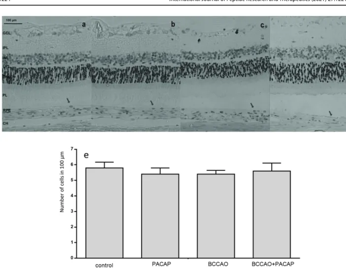
We counted the pigment epithelial cells in 100 µm of the retina of rats after BCCAO. We found 5.8 ± 0.374 cell bodies in 100 µm in average in control retinas. PACAP alone was injected into the right eye of the control animals, but it did not cause any changes in the number of pigment epithelial cells (5.4 ± 0.4 cell bodies in 100 µm). Though in the left eye of BCCAO operated animals (saline treated) the thick- ness of the whole retina, the thickness of each individual layer, and the number of ganglion cells in 100 µm strongly decreased (Werling et al. 2014), the pigment epithelial cells did not suffer any changes in number (5.4 ± 0.245 cell bodies in average). The right eyes (PACAP treated) had 5.6 ± 0.51 pigment cells in 100 µm (Fig. 1).
Cell Stress Array
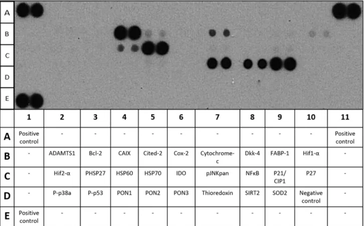
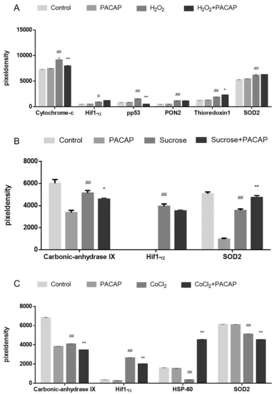
By using cell stress array, we detected the changes in the expression of 26 different molecular biological markers (Fig. 2). Hif1-α levels were strongly elevated following sucrose, H2O2, and CoCl2 administration. In case of hypoxia a significant decrease could be observed with PACAP co- administration, while only a slight decrease occurred in hyperosmotic conditions (Fig. 3c). The H2O2-induced oxi- dative stress activated cytochrome-c and phosphorylated p53 levels. PACAP significantly lowered the expression of both, thus proved to be anti-apoptotic in this model. Following
H2O2 administration, the levels of three important anti- oxidants, namely PON2, SOD2 and thioredoxin increased, PACAP further elevated the expression of the latter one (Fig. 3a). In hypoxia, the concentration of carbonic-anhy- drase IX (CAIX) and SOD2 was significantly lower com- pared to controls, while in hyperosmosis the level of HSP-60 was decreased in addition to CAIX and SOD2. While CAIX concentration was attenuated by PACAP in both hypoxic and hyperosmotic conditions, the expression of SOD2 was decreased after PACAP treatment in hypoxia, but increased in hyperosmosis (Fig. 3b). In CoCl2-induced stress HSP-60 was also significantly upregulated by PACAP.
Discussion
Several papers suggest that DR is not only a vascular dam- age, but neurodegeneration also plays a pivotal role in its pathogenesis (Zhang et al. 2013; Simo and Hernandez 2015;
Rossino et al. 2019). Various neuropeptides and their recep- tors, including VIP and PACAP, were detected in the retina (Nakamachi et al. 2012) and proved to be neuroprotective against different harmful stimuli, thus they can be considered as potential therapeutic agents in DR (Atlasz et al. 2010a, b;
Nakamachi et al. 2012; Lakk et al. 2015; Shioda et al. 2016;
Ye et al. 2019). In vivo studies confirmed that intravitreal or topical administration of PACAP not only improves ERG responses, but attenuates inflammatory processes, protects neurons in the ganglion cell layer and diminishes the thin- ning of the rat retina (Danyadi et al. 2014; Vaczy et al. 2016;
Werling et al. 2017; Atlasz et al. 2018). In streptozotocin- induced diabetes after intravitreal PACAP administration attenuated levels of IL-1β and VEGF were found (D’Amico et al. 2017). PACAP was also able to prevent the damage of the outer blood–retina barrier in diabetic rats (D’Amico et al.
2019; Scuderi et al. 2013). In previous studies, we dem- onstrated the anti-apoptotic and anti-angiogenic effects of PACAP (Mester et al. 2011; Szabo et al. 2012; Fabian et al.
2012). We have recently suggested that PACAP administra- tion might be a possible therapy against complications of DR by maintaining the cellular junctions between the retinal pigment epithelial cells and by inhibiting their VEGF secre- tion (Fabian et al. 2019). In this present study, we confirmed further molecular mechanisms, through which PACAP could exert these functions. Hif1 is a major transcriptional regula- tor, composed of α and β subunits (Semenza 2003; Ziello et al. 2007). Both are expressed at a constant rate in all cells, except for the cells of the peripheral blood, whereas the α subunit is promptly degraded by an oxygen-dependent mech- anism. In hypoxic conditions, Hif1-α is stabilized, dimerizes with Hif1-β, and translocates to the nucleus, where, depend- ing on the cell type, it induces the transcription of over 60 different genes, including VEGF (Forsythe et al. 1996;
Kurihara et al. 2014). Thus, downregulation of Hif1-α could be in the molecular background of the anti-VEGF function of PACAP. In a recent study, the protective function of PACAP was shown in a combined hyperglycemic/hypoxic environ- ment in ARPE-19 cells (Maugeri et al. 2017). The RPE cells were kept in normal glucose medium (5.5 mM) for a week, then, half of the cells were switched in high glucose medium (25 mM) for another 7 days. On the second week, the cells were exposed to 100 µM deferoxamine mesylate salt alone, or in combination with 100 nM PACAP. The researchers found that PACAP reduced the elevated levels of Hif1-α, while it increased the expression of Hif3-α. In accordance with this, in the present study we also found the same effect of PACAP in two different harmful conditions (hypoxia and hyperosmotic stress), whereas we could not prove this ame- liorating function of PACAP in oxidative stress.
CAIX is a transmembrane protein catalyzing the hydra- tion of CO2. As it is another downstream target of Hif1-α,
it has also been showed to be a cellular marker of hypoxia.
In physiological conditions, especially in dark adaptation, the retina is proposed to be hypoxic (Lahdenranta et al.
2001; Arden et al. 2005; de Gooyer et al. 2006; Hughes et al. 2010). This fact would suggest high control levels of both Hif1-α and its downstream molecules. In ARPE- 19 cells, we did not find elevated Hif1-α expression, but under control conditions, the concentration of CAIX was extremely high. PACAP alone, and in co-administration could diminish the effect of the harmful agents (CoCl2 and sucrose). Oxidative stress, a primary causative event in DR (Hernandez et al. 2016) is associated with increased levels of reactive oxygen species (ROS). In our present study, after H2O2 administration elevated expression of three antioxidants was detected, such as PON2, thioredoxin and
Fig. 1 Retina sections stained with toluidine blue of control (a), PACAP (b), BCCAO (c), and BCCAO + PACAP (d) animals.
Scale = 100 µm. Number of cells/100 µm section length in the retinal pigment epithelial layer (RPE) was measured. No significant differ- ence was observed. Arrows show pigment epithelial cells. e Average
number of RPE cells/100 µm section length. CH choroidea, RPE pig- ment epithelium, ONL outer nuclear layer, OPL outer plexiform layer, INL inner nuclear layer, IPL inner plexiform layer, GCL ganglion cell layer
SOD2. PACAP was able to further induce the thioredoxin synthesis, creating an antioxidant rich environment, which is essential against ROS.
In accordance with our previous findings PACAP could attenuate the elevated concentrations of cytochrome-c and p53 (Atlasz et al. 2010b; Szabadfi et al. 2010; Fabian et al.
2012). In contrast, analysis of the histological structure of the retina did not reveal any numerical changes in the pigment epithelial layer, while the thickness of the entire retina, especially the outer plexiform layer, was significantly
reduced (Werling et al. 2014). It is characteristic for several retinal injuries that the inner retinal layers suffer a more severe morphological lesion, while the pigment epithelial layer is seemingly not damaged (Werling et al. 2014). How- ever, the several biochemical changes indicate the severe functional damage in these cells, probably leading to further damage in the inner layers. In summary, we found that, in accordance with previous studies, PACAP could counteract some of the negative changes in the pigment epithelial layer further confirming the retinoprotective effects of the retina.
Fig. 2 Representative picture and the table of detectable markers of the cells stress array
Acknowledgements This study was supported by NAP2017- 1.2.1-NKP-2017-00002; GINOP-2.3.2-15-2016-00050 “PEPSYS”, KA-2016-03, KA-2017-17 MTA-TKI14016; NKFIH K119759, NKFIH K135457, NKFIH FK129190, FIKPIII, Bolyai Scholarship, PTE AOK- TANDEM 2019 Grant, EFOP-3.6.3-VEKOP-16-2017-00009, EFOP- 3.6.1.-16-2016-00004 Comprehensive Development for Implementing Smart Specialization Strategies at the University of Pécs; New Excel- lence Program, EFOP-3.6.2-00008, “The role of neuro-inflammation in neurodegeneration: from molecules to clinics”, Higher Education Insti- tutional Excellence Program of the Ministry of Human Capacities in Hungary, within the Framework of the 20765-3/2018/FEKUTSTRAT.
Funding Open Access funding provided by University of Pécs.
Data Availability The datasets generated during and/or analysed dur- ing the current study are available from the corresponding author on reasonable request.
Compliance with Ethical Standards
Conflict of interest The authors declare that they have no conflicts of interest.
Fig. 3 a Graph of the cell stress array in oxidative stress.
ARPE-19 cells were treated with PACAP, H2O2 and H2O2+ PACAP, vs. control
*P < 0.5, **P < 0.001, vs. H2O2 treated #P < 0.5, ##P < 0.001.
b Graph of the cells stress array in hyperosmotic stress.
ARPE-19 cells were treated with PACAP, sucrose and sucrose + PACAP, vs. control
*P < 0.5; **P < 0.001, vs.
sucrose treated #P < 0.5,
##P < 0.001. c Graph of the cells stress array in hypoxic stress. ARPE-19 cells were treated with PACAP, CoCl2 and CoCl2 + PACAP, vs. control
*P < 0.5, **P < 0.001, vs. CoCl2 treated #P < 0.5, ##P < 0.001
Open Access This article is licensed under a Creative Commons Attri- bution 4.0 International License, which permits use, sharing, adapta- tion, distribution and reproduction in any medium or format, as long as you give appropriate credit to the original author(s) and the source, provide a link to the Creative Commons licence, and indicate if changes were made. The images or other third party material in this article are included in the article’s Creative Commons licence, unless indicated otherwise in a credit line to the material. If material is not included in the article’s Creative Commons licence and your intended use is not permitted by statutory regulation or exceeds the permitted use, you will need to obtain permission directly from the copyright holder. To view a copy of this licence, visit http://creat iveco mmons .org/licen ses/by/4.0/.
References
Abdullah SAK (2018) Role of oxidative stress, inflammation, hypoxia and angiogenesis in the development of diabetic retinopathy.
Saudi J Ophthalmol 32(4):318–323
Amoaku WM, Ghanchi F, Bailey C, Banerjee S, Banerjee S, Downey L, Gale R, Hamilton R, Khunti K, Posner E, Quhill F, Robinson S, Setty R, Sim D, Varma D, Mehta H (2020) Diabetic retinopa- thy and diabetic macular oedema pathways and management: UK Consensus Working Group. Eye (Lond) 34(Suppl 1):1–51 Arden GB, Sidman RL, Arap W, Schlingemann RO (2005) Spare the
rod and spoil the eye. Br J Ophthalmol 89(6):764–769
Arfken CL, Reno PL, Santiago JV, Klein R (1998) Development of proliferative diabetic retinopathy in African-Americans and whites with type 1 diabetes. Diabetes Care 21(5):792–795
Armstrong BD, Abad C, Chhith S, Cheung-Lau G, Hajji OE, Nobuta H, Waschek JA (2008) Impaired nerve regeneration and enhanced neuroinflammatory response in mice lacking pituitary adenylyl cyclase activating peptide. Neuroscience 151(1):63–73
Atlasz T, Babai N, Kiss P, Reglodi D, Tamas A, Szabadfi K, Toth G, Hegyi O, Lubics A, Gabriel R (2007) Pituitary adenylate cyclase activating polypeptide is protective in bilateral carotid occlusion-induced retinal lesion in rats. Gen Comp Endocrinol 153:108–114
Atlasz T, Szabadfi K, Kiss P, Racz B, Gallyas F, Tamas A, Gaal V, Marton Z, Gabriel R, Reglodi D (2010a) Review of pituitary ade- nylate cyclase activating polypeptide in the retina: focus on the retinoprotective effects. Ann N Y Acad Sci 1200:128–39 Atlasz T, Szabadfi K, Kiss P, Tamas A, Toth G, Reglodi D, Gabriel R
(2010b) Evaluation of the protective effects of PACAP with cell- specific markers in ischemia-induced retinal degeneration. Brain Res Bull 81:497–504
Atlasz T, Vaczy A, Werling D et al (2016) Chap 30: protective effects of PACAP in the retina. In: Reglodi D, Tamas A (eds) Pituitary adenylate cyclase activating polypeptide—PACAP. Springer, Berlin, pp 433–448
Atlasz T, Werling D, Song S, Szabo E, Vaczy A, Kovari P et al (2018) Retinoprotective effects ofTAT-bound vasoactive intes- tinal peptide and pituitary adenylate cyclase activating polypep- tide. J MolNeurosci 68:397–407
Bazan NG (2008) Neurotrophins induce neuroprotective signaling in the retinal pigment epithelial cell by activating the synthesis of the anti-inflammatory and anti-apoptotic neuroprotectin D1.
Adv Exp Med Biol 613:39–44
D’Amico AG, Scuderi S, Saccone S, Castorina A, Drago F, D’Agata V (2013) Antiproliferative effects of PACAP and VIP in serum- starved glioma cells. J Mol Neurosci 51(2):503–513
D’Amico AG, Maugeri G, Reitano R, Bucolo C, Saccone S, Drago F, D’Agata V (2015) PACAP modulates expression of
hypoxia-inducible factors in streptozotocin-induced diabetic rat retina. J Mol Neurosci 57(4):501–509
D’Amico AG, Maugeri G, Rasà DM, Bucolo C, Saccone S, Federico C, Cavallaro S, Agata V (2017) Modulation of IL-1β and VEGF expression in rat diabetic retinopathy after PACAP administra- tion. Peptides 97:64–69
D’Amico AG, Maugeri G, Rasà D, Federico C, Saccone S, Lazzara F, Fidilio A, Drago F, Bucolo C, Agata V (2019) NAP modu- lates hyperglycemic-inflammatory event of diabetic retina by counteracting outer blood retinal barrier damage. J Cell Physiol 234(4):5230–5240
Danyadi B, Szabadfi K, Reglodi D, Mihalik A, Danyadi T, Kovacs Z et al (2014) PACAP application improves functional outcome of chronic retinal ischemic injury in rats—evidence from electrore- tinographic measurements. J Mol Neurosci 54:293–299 de Gooyer TE, Stevenson KA, Humphries P, Simpson DA, Curtis
TM, Gardiner TA, Stitt AW (2006) Rod photoreceptor loss in Rho−/− mice reduces retinal hypoxia and hypoxia-regulated gene expression. Investig Ophthalmol Vis Sci 47(12):5553–5560 Ejaz S, Chekarova I, Ejaz A, Sohail A, Lim CW (2008) Importance
of pericytes and mechanisms of pericyte loss during diabetes retinopathy. Diabetes Obes Metab 10(1):53–63
Fabian E, Reglodi D, Mester L, Szabo A, Szabadfi K, Tamas A, Toth G, Kovacs K (2012) Effects of PACAP on intracellular signaling pathways in human retinal pigment epithelial cells exposed to oxidative stress. J Mol Neurosci 48(3):493–500
Fabian E, Reglodi D, Horvath G, Opper B, Toth G, Fazakas C, Vegh AG, Wilhelm I, Krizbai IA (2019) Pituitary adenylate cyclase activating polypeptide acts against neovascularization in retinal pigment epithelial cells. Ann N Y Acad Sci 1455(1):160–172 Forsythe JA, Jiang BH, Iyer NV, Agani F, Leung SW, Koos RD,
Semenza GL (1996) Activation of vascular endothelial growth factor gene transcription by hypoxia-inducible factor 1. Mol Cell Biol 16:4604–4613
Gaal V, Mark L, Kiss P, Kustos I, Tamas A, Kocsis B, Lubics A, Nemeth V, Nemeth A, Lujber L, Pytel J, Toth G, Reglodi D (2008) Investigation of the effects of PACAP on the composition of tear and endolymph proteins. J Mol Neurosci 36:321–329 Girard BM, Campbell SE, Beca KI, Perkins M, Hsiang H, May V,
Vizzard MA (2020) Intrabladder PAC1 receptor antagonist, PACAP(6-38), reduces urinary bladder frequency and pelvic sensitivity in mice exposed to repeated variate stress (RVS).
J Mol Neurosci. https ://doi.org/10.1007/s1203 1-020-01649 -x Hernandez C, Dal Monte M, Simó R, Casini G (2016) Neuroprotec-
tion as a therapeutic target for diabetic retinopathy. J Diabetes Res 2016:9508–9541
Huang H, He J, Johnson D, Wei Y, Liu Y, Wang S, Lutty GA, Duh EJ, Semba RD (2015) Deletion of placental growth factor pre- vents diabetic retinopathy and is associated with Akt activation and HIF1α-VEGF pathway inhibition. Diabetes 64(1):200–212 Hughes JM, Groot AJ, van der Groep P, Sersansie R, Vooijs M, Diest PJ, Van Noorden CJF, Schlingemann RO, Klaassen I (2010) Active HIF-1 in the normal human retina. J Histochem Cytochem 58(3):247–254
Joussen AM, Smyth N, Niessen C (2007) Pathophysiology of diabetic macular edema. Dev Ophthalmol 39:1–12
Jozsa R, Hollosy T, Tamas A et al (2005) Pituitary adenylate cyclase activating polypeptide plays a role in olfactory memory formation in chicken. Peptides 26:2344–2350
Kook D, Wolf AH, Yu AL, Neubauer AS, Priglinger SG, Kampik A, Welge-Lüssen UC (2008) The protective effect of quercetin against oxidative stress in the human RPE in vitro. Investig Oph- thalmol Vis Sci 49(4):1712–1720
Kovacs AK, Atlasz T, Werling D, Szabo E, Reglodi D, Toth GK (2020) Stability test of PACAP in eye drops. J Mol Neurosci. https ://doi.
org/10.1007/s1203 1-020-01532 -9
Kurihara T, Westenskow PD, Friedlander M (2014) Hypoxia-inducible factor (HIF)/vascular endothelial growth factor (VEGF) signaling in the retina. Adv Exp Med Biol 801:275–281
Lahdenranta J, Pasqualini R, Schlingemann RO, Hagedorn M, Stallcup WB, Bucana CD, Sidman RL, Arap W (2001) An anti-angiogenic state in mice and humans with retinal photoreceptor cell degenera- tion. Proc Natl Acad Sci USA 98(18):10368–10373
Lakk M, Denes V, Gabriel R (2015) Pituitary adenylate cyclase-acti- vating polypeptide receptors signal via phospholipase c path- way to block apoptosis in newborn rat retina. Neurochem Res 40:1402–1409
Leasher JL, Bourne RR, Flaxman SR, Jonas JB, Keeffe J, Naidoo K et al (2016) Global estimates on the number of people blind or visually impaired by diabetic retinopathy: a meta-analysis from 1990 to 2010. Diabetes Care 39:1643–1649
Martínez-Rojas VA, Jiménez-Garduño AM, Michelatti D, Tosatto L, Marchioretto M, Arosio D, Basso M, Pennuto M, Musio C (2020) ClC-2-like chloride current alterations in a cell model of spinal and bulbar muscular atrophy, a polyglutamine disease. J Mol Neu- rosci. https ://doi.org/10.1007/s1203 1-020-01687 -5
Maugeri G, D’Amico AG, Reitano R, Magro G, Cavallaro S, Salomone S, D’Agata V (2016) PACAP and VIP inhibit the invasiveness of glioblastoma cells exposed to hypoxia through the regulation of HIFs and EGFR expression. Front Pharmacol 7:139
Maugeri G, D’Amico AG, Saccone S, Federico C, Cavallaro S, D’Agata V (2017) PACAP and VIP inhibit HIF-1alpha-mediated VEGF expression in a model of diabetic macular edema. J Cell Physiol 232(5):1209–1215
Maugeri G, D’Amico AG, Rasà DM, Saccone S, Federico C, Cavallaro S, D’Agata V (2018) PACAP and VIP regulate hypoxia-inducible factors in neuroblastoma cells exposed to hypoxia. Neuropeptides 69:84–91
Maugeri G, D’Amico AG, Bucolo C, D’Agata V (2019a) Protective effect of PACAP-38 on retinal pigmented epithelium in an in vitro and in vivo model of diabetic retinopathy through EGFR-depend- ent mechanism. Peptides 119:170108
Maugeri G, D’Amico AG, Castrogiovanni P, Saccone S, Federico C, Reibaldi M, Russo A, Bonfiglio V, Avitabile T, Longo A, D’Agata V (2019b) PACAP through EGFR transactivation pre- serves human corneal endothelial integrity. J Cell Biochem 120(6):10097–10105
Maugeri G, D’Amico AG, Rasà DM, Federico C, Saccone S, Morello G, La Cognata V, Cavallaro S, D’Agata V (2019c) Molecular mechanisms involved in the protective effect of pituitary adenylate cyclase-activating polypeptide in an in vitro model of amyotrophic lateral sclerosis. J Cell Physiol 234(4):5203–5214
Maugeri G, D’Amico AG, Amenta A, Saccone S, Federico C, Reibaldi M, Russo A, Bonfiglio V, Avitabile T, Longo A, D’Agata V (2020a) Protective effect of PACAP against ultraviolet B radia- tion-induced human corneal endothelial cell injury. Neuropeptides 79:101978
Maugeri G, D’Amico AG, Musumeci G, Reglodi D, D’Agata V (2020b) Effects of PACAP on Schwann cells: focus on nerve injury. Int J Mol Sci 21(21):8233
Mester L, Kovacs K, Racz B et al (2011) Pituitary adenylate cyclase- activating polypeptide is protective against oxidative stress in human retinal pigment epithelial cells. J Mol Neurosci 43:35–43 Moody TW, Nuche-Berenguer B, Jensen RT (2016) VIP/PACAP, and
their receptors and cancer. Curr Opin Endocrinol Diabetes Obes 23:38–47
Nakamachi T, Matkovits A, Seki T, Shioda S (2012) Distribution and protective function of pituitary adenylate cyclase-activating poly- peptide in the retina. Front Endocrinol 3:145
Rodríguez ML, Pérez S, Mena-Mollá S, Desco MC, Ortega ÁL (2019) Oxidative stress and microvascular alterations in diabetic retin- opathy: future therapies. Oxid Med Cell Longev 2019:4940825
Rossino MG, Dal Monte M, Casini G (2019) Relationships between neurodegeneration and vascular damage in diabetic retinopathy.
Front Neurosci 13:1172
Sasaki M, Ozawa Y, Kurihara T, Kubota S, Yuki K, Noda K, Kobayashi S, Ishida S, Tsubota K (2010) Neurodegenerative influence of oxidative stress in the retina of a murine model of diabetes. Dia- betologia 53(5):971–979
Scuderi S, D’Amico AG, Castorina A, Imbesi R, Carnazza ML, D’Agata V (2013) Ameliorative effect of PACAP and VIP against increased permeability in a model of outer blood retinal barrier dysfunction. Peptides 39:119–124
Semenza GL (2003) Targeting HIF-1 for cancer therapy. Nat Rev Can- cer 3:721–732
Shioda S, Takenoya F, Wada N, Hirabayashi T, Seki T, Nakamachi T (2016) Pleiotropic and retinoprotective functions of PACAP. Anat Sci Int 91:313–324
Simo R, Hernandez C (2015) Novel approaches for treating diabetic retinopathy based on recent pathogenic evidence. Prog Retin Eye Res 48:160–180
Szabadfi K, Mester L, Reglodi D et al (2010) Novel neuroprotective strategies in ischemic retinal lesions. Int J Mol Sci 11:544–561 Szabo A, Danyadi B, Bognar E, Szabadfi K, Fabian E, Kiss P, Mester
L, Manavalan S, Atlasz T, Gabriel R, Toth G, Tamas A, Reglodi D, Kovacs K (2012) Effect of PACAP on MAP kinases, Akt and cytokine expressions in rat retinal hypoperfusion. Neurosci Lett 523(2):93–98
Szegeczki V, Bauer B, Jüngling A, Fülöp BD, Vágó J, Perényi H, Tarantini S, Tamás A, Zákány R, Reglődi D, Juhász T (2019) Age-related alterations of articular cartilage in pituitary adenylate cyclase-activating polypeptide (PACAP) gene-deficient mice.
Geroscience 41(6):775–793
Toth D, Szabo E, Tamas A, Juhasz T, Horvath G, Fabian E, Opper B, Szabo D, Maugeri G, D’Amico AG, D’Agata V, Vicena V, Reglodi D (2020) Protective effects of PACAP in peripheral organs. Front Endocrinol 11:377
Vaczy A, Reglodi D, Somoskeoy T, Kovacs K, Lokos E, Szabo E et al (2016) The protective role of PAC1-receptor agonist max- adilan in BCCAO-induced retinal degeneration. J Mol Neurosci 60:186–194
Wang ZY, Alm P, Håkanson R (1995) Distribution and effects of pitui- tary adenylate cyclase-activating peptide in the rabbit eye. Neu- roscience 69(1):297–308
Werling D, Reglodi D, Kiss P, Toth G, Szabadfi K, Tamas A, Biro Z, Atlasz T (2014) Investigation of PACAP fragments and related peptides in chronic retinal hypoperfusion. J Ophthalmol 2014:563812
Werling D, Banks WA, Salameh TS, Kvarik T, Kovacs LA, Vaczy A et al (2017) Passage through the ocular barriers and beneficial effects in retinal ischemia of topical application of PACAP1-38 in rodents. Int J Mol Sci 18:E675
Ye D, Shi Y, Xu Y, Huang J (2019) PACAP attenuates optic nerve crush-induced retinal ganglion cell apoptosis via activation of the CREB-Bcl-2 pathway. J Mol Neurosci 16:019–01309
Zhang X, Wang N, Barile GR, Bao S, Gillies M (2013) Diabetic retin- opathy: neuron protection as a therapeutic target. Int J Biochem Cell Biol 45:1525–1529
Ziello JE, Jovin IS, Huang Y (2007) Hypoxia-inducible factor (HIF)-1 regulatory pathway and its potential for therapeutic intervention in malignancy and ischemia. Yale J Biol Med 80:51–60
Publisher’s note Springer Nature remains neutral with regard to jurisdictional claims in published maps and institutional affiliations.