1 BIOGENESIS OF THE NEOPROTEROZOIC KREMYDILITE MANGANESE ORES 2 FROM URUCUM (BRAZIL) – A NEW MANGANESE ORE TYPE.
3
4 Running title: Biogenesis of a Neoproterozoic manganese ore 5
6
7 Joăo Carlos Biondi1+, Márta Polgári2,3*+, Ildikó Gyollai2, Krisztián Fintor4, Ivett Kovács2, József
8 Fekete2, Stephen J. Mojzsis2,5*
9 10
11 1Federal University of Paraná State, Polytechnic Center, Geology Department, 81531-980 12 Curitiba, Brazil, e-mail: biondiufpr@gmail.com
13 2Institute for Geological and Geochemical Research, RCAES, Hungarian 14 Academy of Sciences, 1112 Budapest, Budaörsi u. 45, Hungary, e-mail: rodokrozit@gmail.com, 15 gyildi@gmail.com, iv.kovacs@gmail.com, fekete.jozsef@csfk.mta.hu
16 3Eszterházy Károly University, Dept. of Natural Geography and Geoinformatics, 3300 Eger, 17 Leányka u. 6, Hungary
18 4Szeged University, Dept. of Mineralogy, Geochemistry and Petrology, 6722 Szeged, Egyetem u.
19 2-6, Hungary, e-mail: efkrisz@gmail.com
20 5University of Colorado, Department of Geological Sciences, 2200 Colorado Avenue, UCB 399, 21 Boulder, Colorado 80309-0399, United States of America, e-mail: mojzsis@colorado.edu
22 23
24 *corresponding authors:
25 rodokrozit@gmail.com
26 + The first two authors contributed equally to this work 27 mojzsis@colorado.edu
28
30 Significance statement 31
32 The Neoproterozoic Urucum manganese deposit (Brazil) is a ~600 Mt microbially-mediated 33 sedimentary Mn ore. Proto-ore formation via sedimentation and diagenesis occurred under 34 suboxic-oxic and semi-neutral pH conditions in the Ediacaran ocean, wherein microbial Mn(II) 35 oxidation ensued from the fine-grained accumulation of Mn oxides and organic matter. Oxic 36 conditions that facilitated enzymatic Mn oxidation and overwhelmed microbial Fe oxidation 37 appears as a sharp contact between manganese and iron beds. The Urucum deposit arose from a 38 complex suite of diagenetic processes, including decomposition and mineralization of microbially- 39 derived organic matter involving extracellular polymeric substances. Kremydilite – a new type of 40 diagenetic concentric Mn mineral structure – formed by randomly activated heterotrophic cell 41 colonies that generated pores in the microbialite sediment after burial, coincident with lithification.
42
43 Highlights
44 1. Urucum Mn deposit formed in an Ediacaran marginal basin with more than 600 Mt of ore 45 formed from manganiferous microbialite.
46 2. Kremydilite is diagenetic structure that comprises a new type of Mn ore.
47 3. Microbial mediation occurred during Mn ore sedimentation and diagenesis.
48 4. Cellular and extracellular polymeric substances from Fe and Mn bacteria and cyanobacteria 49 were mineralized.
51 Abstract
52 The Urucum district in Mato Grosso do Sul (Brazil), hosts the youngest and largest sedimentary 53 Mn ore of Neoproterozoic age; units Mn-1, Mn-2, and Mn-3 are found in jaspilites and ironstones, 54 and represent approximately 600 Mt of extractable rock with 27–44% Mn and 12–30% Fe. High- 55 resolution optical- and cathodoluminescence microscopy, as well as Raman and FTIR 56 spectroscopy show that the lower Mn-1 is ferruginous, while the upper Mn-1 consists mainly of 57 30–75 vol.% braunite, < 0.5% aegirine, 3–15% quartz, 5–10% feldspar, and 1–5% clay minerals, 58 including apatite, chlorite, and organic matter. Here, we model the control of this ore mineralogy 59 by homogeneous oxidation and microbial processes. Layers Mn-2 and Mn-3 contain kremydilite, 60 as a characteristic ore structure, with 77–95 vol.% cryptomelane, 0–23% hollandite, 9–19 % 61 braunite, 7–21% hematite, and 0–5% pores filled with clay minerals and organic matter. These are 62 present within a micro-nodule matrix composed of cryptomelane and hematite in varying 63 proportions. The first syngenetic products of microbial enzymatic oxidation were, on the Fe side, 64 ferrihydrite and lepidocrocite, and on the Mn side, vernadite, todorokite, birnessite, and manganite.
65 These formed under obligatory oxic (Mn) and suboxic (Fe) conditions and close to neutral pH. We 66 describe the genesis of Urucum via complex diagenetic processes, which include the 67 decomposition and mineralization of cellular- and extracellular-polymeric substances from Fe and 68 Mn bacteria and cyanobacteria. The kremydilite forms in successive stages of oxidation of organic 69 matter mediated by microbes, which generate pores and produce methane and CO2/H2 bubbles.
70 They are a unique type of diagenetic structure formed by heterotrophic cell colonies randomly 71 activated in the microbialite milieu following burial in suboxic neutral/alkaline conditions, side- 72 by-side with the lithification and stabilization of the mineral assemblages. (294 words)
73 74 75
76 Keywords:
77 (1) Urucum Ediacaran manganese deposit; (2) kremydilite; (3) microbialite; (4) enzymatic 78 oxidation, cell and extracellular polymeric substance mineralization.
80 1. INTRODUCTION 81
82 The Urucum mining district occupies an area of approximately 800 km2 and is located in the 83 Pantanal swamps region of west-central Brazil. Three layers of massive manganese oxides, named 84 Mn-1, Mn-2, and Mn-3, occur interbedded with massive jasper, banded iron formations (BIFs), 85 and massive iron formations (IF) that comprise the Santa Cruz Formation of the Neoproterozoic 86 Jacadigo Group (Urban et al. 1992; Frei et al. 2017; Fig. 1 and SI 1-Fig). It was estimated that the 87 Urucum district originally contained more than 600 Mt of rock with the manganese content 88 between 27–44 wt.% and iron content between 12–30 wt.% (Urban et al. 1992).
89 Fig. 1 HERE
90 The stratigraphic sequence of the Urucum region was first defined by Dorr (1945) and Almeida 91 (1946), who also conducted the first systematic studies on the origin of iron and manganese 92 deposits in the region. Urban et al. (1992) mapped the entire mining region, and since that time, 93 the regional geological map has been minimally updated. Following the work of Urban et al.
94 (1992), the most relevant changes to our understanding of the regional geology arose from the 95 work of Freitas et al. (2011), who detailed the Jacadigo Group lithologies and defined their 96 sedimentation environments. Biondi and Lopez (2017) identified faults that acted as conduits for 97 hydrothermal fluids which altered the rocks of the Jacadigo Group basement, and exhaled fluids 98 with iron and other elements at the base of the sedimentary sequence of the Urucum basin. They 99 also correlated the Mn-1, Mn-2, and Mn-3 layers with those recognized at different Urucum sites.
100 Various and mutually-exclusive proposed genetic models for the Jacadigo Group iron and 101 manganese rocks have been a topic of discussion and debate since their discovery. These models 102 can be summarized as follows: (a) marine genesis with sediments of continental origin (Dorr 103 1945); (b) marine genesis with sediments of marine origin (Almeida 1946; Putzer, 1958; Haralyi 104 and Walde, 1986); (c) volcanogenic marine genesis (Walde 1981; Walde et al. 1981; Leonardos 105 and Walde 1982; O'Connor and Walde 1985); (d) formation in a glacio-marine sedimentary
106 environment followed by supergene enrichment (Schneider 1984; Schreck 1984; Leeuwen and 107 Graf 1987; Graf et al. 1994; Costa et al. 2005); (e) sedimentary genesis in a flooded graben with a 108 contribution of hydrothermal leaching from hidden mafic rocks (Haralyi and Walde 1986; Walde 109 1988; Trompette et al. 1998); (f) SEDEX, or sedimentary exhalation (Dardenne 1998); and (g) 110 sedimentary genesis in an oceanic environment with a deep-sea hydrothermal contribution (Klein 111 and Ladeira 2004). Recently, Angerer et al. (2016) proposed a biologically-mediated origin in a 112 glacio-marine environment for the carbonate BIFs of the Santa Cruz Mine region located on the 113 southeastern part of the Santa Cruz plateau. In a recent comprehensive study, Biondi and Lopez 114 (2017) (a) recognized the biogenic mediation during the genesis of manganese ore; (b) described 115 in detail mineral structures termed by them kremydilites and argued that they may represent 116 fossilized microbial colonies from organisms that mediated the formation of the manganese layers;
117 and (c) modified the region's stratigraphy based on the fossil assemblages, showing that the 118 Urucum iron-manganese rocks correlate to the carbonate rocks of the Bocaina Formation, of the 119 Corumbá Group, previously considered post-depositional to those of the Jacadigo Group.
120 The Ediacaran Period of formation is proposed by the authors based on the presence of the 121 Corumbella Verneri fossil, found amidst the ironstones separating Mn-2 from Mn-3 (Figs 2B and 122 Figs 4B to D, Biondi and Lopez 2017). This fossil has always been considered Ediacaran, which 123 establishes a wider interest concerning the Urucum Mn deposit.
124 Here, we explore the origin of kremydilites described in Biondi and Lopez (2017) and 125 present a model that explains the processes of sedimentation and diagenesis that facilitated the 126 origin of these structures and the manganese layers. Recent works have provided a geological 127 setting diagram, mineralogy (low magnification optical microscopy, X-ray diffraction, SEM-EDS- 128 based), and chemistry datasets based on bulk samples and in situ (SEM-EDS) data (e.g. Frei et al.
129 2017), but microbial mediation as a plausible mechanism for the genesis of these rocks is still 130 under debate (Biondi and Lopez 2017). We expand the results of these previous studies with more 131 detailed optical microscopy (OM), cathodoluminescence microscopy (CL), Raman- and Fourier- 132 transform infrared spectroscopy (FTIR) to document the micro-mineralogy, presence, and 133 distribution of embedded organic matter. The goal here is to explore the role of microorganisms
134 in the process of manganese ore genesis from Urucum, and to understand the diagenesis, structures, 135 and process of formation of kremydilites.
136
137 2. GEOLOGICAL AND GEOCHRONOLOGICAL BACKGROUND
138 The Santa Cruz Formation is mainly composed of jaspilitic BIFs, (massive) iron formations 139 (IFs), massive banded jasper, and ferruginous arkosic silt and sandstones. The greatest thickness 140 of the Santa Cruz Formation, 396 m, is documented in drill hole (DH) 44-28, made at the Vetorial 141 Mine, and bookended by a 40 m section at the northern end of the Rabicho plateau (Fig. 2). The 142 massive manganese layers, Mn-1, Mn-2, and Mn-3, occur in the lower half of this formation and 143 are interlayered with BIFs and massive jasper.
144 The Jacadigo and Corumbá Groups are considered coeval (Biondi and Lopez 2017) and of 145 Ediacaran age, based on the presence of stromatolites below Mn-1 (Jacadigo Group) and 146 Corumbella fossils in the rocks of the Bocaina and Santa Cruz Formations (respectively, Corumbá 147 and Jacadigo Groups). The age of this fossil in the ironstones of the Santa Cruz Formations 148 (Jacadigo Group) and limestones of the Tamengo Formation (Corumbá Group) was estimated at 149 ca. 550 Ma (Germs 1972; Grant 1990; Grotzinger et al. 1990; Hofmann and Mountjoy 2001;
150 Bengtson 2002). The proposed age of this horizon was 555–542 Ma by ichnofossils, identified by 151 Parry et al. (2017), in the Bocaina Formation. These ages are consistent with U-Pb geochronology 152 of detrital zircons from a volcanic ash layer intercalated with carbonate rocks of the Tamengo 153 Formation, at 543±3 Ma (Babinski et al. 2008), and the 40Ar/39Ar age of 587±7 Ma for 154 cryptomelane in the Mn-1 to Mn-3 layers (Piacentini et al. 2013; Frei et al. 2017 and references 155 therein).
156 Dating braunite from the Mn-1 layer, Piacentini et al. (2013) interpreted the 547±3 to 513±4 157 Ma (40Ar/39Ar) age as a minimum age, arguing that the Ar/Ar thermo-chronological system was 158 rejuvenated by tectonic warming, which was considered a consequence of the metamorphism 159 underwent by the Jacadigo Group rocks. Also using the 40Ar/39Ar method, they dated 513±3 Ma 160 some crystals of muscovite collected from the arkoses that are interlayered with the BIFs, which 161 was also considered metamorphic. According to Piacentini et al. (2013), these ages are “possibly
162 related to disruption between the Amazon Craton and the Apa River cratonic fragment and they 163 do not reflect the time of Jacadigo Group deposition”, which would be greater than 590 Ma, and 164 concluded that Jacadigo’s rocks would have at least 587 ± 7 Ma.
165 To reconstruct the paleogeography of the sedimentary basin, Mn-2 and Mn-3 were leveled 166 and used as stratigraphic markers. This procedure makes it possible to outline the geometrical 167 differences between the Mn-layers. This reconstruction shows that stratigraphy observed within 168 the Urucum plateaux always includes Mn-1, and that this stratum lines the basin floor wherever 169 the Jacadigo Group is described (e.g., Urban et al. 1992; Biondi and Lopez 2017) (Fig. 2). Yet, 170 unlike Mn-1, both the Mn-2 and Mn-3 layers occur only in the interpreted depocenter of the basin, 171 in the region of Urucum, Santa Cruz and southeast of the Morro Grande plateaux (Fig. 1). In the 172 interior of each plateau, mining of the manganese layers reveals that Mn-2 and Mn-3 are flat and 173 parallel to one another, whereas the Mn-1 unit follows the contours of the basin floor. By 174 positioning Mn-2 and Mn-3 in their respective stratigraphic horizons it is now possible to 175 reconstruct Urucum marginal basin floor (Fig. 2).
176 The origin of the sediments of Urucum has been detailed elsewhere (Walde 1981; Walde 177 et al. 1981; Leonardos and Walde 1982; O'Connor and Walde 1985, Haralyi and Walde 1986;
178 Walde 1988; Trompette et al. 1998, Dardenne 1998, Klein and Ladeira 2004, Angerer et al. 2016;
179 Biondi and Lopez 2017), and we provided a brief synopsis, here. The Santa Cruz Formation formed 180 as an in-fill of an ancient graben with iron and manganese-rich sediments overlying fluvial deposits 181 from the Urucum Formation, while limestones from the Bocaina and Tamengo Formations were 182 deposited in the shallow marginal regions (Biondi and Lopez 2017; Fig. 2). As has been proposed 183 for some Phanerozoic Mn ores (e.g. Polgári et al. 2012ab, 2016b), the most probable sources of 184 the Mn and Fe was hydrothermal exhalations in a submarine environment. The Mn and Fe fluids 185 were transported to the sedimentary basin via basement faults (SI 1-2-Figs) that became activated
186 each time the graben widened. During inundations attributed to sedimentation of the Mn-1, Mn-2, 187 and Mn-3 units, Mn and Fe discharged on basin floor mixed with Mn and Fe brought in by water 188 from the open ocean as well as with that originating from the exhalates located outside the Urucum 189 basin.
190 Fig. 2.
191 A transition between the Urucum and Santa Cruz formations through the Mn-1 horizon 192 exists in all mines from the area (Urban et al. 1992). Furthermore, Biondi and Lopez (2017) 193 showed that there are typically two or more layers of Mn-1 manganese ore with meter- to 194 decimeter-scale thicknesses, locally interlayered with jaspillite-rich clasts. We now describe these 195 relationships in more detail.
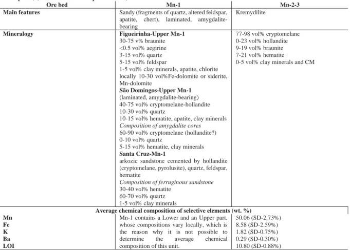
196 The lower Mn-1 ore layer is relatively siliceous and composed mainly of braunite, 197 cryptomelane cement and Mn–Fe-rich carbonate, whereas the Upper Mn-1 layer is a fine-grained, 198 massive, clastic layer of manganese oxides with undulating parallel lamination and numerous 199 decimeter-scale oblate structures, characterized by a massive core and silty clay and arkose wrap 200 dubbed amygdalites(Fig. 3C). The ore layer is bounded by sharp planar contacts typically overlain 201 by conglomerate consisting of angular granite pebbles in an arkosic matrix. Layers Mn-2 and Mn-3 202 contain mostly massive manganese ore with lamination. They are composed mainly of 203 cryptocrystalline manganese oxides and hydroxides, commonly containing kremydilites (Figs. 3D- 204 E and 4) with minor amygdalites (Biondi and Lopez 2017). In Mn-2 and Mn-3, what have been 205 interpreted as the remains of microbial colonies form oblate, 5–15-cm sized concentric kremydilite 206 structures, within the fine-grained and biomass-rich basin floor shale as well as intergranular, 207 oblate gas structures (Figs. 4A, C, F, and H). The main features of the ore beds, including their 208 mineralogical and selective element compositions are summarized in Table 1.
209 Fig. 3
210 Fig. 4.
211 Table 1.
212 The textures of the Mn-2 and Mn-3 layers express as 1–10 millimeter-sized spherical, often 213 zoned manganese oxide micro-nodules that coalesce to form the massive ores. These probably 214 involved the aforementioned kremydilite structures made solely of manganese and minor iron 215 oxides. All observed Mn-2 and Mn-3 outcrops have kremydilites, although they do not constitute 216 all of the ore mass from these layers. As previously described, kremydilites occur between the ore 217 bands (Figs. 4A–J), which are massive or banded (and/or laminated), and are distributed in the 218 layers in an apparently homogeneous manner. It is difficult to estimate the volume occupied by 219 kremydilites owing to the fact that they are complicated to see in discontinuous outcrops. For those 220 observed on the mining fronts, it is likely that they occupy more than about 50% of the ore layer 221 by volume.
222 2.1. Forms and type of kremydilites
223 Kremydilites occur only between the laminations of the massive ore in the Mn-2 and Mn- 224 3 layers, and are absent in banded- or massive ironstones. They are always contoured by the fine 225 laminated ore with micronodular, microbialite micro-texture, in which the diameters of micro- 226 nodules range from 0.2–0.8 mm (Fig. 4; SI 5-Fig, zones 1, 2, 3; and zones 20 to 24). Microbialite 227 and micro-nodule rich layers are in turn contoured by wavy microbialite layers apparently 228 composed by the amalgamation of nodules smaller than 10 μm. Its forms are oblate, centimeter- 229 to-decimeter scale (Fig. 3) and concentrically zoned. Structures of what we interpret to be the 230 different growth development stages also appear to occur together (Figs. 4 and 5), and each stage 231 of growth is marked by the presence of pores, which delineate coarse concentric, sometimes 232 incomplete envelopes (Fig. 4). Kremydilites on the other hand are porous structures absent of 233 micro-nodules. They occur in varied forms as shown in Figures 4A to H.
234 Fig. 5.
235 The simplest kremydilite form consists of a bubble-filled nucleus (Fig. 4A), followed by 236 those with a nucleus having diffuse borders (Figs. 4C). Other forms include a nucleus with one 237 (Fig. 4E) or two (Fig.4G) diffuse concentric laminae (or shells). The more complex kremdylites, 238 with a nucleus and many concentric shells delineated by millimeter to submillimeter pores, 239 crosscut with lighter, massive, and metallic zones (Figs. 4I). In general, the various kremydilite 240 forms contain many oblate structures (Figs. 4A, E, and G). These are less than 20 mm across 241 distributed along the layer containing the kremydilite, and inside and/or near them. Mesoscopic 242 inspections of sawn samples (Fig. 4K) as well as thin and polished sections, show that each layer 243 contains disseminated pores. The quantity of pores increases toward the margin of the shell, and 244 each lamina is surrounded and delimited by areas with high pore density (Biondi and Lopez 2017).
245 The pores are often lined by shiny acicular microcrystals of cryptomelane and/or contain 246 organic matter (Biondi and Lopez 2017). Although kremydilites do not contain micro-nodules, 247 and are instead inside the micronodular bands and contoured by microbialite layers, the outermost 248 zones of kremydilite appear to have a composition similar to that of amalgamated micro-nodules.
249 In these zones, the presence of ring-like structures of carbonate microcrystals are common (SI 5- 250 Fig, zone 16 - detail image, Biondi and Lopez, 2017). The zones closer to the nucleus (zones 9, 251 10, and 11) contain mixed anhedral minerals with metallic luster, but with larger dimensions than 252 the anterior zones. The nucleus of the kremydilites (zones 12 and 13) are microgranular and 253 heterogeneous.
254
255 3. SAMPLES
256 Representative samples and the methods applied (number of photos and spectra) are 257 summarized in Table 2 and Fig. 3–4. Localities of the sample collection are shown in Fig. 2.
258 The samples of Mn-1 are (Fig. 3, SI 3-Fig): COR-4B, a clast-bearing massive ironstone;
259 COR-6, a massive manganese ore; COR-7, a very fine-grained clast-bearing ore with braunite and 260 carbonate; COR-10, a sandy, detritic ore with braunite, quartz, and feldspar; COR-31, an arkosic 261 sandstone with hematite matrix; and COR-32, an amygdalite with cryptomelane massive nucleus 262 surrounded by arkosic sandstone with hematite matrix.
263 Samples of Mn-2 and Mn-3 are: COR-81, a sample of massive manganese ore; COR-78- 264 F3 (Fig. 4F), a nucleus of porous kremydilite with diffuse boundaries, surrounded by two zones 265 also with diffuse boundaries; COR-78-D1 (Fig. 4C), a kremydilite with a diffuse core enveloped 266 by two shells, also with diffuse boundaries; COR-75-B5 (Fig. 4E), a porous kremydilite nucleus;
267 COR-75-2 and COR-48 (Fig. 4J), complex kremydilites with porous core surrounded by many 268 concentric, porous shells; and COR-36-A1 (Fig. 4G), a kremydilite with a nucleus and at least two 269 shells (hereafter the samples are cited without COR).
270 Table 2.
271
272 4. METHODS
273 Thin section and polished section mineralogy was described and quantified using a ZEISS 274 Axio Imager A2m microscope (Federal University of Paraná State, Polytechnic Center, Geology 275 Department, Curitiba, Brazil).
276 Petrographic structural-textural studies by optical rock microscopy (OM) were also made 277 on 12 thin sections in transmitted and reflected light (NIKON ECLIPSE 600 rock microscope, 278 Institute for Geology and Geochemistry, Research Centre for Astronomy and Earth Sciences, 279 Hungarian Academy of Sciences - IGGR RCAES HAS, Budapest, Hungary).
280 Cathodoluminescence (CL) petrography was carried out on 7 thin sections using a 281 Reliotron cold cathode cathodoluminescence apparatus mounted on a BX-43 Olympus
282 polarization microscope (Szeged University, Hungary). The accelerating voltage was 7–7.7 keV 283 during the analysis. Cathodoluminescence spectra were recorded by using an Ocean Optics 284 USB2000+VIS-NIR spectrometer. Spectrometer specifications are a wavelength range of 350–
285 1000 nm and 1.5-nm (FWHM) optical resolution.
286 Mineralogical analyses were performed on three bulk samples using a Rigaku Miniflex-600 287 X-ray diffractometer (XRD), with carbon monochromator and Cu-Kα radiation, at 40 kV and 15 288 mA (IGGR RCAES HAS, Budapest, Hungary). Mineral composition was determined on randomly 289 oriented powdered samples. The diffraction patterns were processed using Siroquant V4 software, 290 and the modal contents determined by the Rietveld method.
291 In situ FTIR microspectrometry used for micro-mineralogy and organic material 292 identification on nine thin sections to determine the mineralogy and characterize the organic 293 material, as well as clarify the concentric structures (415 spectra, IGGR RCAES HAS, Budapest, 294 Hungary), using a Bruker FTIR VERTEX 70 equipped with a Bruker HYPERION 2000 295 microscope with a 20x ATR objective and MCT-A detector. During attenuated total reflectance 296 Fourier transform infrared spectroscopy (ATR) analysis, the samples were contacted with a Ge 297 crystal (0.5-μm) tip with 1 N pressure. The measurement was conducted for 32 s in the 600–4000 298 cm-1 range with 4-cm-1 resolution. Opus 5.5 software was used to evaluate the data. The equipment 299 inappropriate for most of Mn-oxide determinations because those peaks fall in the < 600 cm-1 range 300 (not equipped with that detector). Contamination by epoxy glue and glass was corrected for.
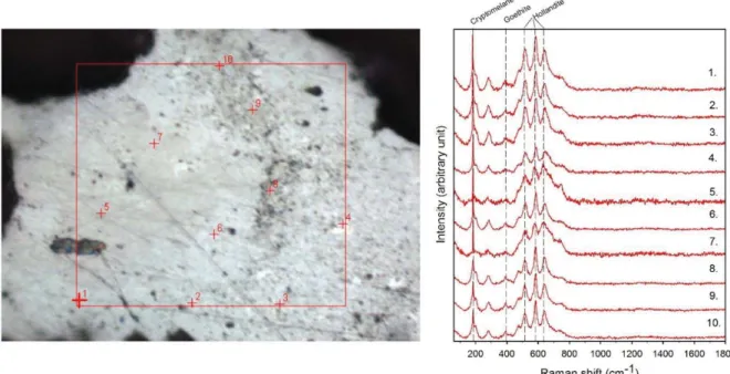
301 High-resolution in situ micro-Raman spectroscopy was used for micro-mineralogy and CM 302 identification and distribution on 9 thin sections (1 polished section) (Szeged University, 303 Hungary). A Thermo Scientific DXR Raman Microscope was used, with a 532-nm (green) diode 304 pumped solid-state (DPSS) Nd-YAG laser, using 1.5-mW laser power and 50x objective lens in 305 confocal mode (confocal aperture 25 μm slit). The acquisition time was 1 min, and the spectral
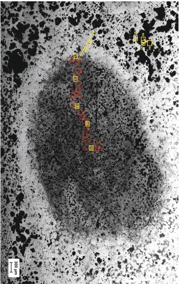
306 resolution was ~2 cm-1 for each measurement. The distance between each point was 10 μm, and 307 the measurement time was 10 min. A composite image of thin sections of Raman microscopy 308 measurements and a series of Raman spectra acquired along the vertical sections are provided in 309 the thin section photomicrographs (arrow points to measurement direction). Diagrams are 310 organized in terms of peak height versus analytical spot number for each of the phases along the 311 Raman-scanned section. Intensities were normalized to the highest peak for each spectrum.
312 Raman measurements were taken on 9 samples (4B, 7, 10, 31, 36-A, 75-2, 75-B5, 48-D1, 313 81). In the case of the homogeneous-like cases 400-500 and in the case of 75-2, 800 spectra were 314 taken along the line shown on section photos (4B, 7, 10, 31). These are systematic investigations 315 along the line profile. Spectra were obtained every 10 μm, providing a high-resolution sensitive 316 study. In samples 75-B5, 78-D1, and 81, the measurements were taken across whole thin sections.
317 The spectra were elaborated in two ways:
318 (1) Diagrams were organized in terms of peak height versus analytical spot number of each of the 319 phases along the Raman scanned section (main minerals and organic matter in general). (2) A 320 detailed determination of all spectra were also made. These results are summarized in tables (Excel 321 files, numbers 1, 2, and 3 indicate the intensity—1-weak, 2-moderate, 3-strong—reference data on 322 detection), in which the mineral composition can be followed from point to point, as well as the 323 type of organic matter. (Supporting Information)
324 Aside from the profile analyses, descriptions of the mineral phase transitions were also 325 constructed for clarification of aegirine (5 photos, 4 mineral spectra, and 1 profile), braunite (3 326 photos, 10 point analyses, and 1 profile across mineral transitional zones), cryptomelane (18 327 photos, 54 point analyses, and 2 profiles across spheres), and the composition of the oblate 328 structures (28 photos, 93 point analyses, and 1 profile).
329 The following Raman bands were used for normalization: rhodochrosite: ~1086 cm-1, 330 dolomite: ~1093-96 cm-1, apatite: ~965 cm-1, quartz: ~463 cm-1; todorokite 633 cm-1; manjiorite 331 641 cm-1; ramsdellite: 650 cm-1; cryptomelane: 183 cm-1 and 580 cm-1; hollandite: 585 cm-1; 332 birnessite: 656 cm-1; ferrihydrite: 707 and 1045 cm-1; goethite: 297 and 385 cm-1; celadonite: 545 333 cm-1; barite: 446 and 985 cm-1; johannite: 785 cm-1; aegirine: 970 cm-1; jacobsite: 620 cm-1; 334 hausmannite: 661 cm-1; braunite: 210, 510, 685 cm-1; and carbonaceous matter: ~1605 cm-1. The 335 identification of minerals was made with the RRUFF Database (Database of Raman – 336 spectroscopy, X-ray diffraction, and chemistry of minerals: http://rruff.info/). Contamination by 337 epoxy glue was taken into consideration. Along with the profile analyses, a detailed determination 338 of all peaks was also made.
339 Comparing the two in situ methods, the AT-FTIR, which did not considerably modify the 340 mineral phases while using the lowest exciting energy, was used to investigate the upper 1–2 μm 341 of the samples. This is the also best method to determine organic matter (Polgári and Gyollai, 342 2019; Polgári et al., 2019). On the contrary, Raman spectroscopy, using higher excitation energy, 343 often caused the transformation of metastable minerals to more stable phases. This method yielded 344 information from the upper 3–4-μm depth of the sample surfaces and was the best method for 345 identifying Mn oxides and hydroxides. The Raman comparative spectra database is more extensive 346 than the AT-FTIR database.
347
348 5. RESULTS
349 5.1. Optical (OM) and cathodoluminescence (CL) rock microscopy 350 5.1.1. Optical rock microscopy
351 Thin sections represent mineralized biomats based on structural observations, which are 352 eminently visible on smaller magnification photos (40x) (Fig. 6, SI 6-, 7-Figs). In all thin sections,
353 adequately high-resolution optical rock microscopy (1000x) supports a series of mineralized 354 biomat microstructures, mineralized microbially produced textures (MMPT) as main constituents 355 (Fig. 6, SI 8-Fig). This microbial microtexture is a basic feature of all the samples, in transmitted 356 as well as reflective light. Well-preserved and mineralized remains of diverse filaments with pearl 357 necklace-like, vermiform inner signatures, and coccoid-like forms embedded in the Mn ore beds 358 are seen, and the whole samples appear densely woven. The minerals are very fine-grained (0.5-1 359 μm) except Mn-1, where clastic contribution occurs. The diameter of the mineralized filaments is 360 around 0.5–1 μm, with variable length (Fig. 6).
361 Fig. 6.
362 Samples 4B, 7, 10, 31, 32 (all from Mn-1) include debris-like components of variable size 363 (20–200 μm). In sample 4B, it seems that the darker gray mineral grains transform to lighter phase 364 (SI 8-Fig). The debris grains are mainly quartz with few fragments of jasper and hydrothermally 365 altered feldspar.
366 5.1.2. Cathodoluminescence microscopy
367 Cathodoluminescence revealed that a part of the debris-like grains (clastic components) is 368 probably composed of real clasts showing the bright, characteristic CL of the mineral (e.g., quartz- 369 blue, feldspar-yellowish) (Fig. 7AB, SI 9-Fig). Some other grains with sizes of some tens of μm 370 resemble clasts but do not show luminescence. These non-luminescent grains are most probably 371 secondary minerals formed via diagenesis (Marshall 1998; Hassouta et al. 1999).
372 Bright blue luminescence is characteristic of kaolinite group-dickite (supported by Raman 373 spectroscopy; Götze et al. 2002), which occurred frequently in our samples (samples 4B, 7, 10, 31, 374 Fig. 7A, B, G, H). The numerous small or larger bright yellow minerals are apatite grains, which 375 often have a lighter margin. These apatites occur along the ore lenses, minerals, and laminae in a 376 woven-like fine-grained biomat-type matrix which mark the borders as accompanying a series of
377 minerals that occurred frequently (Fig. 7, SI 9-Fig) (samples 4B, 7, 10). The fine-grained 378 rhodochrosite (mixed carbonate) show dull reddish (orange) luminescence (Fig. 7A, B) (samples 379 4B, 7, 10). Samples 75-2, 75-B5, and 78-D1 are non-luminescent.
380 Fig. 7.
381 5.2. FTIR spectroscopy
382 Measurements were performed in two ways: (i) randomly, in seven sections (6, 7, 10, 31, 383 32, 36-A1, 78-F3) and (ii) along profiles, in kremydilite sample 48B (Fig. 4K) and in oblate 384 structure (36-A2) (Fig. 4H).
385 5.2.1. Local area analyses
386 Mineral phases and types of organic matter for (i) are summarized in Table 3 and SI 10- 387 Table, according to the measuring area and frequency.
388 Table 3.
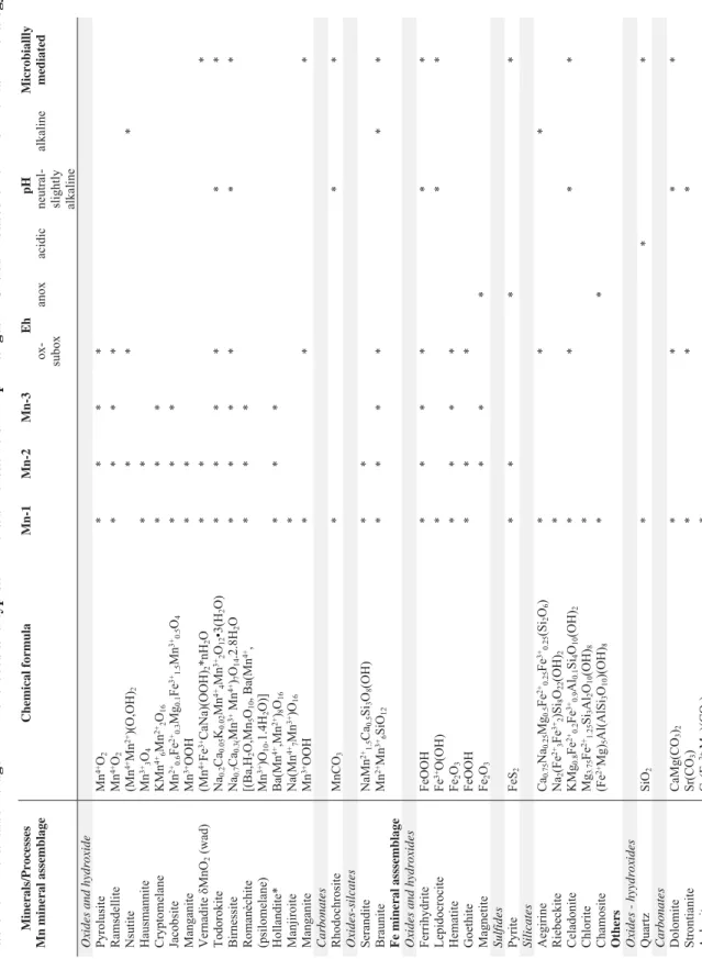
389 In summary, Fe-oxide-hydroxides (ferrihydrite, lepidocrocite, hematite) are common in all 390 the Mn ore beds, Fe-silicates (aegirine) are common in the Mn-1 ore bed, and Fe-sulphide (pyrite) 391 rarely occurs. Variable Mn oxides and hydroxides (todorokite, ramsdellite, pyrolusite, 392 cryptomelane), and oxide-silicates (braunite, serandite) are the main Mn ore minerals. Besides Fe 393 and Mn ore minerals, feldspar, chlorite, celadonite, kaolinite group-dickite, apatite, and quartz are 394 moderate or minor mineral components. Variable types of organic matter occur in all samples.
395 5.2.2. Analyses of kremydilite
396 Three profile analyses in kremydilite sample 48B were made (Fig. 4K and Fig. 8). Two 397 profiles crossed the concentric shells of the kremydilite structure on opposite sides (A and C), and 398 one profile crossed the inner part (B).
399 All concentric shells and the parts intersected between these shells are heterogenous and 400 very fine-grained. Considering that minerals represent the remnants of primary Mn and Fe
401 minerals, each measuring point in the concentric shells and intersected parts resulted in a mixture 402 of minerals, with often poorly crystallized phases.
403 Fig. 8.
404 All shells and the inner part are heterogeneous and very fine-grained. Each measuring point 405 resulted in a mixture of minerals, with often poorly crystallized phases like ferrihydrite. The shells 406 —observed visually—often have the same mineralogy (cryptomelane, hollandite, hematite, 407 rhodocrosite, and pores). The mineralogy of the two sides of the structure are asymmetric (Fig.
408 8D). Profile A (Fig. 8B), from the margin toward the inner part, contain rhodochrosite-goethite, 409 manjiorite-todorokite, minor ferrihydrite-cryptomelane (6 shells), and in the vicinity of the inner 410 part, cryptomelane-ferrihydrite. Profile C, from the margin toward the inner part, contain braunite- 411 rhodochrosite, braunite-goethite-rhodochrosite (3 shells), braunite-cryptomelane-rhodochrosite, 412 braunite-rhodochrosite, braunite, cryptomelane-ferrihydrite-rhodochrosite (2 shells), 413 cryptomelane-ferrihydrite (2 shells), and cryptomelane-braunite, and in the vicinity of the inner 414 part, cryptomelane-ferrihydrite. Profile B, representing the inner part from the shells to the center, 415 contain cryptomelane-ferrihydrite, ramsdellite-rhodochrosite, birnessite-rhodochrosite (2 zones), 416 cryptomelane-quartz-rhodochrosite, cryptomelane-birnessite-dolomite, cryptomelane-quartz- 417 rhodochrosite, ferrihydrite-cryptomelane-dolomite-quartz, and cryptomelane-quartz-dolomite.
418 Varying amounts of pores, with or without organic matter, are characteristic in all layers and in 419 the central parts (Fig. 4).
420 In summary, mineralogical assemblages contain concentric zones (or “shells”) of poorly 421 crystallized, preserved Mn (birnessite, todorokite) and Fe minerals (ferrihydrite), and mainly more 422 stable cryptomelane, hollandite, braunite, hematite, goethite, and rhodochrosite. Profile C mineral 423 components are more stable. More stable minerals represent greater degree of crystallinity.
424 5.2.3. Analyses of oblate (bubble-like) structures
425 Based on OM of sample 36-A1, the outer and inner matrix and also the dark spots of the 426 oblate structures appear very similar, with only the reflective color differing slightly (SI 11-Fig).
427 FTIR analyses resulted in a similar mineralogy and variable organic matter composition, as the 428 sample is very fine-grained and heterogeneous (Fig. 9). The peaks of most of the minerals show 429 broad bands and low intensities, which are characteristic of disordered, poorly crystallized quartz, 430 carbonates, and feldspar.
431 Fig. 9.
432 Out of the oblate structures, the ore contains a matrix, micro-nodules, and dark spots. The 433 micro-nodules and the matrix consist of cryptomelane, ferrihydrite, minor goethite, rhodochrosite, 434 and variable organic matter. The analyzed dark spot in the outer part consists of pores, 435 cryptomelane, ferrihydrite, minor goethite, rhodochrosite, and organic matter.
436 There are no micro-nodules inside the oblate structure. The light part of the matrix inside 437 the oblate structure contains cryptomelane, ferrihydrite, quartz, minor dolomite, and organic 438 matter. The dark part comprises dolomite, ferrihydrite, cryptomelane, and organic matter. Inside, 439 the dark spot consists of ramsdellite, quartz, minor dolomite, and organic matter (SI 11-Table).
440 The oblate, rim structure, separating the outer and inner parts, consists of two phases: (1) 441 the fine-grained rim built up of ferrihydrite, minor goethite, and organic matter; and (2) the coarse- 442 grained phase, which is a mixture of cryptomelane, disordered quartz, rhodochrosite, dolomite, 443 traces of braunite, and variable organic matter. Comparing the outer and inner parts, differences in 444 mineralogy are reflected in the type of carbonate (rhodochrosite outside and dolomite inside), the 445 Mn oxides of the dark spots (cryptomelane outside and ramsdellite (γ-MnO2) inside), and the 446 occurrence of quartz in the inner part and rim, and feldspar in the outer part. On the outside of the 447 oblate structure, the rock contains pores and the typical (micronodule-bearing) microtexture of
448 Mn-2 and 3, whereas inside, the micro-noduliferous textures do not exist, and pores are partially 449 filled by hollandite.
450 5.3. Raman spectroscopy
451 Nearly 11,000 spectra were taken for micro-mineralogical and organic matter composition 452 determinations as well as for the distribution of minerals according to the thin section profiles.
453 Representative analyzed profiles are shown in Fig. 10 and SI 12-Fig. The mineral distribution was 454 evaluated visually based on a series of Raman profiles at the 10-μm scale (Fig. 10, SI 12-Fig). The 455 determined minerals, including FTIR data, are summarized in Table 4. Variable Mn oxides and 456 hydroxides, Mn oxides-silicates, Mn carbonates, variable Fe oxides hydroxides, Fe silicates, Fe 457 sulfide, ore minerals, apatite, feldspar (albite and orthoclase), mica (muscovite, chlorite, 458 celadonite), kaolinite-dickite, barite, carbonates (strontianite, dolomite, ankerite), and quartz occur 459 in the Mn ore beds. Variable organic material is also an important constituent. Based on low 460 intensity and broad peaks, the minerals are poorly crystallized and cryptocrystalline. The 461 representative samples contain a mixture of poorly crystallized mineral phases and organic matter.
462 Table 4.
463
464 5.3.1. Mineral distribution in profiles by Raman spectroscopy
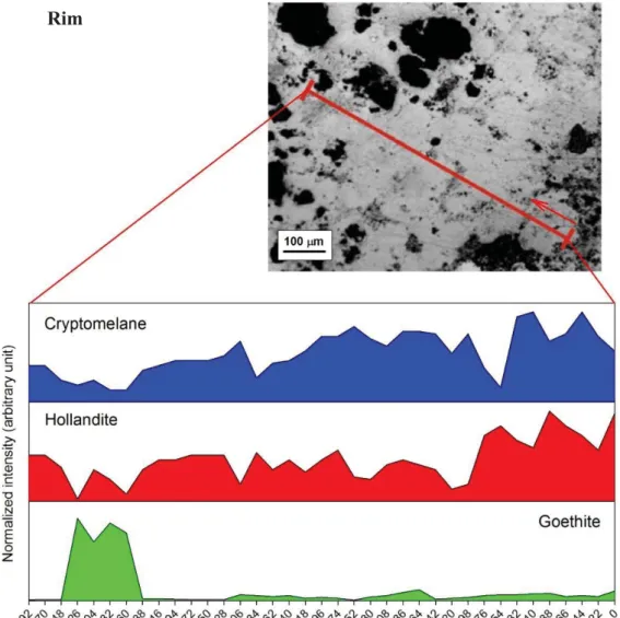
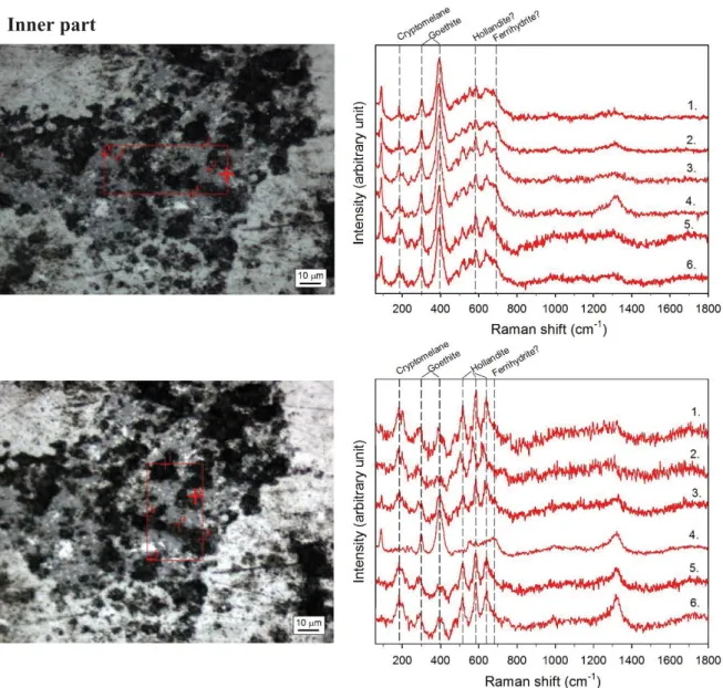
465 A distribution of minerals is evident in all samples, alternating micro-laminae (a few tens 466 of μm thick) along with the kremydilite inner part (Fig. 10, SI 12-Fig). This alternating micro- 467 lamination refer to mineralized microbial cycles in the sediment pile. The documented distribution 468 of minerals in the Mn ore beds is the following:
469 Mn-1 from Figueirinha Mine
470 - Sample 4B - Hematite (rarely aegirine)/rarely quartz alternation, starting with Mn 471 (braunite) alternation and random apatite, and K-feldspar.
472 - Sample 7 - Aegirine/braunite cycles with randomly occurring apatite, mica, and K-feldspar.
473 - Sample 10 - Aegirine-hematite/quartz alternation (Fe cycles) and Mn cycles superposed 474 (braunite, serandite, hausmannite) occur with randomly occurring apatite, barite, feldspar 475 (albite, K-feldspar), and strontianite.
476 Mn-1 from São Domingos Mine
477 - Sample 31 - Hematite (rarely kaolinite/dickite)/quartz alternation (Fe cycles), and Mn 478 cycles superposed (braunite, manjiorite, jacobsite, todorokite, romanèchite).
479 Mn-2 from Urucum Mine
480 - Sample 75-2 - Only Mn minerals occur, but jacobsite and hollandite contain Fe. Jacobsite 481 alternate with cryptomelane, ramsdellite, and hollandite. Ramsdellite is the most oxic 482 phase. In the zone of kremydilite, the micro-lamination turns into random mineral 483 distribution. Accessory minerals are: romanèchite (psilomelane), manganite, todorokite, 484 pyrite, and pyrolusite.
485 - Sample 75-B5 - Goethite is frequent only in this sample. Representative Mn cycles are 486 composed of cryptomelane, hollandite, and occasionally, braunite. Micro-lamination is 487 disordered, and in those zones, random mineral distribution occurs, but locally micro- 488 lamination is well visible. Accessory minerals are: jacobsite, manganite, ramsdellite, 489 todorokite, hausmannite, romanèchite, pyrolusite, ferrihydrite, apatite, and mica.
490 - Sample 78-D1 - Hematite (Fe cycle) alternate with Mn oxide cycles (cryptomelane- 491 hollandite) forming double microbial ore forming lamination. Cryptomelane and hollandite 492 occur together. Hematite and braunite also occur together, but braunite occurs separately, 493 too. Braunite binds to hematite. Locally, pyrolusite, birnessite, romanèchite, jacobsite,
494 manganite, ramsdellite, hausmannite, serandite, ferrihydrite, goethite, mica, and apatite
495 occur.
496 Mn-3 from MCR Mine
497 - Sample 81 - Hematite alternates with Mn oxide (cryptomelane-hollandite). Accessory 498 minerals are: todorokite, ramsdellite, jacobsite, rancieite, pyrolusite, birnessite, braunite, 499 ferrihydrite, magnetite, and mica.
500 Fig. 10.
501 5.3.2. Mineral phase transitions by Raman spectroscopy
502 Microscale mineral phase transitions offer very important information on syngenetic and 503 diagenetic formation processes. Mineral compositions of Urucum samples also provide 504 information on this aspect, which explains the focus on specific mineral transitions.
505 5.3.2.1. Aegirine
506 Aegirine is common in Mn-1, occurring as an alternating mineral with braunite. A detailed 507 study on the phase transition was made for sample 4B (Fig. 11). The microtexture of aegirine 508 resembles a vermiform network that intrudes into the quartz. The quartz occurs in the undulating 509 hematite network as a gel-like segregated silica. Aegirine is present at the contact of quartz, and 510 riebeckite seems to consume aegirine. This relationship shows that aegirine and riebeckite 511 consume quartz. At the contact of segregated quartz, hollandite/vernadite and apatite occur.
512 Braunite binds to hematite in the vicinity of quartz.
513
514 Fig. 11.
515 5.3.2.2. Braunite
516 Braunite also consumes segregated quartz similarly to aegirine, in a vermiform habit, and 517 is in close contact with the hematite network (Fig. 12) (sample 4B). Segregated quartz also contains 518 K-feldspar. Hematite occur as small clusters and contains an undulating network as mineralized 519 biomats.
520 Fig. 12.
521 5.3.2.3. Cryptomelane
522 All the spectra taken in the micro-nodules (cell colonies) and matrix material show 523 dominant vernadite/hollandite-type Mn-oxides-hydroxide composition and a greater or less 524 amount of cryptomelane and variable organic matter (sample 75-2). The minerals are in a 525 cryptocrystalline mixture with variable amounts. The textural differences do not correspond to 526 significant mineralogical differences (Fig. 13, SI 13-Fig). The central part of the micro-nodules 527 consists of hollandite/vernadite and organic matter, around which cryptomelane, pyrolusite and 528 ramsdellite occur.
529 Fig. 13.
530 5.3.3. Oblate structures
531 Detailed Raman measurements were elaborated on a representative oblate structure, used 532 to compare the mineral composition and distribution inside the oblate structure, in its vicinity, and 533 in the rim (sample 36-A1, Fig. 14, SI 14-Fig).
534 The dark spots in the outer matrix are mainly pores, except dark porous inner rims with 535 variable thicknesses, which are composed of a hollandite-type Mn oxide phase (dominant phase), 536 cryptomelane, and goethite. The matrix among the dark spots is built up by hollandite, 537 cryptomelane, and goethite.
538 The non-porous rim of the oblate structure mainly consists of hollandite and cryptomelane 539 in variable amounts. Rarely fine-grained clusters of goethite occur among the hollandite- 540 cryptomelane flakes.
541 In comparing the mineral phases and distribution in the outer, inner, and rim areas of the 542 measured oblate structure, we find that they are similar. The matrix of the inner part of the oblate 543 structure is composed of very fine-grained goethite (ferrihydrite) and small particles of Mn oxides
544 (hollandite and cryptomelane in variable amounts); however, hollandite is dominant in the dark 545 spots. The mineral composition of the matrix and dark spots show a unified distribution, as any 546 difference or significant trend in the matrix or in the dark spots was not detected.
547 Fig. 14.
548 5.3.4. Organic matter
549 The organic matter of Mn-1 ores from Figueirinha (samples 4B, 7, and 10) and São 550 Domingos (sample 31) area are dominated by two bands near 1320 and 1610 cm-1, which are D 551 and G bands of hydrogenated amorphous carbon (Chen et al. 2007). This ore bed contains also 552 traces of aromatic hydrocarbons (825 cm-1), and skeletal stretching of C=C and C=O molecules.
553 Bands of aliphatic hydrocarbons occur at 1000–1280 cm-1 (Okolo et al. 2015) (samples 4B, 7, 10, 554 31), 1300–1390 cm-1 represents CH3 (Jehlička et al. 2009), and 1487 cm-1 refer to CH2/CH3 555 vibrational mode (Jehlička et al. 2009) (samples 7, 10). The band at 1518 cm-1 refers to the C=C 556 stretching in polyenes (sample 31), while 1620–1820 cm-1 show the C=O vibration of oils (Orange 557 et al. 1996) (sample 4B).
558 The organic matter of ores of Mn-2 (Urucum West Mines) (samples 75-2, 78-D1, and 75- 559 B5), contain bands of aliphatic hydrocarbons (1104 cm-1), CH3, and the D and G band of 560 hydrogenated amorphous carbon based on bands near 1320 and 1610 cm-1. The sample 75-2 561 contains only the D and G band of amorphous hydrocarbon, whereas sample 75-B5 contains the 562 aromatic hydrocarbon (825 cm-1) bands of CH2/CH3 vibration (1386, 1469 cm-1), and C=O 563 vibration of oils (1750–1800 cm-1). The sample 78-D1 has bands D and G of hydrogenated 564 amorphous carbon and traces of CH2/CH3 vibrational mode of aliphatic hydrocarbon (1345, 1362 565 cm-1). The sample of the Mn-3 ore bed (MCR Mine, sample 81) contains mostly hydrogenated 566 amorphous carbon (D and G bands at 1317 and 1600 cm-1) and traces of aliphatic (1000–1200
567 cm-1, 1469 cm-1) and aromatic hydrocarbons (825 cm-1). Only 60 of 1903 spectra contains organic 568 material.
569
570 6. DISCUSSION
571 6.1. Sedimentation age and environments
572 The presence of long chain oil type in manganese layers with kremydilite indicates that 573 temperatures were hardly larger than 90°C, which eliminates the possibility that the Jacadigo 574 Group’ rocks have been metamorphosed. This find makes it likely that the 40Ar/39Ar age of the 575 Mn-1 layer is effectively 547 ± 3 to 513 ± 4 Ma, the ages of braunite and muscovite determined 576 by Piacentini et al. (2013). This age seems to be reinforced by that determined by Babinsky et al.
577 (2008), which dated detrital zircons (U – Pb SHRIMP) from a volcanic ash layer intercalated with 578 carbonate rocks of the Tamengo Formation at 543 ± 3 Ma.
579 Corumbella and stromatolite occurrences and field information published by Biondi and 580 Lopez (2017) indicate that the Santa Cruz Formation (BIFs) and the manganese layers sedimented 581 at the same time or after the Bocaina Formation; and that the ages of these rocks are about 550 582 Ma. The age of this fossil in the ironstones of the Santa Cruz Formations (Jacadigo Group) and 583 limestones of the Tamengo Formation (Corumbá Group) was estimated at ca. 550 Ma (Germs 584 1972; Grant 1990; Grotzinger et al. 1990; Hofmann and Mountjoy 2001; Bengtson 2002). Also, 585 the proposed age of this horizon was 555–542 Ma by ichnofossils, identified by Parry et al. (2017), 586 in the Bocaina Formation.
587 There is no diagnostic evidence that sedimentation occurred during some glacial period or 588 during some glaciation, as initially proposed by Urban et al. (1992). The only arguments of these 589 authors were: (a) the presence of the granite blocks they interpreted as dropstones, without even 590 observing whether any of these blocks have faceted, friction-sectioned sides, and/or have striated 591 faces, as is typical of dropstones. These characteristics were never observed in the Urucum 592 (Trompette et al., 1998; Freitas et al., 2011; Biondi and Lopez, 2017). (b) To consider the Santa 593 Cruz Formation, with at least 400 m thick BIFs, as similar to the Rapitan Formation, with less than 594 10 m thick BIFs (Young, 1976). As the Rapitan Formation would be Ediacaran and of glacial 595 origin (Young, 1976), Urban et al. (1992) inferred that the Santa Cruz would have the same origin;
596 and most subsequent authors adopted this idea. However, keeping in mind that this hypothesis is 597 traditionally defended by many authors (e.g. Angerer et al., 2016), it should be discussed.
598 The last glacier related to snowball earth, and the sedimentation of Rapitan-denominated 599 BIFs, was the Marinoan glaciation, which began at about 650 Ma and ended at about 635 Ma.
600 Considering all the information presented above, the Urucum’s BIFs, ironstones and manganese 601 layers sedimented about 550 Ma ago, 85 Ma after the end of the Marinoan and the snowball 602 glaciations. The Gaskiers glaciation, which existed for 340,000 years (579.9 to 579.6 Ma), has 603 occurred about 29 Ma before the end of the Jacadigo Group sedimentation, and could hardly 604 influence its sedimentation. It remains, therefore, to relate the formation of the Jacadigo Group 605 with Baykonurian glaciation (547 to 545.5 Ma), so far recognized only in Asia and Africa 606 (Chumakov, 2009; and Chumakov, 2011; Germs and Gaucher, 2012). We therefore propose to 607 consider the possibility that the sedimentation of the BIFs and manganese layers of the Santa Cruz 608 Formation occurred during the Baykonurian glaciation, which would explain the existence of what 609 is interpreted by Urban et al. (1992), among other authors, as dropstones.
610 We hold the view that Mn-1 was most likely formed during the first inundation of the 611 ancient graben by the fluvial, oxidative sediments that gave rise to the Urucum Formation. Unit 612 Mn-1 contains predominantly siltic and sandy, ferruginous clastic rocks, cemented by microbially 613 mediated Fe minerals (e.g., aegirine), and Mn-oxide and silicate (braunite, serandite, and 614 hollandite). The areas of Figueirinha and São Domingos mines have a larger concentration of 615 manganese in Mn-1, which are contained in clast-bearing massive ores. The upper Mn-1 layers in 616 the Figueirinha and São Domingos mines, which include amygdalites, were probably deposited in 617 the basin depocenter, where the amygdalites formed from hydrodynamic flux. Layers Mn-2 and 618 Mn-3 formed in “offshore” (= greater depth) environments during periods of tectonic quiescence, 619 when fine, clastic quartz fragments and other detrital sedimentation ceased.
620 6.2. Mineralogical interpretations
621 Microtextural evidence in all the studied samples appears as dense features, and the mineral 622 types and embedded variable organic matter raise the microbially-mediated formation of the ore 623 beds, which we argue occur as microbialites (MMPT). Two microbial ore forming systems are 624 proposed as dual systems, characterized by Fe- and Mn-oxidizing metabolic processes (Fe- 625 oxidizing bacteria (FeOB) and Mn-oxidizing bacteria (MnOB)).
626 Several studies on the genesis and preservation of oil and natural gas have shown that long 627 chain hydrocarbons are decomposed at temperatures above 90ºC (Chilingar et al. 2005, p.138- 628 142). Preservation in Mn-2 and Mn-3 of aromatic and aliphatic hydrocarbons, C=C stretching in 629 polyenes, C=O vibration of oils, among others, indicates that temperatures during diagenesis were 630 low (<90ºC) and that syngenetic as well as diagenetic minerals were preserved, as identified by 631 the Raman and FTIR analyses. Along with hydrocarbons, these analyses identified minerals such 632 as birnessite and ferrhydrite, which we consider remnants of the original sedimentation, (i.e. they 633 were not entirely destroyed during diagenesis). Remnants of syngenetic and diagenetic minerals 634 interpreted as complex systems give a plausible series of processes and environmental formation 635 conditions during sedimentation and diagenesis (Table 4, Fig. 15, SI 15-Table). The frequency of 636 the minerals is different: the main minerals, such as cryptomelane, hollandite, hematite, and 637 braunite, form the ore beds, but the moderate and minor minerals have also genetic importance.
638 The Mn layers are the result of complex diagenetic processes and formation of diagenetic minerals, 639 which include the components of the decomposition of cells and extracellular polymeric substance 640 material (Fe and Mn bacteria, cyanobacteria, and other types; see Ewers 1983; Wignall 1994;
641 Konhauser 1998; Villalobos et al. 2003; Dupraz and Visscher 2005; Dupraz et al 2009; Chan et al.
642 2011; Gyollai et al. 2017).
643 Some syngenetic poorly crystallized minerals were preserved, and that serves as a starting 644 point. For a clear understanding, a short review on the most important mineral assemblages and 645 primary minerals is needed. Many types of minerals occur, and these can be grouped as follows.
646 6.2.1. Remnants of syngenetic minerals – Syngenetic Fe- and Mn-rich biomat 647 formation
648 Remnant syngenetic minerals are reported as microbially mediated minerals forming under 649 obligatory oxic (Mn) and suboxic (Fe) conditions, with neutral and semi-neutral pH. The
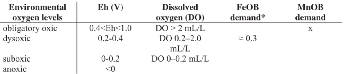
650 microbially mediated Mn and Fe oxidation have different oxygen demand, and the diagenetic 651 zones represent different oxygen conditions. The nomination “suboxic” has a double meaning 652 which can cause discrepancies. To avoid misunderstanding, definitions are listed in Table 5. In 653 general, Eh > 0 represent oxic conditions, but the concentration of oxygen can be different, as 654 shown in Table 5 and Fig. 15, and the microbially mediated processes occur at a given oxygen 655 content. Diagenetic zonation also separates the oxic, suboxic, and anoxic zones, and the oxidizing 656 agent is O2 in the oxic zone, NO3-, MnO2, and Fe2O3 in the suboxic zone, and SO42- in the anoxic 657 zone (Berner 1980; Coleman 1985; Wignall 1994; Polgári et al. 2012ab).
658 Table 5.
659 Ferrihydrite and lepidocrocite on the Fe side, and vernadite, todorokite, birnessite, and 660 manganite on the Mn side, are regarded as syngenetic minerals (Ehrlich 2002). Accordingly, it is 661 obvious that ore formation started with microbial Fe oxidation. That is why interpretation starts 662 with a description of the Fe system.
663 Syngenetic Fe system
664 Understanding the biochemistry of the biomat formation is a key factor in determining the 665 type of Fe-rich biomat that may have been involved in the formation of the Urucum Mn layers, 666 and to define the environmental conditions. There are various types of microbial metabolisms that 667 can oxidize Fe2+ in nature, which occur under varying states of oxygen-deficient conditions. Three 668 types of Fe-rich biomats are considered for the Urucum; all are neutrophilic and consistent with 669 basin conditions (Fig. 15): (1) Microbial neutrophilic, micro-aerobic Fe(II) oxidizing bacteria (pH 670 ~8; Eh +0.3 V) (Hallbeck and Pedersen 1990; Ehrenreich and Widdel 1994; Konhauser 1998;
671 Ehrlich 2002) supported by mineral assemblage (ferrihydrite, goethite, hematite, celadonite); (2) 672 Nitrate-reducing Fe(II) oxidizers in suboxic/anaerobic conditions (lack of filaments; Straub et al.
673 1996); and (3) Photoferrotroph metabolism in anoxic/anaerobic light-demanding conditions,
674 which is not plausible based on mineral assemblage, which support suboxic-oxic conditions. The 675 fourth (4) type, strongly acidic, oxic metabolism is also not plausible in the Urucum basin, and 676 does not fit with the mineral assemblage. However, a further process we have to consider, is the 677 non–Fe-oxidizing microbes later overgrown by Fe oxides via microbial processes (Konhauser 678 1998). This cannot be excluded, but the homogenous Fe-precipitation on filamentous forms do not 679 support this scenario as a principal process.
680 The rhythmic developmental stages via microbial mediation is basic. Free-living Fe(II) 681 oxidizing bacteria exist in the lag and log phases (Novick 1955; Zwietering et al. 1990), and stalk 682 formation (Fe-rich biomat–mineralization) occurs during the stationary (abbrev.: stat) phase under 683 optimal conditions (pH > 6, aerobic, cell number > 6 × 10⁵ mL–1, low organic C content, 1–3 week 684 whole microbial population growth period; e.g., Gallionella-like freshwater types and 685 Mariprofundus-like marine types) (Hallbeck and Pedersen 1990; Chan et al. 2011; Polgári et al.
686 2012a).
687 Organic biomarkers were not directly associated with Fe-rich biomat structures. Raman 688 and FTIR data show organic matter in the biomat lacework but is not diagnostic as to its type.
689 Based on these data, the diagenesis developed more in rocks represented by sample 75-2, where 690 only amorphous carbon remained in traces, and other type of organic material was consumed.
691 Preservation of organic material was best in sample 75-B5, in which organic material occurs in 692 180 of 2447 spectra, and more complex organic material, like oils and aromatic hydrocarbons, 693 were detected. Sample 78-D1 (SI 3-Fig) is more altered, because only traces of complex organic 694 material were preserved, and organic material—mostly D and G bands—occur in only 60 of 3456 695 spectra. Biomarkers cannot be isolated because of multiphase microbial activity and extensive 696 diagenetic overprinting.
697 Fig. 15.
698 Syngenetic Mn system
699 Mn-oxide formation in Mn-1 ore bed can be explained by the oxide surface catalysis model 700 advocated by Morgan (2005). Metal-oxide surfaces are able to accelerate Mn(II) oxidation by 701 redox reactions (e.g., hematite, goethite, lepidocrocite, and manganese dioxide; Wilson 1980; Sung 702 and Morgan 1981; Davies and Morgan 1989). Raman spectroscopy detected vernadite as poorly 703 crystallized mineral phase in the vicinity of hematite biomat lacework (Fig. 11).
704 In the case of Mn-2 and Mn-3, during the development of the Mn-oxide proto-ore, the first 705 product of microbial enzymatic Mn(II) oxidation probably was a bio-oxide (e.g., vernadite, 706 todorokite, birnessite), similar to the experimental studies of Villalobos et al. (2003); Bargar et al.
707 (2005); and Bodeï et al. (2007). This enzymatic Mn oxidation can be referred to as Cycle I. The 708 demand of microbial (enzymatic) Mn(II) oxidation is obligatory oxic conditions (> 2 mL/L 709 dissolved oxygen). This bio-oxide is an X-ray amorphous oxide similar to δ-MnO2 (vernadite, 710 todorokite, birnessite; all detected by Raman), which is thought to be a disordered 711 thermodynamically unstable 7-Å-vernadite (hexagonal phyllomanganate) containing Mn(IV) 712 vacancy defects, with very small particle sizes (< 20 nm lateral dimensions), and having only two 713 or three MnO2 layers stacked along the c-axis (Villalobos et al. 2003). A decrease in the dissolved 714 Mn(II) appears to act as a reductant for the biogenic oxide and control the stability of secondary 715 abiotic reaction products (Mn2+ components in minerals of Urucum support this process). Cation 716 binding, like Mg, supports phyllomanganate transformation to stable tectomanganate (Bodeï et al.
717 2007). Experimental studies showed that extracellular polymers from bacteria catalyze the 718 adsorption of Mg on the surface of the cells (Mandernack et al. 1995). Thus, the bacterial cells not 719 only directly oxidize Mn(II) to Mn(IV), but also, in the early stages of oxidation, influence the 720 cation composition of the Mn-oxide mineral being produced. Mineralogical changes similar to 721 these are likely to be commonplace in natural settings where bacterial oxidation of Mn(II) occurs
722 and may liberate sorbed metal ions or alter the rates of Mn-oxide surface processes, such as the 723 degradation of organic molecules. It is noteworthy that microbes may exploit such mineral 724 transformation reactions to indirectly control chemical conditions in the vicinity of the cell 725 (Mandernack et al. 1995).
726 A series of detailed mineralogy and micro-textures are shown in Fig. 13 (sample 75-2 from 727 Mn-2 ore bed). The studied part is representative for syngenetic microbial Mn oxidation. The 728 distribution of vernadite/hollandite and very early diagenetic cryptomelane and nsutite show 729 micro-nodules with mineralized microbial colonies with embedded organic matter, which appear 730 to support this scenario. Recent results also comport with the study of Piacentini et al. (2013) who, 731 based on petrographic evidence, reported that cryptomelane may not be the primary Mn mineral 732 precipitated in the Neoproterozoic ocean floor.
733 6.2.2. Diagenetic minerals 734 Diagenetic Fe system
735 According to the diagenesis of Fe-rich biomats, the microbes produce poorly ordered 736 ferrihydrite (lepidocrocite) as a primary mineral, which transforms to more ordered minerals, such 737 as goethite or hematite (reduced form as magnetite), within a few months or years via dissolution- 738 dehydration processes, as mentioned before (Konhauser 1998; Schwertmann and Cornell 2007;
739 Gyollai et al. 2015). The main Fe oxide mineral in the filaments of our samples is hematite, but 740 Raman analyses indicate that goethite also occurs (sample 75-B5, SI 12-Fig). In Mn-1, aegirine, 741 and in Mn-2, rare jacobsite can represent mineralized Fe-biomats. In other rare occurrences, pyrite 742 also occurs reflecting that locally anoxic conditions existed but did not become dominant. The 743 fossilized Fe-rich biomats were rapidly and extensively encrusted by minerals, such as dolomite 744 and silica, similar to what has been reported by Baele et al. (2008). Amorphous silica segregation
745 is derived by either the destruction of organic complexes or the transformation of ferrihydrite 746 (Baele et al. 2008).
747 Aegirine, occurring in cyclic microlaminae alternating with braunite in Mn-1, is the 748 diagenetic mineral form of FeOB (Fig. 10). Aegirine forms via early diagenesis from the 749 syngenetic Fe-oxi-hydroxides (ferrihydrite) and segregated silica, and represents a more stable 750 mineral phase. Aegirine micro-laminae represent the Fe-oxidizing microbial cycles, and braunite 751 represents the Mn cycle in silicified and stable form, also reported by Johnson et al. (2016). This 752 aegirine-braunite microbialite represents oxic/suboxic conditions (Listova, 1961). High-resolution 753 Raman investigations show that aegirine consumes segregated silica from hematitic biomat toward 754 the segregated silica via the transitional mineral riebeckite (Fig. 11). Riebeckite is also a common 755 constituent in BIF with aegirine reported by Savko (2006), who proposed metamorphic formation, 756 which does not fit with our observations. In Mn-1, aegirine forms a woven network (Fig. 11), the 757 hematitic proforma of biomat with the segregated silica. At the contact between hematite and silica, 758 apatite, vernadite/hollandite, and braunite occur. Similar to aegirine, braunite also consumes the 759 segregated silica. Our results fail to fit with the hydrothermal origin of aegirine proposed for 760 Paleoproterozoic Hotazel iron-formation, South Africa (Tsikos and Moore 2005); the cyclic 761 occurrence, worm-like consuming behavior, and also the mineral assemblage contradicts with that 762 scenario. Comparison with other natural aegirine occurrences, however, such as authigenic 763 aegirine in the lacustrine Green River Formation of Wyoming, U.S.A. (Fortey and Michie 1978), 764 shows a close similarity. In short, the reported authigenic formation of aegirine fits well with our 765 results, but the source of Na instead of volcanic activity was more probably the decomposition of 766 cell and extracellular polymeric substance organic material.
767 As the depth of the basin is not known, fragments of slightly lithified and re-sedimented 768 and cemented biomats occur, a shallow marine condition cannot be excluded. In such
769 environments, evaporitic alkaline sodium-rich conditions cannot be ruled out and indeed, are 770 preferred for aegirine formation. The high silica concentration favors aegirine formation instead 771 of clay minerals (Decarreau et al. 2004, 2008), which only sporadically occur in the samples 772 studied here (celadonite, chamosite).
773 Celadonite, a dominant mineral phase in the Mesozoic Úrkút Mn-deposit (Polgári et al.
774 2012b; 2016ab), is an Fe-mica reflecting suboxic neutral conditions. It is rare in Urucum 775 presumably because of high silica content.
776 Chamosite formation is favored by seawater solutions at low temperatures with a relatively 777 reduced pH, a low amount of SiO2, high content of Fe2+ and Fe3+, and a relatively high amount of 778 Al and Mg. Aluminum may be donated to the system by organic matter as reported by Maliva et 779 al. (1999) who showed that the aluminum content is greatly increased by complexation with 780 organic acids. Low silica concentration in solution is the most important condition for low- 781 temperature synthesis of clay minerals, as high silica concentration in solution inhibits their 782 formation (Harder 1976).
783 Diagenetic Mn system
784 In diagenesis, the stabilization of the syngenetic Mn oxide hydroxides proceeded and pure 785 forms, such as pyrolusite, ramsdellite, nsutite, hausmannite, manganite, and variable-cation-bound 786 forms (e.g., Na, K, Ca, Mg, Ba, Fe) such as cryptomelane, jacobsite, romanèchite, and manjiorite 787 grew (Giovanoli 1980; Mandernack et al. 1995; Villalobos et al. 2003; Bargar et al. 2005; Bodeï 788 et al. 2007; Johnson et al. 2016). Of note, as described by Polgári et al. (2012b), Maynard (2014), 789 and Johnson et al. (2016), rhodochrosite can result from the sporadic activity of heterotrophic 790 microbes during the early stages of diagenesis. Rhodochrosite is, however, only frequent in the 791 only fully analyzed kremydilite sample shown in Fig. 8. Otherwise, these poorly mineralized 792 cryptocrystalline mineral phases mix in a variable amount in the microlaminae as a manifestation
793 of mineralized Mn cycles. Similarly, pyrolusite, ramsdellite, and romanèchite indicate Mn 794 precipitation via diagenesis from low-temperature pore fluids as reported by Rajabzadeh et al.
795 (2017).
796 6.3. Combined diagenesis of the two ore-forming microbial systems and other 797 microbial forms
798 Harder (1978) noted that “…the silica content of sedimentary iron ores is found in quartz 799 and different iron-containing clay minerals. Chamosite, greenalite, cronstedtite, nontronite, 800 glauconite, and thuringite are common minerals in sedimentary iron ores. In general, all these 801 minerals are extremely fine-grained.” We find that the formation of Mn-1 manganese ore in 802 Urucum influenced and changed this general protocol, despite the observation of quartz, local Fe 803 mica, and Fe clay. Highly alkaline pore water conditions in diagenesis (accompanied by elevated 804 Na content) caused aegirine precipitation instead of smectite (Decarreau et al. 2004, 2008). Low 805 silica content and Si/Fe ratio lead to chamosite formation under reduced pH and Eh conditions.
806 The high silica content probably influenced silica uptake of variable Mn oxide-hydroxide minerals.
807 Through stabilization caused by diagenetic processes, the Mn oxide hydroxide bound not only Fe2+
808 and Fe3+ (e.g., jacobsite, hollandite minerals), but also silica (braunite, serandite), to form a highly 809 variable content of oxide-silicate mixed minerals. These are characterized by highly variable 810 composition. Texturally, mineral habits are strongly modulated (and perhaps templated) by 811 extracellular polymeric substances that form a network of pore spaces.
812 Braunite alternates with aegirine in Mn-1 and also occurs in Mn-2 representing the 813 mineralized Mn cycle (Fig. 10). The principal reasons for this viewpoint is that the system acts as 814 a diagenetic cycle owing to the fact that in Mn-1 an active oxide surface catalyst is likely 815 responsible for the mineral assemblage, as opposed to enzymatic Mn oxidation. The interpretation 816 is that braunite formation is due to combined diagenesis, as the segregated silica needed for