SUPPORTING INFORMATION
ORIGIN OF THE URUCUM IRON FORMATIONS (NEOPROTEROZOIC, BRAZIL):
TEXTURAL AND MINERALOGICAL EVIDENCE (Mato Grosso do Sul – Brazil)
Running title: Fe-rich rocks from Urucum
Márta Polgári
1,2*, Joăo Carlos Biondi
3, Ildikó Gyollai
1, Krisztián Fintor
4, Máté Szabó
11
Institute for Geological and Geochemical Research, RCAES, Eötvös Loránd Research Network, 1112 Budapest, Budaörsi u. 45, Hungary, e-mail: rodokrozit@gmail.com, gyildi@gmail.com, szmatez@gmail.com
2
Eszterházy Károly University, Dept. of Natural Geography and Geoinformatics, 3300 Eger, Leányka u. 6, Hungary
3
Federal University of Paraná State, Polytechnic Center, Geology Department, 81531-980 Curitiba, Brazil, e-mail: biondiufpr@gmail.comS
4
Szeged University, Dept. of Mineralogy, Geochemistry and Petrology, 6722 Szeged, Egyetem u. 2-6, Hungary, e-mail: efkrisz@gmail.com
*corresponding author:
rodokrozit@gmail.com Content
SI 1. Iron deposits similar to Urucum
SI-2. Panorama and high resolution optical rock microscopic photos of thin sections SI 3. FTIR measurement of Urucum iron ore deposit
SI 4. FTIR dataset of samples
SI 5. Raman spectroscopy dataset of samples
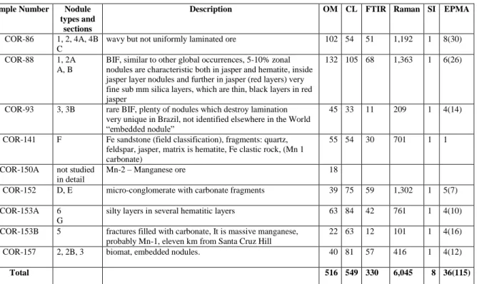
SI 6. Mineral composition of nodules and sections based on Raman spectroscopy SI 7. Raman profiles of nodules
SI 8. Raman profiles of sections
SI 9. Back scattered electron images of samples with mineral composition
SI 10. Element map images in a complex system with OM, CL, FTIR, Raman and stable isotope datasets
SI 11. Carbonate composition of samples based on EPMA analyses SI 12. Carbonate composition of samples based on EPMA analyses
SI 13. Temperature calculation on δ
18O
SMOWcarb(based on Kim et al., 2009; Hodel et al., 2018) Table S1. Samples and methods used
Table S2. Mineral and organic matter composition and frequency
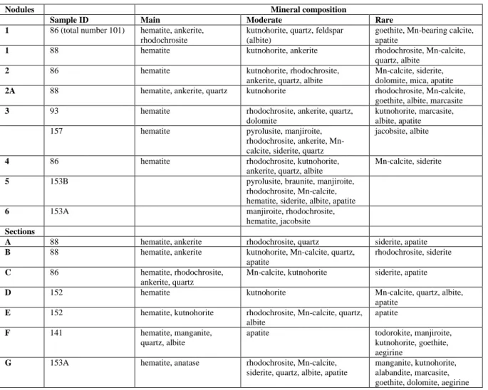
Table S3. Raman profiles, point dataset and FTIR mineralogy both for nodules and sections according to Fe and Mn cycles and syn-and diagenetic formation
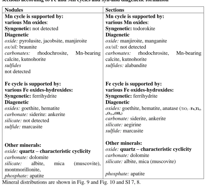
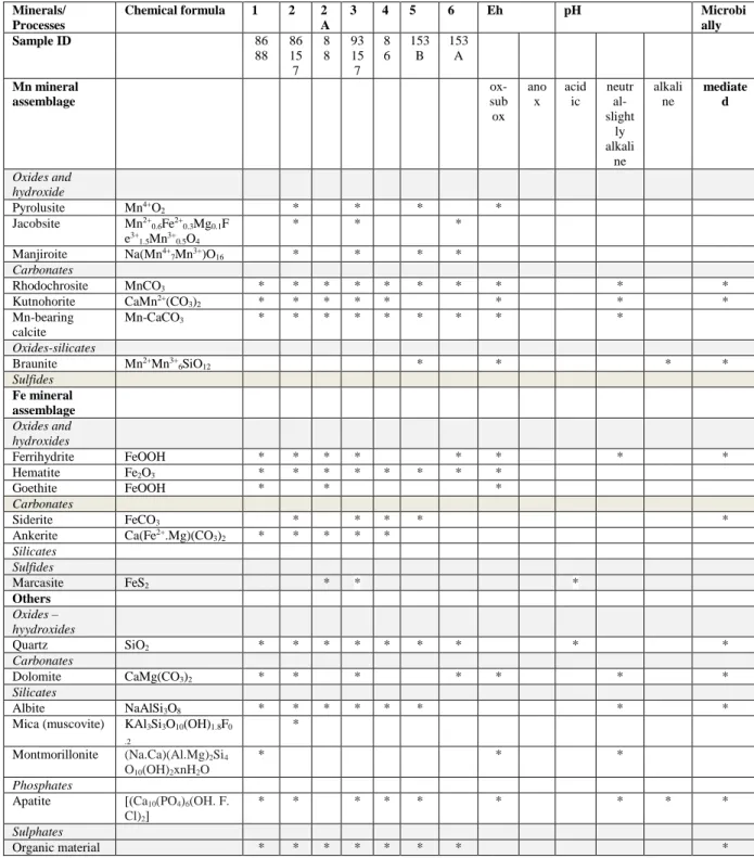
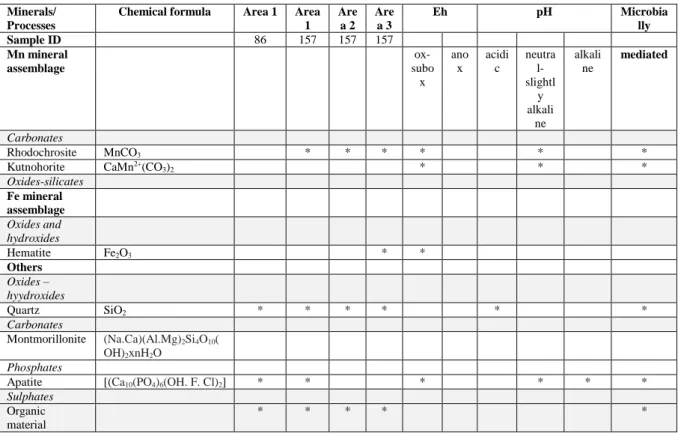
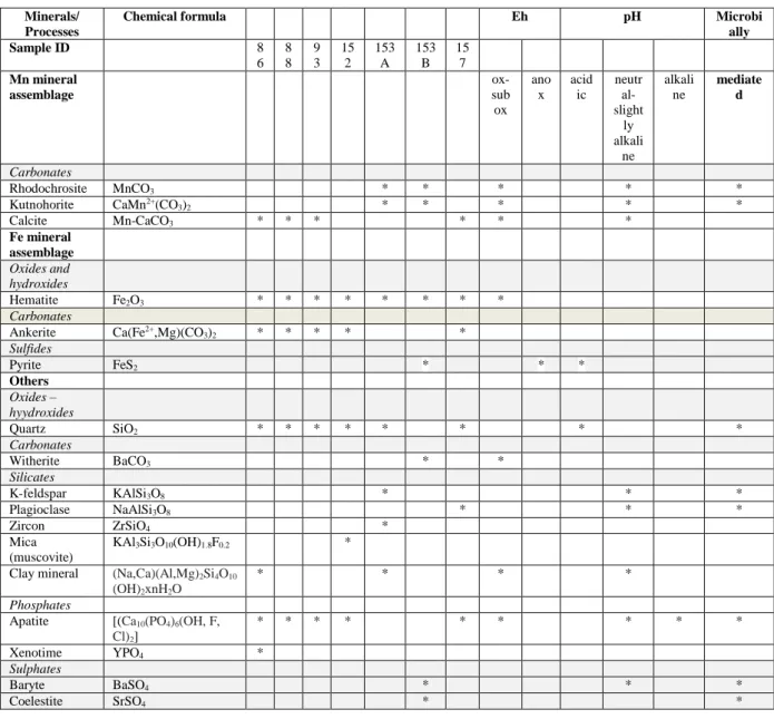
Table S4. Mineral assemblage in Urucum Fe ore, Brazil. and typical minerals indicative of Eh-pH ranges based on environmental mineralogy (low T)
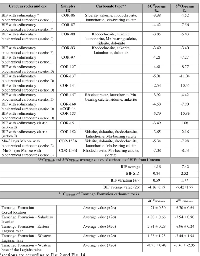
Table S5. Carbonate types and δ
13C
PDBcarband δ
18O
PDBcarbvalues of sedimentary carbonates in
BIFs and Mn ore from Urucum and Vetorial mines, Urucum region (MS, Brazil)
SI 1. Iron deposits similar to Urucum
Ever since Young (1976) described the iron formations of the Rapitan Group (Canada), and genetically related them to Neoproterozoic glaciations, authors who work with iron formations across the planet have adopted the term "Rapitan-type iron formation" to designate all BIFs formed at the end of the Neoproterozoic. Klein and Beukes (1993a, 1993b) showed that the Rapitan Group is aged between 755 and 730 Ma and that it is coeval with several other extensive iron-formations worldwide, all of which are associated with glaciogenic sequences. These sequences would have be related in origin to the enrichment in Fe2+ of seawater, isolated from the atmosphere by ice crusts generated by regional glaciations from the end of the Neoproterozoic (according to the Snowball Earth Theory of Kirschvink, 1992). With the end of the glaciation and the disappearance of glaciers, in some restricted places on the continental shelves there would have been the oxidation of Fe2+ to Fe3+ which, being insoluble in sea water, would sediment together with a large amount of jasper, forming BIFs.
Included in the Rapitan Group type are the following iron-manganesiferous units: (1) Urucum district of Brazil (Urban et al., 1992; Trompette et al., 1998; Klein and Ladeira, 2004), (2) Mutum in Bolivia (Trompette et al., 1998), (3) MacKenzie and Ogilvie Mountains of North America (Young, 1976; Klein and Beukes, 1993a, b), (4) southeastern Uruguay (Pecoits et al., 2008), (5) Damara orogen of Namibia (Breitkopf, 1988; Bühn et al., 1992), (6) Adelaide geosyncline of South Australia (Lottermoser and Ashley, 1999), (7) Erzin basin in Tuva, Russia, and in adjacent Mongolia (Ilyin, 2009), (8) Malyi Khingan in the southern part of the Russian Far East (Ilyin, 2009), and (9) middle Tien-Shan in Kazakhstan, Kyrgyzstan.
Urucum, located in Brazil, and Mutum, located in Bolivia, are different names given to the same geological unit. The eastern part of the Jacadigo Hill (Fig. 1A), located in Brazil, belongs to the Santa Cruz Formation of the Jacadigo Group, which contains several iron-manganese units, including those from the Urucum mine, the name that was historically adopted to designate around 10 other iron and manganese deposits and mines of the Santa Cruz Formation. Crossing the border to the west and entering Bolivia, the western part of Jacadigo Hill is called Mutum (Trompette et al., 1998). The geology and genesis of banded iron formations (BIFs) of the Urucum-Mutum and of the manganese deposits interstratified in these iron formations have been discussed in many publications, among which the most recent and comprehensive are Klein and Ladeira (2004), Angerer et al. (2016), Biondi and Lopez (2017), and Biondi et al. (2020).
Urban et al. (1992) considered the iron formations of the Urucum region similar to the iron formations of the Rapitan Group. By similarity, Urban et al. (1992) considered that the Urucum BIFs would be Cryogenian (720- 635 Ma), and their genesis, like the Rapitan Group, would be related to the great glaciations of the late Neoproterozoic, especially to the Marinoan glaciation (650 to 635 Ma).
After Urban et al. (1992), and Klein and Beukes (1993) several authors adopted the same ideas about the Urucum, even though Biondi and Lopez (2017) and Biondi et al. (2020) describe several geological features that indicate that the BIFs of Urucum (and Mutum) are Ediacarans, aged 560-550 Ma (Piacentini et al., 2013; Biondi and Lopez, 2017; Biondi et al., 2020). Indeed, the iron formations of the Rapitan Iron Formation Group, especially at Cranswick River, Mackenzie Mountains, of the Canada Northwestern Territories, are very similar to those of the Urucum. Layers with nodular and banded jasper interlayered with hematite bands and overprinted by anastomosing hematite are similar to the arkosean iron formations located at the base of the Santa Cruz Formation (Biondi and Lopez, 2017), and the BIFs with jasper nodules in massive hematite (Bekker et al., 2010) are very similar to the BIF types 4 and 4A (Fig. 3), one of the most common BIF-type in the Urucum. On the other hand, besides their ages, Urucum and Rapitan have some other important differences. The total thickness of the Rapitan Group's BIFs rarely exceeds 70 m, but it is more than 400 m in Urucum. In Rapitan there are diagnostic evidences of sedimentation in a glacial environment, with frequent diamictites, dropstones, striated pebbles, etc., while in Urucum no diagnostic evidence was found for regional glacial environments, and the coarse grained rocks are conglomerates formed by avalanches. There are no manganese layers interspersed in the Rapitan Group's BIFs, while in Urucum there are three layers of manganese that are economically very important, with thicknesses between 0.5 and 6.0 m, of biogenic origin (Biondi and Lopez, 2017; Biondi et al. 2020), interspersed in the BIFs of the basal portion of the Santa Cruz Formation. Another important difference between the two units is indicated by multi-element REE diagrams. Urucum BIFs have negative Ce anomalies ranging from discrete to large (Angerer et al., 2016; Biondi and Lopez, 2017) indicating a marine sedimentary environment with hot water (Angerer et al., 2016; Biondi and Lopez, 2017), which contrasts with the sedimentation in the glacial environment of Rapitan BIFs.
According to Klein and Beukes (1993a, b), the Rapitan Group is well exposed in the Mackenzie Mountains and in the eastern Wernecke Mountains of the northeastern Cordillera of North America, with correlatives in the upper Tindir Group (Young, 1982) in the Ogilvie Mountains on the Alaska-Yukon border. It unconformably overlies a thick platform assemblage of carbonates and relatively mature siliciclastics of the Mackenzie Mountains Supergroup (Young et al., 1979) and is paraconformably overlain by shale of the Twitya Formation, forming the base of the Hay Creek Group (Young et al., 1979). Young (1992) suggests that the glaciogenic Rapitan sequence correlates with the Mackenzie Mountains Supergroup in the northern Cordillera. The Mackenzie Mountains
Supergroup and the Ogilvie Mountains differ from the Rapitan Group only because the lower mixtite unit of the Rapitan is not developed in Mackenzie and Ogilvie Mountains. The BIFs, the stratigraphic sequence and the genetic process appear to be the same as those of the Rapitan Group.
The Arroyo del Soldado Group, in Uruguay, is a mixed siliciclastic-carbonate succession, mainly represented by an intercalation of basal pink dolostones, banded siltstones, rhythmites of dolostone-limestone, iron formations, cherts and conglomerates. According to Pecoits et al. (2008), these stratigraphic and chemostratigraphic features are suggestive of a Gaskier age (ca 580 Ma) for the basal glacial-related units. Gaucher et al. (1998) identified two different facies associations, shallow-water and deep-water. The shallow-water facies association is the most widely distributed and consists of basal conglomerates, followed by intercalations of sandstones and pelites in the middle of the succession, grading to banded siltstones at the top. The deep-water facies association comprises an alternation of finely laminated dark shales and arkoses; which are turbidites. More recently Gaucher et al. (2004) reported the presence of oxide-facies, a 50 m thick BIF with up to 24%
magnetite/hematite. These rocks display centimeter alternation of chert and iron-rich layers that do not show the characteristic microbanding of Archean-Palaeoproterozoic (Pecoits et al., 2008), but which is similar to the BIF type 2 (Fig. 3) that occurs in Urucum. Gaucher et al. (1996) suggested that the succession was deposited on a stable continental shelf undergoing tectonic quiescence. Gaucher et al. (2004) suggested that deposition of the BIF took place on a shelf, due to enhanced upwelling of nutrient-rich waters and consequent production of phytoplankton blooms during greenhouse conditions. However, the depositional geological setting of the group does not correspond to an Atlantic-type continental shelf, in which the model was developed, and the age of the whole Arroyo del Soldado Group is younger than the Gaskiers glacial event, which is problematic for that model. Thus, in the absence of more evidence, the mechanism that triggered the iron precipitation remains unresolved (Pecoits et al., 2008).
Otjosondu, Namibia, is a region known mainly for its mines with ferruginous manganese ore, but which also contain some iron deposits derived from intensely metamorphosed BIFs, formed and deformed during the Neoproterozoic Damara orogenesis (Breitkopf, 1988; Bühn et al., 1992). Iron formations occur within the metasedimentary/metavolcanic sequence of the Chuos Formation, with age estimated between 750 and 650 Ma (Miller 1983). Hematite-feldspar-quartzites are considered impure and finely banded hematite-quartzites are recognized as pure iron-formations (Bühn et al., 1992). According to Breitkopf (1988), the iron-rich lithologies comprise magnetite-quartz, hematite-quartz and magnetite-quartz-silicate±carbonate rocks. Iron formation in amphibolite was developed both as a chemical sediment and as a composite rock consisting of chemical iron-rich sediment with admixture of varying amounts of volcanic detritus, while iron formation in diamictite represents a pure chemical sediment. Both of the iron-rich rocks contain abundant apatite and their manganese contents are low. The iron-rich horizons are reported to have a thickness of 20-30 m. The proposed depositional model involves chemical precipitation of dissolved iron (+ manganese) in high-energy, estuarine-deltaic environments. Lack of impurities of the iron-rich layers and their sharp upper and lower contacts indicate chemical precipitation in restricted basins during intervals of interrupted clastic deposition. For interpretation of sharp contacts, Polgári et al. (2016) and Biondi et al (2020) proposed changes of oxygen supply on a fine level between suboxic and obligatory oxic conditions, which determine microbial Fe(II) as suboxic and microbial Mn(II) oxidation as obligatory oxic, and enrichment of Fe- and Mn-ores. Breitkopf (1988) suggests an exhalative origin for the iron formations related to mafic volcanism and ensialic rifting. Because the banded ferriferous formations of Chuos Formation were destroyed by metamorphism and the associated deformation, it is not possible to compare them with those of Urucum.
The eastern part of the Adelaide Geosyncline (South Australia) contains well preserved glaciomarine sequences of the Sturtian glaciation (≈ 750–700 Ma) including calcareous or dolomitic siltstone, manganiferous siltstone, dolostone and diamictite units and the associated Braemar ironstone facies (Lottermoser and Ashley, 1999). The ironstone facies occurs as matrix to diamictites and as massive to laminated ironstones and comprises abundant Fe oxides (hematite, magnetite) and quartz, minor silicates (muscovite, chlorite, biotite, plagioclase, tourmaline), carbonate and apatite, and detrital mineral grains and lithic clasts. Laminated ironstones looking like BIF have magnetite and hematite-rich darker laminae and lighter laminae in siliciclastic and carbonate components. Interbedded lighter coloured siltstone displaying cross-laminations and soft-sediment deformation are common features. Micro-textures indicate that magnetite and hematite are of metamorphic origin. Chemical compositions of ironstones vary greatly and reflect changes from silica-, alumina-poor ironstones, formed by predominantly chemical precipitation processes, to silica-, alumina-rich examples with a significant detrital component. The intimate association of dolostones, manganiferous siltstones, ironstones and diamictites was explained by a transgressive event during a postglacial period. Hydrothermal exhalations added significant amounts of Fe and other metals to Neoproterozoic seawater. Release of CO2 to the atmosphere from the oxygenated waters resulted in the precipitation of carbonate as dolostones, and oxygenation of ferriferous (± manganiferous) waters led to the precipitation of Fe3+ oxides as laminated ironstones and as matrix of diamictic ironstones (Lottermoser and Ashley, 1999). If the iron-rich rocks of the Adelaide Geosyncline are jaspilites, the abundance of cross-laminations and soft-sediment deformation would characterize them as unusual banded iron formations,
devoid of jasper nodules and small jasper lens, and very different from those existing in Urucum and the Rapitan Group.
The Erzin basin is likely to extend over several hundreds of kilometers southwestward, beneath the sand cover of the Borig Del Basin from Tuva to Mongolia, where BIFs are also found on the northern slopes of Khangai.
Quite recently rocks hosting BIFs of the basin have been dated by the 206Pb/238U zircon method at 767 ± 15 Ma based on garnet–biotite plagio-gneiss underlying the quartzites (Ilyin, 2009).
The Erzin BIF, similar to those of the Rapitan Group (Ilyin, 2009), is localized in a tectonically active zone of conjunction of the Precambrian Tuva–Mongolia Massif and Caledonian structures. They differ from the BIFs in other regions mainly by a higher grade of metamorphism (amphibolite facies). They exhibit a clear banded structure related to the alternation of interbeds of magnetite and fine quartz grains. The Erzin BIF are divided into coarse- and thin-banded varieties. In some places, the thin-banded varieties comprise cummingtonite and pass into amphibole–magnetite rocks. Laminae are up to 2 cm thick in the coarse banded varieties and less than 0.7 mm thick in the thin banded varieties. The average ratio of magnetite and quartz laminae is 1:1 (Ilyin, 2009).
Apparently the BIFs in the Erzin region were laminated and banded jaspilites (BIFs types 1 and 2, from Fig.3), which were metamorphosed and acquired a different mineral composition from that of the Urucum. No mention is made of the presence of nodular jaspilites (BIFs type 4 and 4A, Fig. 3).
The Erzin iron ore basin has the following specific features: (1) Stratigraphy section is composed of the lower meta-terrigenous and upper carbonate (conformably overlying) complexes. A small stratigraphic hiatus is likely to separate the complexes. Terrigenous rocks of the lower complex are metamorphosed up to the amphibolite–granulite facies. In the Mugur area, 200–300 m above glacial deposits, dolomites and black graphite marbles include several horizons of organic-rich phosphorites with a small amount of phosphoric anhydrite (10–
15%). The 87Sr/86Sr absolute age of the phosphorite was estimated at ≈600 Ma. Therefore, BIFs belong to the Cryogenic interval of the Neoproterozoic, whereas phosphorites belong to the Ediacaran (Ilyin, 2009). (2) BIFs of the Mugur deposit occur 100–200 m below glacial deposits. The Mugur deposit has proven BIF reserves of approximately 300 Mt with bulk Fe content of 39–40%. The deposit encloses two contiguous, ≈10 m thick BIF beds extending over 60 km. The ore component is mainly represented by hematite or magnetite. The REE distribution and Co + Ni + Cu index suggest an influence of hydrothermal sources of Fe, although it was subordinate to the continental washout. According to Ilyin (2009) iron accumulated in seawater during glaciations, whereas iron mineralization took place at the earliest stages of postglacial transgressions. (3) BIFs are associated with graphitic shales and “red” muscovite schists with powdery pyrite dissemination. Similar occurrence of phosphorites and Neoproterozoic BIFs and their closeness in the stratigraphic section are also characteristic of the Malyi Khingan Ridge, Khankai region, Buryatia (Ilyin, 2009). Bekker (2010) mentions the existence of Neoproterozoic BIFs also in the Tien-Shan in Kazakhstan, Kyrgyzstan region, but no publications were found on these occurrences.
Only Urucum-Mutum has BIFs with mineral composition and structures similar to those of the Rapitan Group region, although apparently formed in a different time and geological environment. Among the diverse geological characteristics of the BIFs described above, the only characteristic common to all is that they are Neopoterozoic, with ages estimated between 767 ± 15 (Erzin region) and 550 Ma (Urucum). The second common feature, though not valid for all, is the origin in a glacial environment following the "Terra Bola de Neve" iron deposits model. Apparently an ice cap separating ocean water from the atmosphere was not necessary to accumulate large concentrations of iron after the GOE (Great Oxidation Event). This model probably does not apply to the Urucum, and also does not apply to BIFs in the Erzin region, where BIFs are 100–200 m stratigraphically below glacial deposits, to the magnetite or siderite iron formation of the Aok Formation, in Canada, with 840 Ma (Rainbird et al., 1994), or to the 1,400 Ma iron formation (IF) from the Xiamaling Formation of the North China Craton (Canfield et al., 2018). Therefore, substantial IFs formed without relation with glacial periods, in the time window between 1,800 and 800 Ma, where they are generally believed to have been absent, and during the transition between Neoproterozoic and Cambrian, at 660-650 Ma (Biondi et al., 2020).
REFERENCES
Bekker, A., Slack, J.F., Planavsky, N., Krapež, B., Hofmann, A., Konhauser, K.O., Rouxel, O.J., 2010, Iron Formation: The Sedimentary Product of a Complex Interplay among Mantle, Tectonic, Oceanic, and Biospheric Processes: Economic Geology, v. 105, pp. 467–508.
Biondi, J.C., Polgári, M., Gyollai, I., Fintor, K., Kovács, I., Fekete, J. & Mojzsis, S.J. 2020. Biogenesis of the Neoproterozoic kremydilite manganese ores from Urucum (Brazil) – a new manganese ore type.
Precambrian Research 340, 105624. https://doi.org/10.1016/j.precamres.2020.105624
Biondi, J.C.; Lopez, M., 2017. Urucum Neoproterozoic–Cambrian manganese deposits (MS, Brazil): Biogenic participation in the ore genesis, geology, geochemistry, and depositional environment. Ore Geology Reviews 91:335-386.
Breitkopf, J.H., 1988, Iron formations related to mafic volcanism and ensialic rifting in the southern margin zone of the Damara orogen, Namibia: Precambrian Research, v. 38, p. 111−130.
Bühn, B., Stanistreet, I.G., and Okrusch, M., 1992, Late Proterozoic outer shelf manganese and iron deposits at Otjosondu (Namibia) related to the Damaran oceanic opening: Economic Geology, v. 87, p. 1393−1411.
Gaucher, C., Sial, A.N., Blanco, G., Sprechmann, P., 2004, Chemostratigraphy of the lower Arroyo del Soldado Group (Vendian, Uruguay) and palaeoclimatic implications. Gondwana Research., v.7, p.715–730.
Gaucher, C., Sprechmann, P. and Schipilov, A., 1996, Upper and Middle Proterozoic fossiliferous sedimentary sequences of the Nico Pérez Terrane of Uruguay: Lithostratigraphic units, paleontology, depositional environments and correlations. Neues Jb. Geol. Palaeontol. Abh., v. 199, p. 339–367.
Ilyin, A.V., 2009, Neoproterozoic banded iron formations: Lithology and Mineral Resources, v. 44, p. 78–86.
Kirschvink, Joseph L., 1992, Late Proterozoic Low-Latitude Global Glaciation: the Snowball Earth. Late Proterozoic Low-Latitude Global Glaciation: the Snowball Earth. In: The Proterozoic biosphere: a multidisciplinary study. Cambridge University Press, New York, pp. 51-52. ISBN 9780521366151 Klein, C., and Beukes, N.J.,1993a, Sedimentology and geochemistry of the glaciogenic Late Proterozoic Rapitan
Iron Formation in Canada: Economic Geology, v. 88, p. 542–565.
Klein, C., and Beukes, N.J.,1993b, Proterozoic iron-formations, in Condie, K.C., ed., Proterozoic crustal evolution:
Amsterdam, Elsevier, p. 383−418.
Klein, C., and Ladeira, E.A., 2004, Geochemistry and mineralogy of Neoproterozoic banded iron-formations and some selected, siliceous manganese formations from the Urucum district, Mato Grosso do Sul, Brazil:
Economic Geology, v. 99, p. 1233−1244.
Lottermoser, B.G., and Ashley, P.M., 1999, Geochemistry, petrology and origin of Neoproterozoic ironstones in the eastern part of the Adelaide geosyncline, South Australia: Precambrian Research, v. 101, p. 49−67.
Miller, R. McG., 1983, The Pan-African Damara Orogen of South West Africa/Namibia. In: R McG Miller (Editor), Evolution of the Damara Orogen of South West Africa/Namibia. Special Publication of the Geological Society of South Africa, v. 11, p. 431-515.
Pecoits, E., Gingras, M.K., Aubet, N., and Konhauser, K.O., 2008, Ediacaran in Uruguay: Palaeoclimatic and palaeobiologic implications: Sedimentology, v. 55, p. 689−719.
Pecoits, E., Ingras, M.G., Aubet, N., Konhauser, K.O., 2008, Ediacaran in Uruguay: palaeoclimatic and palaeobiological implications: Sedimentology (2008) 55, 689–719.
Polgári, M., Hein, J.R., Bíró, L., Gyollai, I., Németh, T., Sajgó, C., Fekete, J., Schwark, L., Pál-Molnár, E., Hámor- Vidó, M., Vigh, T., 2016. Mineral and chemostratigraphy of a Toarcian black shale hosting Mn-carbonate microbialites (Úrkút, Hungary). Palaeogeography, Palaeoclimatology, Palaeoecology 459:99-120.
Trompette, R., Alvarenga, C.J.S., and de Walde, D., 1998, Geological evolution of the Neoproterozoic Corumbá graben system (Brazil): Depositional context of the stratified Fe and Mn ores of the Jacadigo Group:
Journal of South America Earth Sciences, v. 11, p. 587−597.
Urban, H., Stribrny, B., and Lippolt, H.J., 1992, Iron and manganese deposits of the Urucum district, Mato Grosso do Sul, Brazil: Economic Geology, v. 87, p. 1375−1392.
Young, G.M., 1976, Iron formation and glaciogenic rocks of the Rapitan Group, Northwest Territories, Canada:
Precambrian Research, v. 3, p.137−158.
Young, G.M., 1982, The Late Proterozoic Tindir Group, east-central Alaska: Evolution of a continental margin:
Geological Society of America Bulletin, v. 93, p. 759-783.
Young, G.M., 1992, Late Proterozoic stratigraphy and the Canada-Australia Connection: Geology, v. 20, p. 215- 218.
Young, G.M., Jefferson, C.W., Delaney, G.D., and Yeo, G.M., 1979, Middle and Late Proterozoic evolution of the northern Canadian Cordillera and Shield: Geology, v. 7, p. 125-128.
SI 2. Panorama and high resolution optical rock microscopic photos of thin sections
86 (transmitted light)
88 (transmitted light)
93 (transmitted light)
141 (transmitted light)
152 (transmitted light)
153A (transmitted light)
153B (reflected light)
157 (reflected light)
SI-2. Panorama microscopic photos of thin sections
86
86
Ref
Biomats
Biomats
88
88
Biomats
Biomats
Biomats Biomats
Nodule embryos – micro-diapirism
Nodule embryos – micro-diapirism 500 µm
200 µm
88
88
Ref Biomats
Biomats
Nodule embryos – micro-diapirism
200 µm
93
93
93
93 Ref
Nodules
86
86 Ref
Mineralized microbial biosignatures
filaments with pearl necklace-like, vermiform inner signatures, and coccoid-like forms
Mineralized microbial biosignatures
86
88
Mineralized microbial biosignatures
filaments with pearl necklace-like, vermiform inner signatures, and coccoid-like forms
Mineralized microbial biosignatures
Densly woven filaments with pearl necklace-like, vermiform inner signatures
88
93
Mineralized microbial biosignatures
Mineralized microbial biosignatures
93
93
Mineralized microbial biosignatures
Mineralized microbial biosignatures
152
157
Mineralized microbial biosignatures
Mineralized microbial biosignatures embedded in idiomorphic carbonate
SI 2. High resolution optical rock microscopic photos of thin sections
SI 3. FTIR measurement of Urucum iron ore deposit
Mineral phase
Referenc e*
Sample ID cor 88 86 93
Nodule types (numbers) and profiles (letters) B1 B2 B3 2A A1 A2 A3 A4 1
Area1 1 2 4A 4B C 3B 3
Total No. of spectra→ 6 7 9 7 8 4 6 9 12 6 16 7 8 6 8 5 6
Mineral phase
Referenc e*
Wavelength [cm-1]
dolomite 1 720. 888. 1425. 2 6
siderite 1 740. 864. 1402 8 6
rhodochrosite 1 729. 860. 1394 1 7 1 6 3 5
montmorillonite 696. 798. 985s. 1633.
3623
2
ferrihydrite 2 692. 878. 3400 3 2 3 2 3 2 2 16 3
hematite 2 600. 1019 1 2 3 1 2 1 2 2 3 2 2
apatite 3 790. 1012. 1093 3 4 1 3 5 6 7 8 6 4 5 6
albite 4 798. 950. 1000 3 3 2 1
quartz 4 701. 776. 1059 2 6 5 5 1 2 12 6 10 6 4 5 6
Organic compounds
C-N. CH deformation
5 1526 6 6 9 7 8
C-N N-H amide II
5 1540-1550 3 12 6 2 8 6
amide I C=O.
C-N. N_H
5 1632-1652 6 7 9 7 8 6 12 6 2 8 6
CO 5 2343 6 7 9 7 8 4 6 9 12 6 16 7 8 6 8 5 6
CO 5 2365 6 7 9 7 8 4 6 9 12 6 16 7 8 6 8 5 6
OH 6 3230-3700 6 7 7 8 12 16 8 8 6
Mineral phase
Referen ce
Sample ID cor 141 152 152 153A 153A
153B 157
157 157 157 157 157
Nodule types (numbers) and profiles (letters) F1
F2 F3 D1 D2 D3 E1 E2 E3 E4 E5 6
G1 G2 G3
5
Area 1 Area 2 Area 3 2
2B
3
Total No. of spectra→ 7 9 14 8 9 6 4 9 8 10 5 14 9 7 12 12 12 7 9 8 11 10
Wavelength [cm-1]
dolomite 1 720. 888. 1425. 5 5 5 9 7
ankerite 720. 884. 1405 4
siderite 1 740. 864. 1402 1 1 4 5 5 4 12 7 12
rhodocrosite 1 729. 860. 1394 12 12 7 9 6
ferrihydrite 2 692. 878. 3400 2 6 5 8 11 7
hematite 2 600. 1019 4 4 6 5 6 3 1 2 6 4 3 3
aegirine 1 642. 730. 967 4 2 2 3
apatite 3 790. 1012. 1093 7 9 3 5 5
albite 4 798. 950. 1000 3 3 9 7
quartz 4 701. 776. 1059 1 5 6 4 6 3 5 3 9 12 12 7 9 8 11 5
mica 1 653. 976 4
Organic compounds
C-N. CH deformation
5 1526 7 9 14 8 9 6 4 9 8 10 5 16 7 12 12 12 7 9 8 8 10
C-N N-H amide II
5 1540-1550 7 9 14 8 9 6 4 9 8 10 5 16 7 12 12 12 7 9 8 8 10
amide I C=O.
C-N. N_H
5 1632-1652 7 9 14 8 9 6 4 9 8 10 5 7 12 12 12 7 9 8 8 10
CO 5 2343 7 9 14 8 9 6 4 9 8 10 5 16 7 12 12 12 7 9 8 11 10
CO 5 2365 7 9 14 8 6 4 9 8 10 5 16 7 12 12 12 7 9 8 11 10
OH 6 3230-3700 7 9 14 8 9 6 4 8 11 10
For details see Fig. 6 (thin sections with signed nodule types and profiles)
References are marked by numbers, details in reference list. The number of spectra for minerals and organic materials in measuring area is added in columns. Total number of spectra: 330 References:
[1] RRUFF Database
[2] Glotch, T. D. & Rossman, G. R. (2009). Mid-infrared reflectance spectra and optical constants of six iron oxide/oxyhydroxide phases. Icarus, 204(2). 663-671.
[3] Beasley, M. M., Bartelink, E. J., Taylor, L. & Miller, R. M. (2014). Comparison of transmission FTIR. ATR and DRIFT spectra: implications for assessment of bone bioapatite diagenesis. Journal of Archaeological Science, 46. 16-22.
[4] Müller, C. M., Pejcic, B., Esteban, L., Delle Piane, C., Raven. M. & Mizaikoff, B. (2014). Infrared attenuated total reflectance spectroscopy: an innovative strategy for analyzing mineral components in energy relevant systems. Scientific reports, 4. 6764.
[9] Parikh, S. J. & Chorover, J. (2006). ATR-FTIR spectroscopy reveals bond formation during bacterial adhesion to iron oxide. Langmuir, 22(20). 8492-8500.
[5] Madejova, J. & Komadel, P. (2001). Baseline studies of the clay minerals society source clays: infrared methods. Clays and clay minerals, 49(5). 410-432.
SI 4. FTIR dataset of samples
SI 5. Raman spectroscopy dataset of samples
Sections. Raman vibration of minerals in sections of Urucum banded iron formation samples with references and detected types of organic materials.
The number of spectra detected in samples are given in columns
Minerals Sample ID
Chemical formula Bands References cor86_
01
cor88_
01
cor88_
02
cor141_
01
cor152_
01
cor152_
02
cor153_
01
Section C A B F E D G
No. Spectra 701 566 550 701 701 601 701
Mn mineral assemblage
Oxides and hydroxide
Todorokite Na0.2Ca0.05K0.02Mn4+4Mn3+2O12• 3(H2O)
633s RRUFF 0 0 0 1 0 0 0
Manjiroite Na(Mn4+7Mn3+)O16 641s RRUFF 0 0 0 2 0 0 0
Manganite Mn3+OOH 387. 528. 552s. 621 RRUFF 0 0 0 245 0 0 2
Carbonates
Rhodochrosite MnCO3 181. 287. 721. 1087 RRUFF 165 0 7 0 82 0 28
Kutnohorite CaMn2+(CO3)2 160. 286. 712. 1093 RRUFF 228 15 25 4 395 12 8
Mn-bearing calcite
Mn-CaCO3 153. 275. 712 1017w. 1085 RRUFF 16 0 19 0 95 1 28
Sulfides
Alabandite MnS (167m. 193m). (297s. 329s). 471 RRUFF 0 0 0 0 0 0 1
Fe mineral assemblage
Oxides and hydroxides
Hematite Fe2O3 222. 290. 408. 490. 607 hem Das and Hendry (2011) 586 565 548 417 381 600 690
Goethite FeOOH 162. 243. 297s. 385s. 477. 545 Das and Hendry (2011) 0 0 0 3 0 0 4
Anatase TiO2 - FexTi(1-x)O(2-x)OHx 145s ® RRUFF 0 0 0 0 0 0 204
Carbonates
Siderite FeCO3 180. 282. 722. 1082 Das and Hendry (2011) 3 1 4 0 0 0 60
Ankerite CaFeCO3 179. 299. 721. 1095 RRUFF 49 213 202 0 0 0 0
Sulfides
Marcasite FeS2 292w. 321. 386 RRUFF 0 0 0 0 0 0 2
Others
Oxides – hyydroxides
Quartz SiO2 125.207. 353. 393 w. 464s RRUFF 432 89 90 307 20 8 15
Carbonates
Dolomite CaMg(CO3)2 179. 299. 721. 1097 RRUFF 0 0 0 0 0 0 2
Silicates
Aegirine Ca0.75Na0.25Mg0.5Fe2+0.25Fe3+0.25( Si2O6)
185. 212 341. 365. 387. 541. 661. 970s. 1040 RRUFF 0 0 0 0 0 0 1
Albite NaAlSi3O8 478s. 507s. 287m. 330. 244w. 207sh. 182m.
161 sh
RRUFF 0 0 0 141 15 1 27
Phosphates Ca0.75Na0.25Mg0.5Fe2+0.25Fe3+0.25
(Si2O6)
Apatite [(Ca10(PO4)6(OH. F. Cl)2] 427. 587. 605 w. 965s. 1040 w. 1078 w RRUFF 6 8 28 16 2 3 15
Organic material
Orange et al. (1996); Chen et al.
(2007); Jehlicka et al. (2009);
Okolo et al. (2015)
graphite 1600 C=C 0 0 0 5 15 0 0
org1 1330. 1606. 1754. 1824 CH3. fluerene. carboxyls. C=O in
oils
3 1 1 55 67 3 70
org2 1245. 1330. 1600 CH2 scissors mode CH3. C=C
fluerene
11 13 32 56 148 14 72
org3 1326. 1717 CH3 aliphatic. carboxyls 33 4 5 2 43 8 24
org4 1412. 1727 CH3/CH2. carboxyls 3 4 4 1 0 0 16
org5 1267. 1332. 1363. 1484. 1606. 1847 CH2 scissors mode. CH aliphatic
band. CH3. CH2/CH3 vibrational mode. C=C in fuerene C=O in oils
15 13 4 34 26 10 12
org6 1387. 1522. 1607 CH3. C=C stretching of polyene
chain. aromatic C=C in fluerene
0 0 0 1 2 0 0
org7 1336. 1610 CH3. aromatic C=C in fluerene 1 0 0 0 0 0 3
Nodules. Raman vibration of minerals in the nodules of Urucum banded iron formation samples with references and detected types of organic materials. The number of spectra detected in samples are described in columns.
Minerals Sample ID
Chemical formula
Bands References cor86 cor88 cor93 cor153
A
cor153 B
cor157
No. Spectra 101 138 126 126 146 101 41 168 60 101 141 118 157
Mn mineral assemblage
Nodule type 1 2 4A 4B 1 2A 3 3B 6 5 2 2B 3
Oxides and hydroxide
Pyrolusite Mn4+O2 219w. 291w. 404w.
533s. 655s. 756w
Sepulveda et al.
(2015)
0 0 0 0 0 0 0 0 0 26 26 26 26
Jacobsite Mn2+0.6Fe2+0.3Mg0.1
Fe3+1.5Mn3+0.5O4
620s RRUFF 0 0 0 0 0 0 0 0 30 0 9 9 9
Manjiroite Na(Mn4+7Mn3+)O16 641s RRUFF 0 0 0 0 0 0 0 0 60 22 22 22 22
Carbonates
Rhodochrosite MnCO3 181. 287. 721. 1087 RRUFF 23 33 28 29 8 4 0 11 37 25 21 20 20
Kutnohorite CaMn2+(CO3)2 160. 286. 712. 1093 RRUFF 50 80 55 49 84 60 16 6 0 0 64 47 66
Mn-bearing calcite
Mn-CaCO3 153. 275. 712 1017w. 1085
RRUFF 7 7 4 5 2 1 0 0 0 13 7 14 13
Oxides-silcates
Braunite Mn2+Mn3+6SiO12 210s 331. 376w.
510m 622. 685. 970
RRUFF 0 0 0 0 0 0 0 0 0 74 0 0 0
Fe mineral assemblage
Oxides and hydroxides
Hematite Fe2O3 222. 290. 408. 490.
607 hem
Das and Hendry (2011)
78 116 102 100 132 59 40 159 58 36 51 106 150
Goethite FeOOH 162. 243. 297s.
385s. 477. 545
Das and Hendry (2011)
2 0 0 0 0 1 0 0 0 0 0 0 0
Carbonates
Siderite FeCO3 180. 282. 722. 1082 Das and Hendry
(2011)
0 1 2 1 0 0 0 0 0 13 17 28 29
Ankerite CaFeCO3 179. 299. 721. 1095 RRUFF 15 15 17 26 49 36 0 2 0 0 0 1 0
Sulfides
Marcasite FeS2 292w. 321. 386 RRUFF 0 0 0 0 0 2 2 0 0 0 0 0 0
Others Oxides –
hyydroxides
Quartz SiO2 125.207. 353. 393
w. 464s
RRUFF 27 32 42 45 3 70 40 25 0 0 70 24 26
Carbonates
Dolomite CaMg(CO3)2 179. 299. 721. 1097 RRUFF 0 4 0 0 0 0 25 20 0 0 0 0 0
Silicates
Albite NaAlSi3O8 478s. 507s. 287m.
330. 244w. 207sh.
182m. 161 sh
RRUFF 35 71 45 30 1 2 0 1 0 81 30 26 3
Mica (muscovite) 259s. 400. 703 RRUFF 0 2 0 0 0 0 0 0 0 0 0 0 0
Phosphates
Apatite [(Ca10(PO4)6(OH.
F. Cl)2]
427. 587. 605 w.
965s. 1040 w. 1078 w
RRUFF 1 1 0 0 0 0 3 1 0 35 0 0 0
Organic material
Jehlicka et al. (2009);
Chen et al. (2007);
Okolo et al. (2015)
org1 1330. 1606. 1754.
1824
CH3. fluerene.
carboxyls. C=O in oils
8 11 5 13 4 1 2 3 0 0 0 0 1
org2 1245. 1330. 1600 CH2 scissors mode
CH3. C=C fluerene
5 13 3 15 5 3 1 0 0 0 0 0 0
org3 1326. 1717 CH3 aliphatic.
carboxyls
9 9 3 4 3 3 0 0 1 1 0 0 0
org4 1412. 1727 CH3/CH2. carboxyls 1 1 0 0 0 0 0 0 0 0 17 9 9
org5 1267. 1332. 1363.
1484. 1606. 1847
CH2 scissors mode.
CH aliphatic band.
CH3. CH2/CH3 vibrational mode.
C=C in fuerene C=O in oils
3 3 3 3 3 4 4 4 0 0 11 9 1
org6 1387. 1522. 1607 CH3. C=C stretching
of polyene chain.
aromatic C=C in fluerene
0 0 0 0 0 0 0 0 0 0 0 0 0
org7 1336. 1610 CH3. aromatic C=C
in fluerene
4 4 4 4 4 4 1 4 0 3 3 3 3
References
Chen. J.Y.. Schopf. J.W.. Bottjer. D.J.. Zhang. C.Y.. Kudryavtsev. A.B.. Tripathi. A.B.. Wang. X.Q.. Yang. Y.H.. Gao. X. and Yang. Y.. 2007. Raman spectra of a Lower Cambrian ctenophore embryo from southwestern Shaanxi. China. Proceedings of the National Academy of Sciences. 104(15). pp.6289-6292.
Das. S. and Hendry. M.J.. 2011. Application of Raman spectroscopy to identify iron minerals commonly found in mine wastes. Chemical Geology. 290(3). pp.101-108.
Jehlička. J.. Edwards. H.G.M. and Vítek. P.. 2009. Assessment of Raman spectroscopy as a tool for the non-destructive identification of organic minerals and biomolecules for Mars studies. Planetary and space Science. 57(5). pp.606-613.
Okolo GN. Neomagus HW. Everson RC. et al. (2015) Chemical–structural properties of South African bituminous coals: insights from wide angle XRD–carbon fraction analysis. ATR–FTIR. solid state 13 C NMR. and HRTEM techniques. Fuel 158: 779–792.
Orange. D.. Knittle. E.. Farber. D. and Williams. Q.. 1996. Raman spectroscopy of crude oils and hydrocarbon fluid inclusions: A feasibility study. The Geochemical Society. Special Publication. 5. pp.65-81.
Sepúlveda. M.. Gutiérrez. S.. Vallette. M.C.. Standen. V.G.. Arriaza. B.T. and Cárcamo-Vega. J.J.. 2015. Micro-Raman spectral identification of manganese oxides black pigments in an archaeological context in Northern Chile. Heritage Science. 3(1). p.32.
RRUFF