https://doi.org/10.1177/1479164118757922 Diabetes & Vascular Disease Research 1 –9
© The Author(s) 2018 Reprints and permissions:
sagepub.co.uk/journalsPermissions.nav DOI: 10.1177/1479164118757922 journals.sagepub.com/home/dvr
Introduction
Diabetes mellitus is the most frequent cause of severe vas- cular disorders worldwide.1 Prediabetes and poorly con- trolled diabetes mellitus are characterized by frequent hyperglycaemic episodes. Large clinical trials have con- firmed that strict blood glucose control shows strong cor- relation with a reduction in the vascular complications of diabetes.2,3 In a new longitudinal study, cardiovascular burden was associated with higher fasting plasma glucose values.4 Even a short-term hyperglycaemia is able to cause endothelial dysfunction of the vascular wall of healthy subjects5,6 or of diabetic patients.7 Therefore, investigation of potential mediators that are able to pre- vent hyperglycaemia-induced vascular dysfunctions is of major importance.
Pituitary adenylate cyclase-activating polypeptide ameliorates vascular
dysfunction induced by hyperglycaemia
Margit Solymar
1, Ivan Ivic
2, Marta Balasko
1, Balazs D Fulop
2, Gabor Toth
3, Andrea Tamas
2, Gyongyver Reman
2, Akos Koller
4,5,6and Dora Reglodi
2Abstract
Background: Short-lasting hyperglycaemia occurs frequently in prediabetes and poorly controlled diabetes mellitus leading to vascular damage. Pituitary adenylate cyclase-activating polypeptide (PACAP) has been shown to play a protective role in vascular complications of diabetes; moreover, antioxidant effects of PACAP were also described. Therefore, we hypothesized that PACAP exerts protective effects in short-term hyperglycaemia-induced vascular dysfunctions.
Methods: After short-term hyperglycaemia, acetylcholine-induced and sodium nitroprusside–induced vascular relaxation of mouse carotid arteries were tested with a myograph with or without the presence of PACAP or superoxide dismutase.
Potential direct antioxidant superoxide-scavenging action of pituitary adenylate cyclase-activating peptide was tested with pyrogallol autoxidation assay; furthermore, the effect of pituitary adenylate cyclase-activating peptide or superoxide dismutase was investigated on hyperglycaemia-associated vascular markers.
Results: PACAP administration resulted in reduced endothelial dysfunction after a 1-h hyperglycaemic episode.
PACAP was able to restore acetylcholine-induced relaxation of the vessels and improved sodium nitroprusside–induced relaxation. This effect was comparable to the protective effect of superoxide dismutase, but PACAP was unable to directly scavenge superoxide produced by autoxidation of pyrogallol. Endothelial dysfunction was associated with elevated levels of fibroblast growth factor basic, matrix metalloproteinase 9 and nephroblastoma overexpressed gene proteins. Their release was reduced by PACAP administration.
Conclusion: These results suggest a strong protective role of PACAP in the vascular complications of diabetes.
Keywords
High-glucose stress, PACAP1-38, pituitary adenylate cyclase-activating peptide, vasomotor dysfunction, hyperglycaemia
1 Institute for Translational Medicine, Medical School, University of Pécs, Pécs, Hungary
2 Department of Anatomy, MTA-PTE PACAP Research Team, Centre for Neuroscience, Medical School, University of Pécs, Pécs, Hungary
3 Department of Medical Chemistry, University of Szeged, Szeged, Hungary
4Department of Physiology, New York Medical College, Valhalla, NY, USA
5 Department of Neurosurgery, Medical School, University of Pécs, Pécs, Hungary
6 Institute of Natural Sciences, University of Physical Education, Budapest, Hungary
Corresponding author:
Dora Reglodi, Department of Anatomy, MTA-PTE PACAP Research Team, Centre for Neuroscience, Medical School, University of Pécs, Pécs, Szigeti Str. 12, 7624 Hungary.
Email: dora.reglodi@aok.pte.hu
Original Article
Pituitary adenylate cyclase-activating poly peptide (PACAP) discovered in 19898 is a neuropeptide with two biologically active forms, PACAP1-38 (dominant form) and PACAP1-27. Due to high structural similarity between PACAP and vasoactive intestinal peptide (VIP), PACAP is classified as a member of the VIP-secretin-growth hor- mone–releasing hormone (GHRH)-glucagon superfamily.9,10 PACAP exerts its effects through G protein-coupled recep- tors, namely, through the PACAP-specific PAC1 receptor, and through the VPAC1/VPAC2 receptors, which transmit the effects of both PACAP and VIP. PACAP has been remarkably conserved throughout evolution indicating essential roles of the peptide. In the vasculature, these receptors are mainly found in the smooth muscle cells of arteries and arterioles but are also expressed on the endothelial cells.9,11 Previously, PACAP has been shown to play a protective role in various diabetic complications such as diabetic nephropathy12 or retinopathy.13 Minor direct antioxidant effects of PACAP were found only in high concentrations in an in vitro system,14 but PACAP is able to increase the antioxidant capacity after injuries.14 For example, PACAP enhanced antioxidant activity in traumatic brain injury in mice.15 In intestinal ischemia/rep- erfusion, PACAP treatment decreased malondialdehyde levels (an indicator of oxidative stress), while it increased that of superoxide dismutase (SOD) and reduced glutathione.16 In addition, a decrease in the biological antioxidant poten- tial has been described in PACAP-deficient mice during ageing or in relation with tissue injury, indicating the role of endogenous PACAP in increasing the antioxidant capacity.17,18
PACAP plays direct protective effects on endothelial cells. In cell cultures, it has been shown that PACAP pro- tects against oxidative stress and enhances the barrier properties of cerebral microvessel endothelial cells.19,20 Therefore, PACAP could be a potentially protective pep- tide regarding vascular complications of diabetes, which vascular dysfunctions are, at least partially, caused by oxi- dative stress.19
In this study, we hypothesized that PACAP exerts pro- tective effects in hyperglycaemia-induced dysfunctions in relaxation of carotid arteries. Both endothelium-dependent and endothelium-independent vasodilation ability of the vessels were tested following 1-h incubation with high- glucose (HG) solution. We also investigated the protective effect of PACAP on vascular dysfunction caused by this HG stress. For positive control, we applied both SOD and catalase (CAT), as SOD is an enzyme responsible for con- verting the free radical superoxide anion into hydrogen peroxide, which is then neutralized by CAT into water. We also aimed to analyse underlying mechanisms of PACAP- induced protection. Therefore, in addition to vasomotor experiments, potential direct antioxidant actions of PACAP were tested in vitro with spectrophotometry. Diabetic hyperglycaemia promotes vascular complications with
abnormal angiogenesis.21 It has been suggested that vascu- lar dysfunction coinciding with elevated levels of many angiogenic growth factors may point to their malfunction- ing due to oxidative stress and/or protein glycation at the factor and the receptor levels. Therefore, the effects of PACAP on hyperglycaemia-associated angiogenic vascular markers were also measured with protein array analysis.
Methods Animals
The 8- to 10-week-old male mice on Balb/C background (total n = 31) from the Colony of the Department of Immunology and Biotechnology of the Medical School, University of Pecs, Hungary, were used in this study. Mice weighed 26.75 ± 0.44 g at the day of the experiment. In all experimental interventions and procedures, both the gen- eral regulations and the special rules of the permit obtained from the University of Pecs Ethical Committee for the Protection of Animals in Research (BA02/2000- 15024/2011) were strictly followed. All procedures were in accordance with the main directives of the National Ethical Council for Animal Research and those of the European Communities Council (86/609/EEC, Directive 2010/63/EU of the European Parliament and of the Council).
Surgery
Common carotid arteries were isolated using a surgical microscope (Olympus SZX7; Olympus, Inc., Japan) under anaesthesia induced by intraperitoneal 81.7 mg/kg keta- mine and 9.3 mg/kg xylazine administration (ketamine:
Gedeon Richter Plc., Budapest, Hungary, xylazin: Eurovet Animal Health B.V., Bladel, The Netherlands). The proxi- mal and distal ends of the isolated vessel segments were ligated; the vessel was excised between the ligations and then transferred to refrigerated Krebs solution. Carotid arteries from both sides were used. From one mouse, four vessel rings were obtained. After the removal, mice were sacrificed with an intraperitoneal injection of pentobarbital (100 mg/kg; Ceva Sante Animale, Libourna, France).
Measurement of isometric force of isolated arteries
After removal of the vessel segments, they were quickly transferred into ice-cold (~4°C) physiological Krebs solu- tion [NaCl: 119 mM, KCl: 4.7 mM, KH2PO4: 1.2 mM, NaHCO3: 25 mM, Mg2SO4: 1.2 mM, CaCl2 × 2H2O: 1.6 mM, ethylenediaminetetraacetic acid (EDTA): 0.026 mM, glucose: 11.1 mM]. NaCl and KCl were obtained from VWR International (Radnor, PA). All other chemicals and drugs were purchased from Sigma–Aldrich (St Louis,
MO). The arteries were dissected into 2-mm-long rings.
Each vessel ring was positioned between two tungsten wires in the organ chamber of the myograph in 5-mL Krebs bath solution. The diameter of the wire was 0.04 mm. The bath solution was continuously oxygenated with a gas mixture of 95% O2 and 5% CO2 (Linde, Repcelak, Hungary) and kept at 36.9°C ± 0.1°C during the whole experiment.
Isometric contraction forces were measured with a four-chamber system, a DMT 610M Wire Myograph (Danish Myo Technology, Aarhus, Denmark). LabChart 8 (AD Instruments, Dunedin, New Zealand) and Myodaq 2.01 (Danish Myotechnologies, Denmark) software were used for data acquisition and display. Before the start of the experiment, normalization was performed on the ves- sel rings with the Myodaq 2.01 software (Danish Myo Technology A/S) according to Mulvany and Harpern,22 and to our previous study,23 and then the rings were allowed to stabilize for 60 min.
Pharmacological agents and experimental protocol
As shown in Figure 1, at the beginning of the experiments, the functional integrity of the vessels was verified with viability tests. To study the endothelium-dependent [ace- tylcholine (Ach)-induced] and endothelium-independent [sodium nitroprusside (SNP)-induced] relaxation ability of the vessels, first the vessels were pre-contracted with 60 mM KCl. When the contraction reached the plateau phase,
50 µL of increasing doses of either Ach or SNP were administered to the bath solution to reach final concentra- tions of 10−9 to 10−5 M. After this viability test, the cham- bers were washed out before a 1-h incubation period with Krebs solution. For the four vessel rings obtained from the same mouse, we used four different incubation solutions:
(1) a control Krebs solution, (2) a Krebs solution contain- ing 30 mM glucose, (3) a solution where 10−8 M PACAP1- 38 was administered to the Krebs solution containing 30 mM glucose and (4) a solution where combination of SOD and CAT (120 and 200 U/mL, respectively) was adminis- tered to the Krebs solution containing 30 mM glucose.
PACAP1-38 was always administered twice during the incubation period: one dose at the beginning of the incuba- tion period to reach a final concentration of 10−8 M and another similar one after 30 min.
Following the incubation, the four chambers were washed out again with fresh Krebs solution. During the measurement period, vascular responses of the vessels were tested again in a similar fashion as described above.
Thus, we verified that no change in vascular responsive- ness developed due to the 1-h control incubation. After the administration of each dose of a specific substance, the isometric force was registered.
All drugs were dissolved in distilled water. When only the solvent (distilled water) was applied, there was no sig- nificant change in isometric force. In the beginning of the incubation period, vessels of the control group received also 50 µL of distillated water.
Calculations
Changes in the relaxation activity were calculated accord- ing to the following formula: (Fdrug/Fmax) × 100, where Fdrug is a force obtained with the vasorelaxant agent (Ach or SNP), and Fmax is maximal vasoconstriction obtained with 60 mM KCl in that specific group. Calculations were made separately for each administered drug (and dose) and group of mice.
Measuring direct superoxide-scavenging activity of PACAP
Both the classic24 (at pH 8.2) and the improved25 (pH is modified to physiological pH 7.4) pyrogallol autoxidation methods were used to determine superoxide-scavenging activity of PACAP. Pyrogallol can autoxidize in solutions to produce superoxide anion radicals. The reaction is detected by a spectrophotometer; the absorbance reflects the generation of superoxide radicals, where lower absorb- ance indicates higher inhibition of radicals.24,25 Absorbance changes were measured by spectrophotometry (JASCO V630). For the classic assay, 200 µM pyrogallol was dis- solved in 50 mM Tris-HCl buffer at pH 8.20 with 1 mM Na2EDTA. Rate of auto-oxidation was determined from Figure 1. Demonstration of our experimental protocol.
Vascular responses of carotid arteries investigated in several sets of experiments. In all cases, a viability test was performed before starting an experimental session. We used four different incubation solutions: CONTROL – a control Krebs solution, 30 mM HG – a Krebs solution containing 30 mM glucose, 30 mM HG + SOD/CAT – a solution where combination of SOD and CAT (120 and 200 U/mL, respectively) was administered to the Krebs solution containing 30 mM glucose, 30 mM HG + PACAP – a solution where 2 × 10⁻⁸ M PACAP1-38 was administered to the Krebs solution containing 30 mM glucose and WO – washout.
the changes in absorbance measured at 420 nm (A420).
For the improved assay 250 µM pyrogallol of a 60 mM pyrogallol solution (dissolved in 1 mM HCl) was mixed right before the measurement with 50 mM Tris-HCl buffer containing 1 mM Na2EDTA at pH 7.4, and A325 is meas- ured. Both assays were performed at room temperature (24°C). Inhibition of the absorbance changes was meas- ured after the administration of different doses of PACAP1- 38 (2 × 10⁻⁸, 10−7, 10−6 and 10−5 M) to the pyrogallol solution. The initial dose was chosen based on our prior functional experiments and then we increased the dose to reveal any possible superoxide-scavenging effect of PACAP.
Angiogenesis-related protein array analysis
To examine the change in angiogenesis-related protein levels, arteries were collected after using the protocol of 1-h incubation and washout without administration of any vasoactive substances. The arteries were then transported to −70°C. For the measurement, proteins were investi- gated from pooled tissue homogenates by semi-quantita- tive mouse angiogenesis array kit (R&D Systems, Biomedica, Budapest, Hungary), which can detect expres- sion levels of 53 mouse angiogenesis-related proteins. In these arrays, the sample proteins bind to selected captured antibodies spotted on nitrocellulose membranes. The kits contain all buffers, detection antibodies and membranes necessary for the measurement. The arrays were per- formed as described by the manufacturer’s protocol.
Expression of proteins was measured in all four groups.
We used two mice per group and eight mice for one micro- array measurement. For both measurements, 16 mice were used. The samples were homogenized in phosphate- buffered saline with protease inhibitors. The nitrocellu- lose membranes were blocked for 1 h and incubated with reconstituted detection antibody cocktail at room temper- ature for another 1 h. The membranes were incubated overnight with tissue homogenates containing 200 µg/mL of proteins. After washing with buffer 3 times and adding streptavidin-horseradish peroxidase to each membrane, the plates were spread to a chemiluminescent detection reagent (Amersham Biosciences, Hungary). The mem- branes were placed facing up to an X-ray film cassette.
Developed films were scanned and analysed by densitom- etry using the Image J software. The array was repeated 2 times and then averaged.
Statistical analysis
All data were collected as single-point measurement, across different groups and doses. Comparison of groups was performed by two-way analysis of variance (ANOVA), while protein array was analysed with one-way ANOVA [post hoc – Fisher least significant difference (LSD)].
Analyses were performed using Sigma Plot 12.5 (Systat, Chicago, IL). Differences were considered to be signifi- cant when p values were <0.05. All data are given as mean
± standard error of the mean (SEM).
Results
Protective effect of PACAP1-38 against HG stress regarding Ach- and SNP-induced vascular relaxation
Endothelium-dependent relaxation was tested with the administration of cumulative doses of Ach (Figure 2). As expected, HG incubation reduced the endothelium- dependent Ach-induced relaxation of the vessels (p < 0.05, HG vs control at 10−7–10−5 M). The addition of PACAP1- 38 to the HG incubating solution restored vasorelaxation (p < 0.05, HG + PACAP1-38 vs HG at 10−6–10−5 M).
Application of SOD/CAT could also restore Ach-induced relaxation impaired by HG (p < 0.05, HG + SOD/CAT vs HG at 10−6–10−5 M). Administration of both PACAP1-38 or SOD/CAT restored relaxation properties to levels cor- responding to control (p > 0.05, HG + PACAP1-38 and HG + SOD/CAT vs control).
Endothelium-independent relaxation was tested by SNP administration (Figure 3). HG incubation reduced the endothelium-independent SNP-induced relaxation of the vessels (p < 0.05, HG vs control at 10−8–10−5 M). Both SOD/CAT (p < 0.05, at 10−6–10−5 M) and PACAP1-38 (p < 0.05, at 10−7–10−5 M) improved SNP-induced relaxation Figure 2. Ach concentration–response curves of isolated
carotid arteries after 1-h incubation with a control Krebs solution (Control) or a Krebs solution containing 30 mM glucose (HG) or a solution where combination of SOD and CAT (120 and 200 U/mL, respectively) was administered to the Krebs solution containing 30 mM glucose (HG + SOD/CAT) or a solution where 2 × 10⁻⁸ M PACAP1-38 was administered to the Krebs solution containing 30 mM glucose (HG + PACAP1-38).
*p < 0.05 versus control, Δp < 0.05 HG versus HG + SOD/CAT, +p < 0.05 HG versus HG + PACAP1-38, ANOVA, post hoc Fisher LSD, n = 6/group.
response compared to HG; however, relaxation was still significantly reduced compared to control (p < 0.05, SOD/
CAT and PACAP1-38 vs control, at 10−6–10−5 M).
Superoxide-scavenging effect of PACAP1-38
PACAP1-38 was not effective at inhibiting changes in absorbance at pH 8.2 (data not shown). At pH 7.4, absorbance
change was 1.77 ± 0.042 after 5 min in the control condi- tion. Administration of 2 × 10⁻⁸, 10−7, 10−6 or 10−5 M PACAP1-38 was not able to change absorbance signifi- cantly (at pH 7.4, the changes in absorbance were 1.78 ± 0.054, 1.79 ± 0.073, 1.77 ± 0.038 or 1.73 ± 0.012 for 2
× 10−8, 10−7, 10−6 or 10−5 M PACAP1-38, respectively).
Both techniques of superoxide-scavenging assay showed that PACAP1-38 in the observed concentrations is unable to scavenge superoxide produced by autoxidation of pyrogallol.
Effect of PACAP1-38 on the HG-induced rise of angiogenesis markers
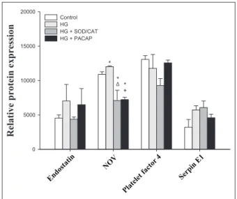
To assess the underlying mechanisms of the above- described protective vascular effects of PACAP1-38, we used angiogenesis array kit. In total, 11 proteins were expressed in the vessels, namely, angiopoietin-1 (Ang-1), tissue factor, fibroblast growth factor (FGF) acidic, FGF basic, matrix metalloproteinase 9 (MMP-9), osteopontin, stromal cell–derived factor 1 (SDF-1), endostatin, nephro- blastoma overexpressed gene (NOV), platelet factor 4 and serpin E1 (Figures 4 and 5). FGF basic (p = 0.012 HG vs control), MMP-9 (p = 0.003 HG vs control) and NOV (p = 0.032 HG vs control) reacted with a rise to the 1-h HG exposure.
Ang-1 declined on HG exposure (p = 0.045 HG vs con- trol), while HG had no significant effect on levels of tissue factor (p = 0.061, HG vs control), FGF acidic (p = 0.082 HG vs control) and osteopoetin (p = 0.108, HG vs control). No treatment could change the level of SDF-1 (p = 0.671), endostatin (p = 0.600), platelet factor 4 (p = 0.130) and serpin E1 (p = 0.091) proteins.
Figure 3. SNP concentration–response curves of isolated carotid arteries after 1-h incubation with a control Krebs solution (Control) or a Krebs solution containing 30 mM glucose (HG) or a solution where combination of SOD and CAT (120 and 200 U/mL, respectively) was administered to the Krebs solution containing 30 mM glucose (HG + SOD/CAT) or a solution where 2 × 10⁻⁸ M PACAP1-38 was administered to the Krebs solution containing 30 mM glucose (HG + PACAP1-38).
*p < 0.05 versus control, Δp < 0.05 HG versus HG + SOD/CAT, +p
< 0.05 HG versus HG + PACAP1-38, ANOVA, post hoc Fisher LSD, n = 6/group.
Figure 4. Relative protein expression in carotid arteries, part 1.
*p < 0.05 versus control; Δp < 0.05 HG versus HG + SOD/CAT; +p
< 0.05 HG versus HG + PACAP1-38, ANOVA, post hoc Fisher LSD, n = 4/group.
Figure 5. Relative protein expression in carotid arteries, Part II.
*p < 0.05 versus control; Δp < 0.05 HG versus HG + SOD/CAT; +p
< 0.05 HG versus HG + PACAP1-38, ANOVA, post hoc Fisher LSD, n = 4/group.
Addition of PACAP1-38 to the HG solution suppressed all increases induced by HG (FGF basic: p = 0.004, MMP-9:
p = 0.041 and NOV: p < 0.001 for HG + PACAP1-38 vs HG). Furthermore, levels of Ang-1 and FGF acidic also decreased significantly compared to HG (Ang-1: p = 0.014 and FGF acidic: p < 0.001 for HG + PACAP1-38 vs HG).
Addition of SOD/CAT in the presence of HG could also reduce the elevation of the hyperglycaemia markers (FGF basic: p = 0.002, MMP-9: p = 0.002 and NOV: p = 0.001 for HG + SOD/CAT vs HG). In addition, levels of Ang-1, FGF acidic and tissue factor also decreased significantly (Ang-1: p = 0.003, FGF acidic: p < 0.001 and tissue fac- tor: p = 0.038 for HG + SOD/CAT vs HG).
Discussion
This study is the first to demonstrate the ability of PACAP to protect vascular function (as indicated by vasorelaxa- tion) following 1-h HG stress in isolated carotid arteries of healthy mice.
In consistency with previous results,26 we showed that short-term HG stress reduces endothelium-dependent vas- odilation ability of the carotid arteries. In the background of this dysfunction, enhanced superoxide production is suggested as enhanced glycolysis results in increased gen- eration of pyruvate, which is transported into the mito- chondria of the cells, and NADH is generated in the tricarboxylic acid cycle. This causes enhanced electron flow through the respiratory chain, which can increase mitochondrial superoxide production. PACAP, similar to the superoxide radical scavenger SOD/CAT, was able to restore Ach-induced vasodilation of the vessels. The pro- tective effect of PACAP was comparable with the protec- tive effect of SOD/CAT, so it was worthwhile to test whether PACAP exerts these positive effects as a direct antioxidant. Pyrogallol autoxidation assay is a classical method to test the superoxide-scavenging ability of differ- ent molecules.24 The assay was originally developed to test/demonstrate the scavenging ability of SOD. As pH is of major importance when testing a possible antioxidant molecule, the improved autoxidation assay25 included a pH correction to 7.4. This improved method was consid- ered appropriate also for PACAP. According to our study, PACAP seems to be unable to scavenge superoxide anions produced by autoxidation of pyrogallol. Thus, PACAP seems to exert its protective effects by activating signal- ling pathways acting on its receptor. These results are con- sistent with the literature. As based on a great number of studies on different cell types (cardiomyocytes,27 intestinal cells,16 astrocytes,28 etc.), it is proposed that the strong pro- tective effects of PACAP against oxidative stress mainly involve not direct, but indirect, antioxidant actions.
According to a recent study, PACAP reverses the decrease in the activity of antioxidant enzymes SOD and CAT in ammonium-vanadate-exposed rat lungs.29 Moreover, a
study of Douiri et al.30 confirmed that PACAP at subna- nomolar concentrations induced a dose-dependent increase in both SOD and CAT activities in astrocytes and simulta- neously blocked H2O2-evoked inhibition of the activity of these two enzymes.
Endothelium-independent relaxation was examined with the nitric oxide donor SNP. Administration of SNP also induced smaller dilation after 1-h HG stress compared to controls. As a possible explanation for this phenomenon, it has been suggested that hyperglycaemia-induced vascular oxidative stress demonstrated by Bagi et al.31 can interfere with the bioavailability of NO.26 According to our results, the addition of SOD/CAT or PACAP to the HG solution could effectively improve SNP-induced relaxation.
The levels of the proteins, FGF basic, MMP-9 and NOV, were increased by a 1-h HG stress. Administration of SOD/CAT alongside with the HG solution markedly decreased the level of these proteins. PACAP together with the HG solution was also able to decrease the level of these markers. FGF basic is synthesized and released by endothe- lial cells and has an autocrine role. It was suggested that endothelium stores FGF basic capable of autocrine growth promotion both by storing growth factor within the cell and by incorporating it into the underlying extracellular matrix.32 In a recent study, the role of FGF basic was sug- gested in type 2 diabetic rats where upregulation of expres- sion of FGF basic proteins were shown.33 In that study, resveratrol promoted the attenuation of left ventricular diastolic dysfunction by decreasing FGF basic. MMP-9 is a dysglycaemia marker34 that fits with an early stage of cardiovascular disease as MMPs are synthetized in athero- sclerotic plaques, and MMP activity is correlated with unstable angina and plaque rupture.34 Diabetes mellitus increases vascular MMP activity.35 MMPs are stored in cytoplasmic secretory granules in endothelial cells, ready for rapid release by angiogenic stimuli.36 Furthermore, oxidative stress regulates their expression.34 NOV is an adipocytokine involved in obesity-associated insulin resistance. According to a recent study37 NOV−/− mice on high fat diet had lower body weight, lower fat mass and improved glucose tolerance and insulin sensitivity. Our data show that FGF basic, MMP-9 and NOV are sensitive vascular markers and are able to indicate already a short- term hyperglycaemia. Both PACAP and SOD/CAT reduced the level of NOV protein below baseline levels. As an explanation of these surprising findings, we suggest that the isolation and the mounting of the vessels (despite the very careful approach of experienced researchers) may induce some low-level damage and oxidative stress (other- wise undetectable). This low-level oxidative stress must have been reduced by the administration of PACAP and SOD/CAT resulting in the fall of angiogenesis-related pro- teins below baseline.
The very fast change in protein concentrations of angi- ogenic factors and proteases (FGF basic, MMP-9, FGF
basic or ANG-1) in our study implies enhanced protein degradation. Previous findings of the literature also dem- onstrate a very short half-life (3–30 min) of angiogenetic factors such as FGF or vascular endothelial growth factor (VEGF).38 Thus, vessels appear to be characterized by very fast angiogenic protein turnover.
However, hyperglycaemia appears to slow down this rapid protein turnover. Previous reports attributed the glu- cose-induced increase in basement membrane FGF basic to the enhancement of its release due to increased perme- ability of endothelial cells.39 In this case, the existence of slow turnover reservoirs (e.g. the basement membrane) were suggested as an explanation of the lasting vascular consequences of short-term exposure to glucose.
Other studies focusing on the mesangial cells of the kidney reported that in hyperglycaemic media, several proteases (e.g. MMP-9) show significantly lowered activ- ity leading to decreased protein degradation. This occur- rence rather contributes to impaired protein turnover and consequent induction of cellular hypertrophy and accumu- lation of extracellular matrix.40
Future studies should investigate the effects of glucose exposure on vascular protein turnover affecting angio- genic factors.
Diabetes mellitus patients frequently have postprandial hyperglycaemia in which situations endothelium is directly exposed to HG environment. Endothelial cells are highly susceptible to both proliferating and antiproliferative sig- nals. In the pathogenesis of atherosclerosis, endothelial dysfunction plays a critical role, leading to vascular inflammation, thrombosis, arterial stiffness and impaired regulation of arterial tone and flow.41 Several studies investigated endothelium-dependent vasodilation to assess the health of endothelium; endothelium-dependent dilation is compromised in patients with coronary artery disease.42 A connection between some of these endothelium damage- inducing/indicating signals and short-lasting hyperglycae- mia was tested in this study. Excessive angiogenesis plays a role in many of the clinical manifestations of diabetes, such as diabetic retinopathy43 or nephropathy.44 As abnor- mal angiogenesis is a key factor of vascular tissue damage in diabetes, it is important to find molecules that can help reduce such trophic effects. A previous study confirmed the role of PACAP and VIP peptides on endothelial cell growth after exposure to a hyperglycaemic environment and suggests the potential involvement of both PAC1 and VPAC2 receptors in inhibiting a harmful proliferative response.45
In summary, the important findings of this study dem- onstrate that PACAP administration results in reduced endothelial dysfunction after a short-term hyperglycaemic episode. This protective effect is comparable to the anti- oxidant effect of SOD. Furthermore, here we reported three vascular markers of short-term hyperglycaemia.
Endothelial dysfunction was associated with the elevation
of FGF basic, MMP-9 and NOV proteins. Their release was reduced by PACAP administration. These results sug- gest a strong protective role of PACAP in diabetic vascular complications.
Perspectives
In the future, investigations should focus on the potential mechanisms of the protective vascular effects of PACAP in hyperglycaemia. Since vascular dysfunctions demon- strated by our study correlate with hyperglycaemia- induced free radical production, antioxidant actions of PACAP should be further characterized. Based on the available data in the literature,17,46,47 this analysis of PACAP’s protective antioxidant actions could include investigation of various enzymes (such as glutathione per- oxidase, heme oxygenase-1 and peroxiredoxin-2) and that of antioxidant small molecules (such as vitamins E and C, glutathione, sulphides or polyphenols).
In addition, we also plan further investigations to char- acterize the PACAP-dependent positive vascular effects in diabetic animal models. Such investigations may reveal a potential contribution of PACAP in the prevention and even in the treatment of the chronic vascular complications of type 2 diabetes mellitus that present a major public health burden in modern societies.
Key messages
•
• Pituitary adenylate cyclase-activating polypep- tide (PACAP) diminishes 1-h high-glucose (HG) stress-induced endothelial dysfunctions in carotid arteries.
•
• Vasoprotective effects of PACAP are independ- ent of direct superoxide scavenging.
•
• PACAP suppresses vascular markers [fibroblast growth factor (FGF) basic, matrix metallopro- teinase 9 (MMP-9) and nephroblastoma overex- pressed gene (NOV)] raised by 1-h HG stress.
Declaration of conflicting interests
The author(s) declared no potential conflicts of interest with respect to the research, authorship and/or publication of this article.
Funding
This work was supported by Hungarian National Science Research Fund (OTKA) K119759, K115874, 2017-1.2.1-NKP-2017-00002 and GINOP-2.3.2-15-2016-00050 ‘PEPSYS’ (D. Reglodi, A.
Tamas); University of Pecs Medical School KA Research Grant (D. Reglodi, A. Tamas); OTKA K108444 (A. Koller); University of Pecs Medical School PTE-AOK-KA-2015-14 (M. Solymar);
PTE AOK-KA 2017/13 (M. Balasko); MTA TKI Grant MTA TKI 14016 (D. Reglodi); Bolyai Scholarship (A. Tamas); EFOP-3.6.3- VEKOP-16-15 2017-00008/EFOP-3.6.2-16-2017-00008 project;
and FP7 Marie Curie project-Small Artery Remodeling (SmART) ITN (I. Ivic, A. Koller). The present scientific contribution is dedi- cated to the 650th anniversary of the foundation of the University of Pecs, Hungary.
References
1. Steiner G. Implications of the global diabetes epidemic.
Diab Vasc Dis Res 2006; 3: S2–S5.
2. The Diabetes Control Complications Trial Research Group.
The effect of intensive treatment of diabetes on the develop- ment and progression of long-term complications in insu- lin-dependent diabetes mellitus. N Engl J Med 1993; 329:
977–986.
3. Intensive blood-glucose control with sulphonylureas or insulin compared with conventional treatment and risk of complications in patients with type 2 diabetes. UK and Prospective Diabetes Study (UKPDS) Group. Lancet 1998;
352: 837–853.
4. Sitnik D, Santos IS, Goulart AC, et al. Fasting glucose levels, incident diabetes, subclinical atherosclerosis and cardiovascular events in apparently healthy adults: a 12-year longitudinal study. Diab Vasc Dis Res 2016; 13:
429–437.
5. Kawano H, Motoyama T, Hirashima O, et al. Hyperglycemia rapidly suppresses flow-mediated endothelium-dependent vasodilation of brachial artery. J Am Coll Cardiol 1999; 34:
146–154.
6. Title LM, Cummings PM, Giddens K, et al. Oral glucose loading acutely attenuates endothelium-dependent vasodila- tion in healthy adults without diabetes: an effect prevented by vitamins C and E. J Am Coll Cardiol 2000; 36: 2185–
2191.
7. Sorensen VR, Mathiesen ER, Clausen P, et al. Impaired vas- cular function during short-term poor glycaemic control in type 1 diabetic patients. Diabet Med 2005; 22: 871–876.
8. Miyata A, Arimura A, Dahl RR, et al. Isolation of a novel 38 residue-hypothalamic polypeptide which stimulates adenylate cyclase in pituitary cells. Biochem Biophys Res Commun 1989; 164: 567–574.
9. Vaudry D, Falluel-Morel A, Bourgault S, et al. Pituitary adenylate cyclase-activating polypeptide and its receptors:
20 years after the discovery. Pharmacol Rev 2009; 61: 283–
357.
10. Segre GV and Goldring SR. Receptors for secretin, cal- citonin, parathyroid hormone (PTH)/PTH-related pep- tide, vasoactive intestinal peptide, glucagonlike peptide 1, growth hormone-releasing hormone, and glucagon belong to a newly discovered G-protein-linked receptor family.
Trends Endocrin Met 1993; 4: 309–314.
11. Fahrenkrug J, Hannibal J, Tams J, et al. Immunohisto- chemical localization of the VIP1 receptor (VPAC1R) in rat cerebral blood vessels: relation to PACAP and VIP contain- ing nerves. J Cereb Blood Flow Metab 2000; 20: 1205–1214.
12. Banki E, Degrell P, Kiss P, et al. Effect of PACAP treat- ment on kidney morphology and cytokine expression in rat diabetic nephropathy. Peptides 2013; 42: 125–130.
13. Szabadfi K, Reglodi D, Szabo A, et al. Pituitary adenylate cyclase activating polypeptide, a potential therapeutic agent for diabetic retinopathy in rats: focus on the vertical informa- tion processing pathway. Neurotox Res 2016; 29: 432–446.
14. Reglodi D, Fabian Z, Tamas A, et al. Effects of PACAP on in vitro and in vivo neuronal cell death, platelet aggrega- tion, and production of reactive oxygen radicals. Regul Pept 2004; 123: 51–59.
15. Miyamoto K, Tsumuraya T, Ohtaki H, et al. PACAP38 sup- presses cortical damage in mice with traumatic brain injury by enhancing antioxidant activity. J Mol Neurosci 2014; 54:
370–379.
16. Ferencz A, Racz B, Tamas A, et al. Influence of PACAP on oxidative stress and tissue injury following small-bowel autotransplantation. J Mol Neurosci 2009; 37: 168–176.
17. Ohtaki H, Satoh A, Nakamachi T, et al. Regulation of oxi- dative stress by pituitary adenylate cyclase-activating poly- peptide (PACAP) mediated by PACAP receptor. J Mol Neurosci 2010; 42: 397–403.
18. Laszlo E, Varga A, Kovacs K, et al. Ischemia/reperfusion- induced kidney injury in heterozygous PACAP-deficient mice. Transplant Proc 2015; 47: 2210–2215.
19. Racz B, Gasz B, Borsiczky B, et al. Protective effects of pituitary adenylate cyclase activating polypeptide in endothelial cells against oxidative stress-induced apoptosis.
Gen Comp Endocrinol 2007; 153: 115–123.
20. Wilhelm I, Fazakas C, Tamas A, et al. PACAP enhances barrier properties of cerebral microvessels. J Mol Neurosci 2014; 54: 469–476.
21. Hamed EA, Zakary MM, Reffat MA, et al. Vasculopathy in type 2 diabetes mellitus: role of specific angiogenic modula- tors. J Phys Biochem 2011; 67: 339–349.
22. Mulvany MJ and Halpern W. Contractile properties of small arterial resistance vessels in spontaneously hypertensive and normotensive rats. Circ Res 1977; 41: 19–26.
23. Ivic I, Fulop BD, Juhasz T, et al. Backup mechanisms main- tain PACAP/VIP-induced arterial relaxations in pituitary adenylate cyclase-activating polypeptide-deficient mice. J Vasc Res 2017; 54: 180–192.
24. Marklund S and Marklund G. Involvement of the super- oxide anion radical in the autoxidation of pyrogallol and a convenient assay for superoxide dismutase. Eur J Biochem 1974; 47: 469–474.
25. Li X. Improved pyrogallol autoxidation method: a reliable and cheap superoxide-scavenging assay suitable for all anti- oxidants. J Agric Food Chem 2012; 60: 6418–6424.
26. Toth E, Racz A, Toth J, et al. Contribution of polyol path- way to arteriolar dysfunction in hyperglycemia. Role of oxidative stress, reduced NO, and enhanced PGH2/TA2 mediation. Am J Physiol Heart Circ Physiol 2007; 293:
H3096–H3104.
27. Gasz B, Racz B, Roth E, et al. Pituitary adenylate cyclase activating polypeptide protects cardiomyocytes against oxi- dative stress-induced apoptosis. Peptides 2006; 27: 87–94.
28. Masmoudi-Kouki O, Douiri S, Hamdi Y, et al. Pituitary adenylate cyclase-activating polypeptide protects astro- glial cells against oxidative stress-induced apoptosis. J Neurochem 2011; 117: 403–411.
29. Tlili M, Rouatbi S, Sriha B, et al. Pituitary adenylate cyclase-activating polypeptide reverses ammonium meta- vanadate-induced airway hyperresponsiveness in rats. Oxid Med Cell Longev 2015; 2015: 787561.
30. Douiri S, Bahdoudi S, Hamdi Y, et al. Involvement of endogenous antioxidant systems in the protective activity of
pituitary adenylate cyclase-activating polypeptide against hydrogen peroxide-induced oxidative damages in cultured rat astrocytes. J Neurochem 2016; 137: 913–930.
31. Bagi Z, Toth E, Koller A, et al. Microvascular dysfunc- tion after transient high glucose is caused by superoxide- dependent reduction in the bioavailability of NO and BH(4). Am J Physiol Heart Circ Physiol 2004; 287: H626–
H633.
32. Vlodavsky I, Folkman J, Sullivan R, et al. Endothelial cell- derived basic fibroblast growth factor: synthesis and depo- sition into subendothelial extracellular matrix. Proc Natl Acad Sci U S A 1987; 84: 2292–2296.
33. Strunz CMC, Roggerio A, Cruz PL, et al. Down-regulation of fibroblast growth factor 2 and its co-receptors heparan sulfate proteoglycans by resveratrol underlies the improve- ment of cardiac dysfunction in experimental diabetes. J Nutr Biochem 2017; 40: 219–227.
34. Zayani Y, El Golli N, Zidi W, et al. Inflammations media- tors and circulating levels of matrix metalloproteinases:
biomarkers of diabetes in Tunisians metabolic syndrome patients. Cytokine 2016; 86: 47–52.
35. Uemura S, Matsushita H, Li W, et al. Diabetes mellitus enhances vascular matrix metalloproteinase activity. Circ Res 2001; 88: 1291–1298.
36. Nguyen M, Arkell J and Jackson CJ. Active and tissue inhibitor of matrix metalloproteinase-free gelatinase B accumulates within human microvascular endothelial vesi- cles. J Biol Chem 1998; 273: 5400–5404.
37. Martinerie C, Garcia M, Do TTH, et al. NOV/CCN3: a new adipocytokine involved in obesity-associated insulin resist- ance. Diabetes 2016; 65: 2502–2515.
38. Folkman J. Role of angiogenesis in tumor growth and metastasis. Semin Oncol 2002; 29: 15–18.
39. Morss AS and Elazer R. Glucose modulates basement mem- brane fibroblast growth factor-2 via alterations in endothe- lial cell permeability. J Biol Chem 2007; 282: 14635–14644.
40. Schenk O, Ling H, Sebekova K, et al. High-glucose media enhance the responsiveness of tubular cells to growth pro- moters: effect on lysosomal cathepsins and protein degrada- tion. Miner Electrolyte Metab 1998; 24: 254–260.
41. Tabit CE, Chung WB, Hamburg NM, et al. Endothelial dys- function in diabetes mellitus: molecular mechanisms and clinical implications. Rev Endocr Metab Disord 2010; 11:
61–74.
42. Celermajer DS, Sorensen KE, Gooch VM, et al. Non- invasive detection of endothelial dysfunction in children and adults at risk of atherosclerosis. Lancet 1992; 340:
1111–1115.
43. Wilkinson-Berka JL. Vasoactive factors and diabetic retinop- athy: vascular endothelial growth factor, cycoloxygenase-2 and nitric oxide. Curr Pharm Des 2004; 10: 3331–3348.
44. Osterby R and Nyberg G. New vessel formation in the renal corpuscles in advanced diabetic glomerulopathy. J Diabet Complications 1987; 1: 122–127.
45. Castorina A, Giunta S, Mazzone V, et al. Effects of PACAP and VIP on hyperglycemia-induced proliferation in murine microvascular endothelial cells. Peptides 2010; 31: 2276–
2283.
46. Resch JM, Albano R, Liu X, et al. Augmented cystine-glu- tamate exchange by pituitary adenylate cyclase-activating polypeptide signaling via the VPAC1 receptor. Synapse 2014; 68: 604–612.
47. Reglodi D, Renaud J, Tamas A, et al. Novel tactics for neuroprotection in Parkinson’s disease: role of antibiotics, polyphenols and neuropeptides. Prog Neurobiol 2017; 155:
120–148.