Genetic Deletions
V. N. Iyer
I. Introduction 205 II. Selection and Detection of Mutants Bearing Potential Deletions 208
III. Operational Criteria for Establishing a Mutation as a Deletion 211
A. Direct Chemical and Physical Measurements 212 B. Visualization of Unmatched Regions in Artificially Hybridized D N A
Strands 213 C. Hybridization between Specific Messenger RNA and a D N A Strand . . 213
D. Rescue of Deleted Genes and Their Detection 213
E. Effects on Genetic Maps 216 IV. The Frequency, Topography, and Topology of Deletion Mutations 216
V. Enzymes That Cut and Repair D N A 221 VI. Agents That May Promote Deletions 224
VII. Hypothetical Mechanisms 226 A. Replication Errors 226 B. Repair Errors 228 C. Recombination Errors 229
VIII. Conclusions 230 References 231
I. INTRODUCTION
Most organisms can tolerate and survive an irreversible elimination of structurally or functionally recognizable parts of their genetic mate- rial. So long as the elimination does not result in the loss of a function that is vital to the organism, it may survive and may behave as a recognizable mutant. Classical and current usage (1) sanctions the term deletion in situations where the part that was eliminated was detached from a chromosome with which it previously formed a continuum. The term deficiency has often been used synonymously and especially in
205
the literature involving eukaryotes. Thus, the chromosome suffers the deletion and the cell or organism may eliminate the deleted portion and become a deletion mutant. Conceptually, it is not necessary to as
sume that deleted portions will be eliminated from a cell (Fig. 1). In practice, however, it is rare for deleted portions of a chromosome to contain elements that endow it with replicative autonomy. Therefore, and while bearing the alternative possibility in mind, we shall assume as a first approximation that most deleted portions of a chromosome will not survive and will be eliminated from a cell. In diploid organisms, protection against possible lethality promoted by a deletion in one chro
mosome may be offered by the intactness of the homologous chromosome (recessive lethal deletions). In haploid organisms, many deletions may be lethal. However, it is clear that deletion mutants do occur in haploids at detectable frequencies. "Deletion mapping" and deletion mutants are powerful and unambiguous tools in molecular genetics (2-4). They have been exploited in seeking answers to a wide range of questions and it is likely that their demonstrated value can be extended. This depends on the acquisition of a collection of deletion mutants that belong to a particular genetic or biochemical system. It is the intent of the first
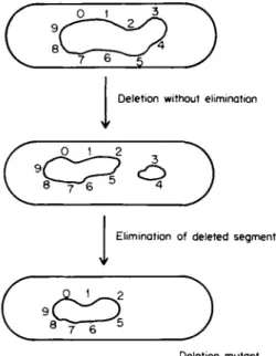
Deletio n withou t eliminatio n
Ψ
Eliminatio n o f delete d segmen t V
Deletio n mutan t
FIG. 1. Diagram illustrating the conceptual distinction between deletion with or without the elimination of the deleted segment.
part of this chapter to describe briefly the techniques that have been successfully used for the isolation and detection of such mutants in those genetic systems that have been studied thus far. These may suggest rationales for the development of new selective techniques for detection, isolation, and use involving other genes, microorganisms, or cells.
Very little is known about the origin of deletions or deletion mutants.
An important limitation to the study of their origin is their low frequency of occurrence. This has made indirect approaches to the question of origin or origins necessary. Given that one can recognize deletion mutants with assurance, several questions may be asked. What circumstances promote or prevent their occurrence? Is the frequency of deletion mutants in an organism related to some other biochemical or physiologi- cal capacity or incapacity of the organism, such as the ability to tolerate or repair structural defects in D N A ? Are there agents that specifically promote deletion mutations? Related to these are other important ques- tions. In what regions of a chromosome do deletion mutants occur and do these have special structural or functional properties? Are there re- strictions to the dimensions or ends of deletions in a specified region?
It is likely that answers to such questions will provide insights into deletion mechanisms and their role in genetic systems.
There are now a number of purified enzymes that can cut or repair D N A in vitro. These, or enzymes similar to them, are potential candi- dates for generating deletions. The properties of those enzymes that have been isolated from E. coli are briefly reviewed. However, we do not know whether any of these known, enzymes are in fact involved.
There is extensive literature on deletion phenomena in the metaphase and more complex chromosomes of some eukaryotic cells. The operational basis for recognizing them has been cytological and cytogenetic, and there is no doubt that they must involve losses of substantial amounts of D N A and other chromosomal constituents. The manner in which D N A is organized within these chromosomes is unclear (recently reviewed in 5, 6) and as long as this is so, the relationship of these interesting phenomena to the deletion of D N A from bacterial or viral chromosomes is obscured. The latter part of this review is concerned with possible mechanisms generating deletions in those few bacterial and bacteriophage systems that have been subjected to intensive genetic and biochemical studies. A corollary of the restricted definition of deletions that we have used here is that autonomously existing genetic material that can never be directly inserted into the continuity of chromosomal genetic material may also be eliminated from cells. The modes by which such elimination may occur is not the subject of this review.
II. SELECTION AND DETECTION OF MUTANTS BEARING POTENTIAL DELETIONS
Any program for the isolation of deletion mutants must first solve the problem of detection. They constitute a fraction, sometimes quite small, of mutants which themselves occur at a very low frequency. This detection can be achieved either by (a) testing a very large number of potential mutants among which there hopefully will be deletion mu
tants or (b) in a particular system, devising and using a technique that has some selectivity for deletions. The first way is not as forbidding as it might seem, provided, as is usually the case, a procedure is available for selecting against unmutated organisms. An appreciable though small fraction (1-10%) of all spontaneous or induced mutants has suffered deletions. Benzer (2), Folsome (7), and Tessman (8) in phage T4, Demerec (9) in Salmonella, Jacob and Wollman (10) and Cook and Lederberg (11) in E. coli have all been able to isolate appreciable num
bers of them in this manner. Agents that are known or suspected to induce deletions are considered separately in a later section.
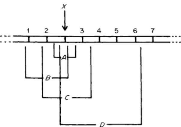
Since deletions must result in the reduction or abolition of at least those genes that they span and because most genes in the examined prokaryotic systems are not redundant, deletion mutants are expected to be usually at a selective disadvantage unless artificially enriched by techniques that suppress the growth of unmutated cells. There are, however, some situations in which the loss of a function can provide, a selective advantage. Such situations can be exploited for the isolation of deletion mutants. In E. coli, one of the most useful of such selection techniques (12, 12a) has been combined selection for tryptophan auxo- trophy and resistance to phage T l (tonB locus). The rare but easily selected Τ1-resistant mutants are frequently deletion mutants. Since the tonB locus is flanked by the attachment site for phage </>80 on one side and the intensively studied trp operon on the other, the end points of these deletions are easily determined (12b, 12c). The principle of this selection technique is shown in Fig. 2. Yanofsky et al. (4) used such deletion mutants to successfully map point mutations in the trpA gene, and Franklin et al. (13) exploited the system to establish that phage φ80 can be inserted into the bacterial chromosome. They did this by showing that, in strains lysogenic for bacteriophage </>80, many Tl-re- sistant mutants (tonB locus) have deletions extending into the prophage genome. This proven exploitability of deletions involving the tonB locus can now be extended to other genes because it has been shown (14,
I
» ΓFIG. 2. A principle of techniques for the isolation of deletion mutants. The loss of gene X will confer a selective advantage (e.g., X may determine a cell surface receptor for the binding of a lethal virus; loss of X will therefore result in a change from phage sensitivity to phage resistance, a change that lends itself to convenient selection). As indicated in the figure, a particular event leading to the deletion of X may simultaneously lead to the deletion of other genes (1-7) flanking X on one or both sides (A, B, C, and D are types of deletion mutants that may be secured).
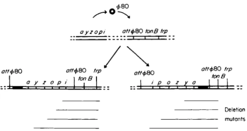
15) that other genes can be transposed to the chromosomal attachment site of </>80. For example, the lac gene has been transposed to the φ80 attachment site and two types of strains lysogenic for φ80 lac con
structed. These two types differ in the orientation of the lac operon genes relative to the genes flanking it (Fig. 3). When Tl-resistant mu
tants were isolated from these two types of lac transposition strains, many bore deletions extending from one or the other end into the lac region [for a more detailed description of techniques using this rationale, see Beckwith et al. (14-18)]. Using the same principle, the feasibility of screening spontaneous anaerobically chlorate-resistant mutants of E.
coli for deletions extending into neighboring genes has been demonstrated (19).
Ames (19a) has recently described a test system for agents that may cause deletions in Escherichia coli and Salmonella typhimurium. The principle of the system is to select for the simultaneous loss of the en
zymes nitrate reductase and galactokinase. Under anaerobic conditions, nitrate reductase converts chlorate to chlorite, which is inhibitory. Muta
tions that eliminate nitrate reductase activity therefore confer anaerobic chlorate resistance. Further, galactose inhibits strains that are mutant for galactose epimerase. This inhibition can be prevented if mutations destroy the galactokinase gene. Mutants that bear deletions spanning
ayzopi σ//φ8 0 tonB trp
σ//φ8 0 trp . I tonB I
/
ott<t>QO trptonB
σ//φ8 0 σ//φ8 0
a y ζ ο ρ i ο ζ y a
Deletio n mutant s
FIG. 3. Diagram illustrating (1) the orientation of lac genes in derivatives of E. coli that have had the lac operon transposed to an atypical chromosomal location close to the tonB locus and (2) how deletion mutants isolated from these derivatives on the basis of Tl resistance will bear deletions extending into one or other end of the lac region.
both nitrate reductase and galactokinase genes can therefore be selected on the basis of ability to grow anaerobically in the presence of galactose and chlorate. Shapiro and Adhya (20) took advantage of the proximity of λ prophage to the gal operon and the existence of thermoinducible mutants of λ which kill the host at high temperature. From a galactose- sensitive mutant unable to synthesize uridine diphosphoglucose and lyso- genic for the thermoinducible λ prophage, they selected galactose-nega- tive and thermoresistant mutants. This double selection yielded λ-gal deletions which rendered the λ prophage nonlethal and adjacent gal genes inactive. Schwartz and Beckwith (21) used a system which takes ad
vantage of the fact that galactoside permease in E. coli (specified by gene y of the lac operon) is necessary for melibiose utilization at 42°C
(15). A lac~ ocher mutation because of its polar effect made no galacto
side permease and the strain was melibiose- at 42°C. Melibiose+ re- vertants were scored at 42°C on a defined agar medium deficient in melibiose, and a fraction of these (0.3-7.0% depending on the mutagen that was used) on further testing proved to have suffered deletions in the lac region. This selection technique could possibly prevent the isola
tion of deletion mutants in the ends extending into the region of genes controlling the operon. Beckwith (3) has pointed out that similar selec
tive techniques may possibly be used for the histidine and tryptophan operons.
Parkinson and Davis (22) and Parkinson and Huskey (28) made the observation that the exposure of a population of phage λ particles
to a chelating agent caused a preferential release of D N A from the heads of wild-type particles so that phages carrying less than normal amounts of D N A survived. B y selecting mutants of wild-type λ that are resistant to heat or chelating agents, it was possible to select a large number of deletion mutants that had lost 2-22% of their D N A randomly through the center segment of the chromosome. It must be noted that approximately one-third of the λ chromosome that is centrally located is not essential for vegetative growth. Most viable deletions would therefore be expected to lie within this region. Also, and as pointed out by Parkinson and Huskey (23), the treatments used caused small deletions to lose viability at a faster rate than large deletions, thus skewing the size distribution toward larger deletions. In an extension of this procedure, Burdon (24) exposed a tryptophan-transducing λ-φ80 phage hybrid to E D T A and isolated phage mutants bearing different deletions in the tryptophan region. The deletion was then transferred back into the E. coli Κ12 chromosome by lysogenizing a trp+ host and then eliminating the prophage, leaving behind a nonlysogenic strain bear
ing the deletion.
Heat resistance in phage T5 is also known to be accompanied by the production of deletions (25, 26). Presumably, this technique of isolat
ing deletion mutants is applicable to all phages, the chromosomes of which have unique ends. The procedures that have been described in this section provide a sampling of the rationale and the techniques that may be developed for selectively isolating deletion mutants. It is unlikely that any of them provides unbiased samples of all deletion mutants that can occur in an organism. However, there is reason to believe that the principles of the techniques that have been described can well be extended to other operons and other genetic systems. Attention has been drawn (3) to one limitation on the mapping of deletion mutants that may be imposed by these isolation techniques. Deletion mutants tend to diminish existing genetic homology on the side of the mutant sites not included in the deletions. It is conceivable that this decreased homology could reduce recombination between the deletion mutant and some mutations that lie close to the deletion region.
III. OPERATIONAL CRITERIA FOR ESTABLISHING A MUTATION
AS A DELETION
Present techniques for the selection of deletion mutants do not as a rule enable us to assume with confidence that an isolated mutant does
in fact bear a deletion. Other mutations that mimic some of the proper
ties of deletion mutants are known to occur (27, 28), and it is therefore desirable to seek definite assurance that a mutation does in fact arise on account of the deletion of genetic material.
A. Direct Chemical and Physical Measurements
If the net amount of deleted genetic material is substantial, a direct chemical or physical measurement of the chromosome or the relevant part of the chromosome may reveal this. This has been possible so far only in the case of some bacteriophages.* The viable b2 deletion mutant of λ was shown by chemical estimation to have suffered a 17% loss of D N A (29); the loss was also estimated by measurement of relative length of D N A in electron micrographs (SO). An estimate of 18% loss was made indirectly from the change in density of the virus particle (31), although a more recent measurement (32) has indicated this to be 13%.
The reproductively defective petite particles of phage T 4 have been shown to bear D N A molecules that are not as long as the normal T4 D N A molecule (33). The mechanisms by which such short molecules of T4 are generated, however, are different from the generation of the typical deletions that occur internally within chromosomes. A direct
demonstration of this type has not thus far been achieved for larger chromosomes, such as those of bacteria. However, it is now possible to conceive of ways by which this may be done. It may be possible, for example, to rescue transducing phages of the λ-ψ80 type that have incorporated into their genome a segment of the bacterial chromosome within which a deletion has been located. A comparison of the length of D N A isolated from such phages with that isolated from the same transducing phage that has incorporated the nondeleted segment could provide a direct estimate of the segment deleted from the bacterial chromosome.
Techniques to selectively label and isolate fragments of D N A repre
sentative of restricted chromosomal regions are being developed (34, 35). If very large deletion mutations can occur in such regions, this may be apparent in the physical properties of such isolated fragments.
Alternatively, the D N A strands of such fragments could be separated and fractionated (36), and heterologous strands, one derived from the deletion mutant and another from the wild-type strain, could be re-
* See note added in proof.
annealed and visualized in electron micrographs. The successful applica
tion of this technique is discussed more fully in the next section.
B. Visualization of Unmatched Regions in Artificially Hybridized DNA Strands
If a D N A strand bearing a deletion can be made to accurately hy
bridize with another that does not bear the same deletion, the resulting double-stranded structure may have unmatched or looped-out regions at the deletion site. This possibility, suggested in 1966 by Nomura and Benzer (37), was recently realized in the case of phage λ. Heteroduplex molecules are formed by mixing the two D N A preparations, dissociating the strands by exposure to alkali, and subsequently gently renaturing the strands in the presence of formamide. Under these conditions, the original homoduplex molecules and a proportion of heteroduplex mole
cules will be formed. When a heteroduplex is formed between a wild-type strand and a deletion strand, the location of the deletion should coincide with an unpaired region in the complementary strand. Depending on the specimen mounting technique used, the unpaired regions appear as
"bushes" (32) or as unpaired "loops" (36). In the formamide mounting technique of Westmoreland et al. (36), who also introduced the refinement of hybridizing with previously fractionated complementary strands, an unpaired region involving a deletion could be distinguished from an un
paired region involving a substitution of nucleotide sequences. The re
sulting loops were also sufficiently resolved to be measurable (Fig. 4).
This straightforward technique can therefore be used not only to localize a deletion, but also to measure its extent and distinguish it from some other kinds of structural aberrations (36, 39, 40).
C. Hybridization between Specific Messenger RNA and a DNA Strand The length of a deletion may be expected to be inversely proportional to the amount of the region-specific messenger R N A that hybridizes with it. This expectation has been met and the technique used to map deletion mutants in bacteriophages T4 (41) and λ (42).
D. Rescue of Deleted Genes and Their Detection
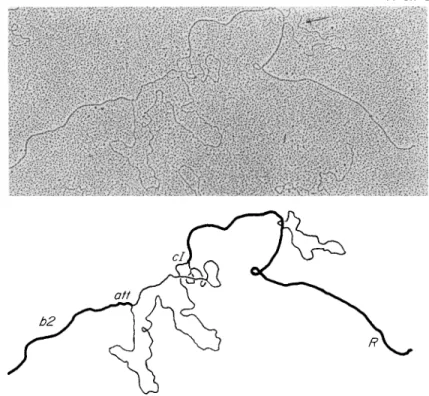
If the deletion of D N A from a chromosome occurs in such a manner that the deleted portion becomes incorporated into an independent repli-
FIG. 4 . An electron micrograph illustrating the technique of cytogenetically de
tecting, mapping, and measuring a deletion in a D N A molecule. The micrograph and accompanying interpretative drawing are of a heteroduplex D N A molecule of coliphage λ formed by annealing separated strands from a λ strain bearing the nind deletion mutation with the complementary strand not bearing this mutation.
The deletion is visualized as a measurable single-strand loop at the right of the figure (indicated by the arrow). The figure also illustrates how this kind of phage DNA deletion can be structurally distinguished from one accompanied by a com
pensatory substitution of bacterial D N A for some of the phage D N A deleted (Xbio mutation), seen as an unrepaired region between att and ci left of the ηίηδ deletion. (Electron micrograph through the courtesy of W. Szybalski and M. Fiandt.)
con, this will provide a mechanism for the rescue of the deleted genes.
The demonstration of such a physical rescue therefore provides evidence for deletion. The induction of prophages, the D N A of which is known to be continuous with that of the chromosome, would involve not only the deletion of phage D N A but occasionally also the deletion of D N A and of bacterial genes adjacent to prophage D N A . This has been referred to as gene "pickup." Gene "pickup" can also be mediated by quiescent episomes that do not kill their host, such as the conjugal fertility factor F. Their detection has most often been by genetic methods and has
relied on other properties of the episome such as the ability of λ phage to transduce the "picked up" bacterial genes or the ability of substituted F factor to promote conjugal transfer of the "picked up" gene. Defective λ phages that have "picked up" portions of the galatose operon have been extensively used in mapping the gal region of E. coli (20, 42a).
The D N A of episomes that have picked up chromosomal material has been shown to have altered physical properties consistent with the genetic length of chromosome that has become associated with them (43, 44)·
Provided it can be shown that the "picked up" genetic material initially had a chromosomal location, gene "pickup" would constitute evidence for deletion from a chromosome, although it may not necessarily result in a deletion mutant.
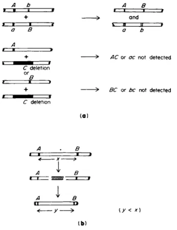
A b α Β
+ > an d
f • I t \ Β II t
a Β a b
A
+ > AC o r ac no t detecte d
C deletio n
+ > BC o r be no t detecte d
C deletio n
(a)
A Β
< x *
A ^ Β
Α ψ
I
Β< y — > (y < χ) ( b )
FIG. 5. Some genetic tests for deletion mutants, (a) Absence of recombination between the test mutation (deletion) and two other recombinable mutations (a and b). (b) Reduced recombination frequency between two recombinable mutations flanking the test mutation (y < x).
Potentially lethal deletion mutants occurring in some bacteriophages can be rescued by multiple infection. For example, although the T4 petite particles have suffered substantial deletions of their genome which make them reproductively defective, the genomes of individual particles are related to one another as random segments of a circle of permuted sequences and can thus potentially reactivate one another (45).
E. Effects on Genetic Maps
Several genetic tests for deletion have proved to be reliable in practice, especially when more than one of them could be used. It must be empha
sized that in most systems, and as yet, these tests constitute the only means of detecting, localizing, and measuring deletions. The absence of recombination between one mutation and two or more other mutations which themselves recombine constitutes evidence that the first mutation genetically spans the sites of the others and may therefore have suffered a deletion (Fig. 5a). If such mutations are deletions they fail to revert, unlike multisite mutations, which may revert. A deletion occurring be
tween two mutations may be expected to decrease the distance between them and lead to a decrease in the frequency of recombination between them (Fig. 5b). This test has been used in phages T4 (37) and λ (46) and in Salmonella (9) and E. coli (28).
IV. THE FREQUENCY, TOPOGRAPHY, AND TOPOLOGY OF DELETION MUTATIONS
It is reasonable to assume that in haploids a substantial fraction of deletions in D N A will go unnoticed because of their lethality. This lethality could arise from two different causes. Either the deletion events themselves are lethal, or the end product of the events is a chromosome that confers a low survival value on the organism. Two other considera
tions arise when trying to make quantitative inferences based on genetic analyses, (a) The assumption that most deletions are continuous has been rarely tested, and (b) most quantitative procedures and estimates on the frequency of deletion mutants are necessarily based on observa
tions on restricted regions of a chromosome. Despite these qualifications, information on the frequency of deletion mutants and their genetic struc
ture is potentially useful.
Although information on the single-stranded D N A viruses is scarce, in all or most organisms that have double-stranded D N A as their genetic material, a small and variable fraction of spontaneous mutations is due to deletions. Drake (47, 48) has compared forward mutation rates in a number of organisms and compiled evidence which suggests that the proportion of all mutations that are deletion mutants may vary consid
erably from one organism to another. In the rll region of phage T4, the frequency of spontaneous deletions is about 12% (2, 7). In the Ζ gene of the lac operon of E. coli, it was reported to be about 3 %
(21). Demerec (9) reported a difference in the frequency of deletion mutations in strains LT2 and LT7 of Salmonella typhimunum and left undetermined the basis of this difference. One possibility is that differ
ences of this type are determined by specific mutations analogous to the mutator genes (49, 50) that affect the frequency of mutations.
Mutators that specifically or preferentially promote deletions have not so far been reported. Bacterial episomes such as the temperate phage λ or the sex factor F may formally be considered to be mutators as they do promote the deletion of chromosomal genes adjacent to their attachment sites.
Although at present there is insufficient information for a rigorous analysis, it seems probable that, in any one strain, different regions of a chromosome may be more or less susceptible than other regions to deletion mutations. Examining a collection of spontaneous mutants involving 23 different genetic loci in Salmonella typhimunum LT2, Demerec (9) observed that the cysC region in this strain showed 10 times as great a proportion of deletion mutants as did other regions.
In Escherichia coli, the region controlling phage T l resistance may be another example (12b, 51). Table I is a compilation of available infor
mation on those genetic regions of E. coli and S. typhimunum where viable deletion mutants have been located (9, 12c, 19, 20, 51-71). The compilation should not be taken to imply that these regions are especially susceptible or that viable deletion mutants cannot occur in another re
gion. Rather, it is a reflection of past and current interest in the functions controlled by these regions and the relative ease with which mutations involving these regions can be detected and examined.
Spontaneous and induced deletions could be repeats of a similar type (12c) and, in some cases, this could be a reflection of the physical or genetic structure of the chromosomal region. In E. coli, the termini of spontaneous deletion mutants that were selected on the basis of trypto
phan dependence and phage T l resistance have been found to vary from one strain to another (12c, 51, 55). In strain B, the deletions removed
T A B L E I
REGIONS OF THE CHROMOSOMES OF Escherichia coli AND Salmonella typhimurium
WHERE VIABLE DELETION MUTATIONS HAVE B E E N LOCATED
Escherichia coli Salmonella typhimurium
Chromosome region0 Reference Chromosome region0 Reference
lac 52 his 63, 64
lac-purB 53 his-rfb 65
lac-trp-tonB 54 cys 9, 66, 67
tonB-trp 51, 55, 56 pro 68
tonB-colB 55
trp 57 leu 69
gal 19, 20 aro 70
gal-aroG 19, 20 trp 71
gal-aroG-chlD 19, 20 mot 12c
gal-chlD-att\-bio-purB-chlA 19, 20
chlD 58
malA 59
ara 60, 61
ara-leu 60, 61
his 62, 62a
glu-argECBH 62b
° Phenotype governed by the gene symbols: lac, lactose utilization; purB, adeny- losuccinase; trp, tryptophan metabolism; tonB, resistance to phages T l , 080, and colicins Β, 1, V ; gal, galactose utilization; aroG, D H A P syntheses; chlD, nitrate- chlorate reductase; att\, integration site for prophage λ; bio, biotin metabolism;
colB, colicin B; chl, nitrate-chlorate reductase; malA, maltose utilization; ara, arabinose utilization; leu, leucine metabolism; his, histidine metabolism; rfb, rough colony (surface polysaccharide) production; cys, cysteine metabolism; pro, proline metabolism; aro, metabolism of aromatic amino acids; mot, motility; arg, arginine;
glu, glutamic acids.
the entire trpB operon. In contrast, mutations selected on a similar basis in strain Κ12 had a random assortment of end points terminating in any of the five structural genes of the trp operon or beyond them. The K 1 2 - B hybrids fell into one or the other of these deletion classes, and it has been suggested (56) that the difference in these two patterns may be due to some unspecified difference in the cysB-trp segment of the respective chromosomes. In the histidine region of S. typhimurium, where large and small deletion mutants occur frequently (72), the genetic ends of the deletions are widely distributed. In the histidine region of
Ε. coli, where deletion mutants are even more common (62, 62a), they seem to be more uniform.
Preferred deletion end points can also be the consequence of the mech
anism of action of a particular inducing agent, when one has been used.
In the rll region of phage T4, both small and large spontaneous deletion mutants were found (2, 8) and their end points were not randomly distributed. The right-hand end points of the mutations tended to extend into the terminus of the Β gene. Among deletion mutants believed to be induced by nitrous acid (8) this bias was even greater and the dele
tions themselves tended to be larger. The left-hand end points of these mutants also tended to recur at sites within the A cistron. These observa
tions suggest that the end-point locations of deletion mutants could be determined both by structural features of the chromosome and by the mechanism by which inducing agents act. The question as to whether nitrous acid specifically induces deletions, however, needs to be re
examined. Experiments by Koch and Drake (73) indicated that only 0.8% of a collection of nitrous-acid-induced mutants of phage T4 were deletion mutants. Recently, Dove (74) reported on a nonessential genetic region adjacent to the rllB gene. This could explain why the end points of the mutants are preferentially located in this region. A comparison of the recombination and integration properties of deletion mutants of phage λ (23, 75) has also suggested basic differences in the mechanism of deletion formation in spontaneous and UV-induced deletion mutants.
There is evidence (reviewed in 75a, 75b) that, in the defective galactose- transducing phage Xdg, the bacterial D N A that is covalently attached to phage D N A was previously located next to the prophage attachment site on the bacterial chromosome. If one examines a number of inde
pendently isolated Xdg particles, one finds that they have suffered a dele
tion of λ genes and λ D N A .
The genetic analysis of transducing phage particles orginating from bacteriophage λ has been revealing in suggesting a possible mode of deletion formation. When the chromosome of phage λ integrates into the bacterial chromosome, it prefers to do so at a defined region of the bacterial chromosome called attx, which is flanked by the bacterial operons gal and bio (Fig. 6). When λ deintegrates from the chromosome it may rarely do so abnormally, so that the resulting phage particle may now include part of gal or bio (Fig. 6b and c). The resulting phages are called Xdg or Xdb, respectively, and a collection of such independently arising xdg or xdb has been genetically analyzed. It was found that members of the Xdg collection lacked genes concerned with head and/or tail synthesis. In contrast, the collection of Xdb always had these genes
( b)
(c)
(d) \bio
\dg
(·)
Deletion s\bio Derivative s
Deletion s
\dg
in Derivative s(f)
Mature DNA end s
Integratio n DNA replicatio n Late Hea d an d tai l Excisio n Early control s Control s synthesi s
Recombin Lysis (gene s A-J)
atio n att
\-PB bio
V — FIG. 6 . Diagram illustrating the mode of formation and the topology of deletions in \dg and Xbio particles. In (a), MO and [ J | are the hybrid attachment sites of bacterium and phage; and represent bacterial and phage chromosomes, respectively; (b)-(d) represent the mode of formation of the two respective defec
tive phages by rare loop-outs; (e) and (f) indicate the topology of deletions in the two types of phages and the variable extents to which deletions may penetrate into the genomes of the respective phages. The arrows in (e) indicate that the deletions may also extend to a variable extent inward into the respective and adjacent chromosomal regions.
220
but could be missing those genes that were on the left of the map (as conventionally drawn; see Fig. 6f). Neither class of deletion mutants extends inward into the λ prophage to delete the ends of the mature phage D N A molecule. In addition to the phage attachment locus attx, the only other essential structural elements of λ appear to be its ends.
Figure 6a-f is a diagram of a model proposed by Campbell (75c) which attempts to explain how such events may occur. It has been useful not only in the special case involving transducing phages but also in situa
tions involving other bacterial episomes. The diagram indicates the de- integration event to involve one break in the integrated episome and another within an adjacent chromosomal region. Alternatively, both breaks could occur in a chromosomal region on either side of the inte
grated episome (76). A transducing particle such as Xdg which has suffered a deletion of λ genes is analogous to a bacterial mutant that may suffer a deletion of genes of the galatose operon, and both may have arisen from the same series of events. In both cases, enzymatic intervention is postulated but detailed mechanisms are unknown.
V. ENZYMES THAT CUT AND REPAIR DNA
Depending on where in a D N A molecule they occur (terminal or inter
stitial), deletions may involve cuts, erosions, or both cuts and erosions, followed by repair in D N A strands. The in vitro properties of the limited number of known nucleases and repair enzymes of bacteria appear to equip them to fulfil these individual roles in a cell, although none of them have been directly implicated. Reviews on the properties of these enzymes have appeared recently (77-79). Table II (80-105) is a sum
mary of those that have been isolated and described from one bacterial species, E. coli, excluding the bacteriophage-induced enzymes that have been reviewed recently by Radding (106) and Koerner (107).
Enzymes or enzyme activities have also been discovered that show specificity for D N A that has suffered mutilations. Escherichia coli exonu- clease II shows specificity for alkylated D N A . Endonucleases with a similar specificity have been detected in Bacillus subtilis (108-110).
Takagi et al. (Ill) and Grossman et al. (112) have independently de
scribed an endonuclease from Micrococcus luteus that produces single- strand cuts in ultraviolet-irradiated D N A that has been denatured. The latter group has also purified from the same species an exonuclease that hydrolyzes single-stranded but not double-stranded D N A . This enzyme
Enzyme Preferred substrate Nature of action (in vitro) End product Reference Endonucleases
Endonuclease I Double-stranded D N A Double-strand breaks with Oligonucleotides 80, 81 a few single-strand
breaks; when complexed with tRNA, single-strand breaks in covalently closed circular D N A
Endonuclease II Double-stranded Single-strand breaks 82
alkylated D N A
Endonuclease R . K Double-stranded and Single-strand break at Large D N A duplexes with 88 (Endonuclease III) specifically unmodified specific sites followed by few single-strand breaks
DNA an opposite break on
complementary chain
Endonuclease R.B Double-stranded and Limited double-strand Large D N A duplexes 84 specifically unmodified breaks at specific sites
DNA
Endonuclease R.N3 Double-stranded and Limited double-strand Large D N A duplexes 85 specifically unmodified breaks at specific sites
DNA
Endonuclease associated Single-stranded D N A rings Endonucleolytic cuts and Linear strands and acid- 86
with recA gene erosion soluble products
Exonucleases
Exonuclease I Single-stranded D N A Erodes from the 3'-hydroxyl Dinucleotides 87, 88 terminus
Exonuclease II Double-stranded D N A Erodes from the 3'-hydroxyl Mononucleotides 89--93
(associated with DNA terminus with a less effi-
polymerase I) cient attack on the 5'-
phosphate terminus
222
Exonuclease III
ATP-Dependent exonuclease activity controlled by recB and recC genes
ATP-Independent exonuclease activity controlled by recB and recC genes
Exonucleases IV A and Β
DNA Polymerase I
D N A Ligase
dimers
Double-stranded DNA
Double-stranded DNA
Double-stranded DNA
Oligonucleotides 3'-Hydroxyl-terminated
primer strand with complementary template strand
Double-stranded DNA containing single-strand breaks displaying 3- hydroxyl and 5'- phosphoryl end groups in juxtaposition
Erodes from the 3' terminus 5'-Nucleotides (up to ap- 95, 96 proximately 50%
digestion)
TCA-Soluble material 97-99
TCA-Soluble material 100
Erodes from the 3' terminus 5'-Mononucleotides 101 Restores double-stranded- Double-stranded duplex 102
ness to partially single- stranded D N A
Esterifies the 5'-phosphoryl Double-stranded D N A with 103-105 group to the 3'-hydroxyl intact strands
group
* See note added in proof.
223
can remove a few nucleotides containing thymine for each single-strand break produced by the UV-specific endonuclease. It is possible therefore that these two enzymes or similar enzymes act in succession to remove radiation-induced dimers. Bacterial transforming D N A that has been inactivated by ultraviolet irradiation can also be partially reactivated by crude bacterial cell extracts [108, 113, 114). Taken together, these observations indicate that bacterial cells do possess enzymatic mecha
nisms that can monitor and remove nucleotides in the vicinity of rare mutilations in D N A . They also possess enzymatic mechanisms to repair the structural damage sustained by the removal of bases. Although there is at present no direct evidence for this idea, it is possible to visualize how the absence of an exact correlation between excision and repair can lead to the perpetuation of a deletion, provided such a deletion can be tolerated by the cell.
VI. AGENTS THAT MAY PROMOTE DELETIONS
There have been relatively few studies undertaken to see if certain agents will induce deletion mutants in preference to other kinds of muta
tions. Limited evidence (114®) suggests that at least some bifunctional alkylating agents may induce deletions in preference to other kinds of mutations. The extension of such observations to a larger list of agents with potential mutagenic properties would be worthwhile if only be
cause deletion mutants have been and can continue to be valuable tools in a variety of studies. Whether such studies would also be useful in attempts to understand how deletions may be produced is a moot point.
It is possible but as yet uncertain {8, 73) that nitrous acid when used in vitro will induce more deletion than point mutants in bacterio
phages. This agent is known to deaminate adenine, guanine, and cytosine and can react with these bases in D N A to eventually yield point muta
tions of the base-transition type. On the other hand, it also produces interstrand cross-links of D N A at frequencies of about one per four deaminations (115). It is possible that the induction of deletion mutants by this agent is a consequence of this cross-linking reaction or of at
tempts to repair the cross-link.
Alkylating agents are a widespread class of compounds with general mutagenic properties, including the potential to produce deletion mu
tants. Their reactions with nucleic acids have been recently reviewed
(116-118). Those with a single reactive group are called monofunctional alkylating agents and those with two or more reactive groups, bifunc- tional alkylating agents. Bifunctional or polyfunctional alkylating agents
also react monofunctionally. Several ways can be imagined by which a monofunctional reaction can lead to a deletion, but it is uncertain whether these mechanisms do in fact operate in vivo. The N-7 position of guanine within D N A appears to be particularly susceptible. It has been observed (119, 120) that the alkyl group on N-7 of guanine labilizes the β-glycosidic bond resulting in depurination. The loss of such bases could lead to deletion either as a direct result or during subsequent replication. Other treatments that cause depurination (e.g., acid and heat) may also promote deletions. It is known (121) that the depurination of guanine in D N A can cause the hydrolysis of an unstable deoxyriboside residue and backbone cleavage, an event which would be highly lethal but which, in the presence of suitable repair potential, could possibly lead to deletion mutants. It has also been sug
gested that the N-3 position of adenine, which is relatively inactive in free nucleotides, is more reactive in double-stranded D N A . With bi
functional alkylating agents, adjacent residues on the same strand could be linked. Alternatively, interstrand cross-linking could occur without detectable hydrolysis of D N A . A number of different agents have been clearly shown or suspected to be able to cross-link in this manner. Among these are nitrogen mustard (115, 122), trichlorotriethylamine (128), the reduced mitomycins, porfiromycin, (124, 125), and triethylenemelamine
(126). Brookes et al. (127) have suggested that bifunctional alkylating agents would be the most likely to produce genetic deletions, and in a recent study involving induced mutagenesis in phage T4, Corbett et al. (114a) found 47% of the mutations induced by nitrogen mustard to involve large deletions. There is some evidence that cross-links pro
duced by nitrogen mustard may be repaired. When a strain of B. subtilis resistant to nitrogen mustard was exposed to nitrogen mustard, its D N A became cross-linked. On further incubation in fresh medium most of the D N A was converted to the normal form (122). It was suggested that a repair mechanism similar to. the excision of ultraviolet-induced thymine dimers (128) may be operative. High doses of ultraviolet are also known to produce interstrand cross-links (129). The fate of inter
strand cross-links has not been extensively studied. If they can be re
paired, and if such a repair will allow cell survival, mistakes in repair could provide a plausible means for generating deletions.
Limited evidence (21, 48) suggests that ultraviolet or X irradiation may not be useful in inducing deletion mutants in phages and bacteria.
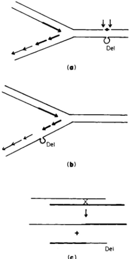
VII. HYPOTHETICAL MECHANISMS
In one context or another, deletion mutants have been speculated to arise as errors in D N A replication, repair, or recombination. These modes of origin could share some common features but are sufficiently different to be distinguishable (Fig. 7).
A. Replication Errors
The mode of replication of bacterial chromosomes probably involves a reiterative interruption and reinitiation of D N A strand elongation complemented by enzyme-mediated ligation (79). If an error occurred in accurately coordinating these processes, a deletion could result. Bren
ner et al. (ISO) suggested that the intercalation of acridines between adjacent bases could induce miscopying errors leading to deletion or addition. The deletions of phage T4 induced by nitrous acid were also thought possibly to originate in this manner (8), although the lesion in this case was believed to be cross-linking rather than intercalation.
Structural aberrations in template strands need not however be an essen
tial prerequisite for promoting miscopying errors. It is conceivable that other components of the replication complex, such as the D N A replicases and ligases, could be prone to spontaneous or induced miscopying of a type that promotes deletion. Mutations in gene 43 of phage T4, which controls T4 D N A polymerase, are known to have mutagenic (131) or antimutagenic effects (132), and a mutation in Bacillus subtilis affecting the temperature sensitivity of D N A synthesis also has mutator effects
(133). In none of these instances, however, has there been any indication so far that deletion mutants are induced. It is unlikely that acridine- induced deletions in phage T4 arise as replication errors in the manner suggested by Brenner et al. (130). The deletion of more than a single base pair could frequently occur on acridine treatment, and the inhibition of D N A synthesis by 5-fluorodeoxyuridine had little effect on proflavine- induced mutagenesis while it did have an effect on base-analog-induced mutagenesis (48, 134). In a thymidine-requiring strain of E. coli Β the frequency of deletion mutants that were simultaneously tryptophan de
pendent and T l resistant was unaffected by the absence of thymidine (56), suggesting that the deletions occur in the absence of D N A synthe
sis. Other experiments by the same authors showed that while thymidine starvation at any time in the cell cycle was without any observable
I I
De l
(b)
i +
De l
(C)
FIG. 7. Three hypothetical modes for the origin of deletions (DEL), (a) As an error during the repair of a spontaneous or induced structural aberration indicated as # ; the two arrows indicate the region where a mispairing reaction may be promoted by excision of the region including the aberration, (b) As an error occur- ring in the vicinity of a chromosomal replicating fork during "normal" semiconserva- tive replication; the arrows represent the 5' to 3' direction of D N A chain elongation;
the most recently replicated region is shaded darker and joined arrows represent the ligation of short pieces that had previously arisen. (The structure of D N A in the replicating region has not been definitively established; the figure represents one plausible situation.) (c) As an error during recombination; the two lines could represent two different mispaired chromosomes or different regions of the same chromosome.
effect, amino acid starvation at certain periods of the cell cycle did reduce the frequency of deletion mutants. For interstrand cross-linking agents that induce deletion mutants (e.g., nitrous acid or nitrogen mus- tard) an implicit assumption has been that replication proceeds normally up to the cross-link, at which site the deletion arises during attempted
replication. In fact, it is not known that deletions do arise at or near the site of cross-links. One must conclude that at the present time there is no evidence to suggest that deletion mutants can arise through the mode of rare mistakes in normal chromosomal replication.
B. Repair Errors
A model initially developed by Streissinger and associates (134) for deletions and duplications of the frame-shift type proposed (a) a single- strand break, (b) local strand separation or limited nucleolytic digestion, (c) erroneous single-strand mispairing, and (d) sealing of the broken and paritally digested strand. This model attributes only a secondary role to inducing agents such as acridines which function mainly to in
crease the time for which an improbably paired configuration may exist.
The primary deletion-inducing event is the mispairing reaction, which should be promoted by the existence of nucleotide redundancies within a chromosome or regions within it. Apparently, a limited number of noncomplementary bases can be accommodated into a double helix with
out intolerable disorganization (135). Frame-shift deletions do have a tendency to occur in the vicinity of regions of local nucleotide re
dundancy and are associated with the origin of duplications. However, all compounds that intercalate or otherwise stabilize D N A do not neces
sarily induce or revert frame-shift mutations. This must not as yet be considered to be a serious drawback to the model. Correlations have not been seriously sought between the degree of stabilization that may be afforded to D N A in vivo by different compounds and their ability to induce deletions or frame-shift mutations in general. The model does not attempt to explain the origin of large deletions.
Available evidence (reviewed in 136) indicates that the excision repair of ultraviolet-induced photoproducts in a cell is not likely to induce mutations. Since under normal circumstances a fraction of mutations induced by ultraviolet is deletion mutant, this would suggest that dele
tion mutants can be produced despite the inability or reduced efficiency of the cells to remove such photoproducts by excision repair. If the suggestion is correct, then the fraction of all mutations that are deletion mutants should be the same in normal strains and in strains mutant at the uvr or her loci that are unable to excise UV photoproducts. Mutants of E. coli deficient in in vitro D N A polymerase I activity (187, 138) are hypersensitive to ultraviolet irradiation because of an apparent par
tial impairment in their ability to rejoin excised and repaired regions
of D N A to the parental strand of D N A (139, 140). The longevity of such an unrepaired lesion may be related to the promotion of deletions.
It was recently reported (141) that, in a mutant deficient in D N A poly
merase I activity (polA~ mutant), the frequency of spontaneous deletion mutants was high. This report, however, needs to be confirmed in view of observations (142) of strong selection effects favoring the growth of mutants in reconstructed mixed culture.
C. Recombination Errors
For the purpose of the present discussion, "recombination events" are defined broadly to include all events that are promoted by the confronta
tion of two different chromosomes or D N A molecules or different parts of a single chromosome. For some "recombination events," homology and presumably homologous pairing between the participants may be an essential prerequisite. For others, little or no homology may be needed.
Potentially, both kinds of events may generate deletions. In cases where homology is essential, the crucial deletion-promoting event would be a mispairing reaction which would then be followed by an unequal cross
over (Fig. 7c). Lerman (143) has hypothesized that the intercalation of acridines between adjacent base pairs promotes unequal crossovers through mistakes in homologous pairing. In some organisms, a correlation has been observed between acridine-induced frame-shift mutations and general recombination potential (144, 14&)- In other organisms, such a correlation has not been found (48, 146). If a certain kind of deletion is promoted by unequal crossovers, one expects these deletions to occur preferentially in those regions of a chromosome where there is sequence similarity. It is clear that some regions of a bacterial chromosome are more prone to deletions than others (e.g., region in the vicinity of cys in Salmonella or trp in E. coli). Within experimental limits, there has been nothing to suggest that these regions have repeating or similar nucleotide sequences. On the contrary, there is now sufficient evidence (reviewed in 72a, 75b, 147) that two chromosomes that share little or no detectable homology may participate in recombination-type breaks and restitutions. Examples are the prophage integration and deintegra- tion events promoted by the int function of phage λ and the transition of the F factor from the integrated to the autonomous state. It is also known that such events can sometimes involve the deletion from the chromosome of genes adjacent to the chromosomal attachment site of the episome. The adjacent genes may be on one or both sides of the
inserted episome and, in the latter case, the breaks and restitutions need involve only chromosomal material. As a working hypothesis, it is attractive to postulate that events of this type may be promoted by episomic elements that are otherwise genetically "silent," that is, elements that have the capacity for chromosomal integration-deintegra- tion but are not otherwise easily recognizable or, at any rate, have not as yet been recognized. This would explain why the frequency of deletion mutants is independent of generalized recombination functions such as those determined by the rec and red genes (H8, 149). In the vicinity of a region that is prone to deletions such as cys in the Sal
monella chromosome, there then ought to be one or more sequences that either directly or indirectly serve to recognize these hypothetical episomes.
VIII. CONCLUSIONS
Genetic deletions are very rare events (about 10~7 or less per cell generation) but a variety of selective techniques can be and has been devised for the isolation of deletion mutants. The unambiguous way in which deletion mutants can be exploited in topological fine-structure mapping fully justifies attention to their isolation and use. Available evidence suggests that such mutants will occur in virtually all regions of a chromosome. The frequency with which they occur will vary with the genetic region as well as the strain involved.
The mechanisms by which deletions are produced are not understood.
The rarity of the events that promote them have made indirect ap
proaches to the question of the mechanisms of their origin necessary.
Some of these indirect approaches suggest that the generation of deletions can be associated with events promoting the deintegration of episomic elements from a chromosome.
It seems reasonable to believe that enzymes identical or similar to the ones that are already described and are known to cut or repair D N A have a role in generating deletions. Mutations in genes controlling some of these enzymes (e.g., D N A polymerases and ligases) may affect deletions, but there is as yet no evidence to suggest that the functions mediated by these known enzymes or any other function are essential for generating deletions.
Note added in proof: Since this chapter was written, pertinent informa
tion has become available and references to this literature are indicated
here, (a) There is now every indication that the technique of physically mapping D N A molecules (Section IIIC) can be extended to genetic elements besides viral D N A {150-152). (b) New enzymes or enzymatic activities involving D N A as the substrate have been discovered in E. coli.
In some cases, the genes determining them have been identified (153- 160). (c) Novel and rare types of interactions between plasmids or a plasmid and chromosome are being observed (161-163). There is a recent and interesting volume on bacteriophage lambda (164); in particular, the article by N. Franklin relates to some of the questions raised here.
ACKNOWLEDGMENTS
Research has been supported by Grant A4429 from the National Research Council of Canada. During the preparation of this chapter, I have received thoughtful criticism from colleagues in this department and from J. Drake, P. Howard-Flanders, R. Iyer, and W. Szybalski.
REFERENCES
1. R. C. King, "A Dictionary of Genetics." Oxford Univ. Press, London and New York, 1968.
2. S. Benzer, Proc. Nat. Acad. Sci. U.S. 45, 1607 (1959).
3. J. R. Beckwith and D . Zipser, "The Lactose Operon." Cold Spring Harbor Lab., Cold Spring Harbor, New York, 1970.
4. C. Yanofsky, B. C. Carlton, J. R. Guest, D. R. Helinski, and U. Henning, Proc. Nat. Acad. Sci. U.S. 51, 266 (1964).
5. H. Ris and D. F. Kubai, Annu. Rev. Genet. 4, 263 (1970).
6. R. Holliday, Symp. Soc. Gen. Microbiol. 20, 359 (1970).
7. C. E. Folsome, Genetics 47, 611 (1962).
8. I. Tessman, / . Mol. Biol. 5, 442 (1962).
9. M. Demerec, Proc. Nat. Acad. Sci. U.S. 46, 1075 (1960).
10. F. Jacob and E. L. Wollman, "Sexuality and the Genetics of Bacteria." Academic Press, New York, 1961.
11. A. Cook and J. Lederberg, Genetics 47, 1335 (1962).
12. Ε. H. Anderson, Proc. Nat. Acad. Sci. U.S. 32, 120 (1946).
12a. M. Demerec, D. H. Gillespie, and K. Mizobouchi, Genetics 48, 997 (1963).
12b. J. S. Gots, W. Y. Koh, and G. R. Hunt, Jr., J. Gen. Microbiol. 11, 7 (1954).
12c. M. Enomoto, Genetics 54, 715 (1966).
13. N. C. Franklin, W. F. Dove, and C. Yanofsky, Biochem. Biophys. Res. Com
mun. 18, 910 (1965).
14. J. R. Beckwith, E< Signer, and W. Epstein, Cold Spring Harbor Symp. Quant.
Biol. 23, 393 (1966).
15. S. Gottesman and J. R. Beckwith, J. Mol. Biol. 44, 117 (1969).
16. K. Ippen, J. H. Miller, J. Scaife, and J. Beckwith, Nature (London) 217, 825 (1968).
17. J. H. Miller, K. Ippen, J. G. Scaife, and J. R. Beckwith, Λ Mol. Biol. 38, 413 (1968).
18. J. Shapiro, L. Machattie, L. Eron, G. Ihler, K. Ippen, J. Beckwith, R. Arditti, W. Reznikoff, and R. MacGillivray, Nature (London) 224, 768 (1969).
19. S. Adhya, P. Cleary, and A. Campbell, Proc. Nat. Acad. Sci. U.S. 61, 956 (1968).
19a. Β. N. Ames, in "Chemical Mutagens" (A. Hollender, ed.), Chapter IX, p.
267. Plenum, New York, 1971.
20. J. A. Shapiro and S. K. Adhya, Genetics 62, 249 (1969).
21. D. 0 . Schwartz and J. R. Beckwith, Genetics 61, 371 (1969).
22. J. S. Parkinson and R. W. Davis, Proc. Nat. Acad. Sci. U.S. 61, 1152 (1968).
23. J. S. Parkinson and R. J. Huskey, / . Mol. Biol. 56, 369 (1971).
24. M. G. Burdon, Mol. Gen. Genet. 108, 288 (1970).
25. R. Hertel, L. Marchi, and K. Muller, Virology 18, 576 (1962).
26. I. Rubenstein, Virology 36, 356 (1968).
27. A. B. Pardee, J. Bacteriol. 73, 376 (1957).
28. J. Davies and F. Jacob, J. Mol. Biol. 36, 413 (1968).
29. G. Kellenberger, M. L. Zichichi, and J. Weigle, J. Mol. Biol. 3, 399 (1961).
30. C. A. Thomas, Jr., Progr. Nucl. Acid Res. Mol. Biol. 5, 315 (1966).
31. G. Kellenberger, M. L. Zichichi, and J. Weigle, Proc. Nat. Acad. Sci. U.S.
47, 869 (1961).
32. R. W. Davis and N. Davidson, Proc. Nat. Acad. Sci. U.S. 51, 883 (1964).
33. G. Mosig, Genetics 59, 137 (1968).
34. R. G. Cutler and J. R. Evans, J. Mol. Biol. 26, 81 (1967).
35. W. Colli and M. Oishi, J. Mol. Biol. 51, 657 (1970).
36. B. C. Westmoreland, W. Szybalski, and H. Ris, Science 163, 1343 (1969).
37. M. Nomura and S. Benzer, J. Mol. Biol. 3, 684 (1961).
38. W. Szybalski, H. Kubinski, Z. Hradecna, and W. C. Summers, in "Methods in Enzymology" (L. Grossman and K. Moldave, eds.), Vol. 21, Part C. p.
383. Academic Press, New York, 1970.
39. R. W. Davies and J. S. Parkinson, J. Mol. Biol. 56, 403 (1971).
40. Z. Hradecna and W. Szybalski, Virology 38, 473 (1969).
41. F. A. Bautz and Ε. K. F. Bautz, J. Mol. Biol. 28, 345 (1967).
42. K. Bovre and W. Szybalski, Virology 38, 614 (1969).
42a. L. Fishbein, H. L. Falk, and W. G. Flamm, "Chemical Mutagens," p. 34.
Academic Press, New York, 1970.
43. J. Scaife and A. P. Pekhov, Genet. Res. 5, 495 (1964).
44. D. Freifelder, Cold Spring Harbor Symp. Quant. Biol. 33, 425 (1968).
45. D. H. Parma, Genetics 63, 247 (1969).
46. E. Jordon, / . Mol. Biol. 10, 341 (1964).
47. J. W. Drake, Nature (London) 221, 1128 (1969).
48. J. W. Drake, "The Molecular Basis of Mutation." Holden-Day, San Francisco, California, 1970.
49. H. P. Treffers, V. Spinelli, and N. O. Besler, Proc. Nat. Acad. Sci. U.S. 40, 1064 (1954).
50. C. Yanofsky, E. C. Cox, and V. Horn, Proc. Nat. Acad. Sci. U.S. 55, 274 (1966).
51. C. Yanofsky and E. S. Lennox, Virology 8, 425 (1959).
52. J. Beckwith, / . Mol. Biol. 8, 427 (1964).
53. F. Jacob, A. Ullman, and J. Monod, J. Mol. Biol. 13, 704 (1965).
54. J. H. Miller, W. S. Reznikoff, A. E. Silverstone, K. Ippen, E. R. Signer, and J. Beckwith, / . Bacteriol. 104, 1273 (1970).