Comparative genomics reveals the origin of fungal hyphae and multicellularity
EnikőKiss1,2, Botond Hegedüs1, Máté Virágh1, Torda Varga 1,2, Zsolt Merényi1, Tamás Kószó1, Balázs Bálint1, Arun N. Prasanna 1,6, Krisztina Krizsán1, Sándor Kocsubé3, Meritxell Riquelme 4, Norio Takeshita 5 &
László G. Nagy1
Hyphae represent a hallmark structure of multicellular fungi. The evolutionary origins of hyphae and of the underlying genes are, however, hardly known. By systematically analyzing 72 complete genomes, we here show that hyphae evolved early in fungal evolution probably via diverse genetic changes, including co-option and exaptation of ancient eukaryotic (e.g.
phagocytosis-related) genes, the origin of new gene families, gene duplications and altera- tions of gene structure, among others. Contrary to most multicellular lineages, the origin of filamentous fungi did not correlate with expansions of kinases, receptors or adhesive pro- teins. Co-option was probably the dominant mechanism for recruiting genes for hypha morphogenesis, while gene duplication was apparently less prevalent, except in transcrip- tional regulators and cell wall - related genes. We identified 414 novel gene families that show correlated evolution with hyphae and that may have contributed to its evolution. Our results suggest that hyphae represent a unique multicellular organization that evolved by limited fungal-specific innovations and gene duplication but pervasive co-option and mod- ification of ancient eukaryotic functions.
https://doi.org/10.1038/s41467-019-12085-w OPEN
1Synthetic and Systems Biology Unit, Institute of Biochemistry, BRC-HAS, Temesvari krt 62, 6726 Szeged, Hungary.2University of Szeged, Faculty of Science and Informatics, Aradi vertanuk tere 1., 6720 Szeged, Hungary.3Department of Microbiology, University of Szeged, Faculty of Science and Informatics, Kozep fasor 52, 6726 Szeged, Hungary.4Department of Microbiology, Centro de Investigación Científica y de Educación Superior de Ensenada, Carr Tijuana- Ensenada 3918, C.I.C.E.S.E, 22860 Ensenada, Baja California, Mexico.5Microbiology Research Center for Sustainability (MiCS), Faculty of Life and Environmental Sciences, University of Tsukuba, 1 Chome-1-1 Tennodai, 305-8572 Tsukuba, Japan.6Present address: Red Sea Science and Engineering Research Center, 4700 King Abdullah University of Science and Technology (KAUST), Thuwal 23955-6900, Saudi Arabia. Correspondence and requests for materials should be addressed to L.G.N. (email:lnagy@fungenomelab.com)
1234567890():,;
T
he evolution of multicellularity (MC) is considered one of the major transitions in the history of life1. Multiple bac- terial and eukaryotic lineages underwent this major tran- sition2–7, in each case arriving at a unique solution to the challenges of multicellular organization6. Among eukaryotes, multicellularity appears to have arisen via either clonal and aggregative mechanisms5,6,8,9, which differ in how multi-celled precursors adhere, cooperate, communicate and functionally diversify3,10,11.Fungi constitute one of the three kingdoms where a majority of extant species are multicellular12, yet, the origins of fungal mul- ticellularity remain obscure. While most multicellular lineages can be recognized as being either clonal or aggregative by com- parisons to their unicellular relatives, fungal multicellularity has been recalcitrant to such categorization6,13. Multicellularity in fungi refers to a thallus made up of hyphae, thin, tubular struc- tures that grow by apical extension to form a mycelium that explores and invades the substrate. Hyphal multicellularity exhibits several unique properties that distinguish it from clonal and aggregative multicellularity, raising the possibility that its evolution may follow markedly different principles7.
First, hyphae might have evolved by the gradual elongation of substrate-anchoring rhizoids of early fungi14–16, through multi- nucleate intermediates, in contrast to clonal and aggregative lineages, where the first multi-celled clusters probably emerged via related cells sticking together (e.g., choanoflagellates17), or gathering to form a syncytial body (e.g., ichthyosporeans)18. Because early hyphae were uncompartmentalized, their evolution could have bypassed the need to resolve group conflicts and align the fitness of individual cells7. Alternatively, it is possible that conflicts are resolved at the level of individual nuclei19. Second, hyphae maximize foraging and nutrient assimilation efficiency and minimize competition for nutrients by a fractal-like growth mode20–22. The mechanism of the origin of hyphae differs from that of other multicellular lineages where selection for increased size possibly helped avoiding predation2. Hyphae might have also facilitated the transition of fungi to terrestrial life, by bridging nutrient-rich and nutrient-poor habitats23 and confer immense medical relevance to pathogenic species24. Hyphae of extant fungi rarely stick to each other in vegetative mycelia and adhesion becomes key only in fruiting bodies25,26—which, in terms of complexity level, resemble multicellular metazoans and plants7,27
—or in the attachment to host surfaces28. Thus, whereas in most multicellular lineages adhesion, cell–cell cooperation, commu- nication and differentiation represent the main hurdles to the emergence of multicellular precursors3,6,29,30, fungi might have had different obstacles to overcome.
While the evolutionary origins of hyphae are obscure, infor- mation on the molecular and cellular basis of hypha morpho- genesis is extensive (for recent reviews see refs.31–34), permitting evolutionary genomic analyses. Hypha morphogenesis builds on cell polarization networks35, the exo- and endocytotic machin- ery36, long-range vesicle transport as well as fungal-specific traits such as cell wall synthesis and assembly37, and the selection of branching points and septation sites38. A key structure of hyphal growth is the Spitzenkörper39, which acts as a distribution center for vesicles transporting cell wall materials and other factors to the hyphal tip. The cytoplasmic microtubule network provides the connection between vesicle cargo from the ER and Golgi and the Spitzenkörper, from where vesicles move to the hyphal tip and secrete their content for building the cell wall and provide surface expansion. Further key processes include the recycling of excess membrane in the subapical zone, the activation of cAMP pathways and mitogen-activated protein kinase (MAPK) cascades and finally the transcriptional control of morphogenesis (reviewed in refs.34,40–42).
A complex hyphal thallus has been reported from a 407 million-year-old fossil Blastocladiomycota43, whereas Glomeromycotina-like hyphae and spores were preserved 460 million years ago44,45indicating that hyphal growth dates back to at least the Ordovician. Most Dikarya and Mucoromycota grow true hyphae, whereas a significant diversity of forms exists in the Blastocladiomycota, Chytridiomycota and to a smaller extent the Zoopagomycota. The Chytridiomycota is dominated by uni- cellular forms that anchor themselves to the substrate by bran- ched, root-like rhizoids22,45. These structures have been hypothesized as the precursors to hyphae14,46. An alternative hypothesis designates hypha-like connections in the thalli of polycentric chytrid fungi (e.g. Physocladia) as intermediates to true hyphae15. Like chytrids, most Blastocladiomycota form mono- or polycentric, unicellular thalli, although some species form wide, apically growing structures resembling true hyphae (e.g., Allomyces) or narrow exit tubes on zoosporangia (e.g., Catenaria spp.)22,45,47. In spite of these intermediate forms, the prevalence of unicellular forms in these phyla indicates their unicellular ancestry and suggests potential convergent origins of hypha-like structures15.
Here we examine how the genetic toolkit of hyphal multi- cellularity was assembled during evolution by reconstructing the evolutionary history of known hypha morphogenesis genes and by systematically searching fungal genomes for gene families whose evolution correlates with that of hyphae. We analyze the genomes of 4 plesiomorphically unicellular, 41 hyphal and 13 secondarily simplified (yeast-like) fungi as well as 14 non- fungal relatives. We identify a multitude of small changes in hyphal-morphogenesis gene families that correlate with the evo- lution of hyphae, including co-option and exaptation of ancient eukaryotic genes, limited gene family diversification and altera- tions of gene structure. Correlated patterns of gene duplication and loss that correlate with the origin of hyphal multicellularity were detected for 414 gene families, providing further candidate key genes. These data indicate that many small changes rather than one major innovation, underlie this key fungal innovation, compatible with evolutionary tinkering48.
Results
Hyphae evolved in early fungal ancestors. To understand the origin of hyphae, we constructed a species phylogeny representing 72 species (Supplementary Data 1) by maximum likelihood and Bayesian MCMC analysis of a supermatrix of 455 single-copy orthologs (75,224 characters, Fig.1a, Supplementary Fig. 1).
Our species phylogeny is strongly supported and recapitulates recent genome-based phylogenies of fungi49–52, with the Rozellomycota, Blastocladiomycota and the Chytridiomycota splitting first, second and third off of the backbone, respectively (ML bootstrap: 100%). We next scored species for their ability to form multicellular hyphae (Fig. 1a, Supplementary Data 1) and performed ancestral character state reconstructions using Baye- sian MCMC. This suggested that hyphae evolved from unicellular precursors in some of the earliest fungal ancestors. The distribution of posterior probability values indicated three nodes as the most likely origins of hyphal multicellularity, which represent the split of Blastocladiomycota, Chytridiomycota and Zoopagomycota, and are referred hereafter to as BCZ nodes (Fig.1a). The posterior probability for the hyphal state started to rise in the most recent common ancestor (MRCA) of the Blastocladiomycota and higher fungi (PP: 0.53, Fig. 1a) and increased to 0.68 and 0.92 in the next two nodes up in the tree.
This suggests that hyphae evolved either in one of the BCZ nodes or that its evolution was a gradual process unfolding in these three nodes. This distribution also reflects the diverse hypha-like
morphologies in the Blastocladio- and Chytridiomycota and is consistent with convergent origins of hypha-like morphologies7,15,16.
To analyze the evolutionary history of putative multicellularity- related genes, wefirst reconstructed gene family origins and gene duplication/loss histories across all the gene families in the examined genomes (Supplementary Fig. 2). In the following sections, we mine this gene duplication/loss catalog for gene families with previously suggested or novel role in multicellularity and hyphal growth.
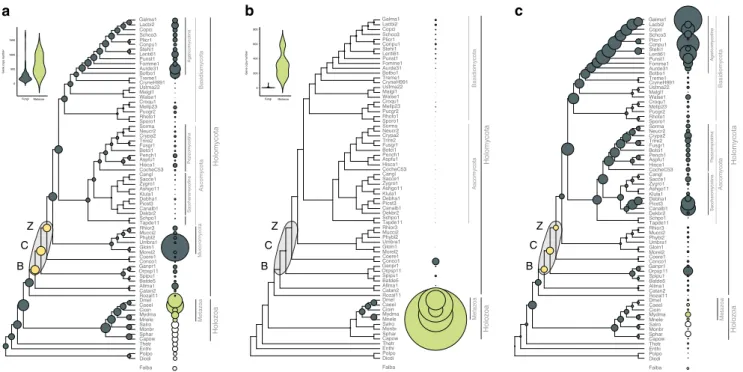
No expansion of kinase, receptor and adhesive repertoires in fungi. The increased sophistication of cell–cell communication and adhesion pathways in multicellular lineages often correlates with expanded repertoires of genes encoding kinases, receptors and adhesive proteins53,54. We thereforefirst tested if these gene families had undergone diversification in BCZ nodes. Ser/Thr kinase (954 clusters), hybrid histidine kinase (96 clusters), receptor (183 clusters) and adhesion (23 clusters) genes (Sup- plementary Data 2) did not show expansions reminiscent of patterns in other multicellular lineages (Fig. 2). Ser/Thr kinase repertoires were similar in unicellular and simple multicellular fungi, with higher kinase diversity found in complex multicellular Basidiomycota (as reported by Krizsán et al. 2019)55 and in Rhizophagus irregularis(Fig.2a). We inferred net contractions in
BCZ nodes, from 572 to 529 reconstructed ancestral kinases (81 duplications, 124 losses, Fig. 2a). Nevertheless, kinase families that duplicated here include all 3 MAPK pathways in fungi, the mating pheromone, cell wall integrity, and osmoregulatory pathways, all of which indirectly regulate hyphal growth40,56.
Overall, fungi had fewer Ser/Thr kinases (mean 257) than metazoans (mean 643), non-fungal opisthokonts (mean 392), including Fonticula alba, the closest relative of fungi (Fig. 2a, Supplementary Note 1). While signal transduction requirements of metazoan MC have been mostly discussed in the context of receptor tyrosine kinases, we found no evidence for domain architectures typical of receptor kinases in fungi. The only group resembling receptor kinases are hybrid histidine kinases (HK), which include a sensor domain, a histidine kinase domain, and a C-terminal receiver domain that acts as a response regulator. We inferred an expansion (24 duplications, 10 losses) of HKs in the MRCA of the Chytridiomycota and other fungi, including class III and X HKs, which are linked to morphogenesis57,58 (Supplementary Fig. 3). Another wave of HK expansion was inferred in the MRCA of Mucoromycota and Dikarya with 11 duplications and 4 losses.
Canonical G-protein coupled receptors (GPCRs) showed an even more extreme difference between fungi and metazoans (Fig.2b). We analyzed 183 GPCR families; a large expansion was observed in animals, resulting 135-583 genes in extant species, whereas, only 19 were found in fungi and only one of them
Chytridiomycota
Zoopagomycota Vesicle transport
Transcriptional regulators
Cell cycle Polarity maintenance
Septum formation
Signaling Actin cytoskeleton
Cell wall biogenesis/
remodeling
Microtubules Vesicle transport
(107)
Actin cytoskeleton (55)
Septum formation (58) Transcriptional regulators
(53)
Signaling (89) Microtubules (32)
Adhesion (45)
b c
Cell cycle (11)
Polarity maintenance (107) Cell wall biogenesis
(94)
Mucoromycota Saccharomycot.
Pezizomycotina
Holomycota
Agaricomycotina
Taphrinomycotina Ustilaginomycot.
Tremellomycetes
Pucciniomycotina
Blastocladiomyc.
BasidiomycotaAscomycota
Rozellomycota
P = 0.003 P = 0.003
P = 0.28 P = 0.21
P = 0.12 P = 0.12
P = 1 P = 0.03*
P = 0.15
Rozella allomycis Catenaria anguillulae Allomyces macrogynus Batrachoch. dendrobatidis Spizellomyces punctatus Orpinomyces sp.
Gonapodya prolifera Conidiobolus coronatus Coemansia erecta Mortierella elongata Rhizophagus irregularis Umbelopsis ramanniana Phycom. blakesleeanus Mucor circinelloides Rhizopus oryzae Taphrina deformans Schizosacch. pombe Dekkera bruxellensis Candida albicans Pichia stipitis Debaryomyces hanseni Kluyveromyces lactis Ashbya gossypi Zygosaccharomyces rouxii Saccharomyces cerevisiae Candida glabrata Cochl. heterostrophus Histoplasma capsulatum Aspergillus fumigatus Pennicilium chrysogenum Botrytis cinerea Fusarium graminearum Trichoderma reesei Cryphonectria parasitica Neurospora crassa Sordaria macrospora Sporobolomyces roseus Rhodosporidium toruloides Puccinia graminis Melampsora laricis-pop.
Cronartium quercuum Wallemia sebi Malassezia globosa Ustilago maydis Cryptococcus neoformans Tremella mesenterica Botryobasidium botryosum Auricularia delicata Fomitiporia mediteranea Punctularia strigosozonata Lentinus tigrinus Stereum hirsutum Coniophora puteana Plicaturopsis crispa Schizophyllum commune Coprinopsis cinerea Laccaria bicolor Galerina marginata
B C
Z
Origin of fungi
Origin of hyphal multicellularity
a
Dictyostelium discoideum Polysphondylium pallidum Entamoeba histolytica Thecamonas trahens Capsaspora owczarzaki Sphaeroforma arctica Monosiga brevicollis Salpingoeca rosetta Mnemiopsis ledyii Hydra magnipapillata Ciona intestinalis Caenorhabditis elegans Drosophila melanogaster
Holozoa
Metazoa Fonticula alba
99/78/*
99/72/*
93/85/*
73/68/*
83/-/*
68/78/*
90/66/*
*/72/*
*/84/*
*/99/*
*/99/*
*/96/*
98/97/*
98/94/*
27/28/*
91/71/*
48/59/*
50/54/*
99/90/*
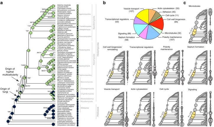
Fig. 1The evolution of hyphal multicellularity and underlying genes in fungi.aPhylogenetic relationships among 72 species analyzed in this study. Pie charts at nodes show the proportional likelihoods of hyphal (green) and non-hyphal (dark blue) ancestral states reconstructed using Bayesian MCMC.
Character state coding of extant species used in ancestral state reconstructions is shown next to species names. BCZ nodes: origins of hyphal growth could be assigned with confidence are highlighted (note the uncertainty imposed byfilamentous Blastocladiomycota). Support values next to branches are given for nodes that received less than maximal support in at least one analysis. Support values are given as ML boostrap (RAxML)/ML bootstrap (IQ-Tree)/
Bayesian posterior probabilities (Phylobayes). Asterisk (*) denotes maximal support in a given analysis.bthe distribution of literature-collected hypha morphogenesis genes among 10 main functional categories.cAncestral reconstructions of gene copy number in 9 main hypha morphogenesis-related categories of genes (see Fig.2c for adhesion). Bubble size is proportional to reconstructed ancestral gene copy number. BCZ nodes are shown in yellow.
P-values of enrichment of duplications are shown next to each tree (Fisher’s exact test, FDR correction). *For vesicle transport theP-value indicates significant depletion of duplications in BCZ nodes
(mating pheromone receptors) was conserved across the kingdom.
Adhesive cell surface proteins are key to the emergence of MC in colonial and aggregative lineages3,5,6, which is reflected in their higher copy numbers in multicellular organisms59. We identified 23 families of putative adhesion-related proteins in fungi, including adhesins, flocculins, hydrophobins, various lectins, and glycosylphosphatidylinositol-anchored cell wall proteins.
These families have undergone a small contraction (from 17 to 14 copies) in BCZ nodes, with expansions observed later, in the Agaricomycotina and in the Saccharomycotina (Fig.2c). The lack of an expansion in BCZ nodes probably reflects a marginal role of these proteins in the evolution of early hyphae, but potentially also the scarcity of adhesive proteins annotated in early-diverging fungi, or the effects of sequence divergence. The expansion in the Agaricomycotina was driven by class 1 hydrophobins and homologs of the Cryptococcus neoformans Cfl1 (with roles in signaling and morphogenesis regulation60) and correlates with the evolution of complex multicellular fruiting bodies7. The higher copy numbers in yeast species relate to yeast-specific adhesin and lectin-like cell wall proteins that have been experimentally characterized in human pathogens (e.g.,Candida spp.)61,62.
Taken together, the evolution of kinase, receptor, and adhesive protein repertoires highlight an important difference between fungi and other multicellular lineages. We observed no significant expansion of these families in filamentous fungi, whereas kinase and adhesion-related genes expanded in complex multicellular Agaricomycotina. This might be explained by the two-step nature of the evolution of complex MC in fungi7,63 that proceeds through an intermediate complexity level, hyphal MC, as opposed to metazoans, where complex MC evolved in a more direct way13. The observation that these’classic’ culprits of multicellular evolution can’t explain the evolution of hyphae prompted us to
examine other gene families, whose evolution might show a better correlate with that of hyphae.
The evolution of hypha morphogenesis genes. We built a dataset of hypha morphogenesis genes to determine whether changes in these gene families correlate with the evolution of hyphae. We identified 651 hyphal multicellularity-related genes belonging to 362 families (from 519 publications, covering our current knowledge on hyphal growth)—mostly derived from well-studied model systems such asA. fumigatus, A. nidulans,N.
crassa, S. cerevisiaeandC. albicans(Supplementary Data 3). We categorized genes into nine functional groups according to the broader function they serve in hyphal growth: actin cytoskeleton regulation, polarity maintenance, cell wall biogenesis/remodeling, septation (including septal plugging), signaling, transcriptional regulation, vesicle transport, microtubule-based transport and cell cycle regulation. The categories “polarity maintenance” and
“vesicle transport” contained the largest number of genes (107 in each), whereas“cell cycle regulation”contained the fewest (11) (Fig. 1b). To account for uncertainty in the exact origin of hyphae, we hereafter focus on BCZ nodes in our analyses of hypha morphogenesis genes. We examined multiple types of evolutionary innovations, to identify the most important mechanisms underlying the emergence of hyphae.
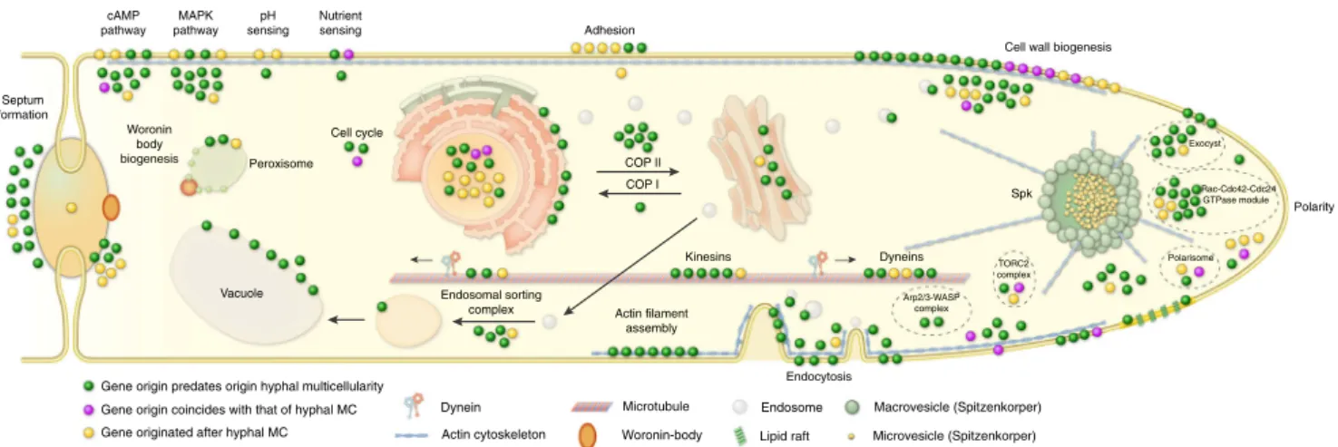
Reconstructions of gene duplication/loss histories for the nine functional categories of hypha morphogenesis gene families are shown on Fig. 1c. A general pattern that emerges from these is that most of the gene families are conserved across fungi (Supplementary Fig. 4) and their origin predate that of hyphae (181 families, 50%, Fig. 3, Supplementary Fig. 5), indicating that fungi have co-opted several conserved eukaryotic functionalities for hyphal growth. Gene families related to septation, polarity maintenance, cell cycle control, vesicle
a b c
Z C B
SaccharomycotinaPezizomycotina HolomycotaHolozoa
Agaricomycotina MetazoaBasidiomycotaAscomycota
Z C B
FungiMetazoa
Gene copy number
1500
1000
500
0
Mucoromycota
SaccharomycotinaPezizomycotina HolomycotaHolozoa
Agaricomycotina MetazoaBasidiomycotaAscomycota
Z C B
Fungi Metazoa
Gene copy number
0 200 400 600 800
HolomycotaHolozoa
MetazoaBasidiomycotaAscomycota
Fig. 2The evolution of kinases, receptors and adhesive proteins in multicellular fungi. The evolution of Ser/Thr kinases (a), canonical GPCRs (b) and adhesion-related genes (c). BCZ nodes (yellow) represent the putative origin of hyphal MC. Bubble size across the tree is proportional to reconstructed ancestral gene copy number (gray bubbles) and extant gene copy number (at the right side of the tree: gray, green and white bubbles represent fungi, metazoa, and protists respectively). Violin plots for kinases (a) and receptors (b) show copy number distribution of gene families in multicellular fungi (gray) and metazoans (green)
transport, and microtubule-based transport are generally more diverse in animals, non-fungal eukaryotes (including Fonticula alba, see Supplementary Note 1) and their ancestral nodes than in fungi, suggesting that despite the key role of these families in hyphal MC, they evolved primarily by gene loss in fungi (Fig.1c).
A significant proportion of hypha morphogenesis families (164 families, 45.3%) emerged after the origin of hyphal MC, indicating lineage- and species-specific genetic innovations. Only 17 families (4.7%) originated in BCZ nodes and were conserved thereafter (Table 1), providing potential candidates that shaped the evolution of hyphal MC. These include two families of transcriptional regulators (StuA and MedA inA. fumigatus), six related to cell wall biogenesis, three to actin cytoskeleton regulation, three to polarity maintenance, two families involved in signaling and one involved in cell cycle regulation. These fungal-specific families are good candidates for being key contributors to the evolution of multicellular hyphae; their functions are summarized in Table1.
Given the low number of gene families specific to multicellular fungi, we were interested in whether evolutionary innovation by duplications shows a peak in BCZ nodes. Ninety-three (25.7%) of the 362 hypha morphogenesis-related gene families showed duplications in BCZ nodes (Supplementary Data 4). Enrichment analyses, however, revealed no individual gene family with significantly increased number of duplications in BCZ nodes relative to the rest of the tree (Benjamini–Hochberg correctedP<
0.05, Fisher’s exact test, Supplementary Data 5). The same analysis on the 9 functional groups showed that duplications are significantly enriched in cell wall biogenesis and transcriptional regulator genes, suggesting that their diversification could have played roles in the evolution of hyphae. The rest of the functional groups showed no such enrichment of duplications, suggesting that the evolution of hyphal growth did not generally coincide with a major burst of gene duplication in BCZ nodes, as some considerations of the evolution of multicellularity predicted27.
Changes in structural properties of genes show a correlation with the evolution of hyphal MC. Significant differences (P< 0.05, two-tailed Welch’s t-test with pooled variance estimation) were observed in gene, coding sequence (CDS) and intron lengths between unicellular and multicellular fungi in 7 out of 9 functional groups (exceptions are cell wall biogenesis and transcriptional regulation genes) (Supplementary Fig. 6, Supple- mentary Data 6). Coding sequences of septation and polarity maintenance genes were significantly longer in multicellular than
in unicellular fungi (P=0.0012-0.00017, Supplementary Fig. 6).
An opposite pattern was observed in introns, which were on average longer in unicellular fungi in actin cytoskeleton, polarity maintenance, septation and vesicle transport-related genes. On the other hand, no significant changes in gene structure were detected in cell wall biogenesis and transcriptional regulation- related genes, the two categories that displayed increased duplicability in early filamentous fungi. This depicts potential complementary mechanisms of evolutionary change in different gene families and functional groups of genes.
We also analyzed changes in domain composition across plesiomorphically unicellular and multicellular fungi. This analysis was inspired by Class V and VII chitin synthases, which evolved higher efficiency in filamentous fungi by gaining a myosin motor domain during evolution64,65. Hypha morphogen- esis gene families, in general, show more change in domain composition between unicellular andfilamentous fungi than do randomly drawn gene families with similar properties (Supple- mentary Fig. 7). This indicates that changes to domain architectures correlate with the emergence of hyphae. We identified 4 gene families (including chitin synthases), in which proteins of multicellular fungi have consistently more domains (P< 0.05, GLM) than do proteins of unicellular fungi in the same family (Supplementary Data 7). Taken together, these analyses revealed several modifications to gene length and domain composition in multicellular fungi, which, although individually are small changes, could have contributed to the evolution of hyphae.
Phagocytotic genes were exapted for hypha morphogenesis.
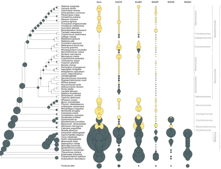
Our set of hypha morphogenesis genes included several entries associated with phagocytosis in non-fungal eukaryotes. This is surprising given that phagocytosis is not known in fungi and their rigid cell wall forms a physical barrier to it. We, therefore, examined the fate of phagocytosis genes in filamentous fungi based on the phagocytotic machinery ofD. discoideum66,67and other eukaryotes68(altogether 106 genes). Filamentous fungi have retained several phagocytotic gene families but lost others (Fig.4).
For example, members of the Arp2/3 complex (involved in actin cytoskeleton rearrangements69) are conserved in filamentous fungi and recycle excess membrane in the subapical zone during hyphal growth70. Engulfment and cell motility (ELMO1/2) genes are found in all filamentous fungi, but are convergently lost in
Cell cycle
Adhesion Signaling
cAMP pathway
MAPK pathway
pH sensing
Nutrient sensing
Peroxisome
Actin filament assembly Woronin
body biogenesis
Endosomal sorting complex
Kinesins Dyneins
Endocytosis COP II
COP I
Spk Septum
formation
Vacuole
Exocyst
Rac-Cdc42-Cdc24 GTPase module
Arp2/3-WASP complex
TORC2 complex
Polarity Cell wall biogenesis
Polarisome
Gene origin predates origin hyphal multicellularity Gene origin coincides with that of hyphal MC Gene originated after hyphal MC
Dynein Microtubule
Actin cytoskeleton
Endosome Woronin-body Lipid raft
Macrovesicle (Spitzenkorper) Microvesicle (Spitzenkorper)
Fig. 3Phylogenetic age distribution of hypha morphogenesis genes. Schematic outline of terminal hyphal cell is shown with genes marked by dots and colored by phylogenetic age. Genes whose origin (based on their containing gene family) predates that of hyphal multicellularity (green) dominate the hypha morphogenetic machinery, followed by genes that originated after hyphal MC (yellow) and genes whose origin approximately coincides with that of hyphae (purple). Data based on onlyA.fumigatusorthologs. See Supplementary Fig. 5 for gene names
budding andfission yeasts, inC. neoformans, M. globosaandW.
sebi, all of which have reduced capacity for hyphal growth. The DOCK (dedicator of cytokinesis, S. cerevisiaeDCK171) protein family, which interacts with ELMO proteins, is retained in fungi.
Of the broader Wiskott–Aldrich syndrome family of proteins, which reorganize the actin cytoskeleton during phagocytosis, the WASP family is conserved across fungi, the WAVE family is only represented in early diverging fungi and non-fungal eukaryotes, whereas the WASH family has been completely lost in fungi (Fig.4), consistent with recent reports72,73. These patterns reveal the conservation of several phagocytotic genes in fungi, despite the loss of phagocytosis itself. This highlights exaptation as another mechanism for the recruitment of genes for hyphal growth.
Genome-wide screen finds novel gene families linked to hyphae. We next asked if there were further gene families that have a potential connection to the evolution of hyphal MC. We reasoned that gene families underlying hyphal MC should origi- nate or diversify in BCZ nodes and be conserved in descendent filamentous fungi. A systematic search for gene families fitting these criteria yielded 414 families (ANOVA, P< 0.05,
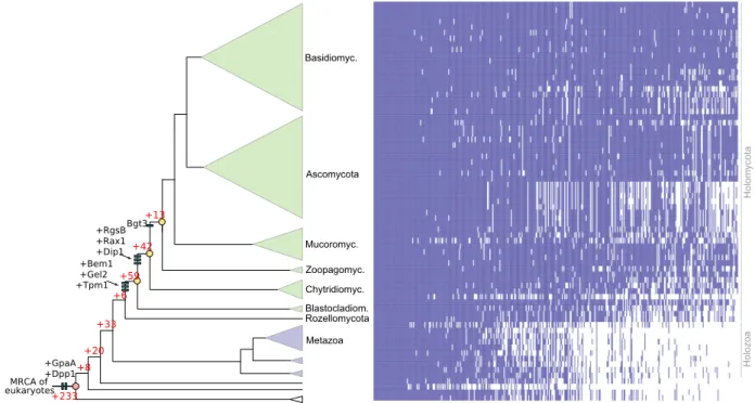
Supplementary Data 8), 114 of which originated in BCZ nodes, while the others showed duplication rates that exceeded the expectation derived from genome-wide figures of gene duplica- tion (Fig. 5). The conservation of these putative hyphal multicellularity-related gene families across fungi is shown on Fig. 5. These families included several known morphogenetic families (e.g. Bgt3, RgsB and Gel2 of A. fumigatus, Bem1 and Rax1 of S. cerevisiae), genes involved in actin cytoskeleton and cell wall assembly, mating, pheromone response (GpaA of A.
fumigatus), sporulation and transport, among others (Supple- mentary Data 8). Several of the identified families contain genes with reported growth defects in A. fumigatus or S. cerevisiae, indicating that our searches recovered genes relevant for hyphal MC. For example, Rax proteins are major regulators of cellular morphogenesis and are involved in bud site selection in budding yeasts74, polarized growth in S. pombe75 and polarity main- tenance in filamentous fungi76. We further detected a fungal- specific cluster of tropomyosins (TPM1 in S. cerevisiae), which originated in the MRCA of Blastocladiomycota and other fungi and is involved in polarized growth and the stabilization of actin microfilaments. The family containingS. pombeDip1 homologs (Afu6g12370 in A. fumigatus) emerged in the node uniting Chytridiomycota with higher fungi and contains a single gene per Table 1 List of the 17 gene families whose emergence coincides with the evolution of hyphae
Emergence of gene family A. fumigatus
ortholog
S.cerevisiae ortholog
Functional category and putative function
mrca of Dikarya, Mucoromycota, Zoopagomycota, Chytridiomycota, Blastocladiomycota
Afu7g03870 PAN1 Actin cytoskeleton: endocytic adaptor that triggers hyphae- specific recruitment of the Arp2/3 complex to sites of endocytosis, for the recycling of excess membrane in the subapical region during hyphal growth41
crh3 UTR2 Cell wall biogenesis: chitin transglycosylase, localized to sites of polarized growth, functions in the transfer of chitin to beta(1–6) and beta(1–3) glucans
gel7 GAS1 Cell wall biogenesis: beta(1–3) glucanosyltransferase, involved in cell wall remodeling during fungal germination or branching Afu6g04940 BNR1 Polarity establishment: mediates actin cable assembly in
filamentous fungi and has a role in diverse morphogenetic processes72
Afu4g04120 BEM1 Polarity establishment: actin cytoskeleton reorganizing factor73,74
stuA PHD1 Transcriptional regulation: mediates yeast–filament transition in S. cerevisiae, developmental modifier inA. fumigatus, that spatially and temporally regulates the central transcription factor cascade
medA NA Transcriptional regulation
mrca of Dikarya, Mucoromycota, Zoopagomycota, Chytridiomycota
Afu6g07910 SLM1 Actin cytoskeleton: effector of PtdIns(4,5)P2, essential for cell growth and actin cytoskeleton polarization
Afu8g04520 SLA1 Actin cytoskeleton: actin cytoskeleton-regulatory complex protein, localized to the actin patches that form the sites of endocytosis
Afu4g06130 WHI2 Cell cycle regulation: required for cell cycle regulation and stimulatesfilamentous growth
Afu4g00620 DFG5 Cell wall biogenesis: mannosidase, involved in bud formation and filamentous growth
Afu8g02320 NA Cell wall biogenesis: ortholog ofN. crassacps1 polysaccharide synthase, functions in cell wall biosynthesis
chsD NA Cell wall biogenesis: class VI chitin synthase, role in chitin biosynthesis
rgsB RAX1 Polarity establishment: bipolar budding inS. cerevisiae75 Afu2g08800 SSY1 Signaling: component of the SPS plasma membrane amino acid
sensor system
ricA NA Signaling: GDP/GTP exchange factor for G proteins, role in regulating fungal development
mrca of Dikarya, Mucoromycota, Zoopagomycota
kre6 KRE6 Cell wall biogenesis: role in beta(1–6) glucan biosynthesis
species afterward, except an expansion in WGD Mucoromycota77 and losses in the Saccharomycotina. In S. pombe, Dip1 activates the Arp2/3 complex without preexisting actin cables78and thus regulates the actin cytoskeleton through a mechanism that seems to be specific to multicellular fungi. Finally, we detected the family containing S. cerevisiae Dpp1 homologs, which regulate morphogenetic transitions in dimorphic fungi through the synthesis of the fungal signal molecule farnesol79, which prompts us to speculate that it might have contributed to the elaboration of farnesol-based communication in fungi. Collectively, the origin of these families in BCZ nodes makes them candidate key con- tributors to the evolution of hyphal MC.
Because the emergence of hyphal multicellularity overlaps with that of other fungal traits, it is challenging to unequivocally separate signals conferred by the emergence of these traits from those by hyphae. It is conceivable that a portion of the 414 gene families were detected because of signals conferred by phylogen- etically co-distributed traits, not necessarily multicellularity itself (see Beaulieu 201680 for a conceptually analogous problem in taxonomic diversification). One such trait could be osmotrophy, feeding by the absorption of soluble’public goods’generated by the activity of secreted extracellular enzymes81. We detected 20 gene families that showed strong correlation with hyphal MC and were annotated as various transporters; such families could conceivably be related to osmotrophy. Further, there were 84 families that are functionally uncharacterized and thus it is impossible to speculate about their role in hyphal MC. These
families suggest that there are many fungal genes that evolved in concert with hyphal MC and await functional characterization to understand their roles.
Yeasts retain genes required for hypha morphogenesis. Yeasts are secondarily simplified organisms with reduced ability to form hyphae, that spend most of their life cycle as unicells16,22,46,82. Our ancestral character state reconstructions imply that yeasts derived from filamentous ancestors (Fig. 1a), and thus they represent a classic example of reduction in complexity. They were hypothesized to have lost MC83, even though rudimentary forms of hyphal growth (termed pseudohyphae) exist in most species.
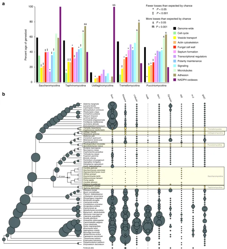
We scrutinized the fate of MC-related genes in five pre- dominantly yeast-like lineages82(Fig.6). Because yeast genomes have undergone extreme streamlining during evolution, we evaluated gene loss among hypha morphogenesis genes in com- parison to genome-widefigures of gene loss.
Yeast species generally have fewer hypha morphogenesis genes and reconstructions indicate more losses than duplications along branches of yeast ancestors (Fig.6). However, when we corrected for genome-wide reductions in gene number, we found that comparatively fewer hypha morphogenesis genes (35–46%) were lost than genes with other functions (46–59%, Fig. 6a, Supple- mentary Data 9). Gene loss in yeast clades is significantly depleted (P< 0.05, Fisher’s exact test) in most groups of hypha morphogenesis genes compared to genome-wide expectations.
Dictyostelium discoideum Polysphondylium pallidum Entamoeba histolytica Thecamonas trahens Capsaspora owczarzaki Sphaeroforma arctica Monosiga brevicollis Salpingoeca rosetta Mnemiopsis ledyii Hydra magnipapillata Ciona intestinalis Caenorhabditis elegans Drosophila melanogaster Rozella allomycis Catenaria anguillulae Allomyces macrogynus Batrachoch. dendrobatidis Spizellomyces punctatus Orpinomyces sp.
Gonapodya prolifera Conidiobolus coronatus Coemansia erecta Mortierella elongata Rhizophagus irregularis Umbelopsis ramanniana Phycom. blakesleeanus Mucor circinelloides Rhizopus oryzae Taphrina deformans Schizosacch. pombe Dekkera bruxellensis Candida albicans Pichia stipitis Debaryomyces hanseni Kluyveromyces lactis Ashbya gossypi Zygosaccharomyces rouxii Saccharomyces cerevisiae Candida glabrata Cochl. heterostrophus Histoplasma capsulatum Aspergillus fumigatus Pennicilium chrysogenum Botrytis cinerea Fusarium graminis Trichoderma reesei Cryphonectria parasitica Neurospora crassa Sordaria macrospora Sporobolomyces roseus Rhodosporidium toruloides Puccinia graminis Melampsora laricis-pop.
Cronartium quercuum Wallemia sebi Malassezia globosa Ustilago maydis Cryptococcus neoformans Tremella mesenterica Botryobasidium botryosum Auricularia delicata Fomitiporia mediteranea Punctularia strigosozonata Lentinus tigrinus Stereum hirsutum Coniophora puteana Plicaturopsis crispa Schizophyllum commune Coprinopsis cinerea Laccaria bicolor Galerina marginata
DOCK ELMO
Sum WASP WAVE WASH
Zoopagomycota Mucoromycota
SaccharomycotinaPezizomycotina HolomycotaHolozoa
Agaricomycotina
Taphrinomycotina Ustilaginomycotina Tremellomycotina
Pucciniomycotina
MetazoaBasidiomycotaAscomycota
Blastocladiomycota Chytridiomycota
Rozellomycota
Fonticula alba
Fig. 4Evolutionary history of phagocytosis-related gene families. Several phagocytotic gene families retained infilamentous fungi (DOCK, ELMO, WASP).
WAVE family retained only in early fungi (Blastocladiomycota and Chytridiomycota), WASH family is represented only in non-fungal eukaryotes. Bubble size is proportional to ancestral and extant gene copy number. Copy numbers offilamentous fungi are labeled with yellow
We recovered only six cases where significantly more MC-related genes were lost than expected (P< 0.05, Fisher’s exact test, Supplementary Data 9). These included proteins related to adhesion and microtubule-based transport, suggesting that these functions are generally dispensable for yeast clades. Contractions (e.g. gamma-tubulin complex, kinesins, dynein heavy chain (nudA)) or complete losses (e.g., dynactin, gcpC, and the dynactin linking protein ro-284) of gene families in microtubule-based transport are particularly interesting from the perspective of long-range transport of vesicles and nuclei along hyphae. The budding and fission yeast lineages show the most losses in these genes, consistent with the strongest reduction of hyphal growth abilities in these clades. Losses of NADPH oxidases were observed in all yeast clades, with complete loss of the family in the Saccharomycotina, Ustilaginomycotina, in S.
pombe and C. neoformans (Supplementary Fig. 8), as reported previously85.
Collectively, these analyses suggest that hypha morphogenesis genes are, in general, less dispensable for yeasts than genes with other functions. This agrees with most yeast-like fungi being able to switch to hyphal or pseudohyphal growth under certain conditions. The inferred gene losses, nevertheless, do indicate reductions in hyphal growth, but this reduction is smaller than that of other functions in the genome. This, in turn means, that the ability for multicellular growth is among the functions preferentially retained by yeast-like fungi.
Discussion
In this study, we analyzed the genetic underpinnings of the evolution of fungal hyphae, the most enigmatic of fungal struc- tures, with a unique multicellular organization but a poorly understood evolutionary origin. Our analyses suggest that hyphae evolved in early fungi (the split of Blastocladio-, Chytridio- and Zoopagomycota, termed BCZ nodes), consistent with the
previous studies15. To understand how the underlying genetics evolved, we reconstructed the evolution of 362 hypha morpho- genesis gene families and predicted a link to hyphae for another 414 families.
The evolutionary picture developed from these analyses has several conspicuous features. Most families were conserved across all sampled eukaryotes with few or no duplications at the origin of multicellular fungi. A second category of gene families show a deep eukaryotic origin and duplications coincident with the evolution of hyphae (e.g., cell wall biogenesis and transcriptional regulation-related genes). However, none of these families had a significantly elevated duplication rate in BCZ nodes, indicating limited innovation via gene duplication. A third category con- sisted of gene families whose origin map to BCZ nodes. These could have evolved de novo or they could have diverged so much that similarity to homologous non-fungal sequences cannot be detected. We find support for both of these possibilities. For example, the MedA or APSES families contain fungal-specific protein domains; these have conceivably evolved in early fungi and represent fungal-specific innovations underlying hyphal growth. On the other hand, the detected formin and RGS families contained only fungal genes, but their characteristic Interpro domains occur outside of fungi too, possibly reflecting common ancestry, with evidence for homology blurred by sequence divergence.
Beyond gene family events, our analyses revealed that changes in the length of genes/introns and domain architectures of MC- related genes in multicellular vs unicellular fungi also correlate with the emergence of hyphae. There is also evidence for changes to amino acid sequence, for example in fungal kinesins that are 2× more processive than other eukaryotic kinesins86, conceivably as a result of selection for efficient long-range transport along the hyphal axis.
Co-option and exaptation may have been the most important source of genes for hyphal MC, followed by gene duplications,
Chytridiomyc.
Zoopagomyc.
Mucoromyc.
HolomycotaHolozoa
Blastocladiom.
Metazoa Basidiomyc.
Ascomycota
Rozellomycota Bgt3
MRCA of eukaryotes
+20 +33
+6 +59
+42 +13
+233 +GpaA+8 +Dpp1
+Bem1 +Gel2 +Tpm1
+RgsB +Rax1 +Dip1
Fig. 5Origin of 414 gene families potentially related to the evolution of hyphal MC, identified by ANOVA (P< 0.05). 114 families originated in BCZ nodes (shown in yellow), including known morphogenesis-related proteins (e.g. Bgt3, RgsB, Gel2 ofA. fumigatus, Rax1, Bem1, Tpm1, and Dpp1 fromS. cerevisiae, Dip1 fromS. pombe) labeled as blue bars. Red numbers represent the number of gene families originated at the respective branches. Heatmap next to the tree shows the conservation of the identified 414 gene families across fungi. Blue and white colors indicate that the gene family is present or absent in the genome, respectively
structural changes to genes and de novo gene family birth.
Thesefindings mirror patterns of the evolution of multicellular animals and plants (e.g. refs. 6,87–89) that gave rise to the hypothesis that, in terms of genetic novelty, transitions to multicellularity represent a minor rather than a major evolu- tionary step90, an idea that finds support in the observations made here on fungi.
Our observations highlight how a wealth of small genetic changes may synergistically lead to a key evolutionary innovation, such as fungal hyphae. This’tinkering’48process is consistent with the stepwise, gradual evolution of hyphae and could explain why transitional hypha-like forms exist in early-diverging fungi (Blastocladiomycota, Chytridiomycota)15. The dynamic cytoske- letal and endomembrane systems have been suggested to underlie
Kinesins γ-tubulin nudA bim1 Dynactin rbl2 ro-2
Dictyostelium discoideum Polysphondylium pallidum Entamoeba histolytica Thecamonas trahens Capsaspora owczarzaki Sphaeroforma arctica Monosiga brevicollis Salpingoeca rosetta Mnemiopsis ledyii Hydra magnipapillata Ciona intestinalis Caenorhabditis elegans Drosophila melanogaster Rozella allomycis Catenaria anguillulae Allomyces macrogynus Batrachoch. dendrobatidis Spizellomyces punctatus Orpinomyces sp.
Gonapodya prolifera Conidiobolus coronatus Coemansia erecta Mortierella elongata Rhizophagus irregularis Umbelopsis ramanniana Phycom. blakesleeanus Mucor circinelloides Rhizopus oryzae Taphrina deformans Schizosacch. pombe Dekkera bruxellensis Candida albicans Pichia stipitis Debaryomyces hanseni Kluyveromyces lactis Ashbya gossypi Zygosaccharomyces rouxii Saccharomyces cerevisiae Candida glabrata Cochl. heterostrophus Histoplasma capsulatum Aspergillus fumigatus Pennicilium chrysogenum Botrytis cinerea Fusarium graminis Trichoderma reesei Cryphonectria parasitica Neurospora crassa Sordaria macrospora Sporobolomyces roseus Rhodosporidium toruloides Puccinia graminis Melampsora laricis-pop.
Cronartium quercuum Wallemia sebi Malassezia globosa Ustilago maydis Cryptococcus neoformans Tremella mesenterica Botryobasidium botryosum Auricularia delicata Fomitiporia mediteranea Punctularia strigosozonata Lentinus tigrinus Stereum hirsutum Coniophora puteana Plicaturopsis crispa Schizophyllum commune Coprinopsis cinerea Laccaria bicolor Galerina marginata
Sum
0 20 40 60 80 100
Saccharomycotina Taphrinomycotina Ustilaginomycotina Tremellomycotina Pucciniomycotina
Genome-wide
Vesicle transport Actin cytoskeleton Fungal cell wall Septum formation Transcriptional regulators
Signaling Microtubules Adhesion NADPH oxidases
Percent age of geneslost
*
* *
****
**
** **
**
**
**
*
**
** **
**
**
*
*
* *
** **
** *
&
&
&
&
&
&&
*
&
&&
**
P < 0.05 P < 0.001
More losses than expected by chance Fewer losses than expected by chance
a
b gcpC
46 losses
71
25
P < 0.05 P < 0.001
Cell cycle
Polarity maintenance
&&
Saccharomycotina
Taphrinomycotina Ustilaginomycotina Tremellomycetes
Pucciniomycotina
Fonticula alba
Fig. 6Secondarily simplified yeast-like fungi retain genes for hyphal MC.athe percentages of lost genes in main morphogenesis-related categories.
Percentages were calculated relative to ancestral copy numbers inferred in the node preceding the origin of 5 yeast-like clades (Saccharomycotina, Taphrinomycotina, Pucciniomycotina, Ustilaginomycotina, and Tremellomycotina). Significance of the enrichment of gene losses in each category relative to genome-widefigures of gene loss were determined by Fisher’s exact test and is shown above bars. (b) ancestral gene copy number reconstruction of microtubule-based transport genes along the fungal phylogeny. Secondarily simplified (yeast-like) clades are highlighted in yellow. Bubble size proportional to reconstructed ancestral and extant gene copy number across 19 gene families. Copy number distribution of each gene family is shown right to the tree