Regular Article
Ambivalent roles of carboxypeptidase B in the lytic susceptibility of fi brin
András Kovács
a, László Szabó
b, Colin Longstaff
c, Kiril Tenekedjiev
d, Raymund Machovich
a, Krasimir Kolev
a,⁎
aDepartment of Medical Biochemistry, Semmelweis University, Budapest, Hungary
bInstitute of Materials and Environmental Chemistry, Research Centre for Natural Sciences, Hungarian Academy of Sciences, Budapest, Hungary
cBiotherapeutics, Haemostasis Section, National Institute for Biological Standards and Control, South Mimms, Potters Bar, UK
dIT Department, N.Y. Vaptsarov Naval Academy, Varna, Bulgaria
a b s t r a c t a r t i c l e i n f o
Article history:
Received 29 July 2013
Received in revised form 10 September 2013 Accepted 17 September 2013
Available online 21 September 2013
Keywords:
Carboxypeptidase Fibrin
Fibrinolysis Plasmin tPA
Background:Removal of C-terminal lysine residues that are continuously exposed in lysingfibrin is an established anti-fibrinolytic mechanism dependent on the plasma carboxypeptidase TAFIa, which also removes arginines that are exposed at the time offibrinogen clotting by thrombin.
Objective:To evaluate the impact of alterations infibrin structure mediated by constitutive carboxypeptidase activity on the function offibrin as a template for tissue plasminogen activator-(tPA) induced plasminogen acti- vation and its susceptibility to digestion by plasmin.
Methods and results:We used the stable carboxypeptidase B (CPB), which shows the same substrate specificity as TAFIa. If 1.5–6μMfibrinogen was clotted in the presence of 8 U/mL CPB, a denserfibrin network was formed with thinnerfibers (the medianfiber diameter decreased from 138–144 nm to 89–109 nm as established with scanning electron microscopy). If clotting was initiated in the presence of 5–10μM arginine, a similar decrease infiber diameter (82 -95 nm) was measured. Thefine structure of arginine-treatedfibrin enhanced plas- minogen activation by tPA, but slowed down lysis monitored usingfluorescent tPA and confocal laser microscopy.
However, if lysis was initiated with plasmin in CPB-treatedfibrin, the rate of dissolution increased to a degree cor- responding to doubling of the plasmin concentration.
Conclusion:The present data evidence that CPB activity generatesfine-meshfibrin which is more difficult to lyse by tPA, but conversely, CPB and plasmin together can stimulatefibrinolysis, possibly by enhancing plasmin diffusion.
© 2013 The Authors. Published by Elsevier Ltd.
Introduction
Binding of tissue-type plasminogen activator (tPA) and plasminogen tofibrin is a prerequisite for efficientfibrinolysis (reviewed in[1]), in the course of which the generated plasmin provides a positive feed- back loop through exposure of new carboxyl terminal lysines that pro- motefibrinolysis primarily through plasminogen and plasmin binding [2]. The amplifying effect of C-terminal lysines on plasminogen activa- tion and the protection of the bound plasmin against its major plasma inhibitor α2-plasmin inhibitor is counterbalanced by the action of thrombin activatablefibrinolysis inhibitor (TAFI, or carboxypeptidase U), an exopeptidase that removes basic amino acids (arginine and
lysine) from the C-terminal of peptides (reviewed in[3]). TAFI is present in blood plasma in zymogenic form and it is activated by thrombin- thrombomodulin complex to TAFIa, but its activity decays with a half- life of several minutes[4]. A distinct, constitutively active enzyme, car- boxypeptidase N (CPN) is also present in plasma[5,6]. It bindsfibrin and can be detected in the structure of plasma clots[7,8]. Despite the identical primary specificity of TAFIa and CPN the anti-fibrinolytic ac- tion of CPN is only a fraction of that of TAFIa even after an activating cleavage by plasmin[9]. The lysis of whole blood clots immersed in tPA is surprisingly insensitive to the action of CPN; no impairment of lysis is observed under conditions when CPN removes lysyl residues fromfibrin at a rate corresponding to 50% of that by TAFIa[10]. The background of the differential anti-fibrinolytic potential of CPN and TAFI is still not clarified.
Another determinant offibrinolytic efficiency is fibrin structure, which as a cofactor for tPA, regulates plasminogen activation, and sus- ceptibility to plasmin digestion (reviewed in[11]). The initial structure and subsequent rearrangements offibrin during lysis mean that binding events, plasminogen activation andfibrin digestion can be affected in complex ways, sometimes in opposite directions[12]. With this in mind, a question arises, can carboxypeptidase activity expressed during clot formation and dissolution induce structural changes that modulate Abbreviations:CPB, carboxypeptidase B; CPN, carboxypeptidase N; TAFI, thrombin
activatablefibrinolysis inhibitor; tPA, tissue-type plasminogen activator.
⁎ Corresponding author at: Semmelweis University, Department of Medical Biochemistry, 1094 Tűzoltó u. 37-47., Budapest, Hungary. Tel.: +36 1 4591500/60035;
fax: +36 1 267 0031.
E-mail address:Krasimir.Kolev@eok.sote.hu(K. Kolev).
0049-3848 © 2013 The Authors. Published by Elsevier Ltd.
http://dx.doi.org/10.1016/j.thromres.2013.09.017
Contents lists available atScienceDirect
Thrombosis Research
j o u r n a l h o m e p a g e : w w w . e l s e v i e r . c o m / l o c a t e / t h r o m r e s
Open access under CC BY license.
Open access under CC BY license.
fibrinolysis? The timeliness of this question also stems from rather contradictory data on the association of TAFI and thrombotic events reported in epidemiological studies. TAFI levels in circulation are regu- lated by known gene polymorphisms, but the connection with coronary heart disease[13]or stroke[14]is less clear. In the absence of any mech- anistic evidence for antithrombotic effects of TAFI the controversy that both high and low thrombotic risk can be accompanied by elevated TAFI levels in plasma[15–19]is currently explained by the different methods used for determination of TAFIa activity or TAFI antigen and the different sensitivity of the detection assays for TAFI isoforms[20].
However, additional levels of complexity may still remain uncovered as suggested by the fact that some aspects of TAFIa mechanism of action establishedin vitroremain a puzzle. For example, iffibrin con- tainingα2-plasmin inhibitor or plasma clots are supplemented with in- creasing concentrations of TAFIa, a threshold concentration can be reached beyond which TAFIa is profibrinolytic[21]. One hypothesis for this phenomenon is the removal of lysine452ofα2-plasmin inhibitor by carboxypeptidases[21], but this is difficult to reconcile with thefind- ings that the C-terminal lysine in the inhibitor is not essential for its in- teraction with plasmin[22]. The completely normal phenotype of TAFI knock-out mice (including hemostasis at basal state or if challenged by a variety of prothrombotic stimuli)[23]also raises the possibility for subtle TAFI effectsin vivobeyond the known mechanism of action.
Thus, the details of TAFI function still require further elaboration despite its well-documented anti-fibrinolytic actionin vitro. The present study addresses the modulation of fibrinolysis related to fibrin structure which is altered as a consequence of basic carboxypeptidase activity.
Materials and methods
Plasminogen-depleted humanfibrinogen, streptokinase and porcine carboxypeptidase B (CPB) were from Calbiochem (La Jolla, CA). The chromogenic substrate for plasmin, Spectrozyme-PL (H-D-norleucyl- hexahydrotyrosyl-lysine-p-nitroanilide) was from American Diagnostica (Pfungstadt, Germany) and tPA was from Boehringer Ingelheim (Germany). Bovine thrombin was purchased from Serva (Heidelberg, Germany) and further purified by ion-exchange chromatography on sulfopropyl-Sephadex yielding a preparation with specific activity of 2100 IU/mg[24]and 1 IU/mL was considered equivalent to approximate- ly 10.7 nM by active site titration[25]. Alexa Fluor® 546-conjugated fibrinogen was the product of Invitrogen Life Technologies, Budapest, Hungary. Human plasminogen was purified by affinity chromatography on Lysine-Sepharose from citrated human plasma provided by the Hungarian Blood Supply Service[26]. The generation of plasmin from the zymogen and determination of its active concentration were performed as previously described[27]. Blood was collected from healthy volunteers with venipuncture in 10 mM trisodium-citrate (final concen- tration) and following 10-min centrifugation at 2,000gthe top ¾ of the plasma layer was used for the measurements within 4 h.
Turbidimetricfibrinolytic assays
In 96-well microtiter plates, 6μMfibrinogen in 10 mM HEPES buffer pH 7.4 containing 150 mM NaCl and arginine or CPB at various concen- trations were mixed with 20 nM thrombin in a total volume of 100μl. In the assays when lysis was initiated by surface tPA,fibrinogen also contained 1μM plasminogen and following 30 min clotting, tPA was applied to the surface of clots at 15 nM. The concentration of CPB that produced maximum effect in this assay (8 U/mL) was applied in the rest of the experiments in this study. In the assays when lysis was initi- ated by tPA dispersed in the clot,fibrinogen contained 0.25μM plasmin- ogen and 0.1 nM tPA was added together with thrombin. In the assays when lysis was initiated by plasmin its concentrations were chosen to yield complete dissolution within 5 h; in the range 2-10 nM for plasmin uniformly dispersed in the clot and 0.5–2μM for plasmin applied to the clot surface as described previously [28,29]. Clot formation and
dissolution was followed by measuring the light absorbance at 340 nm at 37 °C with a Zenyth 200rt microplate spectrophotometer (Anthos Labtec Instruments GmbH, Salzburg, Austria). For adequate comparison of lytic rates from measurements, in which different maxi- mum turbidity values were reached despite the identical quantities of fibrin, the absorbance values were evaluated in normalized form[30].
The time needed to reduce the turbidity of the clot to a given fraction of the maximal value (t0.5to reach 0.5Amax,t0.1reach 0.1Amax) was used as a quantitative parameter of fibrinolytic activity. In certain cases plasma containing various concentrations of added arginine and CPB was clotted with 15 nM thrombin and 12.5 mM CaCl2. If plasma clot dissolution was mediated by tPA dispersed in the clot, tPA was added at 0.8 nM prior clotting, whereas the concentration of tPA ap- plied to the surface of plasma clots was 30 nM.
Scanning electron microscopic (SEM) studies
Fibrin clots of 50μl volume were prepared in duplicate:fibrinogen (at concentration in the range 1.5–6μM) in 10 mM HEPES buffer pH 7.4 containing 150 mM NaCl and the additives (arginine or CPB) was clotted with 20 nM thrombin at 37 °C for 30 min. Thereafter clots were placed into 10 mL 100 mM Na-cacodylate pH 7.2 buffer for 24 h at 4 °C. Following repeated washes with the same buffer, samples werefixed in 1%(v/v) glutaraldehyde for 16 h. Thefixed samples were dehydrated in a series of ethanol dilutions (20–96%(v/v)), 1:1 mixture of 96%(v/v) ethanol/acetone and pure acetone followed by critical point drying with CO2in E3000 Critical Point Drying Apparatus (Quorum Technologies, Newhaven, UK). The specimens were mounted on adhe- sive carbon discs, sputter coated with gold in SC7620 Sputter Coater (Quorum Technologies, Newhaven, UK) and images were taken with scanning electron microscope EVO40 (Carl Zeiss GmbH, Oberkochen, Germany). The SEM images were analyzed to determine the diameter of the fibrin fibers using self-designed program functions running under the Image Processing Toolbox v. 8.2 of Matlab 8.1.0.604 (R2013a) (The Mathworks, Natick, MA) as previously described[12,31].
Plasminogen activation assay
In 96-well microtiter plates, 6μMfibrinogen in 10 mM HEPES buffer pH 7.4 containing 150 mM NaCl, 0.5μM plasminogen and additives (ar- ginine or CPB) was clotted with 25 nM thrombin in a volume of 80μl.
After 30 min at 37 °C 60μl of 15 nM tPA and 0.6 mM Spectrozyme-PL in 10 mM HEPES, 150 mM NaCl pH 7.4 were placed on the surface of the clot. The forming plasmin generated p-nitroaniline, the absorbance of which was continuously recorded at 405 nm (A405) with Zenith 200rt spectrophotometer. The measured values were plotted versus time squared (t2) yielding a linear relationship according to the equation ΔA405= 0.5εk1kcat[tPA] t2[32], whereε= 12.6 mM-1cm-1is the ex- tinction coefficient of p-nitroaniline[33],k1= 350 min-1is the turn- over number of plasmin on Spectrozyme-PL[33],kcatand[tPA]are the catalytic constant for plasminogen activation and the concentration of tPA in the reactive layer on the surface offibrin, respectively[34]. The termVapp= kcat[tPA]is equivalent to the apparent maximal rate of plas- minogen activation in the reactive layer offibrin and was determined from linear regression according to the abovementioned equation (Curvefitting toolbox v. 3.3.1 of Matlab 2013a).
Confocal microscopic imaging
Fibrin clots were prepared from 6μMfibrinogen, 2% of which was Alexa Fluor® 546-labelled in 10 mM HEPES buffer pH 7.4 containing 150 mM NaCl, 1.5μM plasminogen and the tested additives with 15 nM thrombin for 30 min at room temperature in sterile, uncoated IBIDI VI 0.4μ-slides (Ibidi GmbH, Martinsried, Germany). Thereafter 50 nM tPA-YFP (tPA with Yellow Fluorescent Protein fused to its C- terminal expressed using pFastBac-tPA as previously described)[12]
was added to the edge of the clot and thefluorescence (excitation wave- length 488 nm, emission wavelength 525 nm for tPA-YFP detection and excitation wavelength 543 nm, emission wavelength 575 nm for Alexa546-fibrinogen detection) was monitored with Confocal Laser Scanning System LSM710 (Carl Zeiss GmbH, Jena, Germany) taking se- quential images of thefluid-fibrin interface at a distance of approxi- mately 50μm from the chamber surface with identical exposures and laser intensities using a Plan-Neofluar 20x/0.5 objective. It should be noted that the 10–20μmfluorescent aggregates present in the com- mercial Alexa Fluor® 546-labelled fibrinogen were not centrifuged before clotting as done in our earlier work[35], but these were pre- served and used as position markers in thefibrin.
Statistical analysis
The distribution of the data onfiber diameter was analyzed according to an algorithm used previously[12,31]: theoretical distributions were fitted to the empirical data sets and compared using Kuiper test and Monte Carlo simulation procedures. The statistical evaluation of other experimental measurements in this report was performed with Kolmogorov-Smirnov test (Statistics Toolbox 8.2 of Matlab R2013a).
Results
Structural modifications offibrin related to CPB activity or presence of arginine
We have previously shown[12], when CPB was used as a stable an- alogue of TAFIa to evaluate the impact of removal of carboxyl terminal lysines on the kinetics offibrinolysis, that the presence of CPB duringfi- brinogen clotting modifies the turbidity of the clots before initiation of lysis, which suggested changes in the structure offibrin. SEM imaging provided direct evidence for the alteration of thefibrin structure related to the action of CPB (Fig. 1A). Morphometric analysis of the SEM images (Fig. 1B) revealed that thefiber diameter decreased by 21–25% in CPB- treated clots prepared fromfibrinogen at physiologically relevant con- centration (1.5–6μM). Because the nativefibrinogen molecule does not contain any substrate (C-terminal lysine or arginine) for CPB, the single target of CPB action in this purified system could be the arginine residues in thefibrinopeptides newly cleaved by thrombin in the pro- cess of clotting. If all new C-terminal arginines were released from the 4fibrinopeptides derived from eachfibrinogen molecule, an increment of 25–50μM would be expected in the local concentration of arginine in clots prepared from 2–4 mg/mLfibrinogen (a prediction supported by direct measurements of arginine release by TAFIa infibrin: 10μM in 5 min after clotting[36]). As evidenced by the morphometric data (Fig. 1C), addition of arginine at 5–20% of this maximal concentration caused changes in the structure offibrin comparable to those induced by the action of CPB (Fig. 1B). Considering the normal plasma level of ar- ginine of about 100μM[37]and these subtle structural alterations infi- brin, inclusion of micromolar arginine appears to be essential for appropriate modelling of the physiological conditions when fibrin structure-dependent processes are evaluated within vitroassays.
Kinetics of plasminogen activation andfibrin dissolution
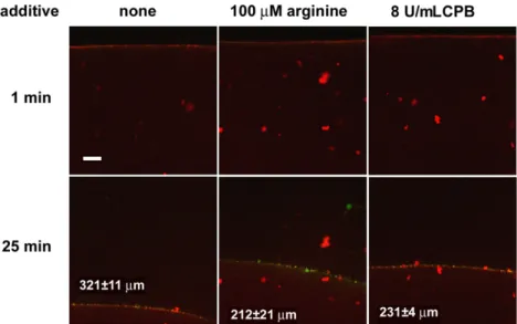
On a microscopic scale (Fig. 2), tPA-induced lysis offibrin modified by CPB was significantly slowed down with a granular pattern of accumula- tion of tPA on the surface of the clot similar to that of nativefibrin. On a macroscopic scale of lytic kinetics, CPB had a maximal inhibitory effect at 8 U/mL in the assay when tPA was added to the surface of pre- formed clots containing plasminogen, as shown inFig. 3. A similar level of inhibition of lysis was also seen in clots formed in the presence of argi- nine at concentration as low as 2μM. Although with either CPB or argi- nine the acceleration seen at later stages of lysis in purefibrin was missing and despite the identical time to complete dissolution, the lysis
kinetics were somewhat different with CPB or arginine, suggesting a dif- ferent mechanism of action. To address this difference two assays were used; one that monitored solely the generation of plasmin (Fig. 4) and a second one that bypassed the stage of plasminogen activation (Fig. 5).
The effects of CPB and arginine on plasminogen activation by tPA on the surface of fibrin was studied in isolation using the substrate Spectrozyme-PL to follow plasmin generation. As expected, plasmin generation was inhibited in the presence of CPB, presumably resulting from removal of newly exposed C-terminal lysine binding sites in fibrin (Fig. 4). In contrast however,fibrin formed in the presence of arginine proved to be a better template for plasminogen activation than the nativefibrin structure (Fig. 4), despite the overall inhibiting effect of arginine on tPA-inducedfibrin degradation seen inFig. 3. It is noteworthy that arginine at identical concentrations does not affect plasminogen activation by tPA infibrin-free systems (data not shown).
When plasmin was added to the surface offibrin, slower lysis was ob- served similarly to the tPA-induced lysis, if clots were formed in the pres- ence of arginine (Fig. 5A). Quite unexpectedly, CPB renderedfibrin more susceptible to lysis by plasmin added to the surface of the clot (Fig. 5B).
The complete lysis of CPB-treatedfibrin was faster (Table 1) and non- treated fibrin was dissolved by 2μM plasmin at approximately the same rate as CPB-modifiedfibrin by 1μM plasmin (no significant differ- ence between the values for these two states in the 4th and 5th column ofTable 1). However, if plasmin was homogeneously dispersed within the clot, neither inhibitory effect of arginine, nor stimulatory effect of CPB could be observed (data not shown). The discordant effects of the modulators in the two assay formats indicate that at least in part their ef- fects arise from the variations in the penetration of plasmin infibrin of dif- ferent structure.
Fibrinolysis in plasma environment
In line with the effects observed with purifiedfibrinolytic compo- nents (Fig. 3), arginine and CPB retarded the dissolution of plasma clots by tPA (Fig. 6). However, CPB inhibited plasma clot lysis at lower concentrations than used in the purified systems above. If tPA was uni- formly dispersed in the plasma prior clotting, 0.1 U/mL CPB prolonged the half lysis time more than two-fold, whereas the effect of arginine at physiologically relevant concentration (100μM) was minimal (Fig. 6B). If tPA was applied to the surface of pre-formed clots, the effect of this arginine was equivalent to 0.4 U/mL CPB (Fig. 6A).
Discussion
Since the discovery of TAFI[38]its anti-fibrinolytic action has been largely attributed to the removal of plasmin-generated C-terminal lysines in thefibrin matrix, but it has been difficult to show convincingly that TAFI levels affect myocardial infarction or stroke outcomes at a population level or to explain the different anti-fibrinolytic impact of TAFI and plasma CPN. In the present study we used CPB, a homologous, but stable carboxypeptidase with specificity for basic amino acids and addressed alternative or additional factors that could modulate its anti-fibrinolytic function. The application of CPB overcomes the short half-life of TAFIa of approximately 10 min[4], has the technical advantage that it does not inactivate plasmin in contrast to TAFIa[36]
and models also the constitutivein vivoactivity of CPN at the stage of fibrin clot formation. Our work identified two consequences of CPB activity infibrin that antagonize the CPB-dependent blockade of the positive feed-back loop in fibrinolysis based on C-terminal lysine exposure: 1) enhancement of plasmin activity when thefluid-borne enzyme attacks the surface of pre-formed clots, probably due to improved diffusion; and 2) fibrin structure-related acceleration of plasminogen activation.
Fig. 1. Modification offibrin structure by arginine and carboxypeptidase B.Fibrin clots were prepared in duplicate fromfibrinogen at various concentrations and followingfixation and drying images were taken with a scanning electron microscope (SEM). A: Representative images of nativefibrin at 6μM (left panel) andfibrin at the same concentration treated with 8 U/mL CPB. Scale bar = 1μm. B: Effect of CPB on the ultrastructure offibrin. Morphometric analysis offibrin was performed on SEM images illustrated in panel A. The diameter of 300fibres was measured and their empiric (black histograms) as well as best-fitted theoretical (gray curves) probability density function (PDF) was determined. Median values and interquartile range (in brackets) are shown for the theoretical distributions of the diameter values. The concentrations offibrinogen (Fg) indicated on the left side refer to both non-treated and CPB-treatedfibrin.
C: Effect of arginine on thefibrin structure. The same morphometric analysis was performed onfibrin prepared from 6μMfibrinogen containing the concentrations of arginine indicated.
Degradation offibrin by plasmin at the surface of the clot
A rather unexpected observation arose from the study of surface ap- plication of plasmin tofibrin containing CPB (Fig. 5B,Table 1), where in- creased potency of plasmin was noted. Under the conditions shown, CPB treatment was equivalent to doubling the concentration of plasmin.
This was in contrast to the lack of CPB effect onfibrin digestion when plasmin was dispersed in the clot (data not shown). Enhanced suscepti- bility to plasmin could be explained by removal of newly exposed C- terminal lysines that serve as binding sites for plasmin and retard its penetration into the lysing clot. The elimination of these retarding inter- actions shifts the dynamic equilibrium of free and bound plasmin in favour of the former which is actually engaged in the degradation of thefibrin matrix. This interpretation of the CPB effect on plasmin- mediatedfibrinolysis is in agreement with earlierfindings that TAFIa increases the susceptibility offibrin-bound plasmin to inactivation by α2-plasmin inhibitor[39]due to elimination of binding sites essential for the reduction of the inhibition rate constant by orders of magnitude compared tofibrin-free solution[27]. This enhancement of plasmin
action provides an explanation for the observed reversal of the anti- fibrinolytic effect of TAFI in situations when abundant plasmin is formed at the surface of the clot and thus the major control point of the overall fibrinolysis is shifted from the stage of plasminogen activation to the ac- tion of plasmin. Such a loss of TAFI-dependent inhibition offibrinolysis has been reported for high concentrations of tPA[40]and other activa- tors[41], as well as when plasma is supplemented with plasminogen [42]. The keyfinding from the present study that CPB activity favours the action of plasmin is in line with the predictions of recent theoretical models offibrinolysis[43]and provides a clue for understanding the dif- ference in the anti-fibrinolytic potential of a transiently active (TAFIa) and constitutive (CPN) carboxypeptidase[9,10].
tPA-mediatedfibrinolysis
Although as expected, CPB slows down the tPA-dependent lysis of both purefibrin (Figs. 2 & 3) and plasma clots (Fig. 6), the route of action Fig. 2. Effects of arginine and carboxypeptidase B on the penetration of tPA-YFP intofibrin in the course of lysis.Clots were prepared fromfibrinogen containing Alexa546-label, plasminogen and the indicated additives. tPA-YFP was added to the surface offibrin (at the top of the images) and thefluid/fibrin interface was monitored by confocal laser scanning microscopy using doublefluorescent tracing (tPA-relatedfluorescence stains in green, whereas thefibrin is shown in red in these images). The time after the application of tPA-YFP is indicated, and the scale bar = 50μm. The numbers in the bottom panels indicate the distance for penetration of tPA-YFP in the clot at 25 min (mean and standard deviation from 3 sam- ples, the values for arginine and CPB differ from purefibrin atpb0.05 level according to Kolmogorov-Smirnov test).
Fig. 3. Arginine and carboxypeptidase B infibrin clot lysis assay.Fibrin clots containing plasminogen and the indicated additives were prepared, tPA was added to the surface and the absorbance was continuously measured at 340 nm (turbidity, A340is presented in rel- ative units, normalized for maximal value of absorbance of each individual curve). Mean values of 8 measurements from 3 independent experiments (continuous lines with sym- bols at every 10th measured point for identification of the curves) and SEM values above and below mean (gray lines) are shown.
Fig. 4. Plasminogen activation on the surface offibrin.Fibrin clots containing plasmin- ogen and the indicated additives were prepared and following addition of tPA and the plasmin substrate Spectrozyme-PL the absorbance was continuously measured at 405 nm (A405). Mean values of 8 measurements from 3 independent experiments (contin- uous lines with symbols at every 5th measured point for identification of the curves) and SEM values above and below mean (gray lines) are shown. Inset: Secondary plots of the raw data the slopes of which represent the apparent maximal activation rates of plasmin- ogen as defined in Materials and methods (1.1 nM/min in the absence of additives, 0.8 nM/min in CPB-treatedfibrin, 1.8 nM/min in arginine-modifiedfibrin).
of the activator (at the surface or within the clot) profoundly affects its sensitivity to the action of CPB. The difference in thefibrinolytic out- come for the two accession routes could be explained by the diffusion- dependent effects of CPB on plasmin as discussed above (the favorable CPB effects on plasmin at the clot surface counteract the inhibition of plasminogen activation, whereas in the absence of such a counter- balancing factor lower CPB activity is sufficient to inhibit the tPA- mediated lysis in a homogenous assay format). In addition, we observed that plasminogen activation onfibrin byfluid-borne tPA is less affected than expected from earlier studies [36]. In different experimental setups, where unidirectional diffusion or matrix penetration is not at issue (e.g. with enzymes uniformly dispersed withinfibrin clots), the cleavage of C-terminal lysines reduces the rate constant of plasminogen
activation by a factor of 2.5[36]. In contrast, only a 27% decrease in the plasminogen activation rate on the surface offibrin can be achieved by CPB treatment (Fig. 4). This discrepancy may be explained by a compen- satory increase in plasminogen activation rate due to the formation of fine meshfibrin related to CPB activity.
Interestingly, the same structural alterations were observed in the presence of arginine at micromolar concentrations (Fig. 1C). Because in the concentration range up to 250μM, arginine does not affect plas- min activity on a small peptide substrate (Spectrozyme PL) and plas- minogen activation by tPA infibrin-free systems (data not shown), measurements with arginine-modified fibrin proved to be a helpful tool to discriminate between cleavage of C-terminal lysines and modifi- cation offibrin structure as a background of the CPB effects discussed Fig. 5. Dissolution offibrin by plasmin applied to the surface of the clots.Fibrinogen
containing the indicated additives was clotted with thrombin and thereafter plasmin was applied to its surface and the absorbance was continuously measured at 340 nm (tur- bidity, A340is presented in relative units, normalized for maximal value of absorbance of each individual curve). Mean values of 8 measurements from 3 independent experiments (continuous lines with symbols at every 10th measured point for identification of the curves) and SEM values above and below mean (gray lines) are shown. A: Effects of argi- nine on lysis by 0.5μM plasmin. B: Effect of 8 U/mL CPB on the lysis offibrin by plasmin.
Table 1
Impact of carboxypeptidase B on the kinetics offibrinolysis by plasmin. Fibrin clots were prepared and lysis initiated with plasmin at various concentrations as illustrated inFig. 5B. The time needed for a decrease in the maximal absorbance to the fraction values shown in the indices oftis presented in min as mean and SD of 8 measurements from 3 independent exper- iments (asterisk indicates differences between values for CPB-treated and non-treated samples atpb0.05 level according to Kolmogorov-Smirnov test).
0.5μM plasmin 1.0μM plasmin 2.0μM plasmin
CPB 0 8 U/mL 0 8 U/mL 0 8 U/mL
t0.9 7.9 ± 0.7 10.4 ± 0.6* 5.8 ± 0.7 3.8 ± 0.3* 5.4 ± 1.4 3.9 ± 0.2
t0.5 75.4 ± 6.7 81.8 ± 2.3 46.5 ± 0.8 37.0 ± 3.5* 33.1 ± 5.6 23.0 ± 2.6*
t0.3 126.0 ± 9.3 126.3 ± 1.2 77.6 ± 0.9 68.8 ± 4.0* 54.4 ± 11.1 35.6 ± 6.4*
t0.1 187.0 ± 9.0 165.0 ± 2.6* 126.5 ± 4.1 110.5 ± 3.4* 90.6 ± 19.2 54.5 ± 8.8*
Fig. 6. Effects of arginine and carboxypeptidase B onfibrinolysis in plasma environ- ment.Citrated plasma containing the indicated additives was clotted with thrombin and recalcification andfibrinolysis was initiated either by 30 nM tPA added to the surface of pre-formed clots (A) or by 0.8 nM tPA mixed with thrombin prior clotting (B). Thereafter the absorbance was continuously measured at 340 nm (A340). Mean values of 4 measure- ments from 3 independent experiments (continuous lines with symbols at every 10th measured point for identification of the curves) and SEM values above and below mean (gray lines) are shown.
above. The minimal inhibitory effect of arginine-modifiedfibrin on plas- min action at the surface (Fig. 5A) precludes a role forfine-mesh struc- ture in the pro-fibrinolytic effect of CPB (Fig. 5B) and thus supports the interpretation of the results based on changes in the penetration pattern of plasmin infibrin devoid of C-terminal lysines. The definite accelera- tion of plasminogen activation on arginine-modifiedfibrin (Fig. 4) sup- ports the conclusion given above that the observed rate of plasmin generation on CPB-modifiedfibrin is the outcome of two opposing ef- fects; a positive one based onfine-mesh structure and a negative one based on elimination of C-terminal lysine binding sites that would oth- erwise promote plasminogen activation.
Out of these findings a hypothesis is emerging for a causative relationship between carboxypeptidase activity, arginine release and structure/function alterations offibrin. Variations of arginine concentra- tion within the range used in the present study could arisein vivofrom TAFIa action onfibrinopeptides A and B released by thrombin as report- ed earlier by direct arginine measurements infibrin clots prepared from physiologically relevant fibrinogen concentrations [36]. Alternative sources of carboxypeptidase activity at the stage offibrin formation (before TAFIa peaks) could be the constitutive plasma CPN and under pathological conditions even pancreatic CPB[44]. A fraction of the argi- nine concentration that can be maximally released from this source was sufficient to modify the structure of fibrin in a similar way to CPB (Fig. 1). It is noteworthy that in the course of tPA-mediated lysis of purefibrin, TAFIa releases both arginine and lysine at increasing concen- trations in the micromolar range and a ratio that is consistently about five-fold in favor of arginine over a period of several hours[36]. This fact has been largely ignored in thein vitrofibrinolytic studies using clots prepared from purifiedfibrinogen or diluted plasma. Because the results of the present study indicate that the effects of arginine onfibrin structure andfibrinolysis are saturable at concentrations approaching the physiological plasma level of 100μM[37], and in vitro the genera- tion of arginine up to 50μM may account for variability related to the application of various concentrations of TAFIa (or CPB).
In vivo considerations
The normal plasma concentration of arginine of about 100μM pro- vides a baseline level of arginine, at which blood clots are formed in vivo. However, in sepsis the systemic arginine concentration falls to values below 50μM[45], which means that locally, around inflammato- ry cells expressing inducible NO synthase, arginine could be severely de- pleted. Furthermore, red blood cells contain high concentrations of arginase I[46], which could deplete the arginine in erythrocyte-rich thrombi. Thus, depending on the cell content of thrombi the arginine concentrations can vary over a range capable of producing the effects evaluated in the present study.
Our study demonstrates that carboxypeptidase-related modulation offibrinolysis is more complex than generally believed. To date, down regulation offibrinolysisin vivohas been ascribed to bypassing of the positive feed-back loop in plasminogen activation or accelerated inacti- vation of plasmin by α2-plasmin inhibitor in the lysing fibrin. The current study suggests inhibition offibrinolysis may be counteracted to some degree by carboxypeptidases (TAFIa, CPN) effecting changes infibrin ultrastructure to improve plasminogen activation rates and by enhancing plasmin diffusion infibrin clots. Consideration of these subtle effects and their balance appears essential in view of the recent interest in TAFIa as a target of pharmacological agents for potential ad- ministration as standalone or adjuvant thrombolytics (reviewed in [47,48]) and the previously suggested regulatory role for the plasma CPN under certain conditions[9,49].
Conflict of interest statement None to declare.
Acknowledgments
This work was supported by the Hungarian Scientific Research Fund [OTKA 83023] and the Wellcome Trust [083174].The authors are grate- ful to Györgyi Oravecz for technical assistance.
References
[1]Kolev K, Longstaff C, Machovich R. Fibrinolysis at thefluid-solid interface of thrombi.
Curr Med Chem Cardiovasc Hematol Agents 2005;3:341–55.
[2]Silva MM, Thelwell C, Williams SC, Longstaff C. Regulation offibrinolysis by C- terminal lysines operates through plasminogen and plasmin but not tissue plasmin- ogen activator (tPA). J Thromb Haemost 2012;10:2354–60.
[3]Nesheim M, Bajzar L. The discovery of TAFI. J Thromb Haemost 2005;3:2139–46.
[4]Boffa MB, Wang W, Bajzar L, Nesheim ME. Plasma and recombinant thrombin- activablefibrinolysis inhibitor (TAFI) and activated TAFI compared with respect to glycosylation, thrombin/thrombomodulin-dependent activation, thermal stability, and enzymatic properties. J Biol Chem 1998;273:2127–35.
[5]Hendriks D, Wang W, Scharpé S, Lommaert MP, van Sande M. Purification and char- acterization of a new arginine carboxypeptidase in human serum. Biochim Biophys Acta 1990;1034:86–92.
[6]Wang W, Hendriks DF, Scharpé SS. Carboxypeptidase U, a plasma carboxypeptidase with high affinity for plasminogen. J Biol Chem 1994;269:15937–44.
[7]Talens S, Lebbink JH, Malfliet JJ, Demmers JA, Uitte de Willige S, Leebeek FW, et al.
Binding of carboxypeptidase N tofibrinogen andfibrin. Biochem Biophys Res Commun 2012;427:421–5.
[8]Talens S, Leebeek FW, Demmers JA, Rijken DC. Identification offibrin clot-bound plasma proteins. PLoS One 2012;7:e41966.
[9]Walker JB, Binette TM, Mackova M, Lambkin GR, Mitchell L, Bajzar L. Proteolytic cleavage of carboxypeptidase N markedly increases its antifibrinolytic activity. J Thromb Haemost 2008;6:848–55.
[10]Redlitz A, Tan AK, Eaton DL, Plow EF. Plasma carboxypeptidases as regulators of the plasminogen system. J Clin Invest 1995;96:2534–8.
[11]Weisel JW, Litvinov RI. The biochemical and physical process offibrinolysis and ef- fects of clot structure and stability on the lysis rate. Cardiovasc Hematol Agents Med Chem 2008;6:161–80.
[12]Longstaff C, Thelwell C, Williams SC, Silva MM, Szabó L, Kolev K. The inter- play between tissue plasminogen activator domains andfibrin structures in the regulation of fibrinolysis: kinetic and microscopic studies. Blood 2011;117:661–8.
[13]Morange PE, Tregouet DA, Frere C, Luc G, Arveiler D, Ferrieres J, et al. TAFI gene hap- lotypes, TAFI plasma levels and future risk of coronary heart disease: the PRIME Study. J Thromb Haemost 2005;3:1503–10.
[14]Ladenvall C, Gils A, Jood K, Blomstrand C, Declerck PJ, Jern C. Thrombin activatable fibrinolysis inhibitor activation peptide shows association with all major subtypes of ischemic stroke and with TAFI gene variation. Arterioscler Thromb Vasc Biol 2007;27:955–62.
[15]Eichinger S, Schönauer V, Weltermann A, Minar E, Bialonczyk C, Hirschl M, et al.
Thrombin-activatablefibrinolysis inhibitor and the risk for recurrent venous throm- boembolism. Blood 2004;103:3773–6.
[16]Meltzer ME, Lisman T, de Groot PG, Meijers JC, le Cessie S, Doggen CJ, et al. Venous thrombosis risk associated with plasma hypofibrinolysis is explained by elevated plasma levels of TAFI and PAI-1. Blood 2010;116:113–21.
[17]Montaner J, Ribó M, Monasterio J, Molina CA, Alvarez-Sabín J. Thrombin-activable fibrinolysis inhibitor levels in the acute phase of ischemic stroke. Stroke 2003;34:1038–40.
[18]Folkeringa N, Coppens M, Veeger NJ, Bom VJ, Middeldorp S, Hamulyak K, et al. Abso- lute risk of venous and arterial thromboembolism in thrombophilic families is not increased by high thrombin-activatablefibrinolysis inhibitor (TAFI) levels. Thromb Haemost 2008;100:38–44.
[19]Juhan-Vague I, Morange PE, PRIME Study Group. Very high TAFI antigen levels are associated with a lower risk of hard coronary events: the PRIME Study. J Thromb Haemost 2003;1:2243–4.
[20]Declerck PJ. Thrombin activatable fibrinolysis inhibitor. Hamostaseologie 2011;31:165–73.
[21]Walker JB, Bajzar L. The intrinsic threshold of thefibrinolytic system is modulated by basic carboxypeptidases, but the magnitude of the antifibrinolytic effect of activated thrombin-activablefibrinolysis inhibitor is masked by its instability. J Biol Chem 2004;279:27896–904.
[22]Wang H, Yu A, Wiman B, Pap S. Identification of amino acids in antiplasmin involved in its noncovalent 'lysine-binding-site'-dependent interaction with plasmin. Eur J Biochem 2003;270:2023–9.
[23]Nagashima M, Yin ZF, Zhao L, White K, Zhu Y, Lasky N, et al. Thrombin-activatable fibrinolysis inhibitor (TAFI) deficiency is compatible with murine life. J Clin Invest 2002;109:101–10.
[24]Lundblad RL, Kingdon HS, Mann KG. Thrombin. Methods Enzymol 1976;45:156–76.
[25]Longstaff C, Wong MY, Gaffney PJ. An international collaborative study to investigate standardisation of hirudin potency. Thromb Haemost 1993;69:430–5.
[26]Deutsch DG, Mertz ET. Plasminogen purification from human plasma by affinity chromatography. Science 1970;170:1095–6.
[27]Kolev K, Léránt I, Tenekejiev K, Machovich R. Regulation offibrinolytic activity of neutrophil leukocyte elastase, plasmin, and miniplasmin by plasma protease inhib- itors. J Biol Chem 1994;269:17030–4.
[28]Kolev K, Komorowicz E, Owen WG, Machovich R. Quantitative comparison offibrin degradation with plasmin, miniplasmin, neurophil leukocyte elastase and cathepsin G. Thromb Haemost 1996;75:140–6.
[29]Kolev K, Tenekedjiev K, Komorowicz E, Machovich R. Functional evaluation of the structural features of proteases and their substrate infibrin surface degradation. J Biol Chem 1997;272:13666–75.
[30]Weisel JW. Structure of fibrin: impact on clot stability. J Thromb Haemost 2007;5(Suppl. 1):116–24.
[31] Nikolova ND, Toneva-Zheynova D, Kolev K, Tenekedjiev K. Monte Carlo statistical tests for identity of theoretical and empirical distributions of experimental data.
Theory and applications of Monte Carlo simulations. Rijeka: InTech; 2013. p. 1–26.
http://dx.doi.org/10.5772/53049.
[32]Rånby M. Studies on the kinetics of plasminogen activation by tissue plasminogen activator. Biochim Biophys Acta 1982;704:461–9.
[33]Tanka-Salamon A, Tenekedjiev K, Machovich R, Kolev K. Suppressed catalytic effi- ciency of plasmin in the presence of long-chain fatty acids. Identification of kinetic parameters from continuous enzymatic assay with Monte Carlo simulation. FEBS J 2008;275:1274–82.
[34]Longstaff C, Whitton CM. A proposed reference method for plasminogen activators that enables calculation of enzyme activities in SI units. J Thromb Haemost 2004;2:1416–21.
[35]Wohner N, Sótonyi P, Machovich R, Szabó L, Tenekedjiev K, Silva MM, et al. Lytic re- sistance of fibrin containing red blood cells. Arterioscler Thromb Vasc Biol 2011;31:2306–13.
[36]Wang W, Boffa MB, Bajzar L, Walker JB, Nesheim ME. A study of the mechanism of inhibition offibrinolysis by activated thrombin-activablefibrinolysis inhibitor. J Biol Chem 1998;273:27176–81.
[37]Baylis C. Arginine, arginine analogs and nitric oxide production in chronic kidney disease. Nat Clin Pract Nephrol 2006;2:209–20.
[38]Bajzar L, Manuel R, Nesheim ME. Purification and characterization of TAFI, a thrombin-activablefibrinolysis inhibitor. J Biol Chem 1995;270:14477–84.
[39]Schneider M, Brufatto N, Neill E, Nesheim M. Activated thrombin-activatablefibrino- lysis inhibitor reduces the ability of high molecular weightfibrin degradation prod- ucts to protect plasmin from antiplasmin. J Biol Chem 2004;279:13340–5.
[40]Colucci M, D'Aprile AM, Italia A, Gresele P, Morser J, Semeraro N. Thrombin activatablefibrinolysis inhibitor (TAFI) does not inhibit in vitro thrombolysis by pharmacological concentrations of t-PA. Thromb Haemost 2001;85:661–6.
[41]Guimarães AH, Barrett-Bergshoeff MM, Criscuoli M, Evangelista S, Rijken DC. Fibri- nolytic efficacy of Amediplase, Tenecteplase and scu-PA in different external plasma clot lysis models: sensitivity to the inhibitory action of thrombin activatablefibrino- lysis inhibitor (TAFI). Thromb Haemost 2006;96:325–30.
[42]Mutch NJ, Moore NR, Wang E, Booth NA. Thrombus lysis by uPA, scuPA and tPA is regulated by plasma TAFI. J Thromb Haemost 2003;1:2000–7.
[43] Bannish BE, Keener JP, Fogelson AL. Modellingfibrinolysis: a 3D stochastic multiscale model. Math Med Biol 2013.http://dx.doi.org/10.1093/imammb/dqs029[in press].
[44]Borgström A, Regnér S. Active carboxypeptidase B is present in free form in serum from patients with acute pancreatitis. Pancreatology 2005;5:530–6.
[45]Druml W, Heinzel G, Kleinberger G. Amino acid kinetics in patients with sepsis. Am J Clin Nutr 2001;73:908–13.
[46]Omodeo-Salè F, Cortelezzi L, Vommaro Z, Scaccabarozzi D, Dondorp AM. Dysregula- tion of L-arginine metabolism and bioavailability associated to free plasma heme.
Am J Physiol Cell Physiol 2010;299:C148–54.
[47]Willemse JL, Heylen E, Nesheim ME, Hendriks DF. Carboxypeptidase U (TAFIa): a new drug target forfibrinolytic therapy? J Thromb Haemost 2009;7:1962–71.
[48]Vercauteren E, Gils A, Declerck PJ. Thrombin activatablefibrinolysis inhibitor: a putative target to enhancefibrinolysis. Semin Thromb Hemost 2013;39:365–72.
[49]Gils A. Which carboxypeptidase determines the antifibrinolytic potential? J Thromb Haemost 2008;6:846–7.